Public Volume Electron Microscopy Data: An Essential Resource to Study the Brain Microvasculature - Frontiers
←
→
Page content transcription
If your browser does not render page correctly, please read the page content below

ORIGINAL RESEARCH
                                                                                                                                                     published: 05 April 2022
                                                                                                                                              doi: 10.3389/fcell.2022.849469

                                            Public Volume Electron Microscopy
                                            Data: An Essential Resource to Study
                                            the Brain Microvasculature
                                            Stephanie K. Bonney 1†, Vanessa Coelho-Santos 1†, Sheng-Fu Huang 2,3†, Marc Takeno 4,
                                            Joergen Kornfeld 5, Annika Keller 2,3* and Andy Y. Shih 1,6,7*
                                            1
                                              Center for Developmental Biology and Regenerative Medicine, Seattle Children’s Research Institute, Seattle, WA, United States,
                                            2
                                              Department of Neurosurgery, Clinical Neuroscience Center, University Hospital Zürich, University of Zürich, Zürich, Switzerland,
                                            3
                                              Neuroscience Center Zürich, University of Zürich and ETH Zürich, Zürich, Switzerland, 4Allen Institute for Brain Science, Seattle,
                                            WA, United States, 5Max Planck Institute of Neurobiology, Planegg, Germany, 6Department of Pediatrics, University of
                          Edited by:        Washington, Seattle, WA, United States, 7Department of Bioengineering, University of Washington, Seattle, WA, United States
                  Christopher Guerin,
Vlaams Instituut voor Biotechnologie,
                             Belgium
                                            Electron microscopy is the primary approach to study ultrastructural features of the
                          Reviewed by:
                                            cerebrovasculature. However, 2D snapshots of a vascular bed capture only a small
                  Craig Edward Brown,       fraction of its complexity. Recent efforts to synaptically map neuronal circuitry using
          University of Victoria, Canada
                                            volume electron microscopy have also sampled the brain microvasculature in 3D. Here, we
                       Baptiste Lacoste,
     Ottawa Hospital Research Institute     perform a meta-analysis of 7 data sets spanning different species and brain regions,
                         (OHRI), Canada     including two data sets from the MICrONS consortium that have made efforts to segment
                  *Correspondence:          vasculature in addition to all parenchymal cell types in mouse visual cortex. Exploration of
                         Annika Keller
                  Annika.keller@usz.ch
                                            these data have revealed rich information for detailed investigation of the
                         Andy Y. Shih       cerebrovasculature. Neurovascular unit cell types (including, but not limited to,
        Andy.Shih@Seattlechildrens.org
                                            endothelial cells, mural cells, perivascular fibroblasts, microglia, and astrocytes) could
†
    These authors share first authorship
                                            be discerned across broad microvascular zones. Image contrast was sufficient to identify
                                            subcellular details, including endothelial junctions, caveolae, peg-and-socket interactions,
                      Specialty section:
           This article was submitted to    mitochondria, Golgi cisternae, microvilli and other cellular protrusions of potential
                   Cellular Biochemistry,   significance to vascular signaling. Additionally, non-cellular structures including the
                 a section of the journal
    Frontiers in Cell and Developmental     basement membrane and perivascular spaces were visible and could be traced
                                 Biology    between arterio-venous zones along the vascular wall. These explorations revealed
           Received: 06 January 2022        structural features that may be important for vascular functions, such as blood-brain
          Accepted: 21 February 2022
                                            barrier integrity, blood flow control, brain clearance, and bioenergetics. They also identified
             Published: 05 April 2022
                                            limitations where accuracy and consistency of segmentation could be further honed by
                             Citation:
        Bonney SK, Coelho-Santos V,         future efforts. The purpose of this article is to introduce these valuable community
   Huang S-F, Takeno M, Kornfeld J,         resources within the framework of cerebrovascular research. We do so by providing
   Keller A and Shih AY (2022) Public
Volume Electron Microscopy Data: An         an assessment of their vascular contents, identifying features of significance for further
     Essential Resource to Study the        study, and discussing next step ideas for refining vascular segmentation and analysis.
               Brain Microvasculature.
    Front. Cell Dev. Biol. 10:849469.       Keywords: cerebrovascular, pericytes, endothelium, blood-brain barrier, astrocyte endfeet, perivascular
       doi: 10.3389/fcell.2022.849469       fibroblasts, microglia, peg-socket interaction

Frontiers in Cell and Developmental Biology | www.frontiersin.org                     1                                               April 2022 | Volume 10 | Article 849469
Bonney et al.                                                                                                       Microvasculature in Volume EM Data

INTRODUCTION                                                                   Mizell et al., 2021) and independent laboratories (Lee et al.,
                                                                               2016; Dorkenwald et al., 2017; Bloss et al., 2018; Shapson-Coe
Electron microscopy is an essential tool in cerebrovascular research. It       et al., 2021) have generated enormous data sets encompassing
is the primary means to visualize and quantify subcellular structures          up to a cubic millimeter of tissue volume. These publicly
central to blood-brain barrier function, blood flow regulation and              available 3D-EM data sets hold immense information on the
neurovascular communication. These functions rely, for example, on             fine-structure of cerebral microvasculature across different
endothelial tight and adherens junctions, caveolar vesicles, organelles        zones of the vascular network, allowing deep exploration of
(e.g., mitochondria), and the vascular basement membrane, which                cellular composition, morphology and subcellular interaction
are all structures on the order of tens to hundreds of nanometers.             between neurovascular cell types. Despite being available in
Two-dimensional (2D) transmission electron microscopy (TEM) is                 online browsers, these data have yet to be mined and interpreted
most often used to resolve these ultrastructures, providing cross              for insight on cerebrovascular biology. The purpose of this
sectional high-resolution views of the vascular wall. However,                 article is to assess large-scale 3D-EM data sets gathered from
vascular cells and their subcellular compartments have                         the brain, and to show vignettes of data on the vasculature held
sophisticated 3D morphologies inadequately captured in 2D                      within them.
images. For example, mural cells (smooth muscle cells and
pericytes) exhibit diverse cellular morphologies and cellular
interactions with the endothelium in different regions of the                  METHODS
microvasculature (Grant et al., 2017; Ornelas et al., 2021). “Peg-
and-socket” contacts between pericytes and endothelial cells, sites            All data sets were explored through the access links provided in
where the direct communication between these cells takes place, can            Table 1. These resources have used Neuroglancer, an open-
only be visualized using electron microscopy. These contacts are               source browser-based viewer for visualization of large-scale 3D
likely important for cerebrovascular function since loss of pericyte-          data. An introduction on how to use Neuroglancer can be found
endothelial interactions are associated with increased blood-brain             at: https://www.microns-explorer.org/visualization. Full details
barrier permeability (Armulik et al., 2010; Daneman et al., 2010),             on Neuroglancer can be found at: https://github.com/google/
brain entry of circulating leukocytes (Török et al., 2021), and                neuroglancer#readme.
impaired cerebral blood flow (Kisler et al., 2017; Nikolakopoulou                   In the figure legends, we provide the x, y, z coordinates for the
et al., 2019; Hartmann et al., 2021a).                                         regions of interest shown in each data set, which can be copied and
    A second limitation of non-automated 2D-EM is that only                    pasted into the x, y, z, query boxes on the top left of Neuroglancer.
fields of view in the micrometer range can be imaged, a scale on                For Figures 1 and 3, web addresses are provided in the figure
which microvasculature is sparse. Further, there are distinct                  legend to view annotated MICrONS Layer 2/3 data. However,
functional zones of the microvasculature, including arterioles,                viewers must first register and agree to terms of service, which can
capillaries, venules or transitional regions between them, that                be prompted through the following link: https://neuromancer-
are difficult to locate and image in 2D. The cellular                           seung-import.appspot.com/?json_url=https://globalv1.dafapis.
composition of the vascular wall, i.e., the neurovascular                      com/nglstate/api/v1/5665719098277888.
unit, differs between the vascular zones. Arterioles are                           The data sets examined here are not exhaustive of those
composed of endothelial cells, smooth muscle cells,                            available for public viewing. The reader is directed to the
perivascular fibroblasts and macrophages, and astrocytic                        following websites for additional volume EM resources: https://
endfeet. In contrast, capillaries are composed of endothelial                  neurodata.io/project/ocp/. https://webknossos.org/publications.
cells, pericytes encased in the basement membrane and
astrocytic end feet. The subcellular structures and organelles
of these cell types and the composition of the basement                        RESULTS
membrane between the cells also differ between
microvascular regions. Therefore, to understand the                            Table 1 provides an overview of the publicly available 3D-EM
vasculature, data must be collected at an ultrastructural                      data sets examined in this study. We first focus on two data sets
level on a scale of hundreds of micrometers to millimeters.                    from the cerebral cortex of adult mice, generated by the
    Recent advances in 3D-EM of brain tissue have overcome                     MICrONS consortium, which can be accessed through www.
these considerable technical challenges (Kornfeld and Denk,                    microns-explorer.org.
2018; Yin et al., 2020), and several high-content 3D-EM data
sets from various species have been generated (e.g., finch,                     MICrONS Layer 2/3
mouse, human). The primary drive behind these efforts has                      An initial study examined a region of adult mouse visual cortex
been to map synaptic connectivity between neurons. Initial data                spanning a volume of 250 × 140 × 90 µm in cortical layers 2/3
sets, such as the C. elegans data captured in the 1980s (White                 (Schneider-Mizell et al., 2021). All parenchymal cell types were
et al., 1986), were of smaller size due to challenges in imaging,              segmented in this data set (neurons, astrocytes, microglia) and the
segmentation and data processing. However, recent                              vascular wall was segmented as a combined element of both
collaborative efforts through the Machine Intelligence from                    mural cells and endothelial cells. The vascular network includes a
Cortical networks (MICrONS) consortium (Dorkenwald                             portion of a single cortical ascending venule and surrounding
et al., 2019; Consortium Microns et al., 2021; Schneider-                      capillaries. Occasionally, neurons or astrocytes are captured

Frontiers in Cell and Developmental Biology | www.frontiersin.org          2                                      April 2022 | Volume 10 | Article 849469
Frontiers in Cell and Developmental Biology | www.frontiersin.org
Bonney et al.
TABLE 1 | Attributes and vascular contents in public 3D-EM data sets from brain tissue.
Source Species Brain region(s) Volume Vascular content Access
MICrONS consortium Mouse (P36 CamKIIa- Primary visual cortex (Layer Size: 250 × 140 × 90 µm volume Portion of ascending venule and https://www.
Layer 2/3 Cre::B6;CBA- 1, 2/3) Resolution: 3.58 × 3.58 × 40 nm surrounding capillary network microns-explorer.
https://doi.org/10.1101/2019.12.29.890319 Tg(Camk2a-tTA) per voxel org/phase1
https://doi.org/10.7554/eLife.73783 1Mmay/j::Ai93)
Lee et al Mouse (9 months, male, Primary visual cortex (Layer Size 450 × 450 × 150 μm volume Portion of penetrating arteriole https://neurodata.io/
https://www.nature.com/articles/nature17192 C57BL/6) 1, 2/3) Resolution: 4 × 4 × 40 nm per and surrounding capillary data/lee16/
voxel network
MICrONS consortium Mouse (P87, SLC17a7- Primary visual cortex and Size: 1.4 × .87 × 0.84 mm volume Numerous penetrating arterioles www.microns-
Cortical MM^3 Cre::Ai162) higher visual areas (All Resolution: 4 × 4 × 40 nm per and ascending venules and explorer.org
https://www.biorxiv.org/content/10.1101/2021.07.28. cortical layers and white voxel extensive microvascular
454025v2.full matter) network
Bloss et al Mouse (12 weeks, male, Hippocampus Size: 350 × 200 × 17 µm Portions of a venule and https://neurodata.io/
https://www.nature.com/articles/s41593-018-0084-6 C57BL/6) Resolution: 3.8 × 3.8 × 50 nm per surrounding capillary network data/bloss2018/
3
voxel
Dorkenwald et al Zebra finch (>90 days Basal ganglia (Area X) Size: 97.9 × 95.6 × 115 μm Portion of venule and https://syconn.esc.
post hatch, male) volume surrounding capillary network mpcdf.mpg.de
j0126 Resolution: 9 × 9 × 20 nm per (not yet public)
https://www.nature.com/articles/nmeth.4206#Sec2 voxel
Schubert et al., in preparation
Schubert et al., in preparation Zebra finch (>90 days Basal ganglia (Area X) Size: 256 × 256 × 384 μm volume Portions of arterioles, venules https://syconn.esc.
post hatch, male) and numerous capillaries mpcdf.mpg.de
j0251 Resolution: 10 × 10 × 25 nm per (not yet public)
voxel
Shapson-Coe et al Human (45 year old, Cerebral cortex, temporal Size: Irregular pentagon shape with Thin tissue slice with small https://h01-release.
female) lobe ~3 mm at longest width, ~2 mm at portions of parenchymal vessels storage.googleapis.
April 2022 | Volume 10 | Article 849469
longest height, and ~0.15 mm in and capillary network com/gallery.html
Microvasculature in Volume EM Data
thickness
https://www.biorxiv.org/content/10.1101/ Resolution
2021.05.29.446289v1 4 × 4 × 33 nm per voxel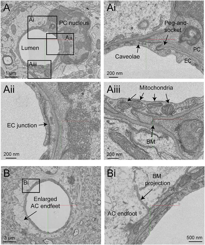
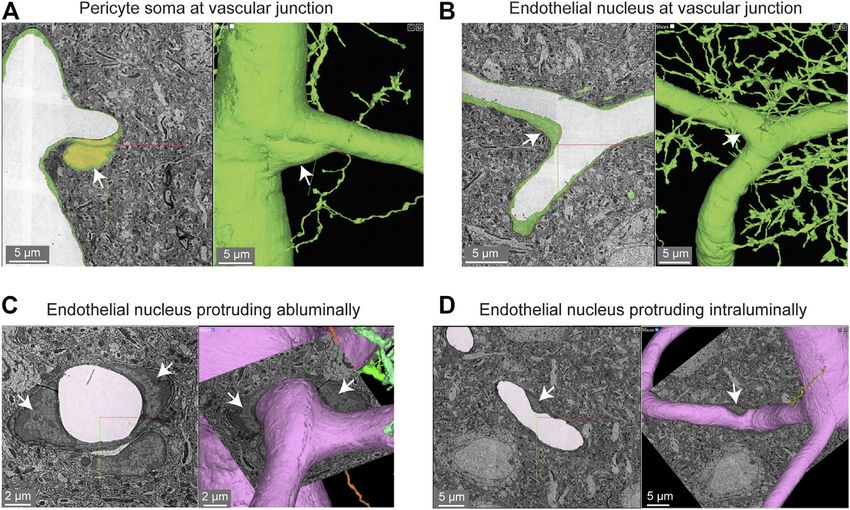
Bonney et al.                                                                                                                             Microvasculature in Volume EM Data

 FIGURE 1 | MICrONS Layer 2/3 pericyte-endothelial interactions. (A) The location of all endothelial and pericyte nuclei identified within the MICrONS Layer 2/3 data
 set. (B) Examples of pericyte pegs extending into the endothelium (left), and conversely, endothelial pegs extending into the pericyte (right). (C) The location of pericyte
 pegs mapped onto the 3D microvascular network reveals that pegs are enriched at sites of pericyte somata. The pericyte somata are labeled with yellow spheres, and
 the individual pegs are labeled with smaller colored spheres, color-coded per pericyte. (D) The location of endothelial pegs are also enriched around pericyte
 somata. Images adapted from (Ornelas et al., 2021). Link to annotations (See methods to register and agree to terms of service): https://neuromancer-seung-import.
 appspot.com/?json_url=https://globalv1.daf-apis.com/nglstate/api/v1/5073246747623424.

 FIGURE 2 | Pericyte and endothelial cell somata can be positioned at vascular junctions. (A) Pericyte soma (white arrow) positioned at the junction between the
 capillary and ascending venule in the MICrONS Layer 2/3 data set. Left panel shows selected vascular wall segmentation in 2D, and the right panel shows 3D rendering of
 the vascular wall. MICrONS Layer 2/3 x, y, z coordinates at: 82286, 68574, 1719. (B) Endothelial nucleus (white arrow) at a capillary junction. Coordinates at: 68291,
 61335, 1788. (C) Example of two endothelial nuclei that protrude abluminally without affecting the shape of the lumen. Coordinates at: 76691, 59357, 1,594. (D)
 Example of an endothelial nucleus that protrudes intraluminally and causes local reduction in capillary diameter. Coordinates at: 71372, 45504, 1,467.

Frontiers in Cell and Developmental Biology | www.frontiersin.org                      4                                               April 2022 | Volume 10 | Article 849469
Bonney et al.                                                                                                                          Microvasculature in Volume EM Data

 FIGURE 3 | Pericyte processes in relation to endothelial features. (A) The edge of a pericyte process annotated in Neuroglancer 3D view with red dots, alongside
 endothelial junctions in cyan dots. (Ai) The 2D cross section of one location (dotted magenta line) within the same capillary segment. (B) The pericyte process tends to
 exhibit greater endothelial coverage near endothelial somata. (Bi) The 2D cross section of one location (dotted green line) within the same capillary segment. EC,
 endothelial cell. Images adapted from (Ornelas et al., 2021). Link to annotations (See methods to register and agree to terms of service): https://neuromancer-
 seung-import.appspot.com/?json_url=https://globalv1.daf-apis.com/nglstate/api/v1/6082177280245760.

within the vascular segmentation due to their proximity to the                           protrusions, similar to pericyte somata (Figure 2C). However, we
vascular wall.                                                                           also noted many instances where the endothelial nuclei encroached
                                                                                         upon the intraluminal space, reducing its diameter (Figure 2D). This
Pericyte-Endothelial Interaction                                                         suggests that the position of endothelial nuclei may influence blood
We noted that the capillary network contains 25 endothelial cell nuclei                  cell flow in the capillary network by imparting blood flow resistance.
and five pericyte nuclei, consistent with individual pericytes covering                   This possibility has not been thoroughly examined in physiological
multiple endothelial cells (Figure 1A). Pericytes were identified as cells                studies, unlike blood flow in relation to the position of pericyte
on the abluminal side of endothelium with protruding cell somata and                     somata.
elongated, slender processes embedded in the vascular basement                               The majority of the capillary length in cortex (~90%) is contacted
membrane. In the volume EM data, their abluminal processes                               by the slender processes of pericytes (Grant et al., 2017;Berthiaume
could be traced back to the cell somata to verify its identity as a                      et al., 2018). To better understand the arrangement of pericyte
pericyte. Physical interlocking between pericytes and endothelial cells                  processes in specific regions of interest, we manually labeled their
via peg-and-socket interactions is important for their communication                     edges with sequential annotations in Neuroglancer (red dots;
and attachment. These structures could be discerned in the data                          Figures 3A,Ai). This allowed us to track the pericyte process
(Figure 1B). By annotating the positions of peg-and-socket                               alongside co-annotated endothelial features (somata, cell-cell
interactions in Neuroglancer, they were found to be concentrated                         junctions) (Ornelas et al., 2021). Pericyte processes were
near the somata of pericytes, implicating the pericyte somata as                         occasionally found to cover and track along with endothelial cell
potential hot spots for direct communication with endothelial cells                      junctions, suggesting that they may provide structural support for
(Figures 1B,C). These interactions included both extension of pericytes                  this feature of the blood-brain barrier. When pericyte processes
pegs toward the adjacent endothelium, and less commonly, endothelial                     encountered the location of endothelial nuclei, they tended to
cells extending toward pericytes (Figures 1B,D).                                         increase their surface coverage and proximity to the nucleus
    Pericyte somata are often positioned at capillary bifurcations                       (Figures 3B,Bi) (Ornelas et al., 2021). Heightened coverage of
(Figure 2A) (Hartmann et al., 2015). This localization may play a                        the endothelial nucleus may provide enhanced communication to
role in how blood flow is partitioned down separate capillary                             influence nuclear functions, such as gene expression.
branches during cerebral perfusion (Gonzales et al., 2020).
Endothelial cell nuclei were located along capillary segments and                        Endothelial Protrusions
also some capillary bifurcations (Figure 2B). The extra volume of the                    “Microvilli” are small protrusions that extend from the
endothelial nuclei often manifested as abluminally-oriented                              endothelium into the intraluminal space (Figure 4A). They

Frontiers in Cell and Developmental Biology | www.frontiersin.org                    5                                              April 2022 | Volume 10 | Article 849469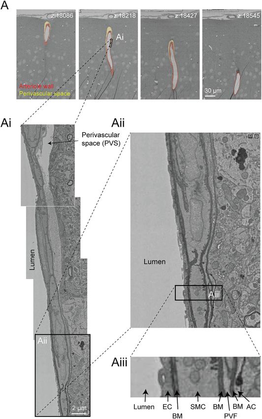
Bonney et al.                                                                                                                  Microvasculature in Volume EM Data

                                                                                         where the endothelium protruded abluminally toward the
                                                                                         brain parenchyma (Figure 4B). Perhaps these are also
                                                                                         specialized appendages for direct sensation of neural or
                                                                                         astrocytic activity. In fact, the protrusion shown is surrounded
                                                                                         by neurites and one axon swells into a bouton shape when in
                                                                                         contact with the endothelium (Figure 4C).

                                                                                         Astrocyte Coverage
                                                                                         Astrocytes are extremely complex in structure and their
                                                                                         multitude of fine processes interact with the vasculature in the
                                                                                         form of “endfeet,” which cover much of the abluminal surface
                                                                                         (Mathiisen et al., 2010; Korogod et al., 2015). Astrocytic coverage
                                                                                         of the vasculature contributes to blood-brain barrier integrity
                                                                                         (Araya et al., 2008), release of vasoactive substances (Attwell et al.,
                                                                                         2010), and water transport (Jin et al., 2013). The segmented
                                                                                         astrocytes in the MICrONS data sets have classic astrocyte
                                                                                         morphology (dense and highly ramified processes) with
                                                                                         endfeet in subregions of the cell that covered the vascular wall.
                                                                                         We observed that astrocytes covered the vasculature in discrete
                                                                                         non-overlapping territories (Figure 5A). Along just one capillary
                                                                                         segment, multiple astrocytes cooperated to establish coverage
                                                                                         (Kubotera et al., 2019). However, it is difficult to know if
                                                                                         individual astrocytes have been wholly segmented, given their
                                                                                         morphological complexity. Thus, it is also possible that the image
                                                                                         shows fragments of individual astrocytes. These data may also be
                                                                                         useful to understand the extent and distribution of regions that
                                                                                         lack astrocyte coverage, which may be relevant as sites of immune
                                                                                         cell entry and interaction (Horng et al., 2017).

                                                                                         Microglial-Capillary Interaction
                                                                                         Microglia associate with the capillary wall and contribute to
                                                                                         regulation of neurovascular structure and function (Joost et al.,
                                                                                         2019;Bisht et al., 2021). Capillary-associated microglia could be
                                                                                         detected in the data set and close inspection revealed that they
 FIGURE 4 | Endothelial protrusions extend luminally and abluminally
                                                                                         abutted the vascular basement membrane without intervening
 from the vessel wall. (A) Example of a luminally-directed endothelial protrusion
 (microvilli) within a capillary segment. Left panel shows the 2D cross section of       contact with astrocytic endfeet (Figure 5B), consistent with prior
 the structure (black arrow). Right panel shows a 3D view of the same                    studies (Mathiisen et al., 2010; Bisht et al., 2021). Individual
 structure in segmented vasculature (white arrow). MICrONS Layer 2/3 x, y, z             microglia are segmented allowing for detailed structure and
 coordinates at: 62431, 48503, 1336. (B) Example of abluminally-directed                 ultrastructural investigation of their interactions with other
 endothelial protrusion. Left panel shows the 2D cross-section of the structure
 (black arrow). Right panel shows a 3D view of the same structure in
                                                                                         neurovascular cells.
 segmented vasculature. MICrONS Layer 2/3 x, y, z coordinates at: 73675,
 48870, 746. The structure is segmented separately from the vascular wall, but           Subcellular Features
 is in fact an extension of the endothelial cell. (C) The abluminally-directed           Two subcellular features are often quantified in studies that assess
 endothelial protrusion is surrounded by neuronal axons and dendrites. One
                                                                                         blood-brain barrier integrity. First are electron dense tight
 axon (cyan) exhibits a region of swelling (potential bouton) in direct contact
 with the endothelial structure.
                                                                                         junctions where individual endothelial cells are bound by a
                                                                                         variety of adhesion proteins to form a barrier (claudins,
                                                                                         occludin, junction adhesion molecules, cadherins) (Haseloff
have been widely observed across mammals and birds (Fujimoto                             et al., 2015). Endothelial cell-cell junctions are not simply rigid
et al., 1975; Makarov et al., 2015), though their physiological role                     physical barriers, but highly dynamic structures regulating the
has remained poorly understood. They could be important for                              paracellular (between cell) passage of cells, water, ions and
sensing the passage of blood cells or impede the movement of                             macromolecules. Second, small endothelial vesicles called
blood cells by creating resistance. Indeed, prior studies have                           caveolae facilitate transcellular (through cell) passage across
shown that microvilli incidence increases in cerebral ischemia,                          the endothelium (Andreone et al., 2017). Both of these classic
perhaps contributing to poor capillary blood flow and no reflow                            blood-brain barrier structures are clearly visualized in the
(Dietrich et al., 1984). 3D-EM data makes it possible to study the                       Cortical Layer 2/3 data set (Figure 6).
shape, prevalence and distribution of microvilli throughout the                             We noticed that caveolae density varied among capillary
capillary network. Curiously, there were also some instances                             segments, which may mean that transcellular permeability

Frontiers in Cell and Developmental Biology | www.frontiersin.org                    6                                       April 2022 | Volume 10 | Article 849469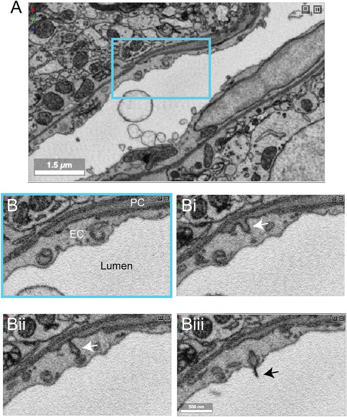
Bonney et al.                                                                                                                             Microvasculature in Volume EM Data

 FIGURE 5 | Astroglial and microglial vascular interactions. (A) Three astrocytes (blue, green, light green) enwrap the capillary wall and their territories form a tiling
 pattern. Dendrites of a neuron (purple) course through the astrocytic territory. The vascular wall is not shown in the 3D view (right) to provide a clearer view of astrocyte
 contact with the vascular wall. MICrONS Layer 2/3 x, y, z coordinates at: 77091, 62413, 784. (B) A putative capillary-associated microglia. The 3D view shows the
 vascular lumen in fuchsia and a microglial soma (red) that drapes along the vessel surface at a bifurcation. Close inspection of this microglia will reveal that it directly
 contacts the vascular basement membrane without intervening astrocytic endfeet. MICrONS Layer 2/3 x, y, z coordinates at: 76302, 59416, 1641.

 FIGURE 6 | Subcellular structures in the vasculature are visible with high contrast in the MICrONS Layer 2/3 data set. (A) A representative cross-section of a
 capillary with magnified insets showing commonly measured subcellular structures, including caveolae and mitochondria (Ai) and endothelial junctions and basement
 membrane (Aii). MICrONS Layer 2/3 x, y, z coordinates at: 109802, 58170, 707.

occurs heterogeneously across the capillary network (not                                   have already highlighted interesting zone-specific differences
shown). 3D-EM data may potentially allow caveolae density                                  that involve caveolae. Venous endothelial cells and capillaries
and their characteristics to be charted across the microvascular                           exhibit greater uptake of plasma proteins through transcytosis
network to better understand their distribution. Recent studies                            than arterioles (Yang et al., 2020). Similarly, post-capillary

Frontiers in Cell and Developmental Biology | www.frontiersin.org                      7                                               April 2022 | Volume 10 | Article 849469

Bonney et al.                                                                                                                                Microvasculature in Volume EM Data

 FIGURE 7 | All vascular zones are represented in the MICrONS Cortical MM^3 data set. (A) The vasculature captured includes 12 penetrating arterioles, 25
 ascending venules, and extensive intervening capillary networks. The bottom panel shows a top-down view of the volume and reveals the originating position of
 penetrating vessels at the pial surface. Link to annotations: https://ngl.microns-explorer.org/#!gs://microns-static-links/mm3/vascular_annotation.json. (B) A
 penetrating arteriole with some smooth muscle cells selected for 3D viewing (arrows). On the right, the three distinct cell types that make up the vascular wall
 (endothelial cells, mural cells, and perivascular fibroblasts) are shown through manual labeling. MICrONS Cortical MM^3 x, y, z coordinates at: 225166, 107266, 18188.
 (C) An ascending venule exhibits a vascular wall that is thinner than the arteriole, but still contains endothelial cells, mural cells, and perivascular fibroblasts. These cell
 types were not well separated by segmentation and therefore only manual labeling of these cell layers is shown. Coordinates at: 262168, 104664, 24791. (D) The
 arteriole-capillary transition zone is a small segment of vasculature that intervenes between the penetrating arteriole and true capillary bed. The endothelium, mural cells
 (sphincter cell and ensheathing pericyte), and perivascular fibroblasts are generally segmented in this region of interest. Here, we show the mural cell layer (left and
 middle) which involved cobbling of many separately segmented structures to visualize the overall morphology of the sphincter cell and ensheathing pericyte. The
 sphincter cell was classified based on prior descriptions by Grubb et al. showing a cuff-like cell at the penetrating arteriole just before branching into the transitional zone,
 with local decrease in vessel diameter. On the right, we add a perivascular fibroblast, which may not have been captured in entirety but shows feasibility of its separate
 segmentation. On the right, the location of different cell layers is shown through manual labeling. Coordinates at: 168985, 165246, 19171. (E) The capillary wall consists
                                                                                                                                                                    (Continued )

Frontiers in Cell and Developmental Biology | www.frontiersin.org                        8                                                April 2022 | Volume 10 | Article 849469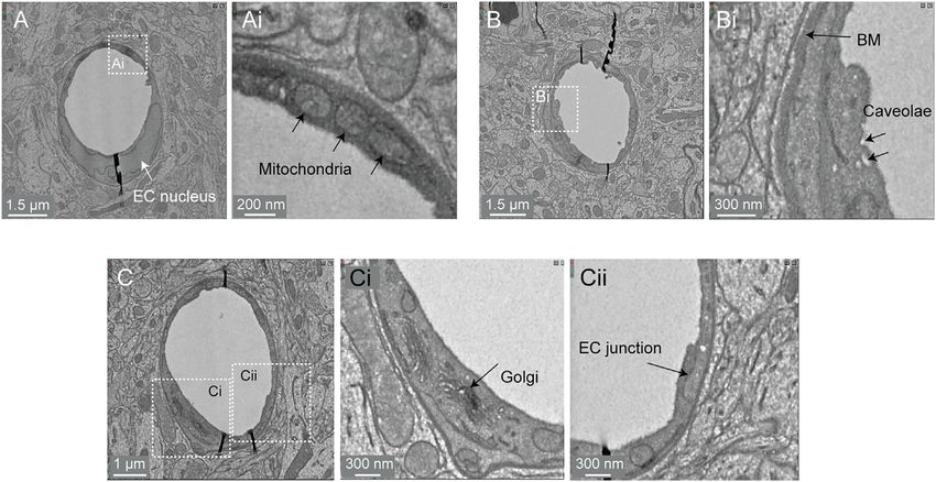
Bonney et al.                                                                                                                    Microvasculature in Volume EM Data

 FIGURE 7 | of only endothelial cells and pericytes. These two cells are sometimes separately segmented, as in the case shown here. However, it is not consistent
 throughout the network. One process of the pericyte is segmented together with the endothelium. On the right, the location of these cells is shown through manual
 labeling. Note the fibroblast layer is absent in capillaries. Coordinates at: 231661, 104310, 24813.

venules show heightened receptor-mediated transport of                               capillary arterioles) that successively ramify into dense and
nanoparticles useful for drug delivery (Kucharz et al., 2021).                       highly interconnected capillary networks. After passing the
Interestingly, arteriole endothelial cells are not devoid of                         dense capillary network, blood then coalesces into ascending
caveolae and in fact show an enrichment in caveolae                                  venules oriented in parallel with penetrating arterioles, that
essential to neurovascular signaling during functional                               drain back to a network of leptomeningeal venules at the pial
hyperemia (Chow et al., 2020). Mitochondria are segmented                            surface.
throughout the data set, including in endothelial cells and                             The vascular network of the MICrONS Cortical MM^3
mural cells (Figures 6A,Ai) (Turner et al., 2020). Prior studies                     data set can be visualized in entirety by selecting the
have shown pericytes to be enriched with mitochondria                                segmentation of the intraluminal space. The data contains
(Mathiisen et al., 2010), which are likely needed to support                         leptomeningeal vessels within the subarachnoid space of
the metabolic demands of solute transport at the blood-brain                         the meninges, which can be followed as they dive into
barrier.                                                                             penetrating vessels of the cortical parenchyma. In total, 12
                                                                                     penetrating arterioles and 25 ascending venules were captured
Non-Cellular Features                                                                within the volume (Figure 7A). The identity of the
An important component of the vascular wall is the basement                          penetrating vessel was determined by examining the
membrane (BM) between cells that contribute to cellular                              morphology of mural cells that lie abluminal to the
adhesion and signaling. The basement membranes are thin                              endothelium. Arterioles are surrounded by concentric,
and highly organized structures (50–100 nm in thickness)                             ring-shaped smooth muscle cells (Figure 7B), whereas
consisting mainly of collagen IV, fibronectin, laminins,                              venules lack these cells (Figure 7C).
nidogen and proteoglycan perlecan (Xu et al., 2019). There
can be multiple BM layers depending on the microvascular                             Endothelial and Mural Cells
zone. In capillaries, there is the endothelial BM that lies                          Unlike the MICrONS Layer 2/3 data set, endothelial and mural
between the endothelium and the pericyte, and the                                    cells have been segmented separately in Cortical MM^3.
parenchymal BM between the pericyte and the astrocyte. The                           However, these segmentations are not consistent throughout
basement membrane is very apparent in 3D-EM data because it is                       the microvasculature. The segmented endothelium is not of
generally more electron dense than the neighboring cells (Figures                    individual cells, but of most of the endothelial layer. Further,
6A,Aii). At sites of pericyte-endothelial interaction, the basement                  mural cells are complex in structure and an individually
membrane may become thin and harder to recognize. Increased                          segmented region may represent fragments of cells.
basement membrane thickness is described in aging and                                Nevertheless, they demonstrate feasibility of segmenting
conditions of poor vascular health such as in the tissues                            vascular cells and provide biological insight. For example,
surrounding an ischemic stroke (Nahirney et al., 2015). This                         smooth muscle cells of the penetrating arteriole viewed in 3D
may indicate a loosening of the protein sheets or adverse                            reveal the position of their somata, which bulge slightly from the
production of BM proteins, both of which would reduce                                vascular wall (Figure 7B, left, middle). The tiling of their
neurovascular communication.                                                         individual territories and details of their circumferential
                                                                                     processes can be examined. In an accompanying 2D image we
MICrONS Cortical MM^3                                                                verified the positions of different cell types along the same
A second study from the MICrONS consortium imaged a 1.4 ×                            arteriole (Figure 7B, right). In contrast to the arteriole, the
0.87 × 0.84 mm volume from visual cortex, which is 400× larger                       wall of ascending venules is much thinner, and as such, the
than the prior Layer 2/3 sample. It spans all 6 cortical layers,                     segmentation of endothelial and mural cells along venules
including some white matter at its base. This significantly larger                    examined is less accurate than arterioles. We provide only
tissue volume provides the unique opportunity to study the                           labeling in 2D to show how vascular cells are positioned along
neurovascular unit across different vascular zones, cortical                         the venular wall (Figure 7C).
layers, multiple cortical areas and between gray vs. white matter.                       The extensive microvascular network could be seen
                                                                                     throughout the data set, including capillaries and the
Microvascular Architecture                                                           transitional zones bridging the capillaries to arterioles and
We first briefly discuss the vascular architecture of the                              venules. In one example, a small penetrating arteriole extends
cerebral cortex. Blood flow to the cortex enters through                              an offshoot toward the capillary network (Figure 7D). A pre-
leptomeningeal arterioles on the brain surface. From the                             capillary sphincter cell (a presumed mural cell type) is seen as a
pial arteriolar network, penetrating arterioles branch and                           cuff around the origin of this offshoot where the vessel diameter is
dive into the parenchyma to perfuse columns of tissue                                locally decreased (Grubb et al., 2020). Sphincter cells are
(Shih et al., 2015). Penetrating arterioles then send smaller                        morphologically distinct from pericytes and exhibit high
offshoots called arteriole-capillary transitions (or pre-                            reactivity to vasoactive mediators during neurovascular

Frontiers in Cell and Developmental Biology | www.frontiersin.org                9                                            April 2022 | Volume 10 | Article 849469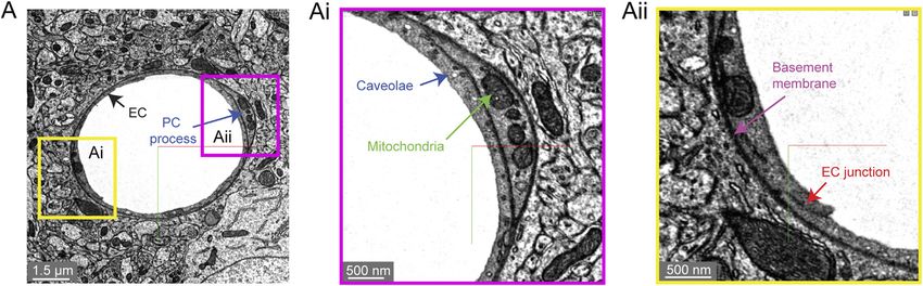
Bonney et al.                                                                                                                    Microvasculature in Volume EM Data

 FIGURE 8 | Subcellular features of the endothelium in the MICrONS Cortical MM^3 data. (A) Capillary cross-section with inset (Ai) showing mitochondria.
 MICrONS Cortical MM^3 x, y, z coordinates at: 141179, 177334, 21310. (B) Capillary cross-section with inset (Bi) showing endothelial caveolae and basement
 membrane. Coordinates at: 167382, 134731, 20338. (C) Capillary cross-section with inset (Ci) showing Golgi cisternae and separate inset (Cii) showing endothelial
 junction. Coordinates at: 129596, 117071, 20179.

coupling (Zambach et al., 2021), as well as aberrant contraction                      may add an additional parameter to consider for in silico
during pathology (Khennouf et al., 2018).                                             studies of capillary flow through cortical vascular networks.
   The protruding soma of an ensheathing pericyte is visible just
downstream of the sphincter (Figure 7D). Unlike the more                              Perivascular Fibroblasts
prevalent capillary pericytes found deeper in the capillary                           Perivascular fibroblasts lie just abluminal to the mural cells on larger
network, and as described for the Layer 2/3 data, this pericyte                       parenchymal vessels in the brain (Bonney et al., 2021). However, they
subtype of the transitional zone exhibits longer processes than                       are absent in the capillary zone. The physiological role of perivascular
smooth muscle cells but shorter than those of capillary pericytes                     fibroblasts remains largely unknown. Single cell transcriptomic
(Hartmann et al., 2021a;Hartmann et al., 2021b). However, the                         analyses of brain vasculature have shed light on their gene
processes completely cover the vascular circumference with fine                        expression profiles, which suggest potential roles in basement
interdigitated projections (Grubb et al., 2020). Critically, in the                   membrane protein production and contribution to cerebrovascular
image shown (Figure 7D, middle), the complexity of this cell is not                   structure (Vanlandewijck et al., 2018). In some pathological scenarios,
captured by a single segmented volume. Rather, multiple segmented                     such as neurodegenerative disease, ischemia and injury, perivascular
regions were cobbled together to obtain a general view of the cell                    fibroblasts proliferate to form scar tissue (Soderblom et al., 2013).
coverage of the transitional segment. Ensheathing pericytes are                       Consistent with prior work, we observed perivascular fibroblasts in
reactive to vasoactive stimuli and the regions they cover dilate                      arteriole, arteriole-capillary transition (Figures 7B,D), but not in
rapidly during functional hyperemia (Hall et al., 2014).                              capillaries (Figure 7E). They are also present on venules
   As described in the MICrONS Layer 2/3 data, capillary                              (Figure 7C), but more superficial of the brain surface and only on
pericytes exhibit protruding somata and long processes that                           venules of larger diameter. Morphologically, perivascular fibroblasts
incompletely cover the circumference of endothelium (Grant                            exhibit flattened somata and thin lamellar processes that cover the
et al., 2017). Pericytes of this morphology could be discerned                        vessel surface.
throughout the extensive capillary beds of the sample.
Pericyte and endothelial cells are occasionally segmented
as individual cell types along the capillary wall                                     Perivascular Macrophages
(Figure 7E). However, the segmentation of individual                                  Perivascular macrophages are resident immune cells of the brain
pericytes may be incomplete, as evidenced by a missing                                with close association to the vasculature. They have been shown
process in the 3D view shown. Throughout the capillary                                to communicate with other neurovascular cell types, are a source
network, the nuclei of pericytes and endothelial cells                                of reactive oxygen species, and contribute to a variety of disease
exhibited clear contrast, providing a unique opportunity to                           processes (Faraco et al., 2016). They occupy the same perivascular
understand        pericyte-endothelial      positioning        and                    space as fibroblasts and may also exhibit morphological
arrangement in the microvasculature. This information                                 similarities. Thus, detailed examination will be required to

Frontiers in Cell and Developmental Biology | www.frontiersin.org                10                                           April 2022 | Volume 10 | Article 849469
Bonney et al.                                                                                                                            Microvasculature in Volume EM Data

 FIGURE 9 | Perivascular space and layers of the arteriole wall. (A) A large penetrating arteriole in the MICrONS Cortical MM^3 data set exhibits a clear perivascular
 space. (Ai) Magnified view of the arteriole wall showing the perivascular space diminishing in size with greater cortical depth. (Aii, Aiii) Magnified view showing layers of
 the arteriole wall, revealing that the perivascular space becomes continuous with the perivascular fibroblast layer as the perivascular space diminishes. EC—endothelial
 cell, BM—basement membrane, SMC—smooth muscle cell, PVF—perivascular fibroblast, AC—astrocyte. Coordinates of the penetrating arteriole: 175231,
 117475, 19297.

understand how to separate these two cell types at the                                     Subcellular Compartments
ultrastructural level. Perivascular macrophages are highly                                 Image contrast in the Cortical MM^3 data was sufficient to resolve
phagocytic and should presumably be distinguishable from                                   a variety of subcellular features (endothelial cell junctions, caveolae,
fibroblasts by a higher density of electron dense lysosomes.                                Golgi cisternae, mitochondria) (Figure 8A,Ai,B,Bi,C,Ci,Cii). In

Frontiers in Cell and Developmental Biology | www.frontiersin.org                     11                                              April 2022 | Volume 10 | Article 849469
Bonney et al.                                                                                                                           Microvasculature in Volume EM Data

 FIGURE 10 | White matter vessels in MICrONS Cortical MM^3 data. (A) Representative cross section of capillary-sized vessels in white matter (left) and gray matter
 (right). White matter capillary cross-section x, y, z coordinates at: 114029, 277354, 17647. Gray matter vessel cross-section x, y, z coordinates at: 109231, 208260,
 17714. (B) A white matter capillary embedded within the axon tracts of the white matter (left). In 3D view, the vascular lumen segmentation is displayed, alongside select
 axons that have been segmented. Coordinates at: 155004, 269113, 21080. (C) A perivascular white matter astrocyte. In 3D view, the coverage of the capillary wall
 can be seen. Coordinates at: 150176, 279591, 18383. (D) A branch of a deep draining venule, principal cortical venule, is present within the data set. Coordinates at:
 195156, 273647, 18192.

fact, instances of caveolar fusion with the endothelial plasma                            arterioles, arteriole-capillary transition zones, and
membrane were observed (Figures 8B,Bi).                                                   capillaries did not exhibit overt perivascular spaces though
                                                                                          they were encased in the basement membrane. This raises the
Perivascular Space                                                                        question of whether CSF flows along the walls of the small
The perivascular space is a fluid filled space surrounding larger                           vessels and capillaries, and if so whether it occurs within the
brain vessels that is vital for influx and efflux of cerebrospinal                          vascular basement membrane. It also suggests that
fluid (CSF) in the clearance of metabolic waste from brain tissue                          perivascular fibroblasts may be involved in adherence of
(Iliff et al., 2012; Wardlaw et al., 2020). However, there remains a                      the vascular basement membranes, and in doing so
lack of clarity on the anatomy of the perivascular spaces,                                influence the extent of the perivascular space.
leading to controversy in pathways for CSF flow and where
they reside in the vascular wall (Bakker et al., 2016). 3D-EM                             White Matter
data may provide an opportunity to define the architecture of                              The Cortical MM^3 data set contains white matter just below the
the perivascular space. Details including the vessel types that                           cortex, where microvasculature is seen alongside densely packed
exhibit a perivascular space (arterioles vs. venules, small vs.                           myelinated axons (Figures 10A,B). There is sufficient contrast to
large diameter vessels), the diameter/volume of these spaces,                             discern the boundaries of endothelial cells and mural cells in
and the neurovascular cell types forming their boundaries                                 white matter, as well as subcellular features, such as caveolae. The
could be examined in detail. However, caution is needed to                                interaction of white matter astrocytes with the vascular wall may
ensure that these spaces are adequately preserved in fixed                                 be accessible to study, as some of the white matter astrocytes have
tissue specimens.                                                                         been segmented (Figure 10C). The vasculature in white matter
    In the Cortical MM^3 data, perivascular spaces could be                               also contains one branch of a principal cortical venule, which is a
observed primarily around larger penetrating arterioles and                               large ascending venule that serves as the primary drainage route
ascending venules, and they were most apparent in the upper                               for juxtacortical white matter (Figure 10D) (Duvernoy et al.,
layers of cortex (Figure 9A). The perivascular space                                      1981).
diminishes as the vessels become smaller in diameter                                         This gray-white matter sampling can help to clarify
(Figure 9Ai). The space eventually becomes continuous                                     differences in composition and subcellular features of the
with the perivascular fibroblast layer, flanked by two layers                               neurovascular unit as blood vessels course into the white
of basement membrane (Figures 9Aii,Aiii). Smaller                                         matter environment. For example, it should be possible to

Frontiers in Cell and Developmental Biology | www.frontiersin.org                    12                                              April 2022 | Volume 10 | Article 849469
Bonney et al. Microvasculature in Volume EM Data
FIGURE 11 | Ultrastructural view of microvasculature in the mouse hippocampus. (A) Representative images from data of Bloss et al. taken from mouse
hippocampus. The image shows a capillary cross-section where a pericyte soma is located. Insets from (Ai–Aiii) show a variety of subcellular structures that can be
visualized. Coordinates at: 20865, 54458, 146. (B) A separate capillary cross-section taken from the same volume exhibits enlarged astrocytic endfeet against the
vascular wall. An inset (Bi) shows a thin projection of the basement membrane extending abluminally. Coordinates at: 25774, 59225, 86.
understand structural differences in pericyte coverage and microvasculature may eventually be linked to neurovascular
association with the endothelium, as well as the ratio of dynamics captured antemortem.
pericytes and endothelial cells per capillary length. Given
the vulnerability of white matter to ischemia and in Additional Studies of Mouse Visual Cortex
neurodegenerative diseases, understanding how the A 2016 study by Lee et al. provided a sample from visual cortex
neurovascular unit differs between gray and white matter layer 1 and layer 2/3 with a volume of 450 × 450 × 150 μm (Lee
will be of great importance. et al., 2016). Image contrast was high, allowing cellular and non-
cellular (basement membrane) components of the vascular wall
Accompanying Physiological Data to be readily discerned. The volume contains a portion of a
Remarkably, the 3D-EM data produced by the MICrONS penetrating arteriole and surrounding capillary networks.
consortium has accompanying multiphoton imaging data on However, the vascular-specific compartments (vascular lumen,
neuronal activity from the same tissues, where the wall, astrocytes) are not segmented.
microvasculature served as a fiduciary to co-register in vivo
and post-mortem data. This physiological data includes Hippocampus
neuronal calcium dynamics during visual stimulation, and The hippocampus has a different vascular supply and
separately, vascular structure (intravenous fluorescent dye). microvascular architecture compared to the cerebral cortex,
This is proof-of-principle that ultrastructural features of the and these differences may underlie vulnerabilities to blood
Frontiers in Cell and Developmental Biology | www.frontiersin.org 13 April 2022 | Volume 10 | Article 849469Bonney et al. Microvasculature in Volume EM Data FIGURE 12 | Pericyte projection into vessel lumen in finch brain capillaries. (A) A capillary from finch basal ganglia (data set: j0251), located at x, y, z coordinates: 19129, 12810, 7,330. (B–Biii) Magnified view of inset in panel A showing a pericyte projecting into the luminal space and ending in a cilia-like structure. The four panels span 10 slices in the z dimension, corresponding to ~200 nm of distance. FIGURE 13 | Human cerebral cortex microvasculature. (A) Broad view of the microvasculature captured in the data set of Shapson-Coe et al. (B) A capillary-sized vessel showing general segmentation of the vasculature. The nuclei of vascular cells were separately segmented into pericytes or endothelial cells, based on their location relative to the blood vessel lumen and basement membrane layers, as well as their appearance in EM. In the example shown, the pericyte nucleus is green and the endothelial nucleus is pink. Left image x, y, z coordinates at: 297270, 74996, 5229. Right image x, y, z coordinates at: 299348, 73663, 5229. Frontiers in Cell and Developmental Biology | www.frontiersin.org 14 April 2022 | Volume 10 | Article 849469
Bonney et al.                                                                                                        Microvasculature in Volume EM Data

flow insufficiency (Shaw et al., 2021). Thus, comparative 3D-EM                 3,549 pericytes, 574 smooth muscle cells and 79 fibroblast-like cells).
studies across different brain regions will be valuable in future             The astrocyte endfeet surrounding the vasculature were swollen,
studies. A study by Bloss et al. imaged a volume of mouse                     which is unlike that seen in the samples prepared from mouse or
hippocampal CA1 region spanning stratum radiatum and                          finch and may be due to the limitations of post-mortem sample
stratum lacunosum-moleculare (Bloss et al., 2018). The sample                 collection (Figure 13B).
is 350 × 200 × 17 µm in size and contains portions of a venule                    A notable feature unique to this human tissue sample is the
network and some surrounding capillaries. The image contrast is               presence of many perivascular oligodendrocytes that lined the
excellent, allowing a clear separation of vascular cell types,                vascular wall, particularly the radially-oriented vessels of white
subcellular structures, and the basement membrane layers                      matter. This may be why oligodendrocytes are a prevalent cell
(Figure 11A). Curiously, some vessels exhibit enlarged                        type isolated from vascular-enriched single cell transcriptomic
astrocytic endfeet not seen in the cortical data sets described               studies (Yang et al., 2022). Another observation was the
above (Figure 11B). Thin projections of the vascular basement                 existence of string capillaries, which are basement membrane
membrane also extended into the parenchyma. Whether these                     tubes with pericytes encased, but no endothelial cells. Their
features are true distinctions of hippocampal vs. cortical                    occurrence is not uncommon in the normal brain (Brown,
vasculature or consequences of tissue preparation remain to be                2010), but prevalence can increase in pathological context
clarified.                                                                     leading to reduced capillary connectivity and blood flow
                                                                              impairment (Challa et al., 2004).
Finch Basal Ganglia
Zebra finch is a widely used model system in behavioral
research, particularly with respect to song development                       DISCUSSION
and auditory processing. Two zebra finch brain data sets
(j0126 and j0251, basal ganglia, Area X) were imaged with                     We surveyed the vasculature in 7 recent large-scale 3D-EM studies
high contrast 3D-EM and show many cellular and subcellular                    (Table 1). These data spanned different species (mouse, finch,
details of the avian vasculature (Figure 12A). The smaller                    human) and brain regions (cortex, callosum, hippocampus, basal
j0126 data set (97.9 × 95.6 × 115 μm) contains a venule and                   ganglia). The data sets captured images across different zones of the
surrounding capillaries. Close inspection of the capillaries                  microvasculature (arterioles, capillaries, venules and transitional
revealed similar arrangement of endothelial cells and                         segments), and were of sufficient quality to visualize subcellular
pericytes as seen in the mouse brain. Pericyte and                            features relevant to blood-brain barrier function (endothelial
endothelial interdigitations were widely observed, and in                     junctions, caveolae, basement membrane, astrocytic coverage),
some cases pericyte pegs contacted the endothelial nucleus                    blood flow (positioning of endothelial and pericyte nuclei, mural
as reported previously in mouse (Ornelas et al., 2021).                       cell coverage, endothelial microvilli, microglial-vascular interaction),
Interestingly, in the larger j0251 data set (256 × 256 ×                      bioenergetics (mitochondria), cellular communication (caveolae, peg-
384 μm), pericyte projections were seen to bypass the                         and-socket interactions) and cerebrospinal fluid flow (perivascular
endothelial layer and protrude into the luminal space and                     space). The biological insight that can be garnered from these data is
end in small cilia-like structures (Figures 12B–Biii), opening                immense. Further work is needed to segment, annotate and quantify
the possibility that pericytes use these projections for direct               cerebrovascular features. These efforts will stimulate the development
sensation of blood flow. This is a feature that we have thus far               of new hypotheses, design of novel physiological studies, and
not seen in the mouse brain.                                                  improved in silico modeling approaches.
                                                                                  To fully leverage 3D-EM for vascular research, it will be
Human Cortex                                                                  necessary to accurately segment the cellular components of the
A recent study from Shapson-Coe et al. examined a small                       vascular wall: endothelial cells, mural cells (pericytes and smooth
fragment of human cortex (temporal lobe), resected from a                     muscle cells), perivascular fibroblasts, and perivascular
patient with drug-resistant epilepsy during surgery to remove an              macrophages. Together with already segmented parenchymal
epileptic focus (Shapson-Coe et al., 2021). Immediately after                 components         (neurons,       astrocytes,      microglia,      and
excision, the sample was immersion-fixed overnight. The                        oligodendrocytes), this would account for all cells that
hippocampus was sclerotic and exhibited signs of pathology                    comprise the neurovascular unit, a framework used to
consistent with epilepsy, but the resected cortical sample                    understand how neurons and other cell types communicate
appeared normal on traditional histopathology.                                with nearby vessels to serve metabolic needs of the brain
    The sample spans ~3 mm at its longest width, ~2 mm at its longest         (Schaeffer and Iadecola, 2021). Advances in the MICrONS
height, and ~0.15 mm in thickness. It includes sections of                    Cortical MM^3 data set have shown feasibility of segmenting
parenchymal arterioles and venules and surrounding capillary                  individual cell types of the vascular wall. However, further work is
networks (Figure 13A). Despite the vascular lumen being mostly                needed to improve accuracy and consistency of the current
collapsed, the nuclei of vascular cells could be categorized as mural         segmentations and to identify the specific challenges posed by
cells or pericytes based on distance from the lumen and position              vascular cells. In addition, existing data sets offer the possibility to
relative to the endothelial and parenchymal basement membranes                identify and map innervating neurons of cerebral vessels,
(Figure 13B). A total vascular length of 22.6 cm was segmented, in            providing insights into flow regulation by direct release of
which the authors detected ~8k vascular cells (4604 endothelial cells,        neurotransmitter on the vascular wall (Hamel, 2006).

Frontiers in Cell and Developmental Biology | www.frontiersin.org        15                                       April 2022 | Volume 10 | Article 849469
Bonney et al. Microvasculature in Volume EM Data
Mapping the prevalence and distribution of vascular with astrocyte endfeet (Mathiisen et al., 2010) while only two-third
ultrastructures is essential for our understanding of vascular coverage is seen with cryo-EM (Korogod et al., 2015). One way to
functions across different zones. For example, the nuclei of optimize tissue preparation is to image the same regions both in vivo
endothelial cells and pericytes are easily distinguished in the and post-mortem, as done in the MICrONS pipeline. The in vivo two-
MICrONS Cortical MM^3 data. How the nuclei localize in the photon imaging data may then be compared to the 3D-EM data to
network and impinge upon the luminal space, may hold insight understand how well vascular diameter and wall structure is
into how blood flow is distributed through complex capillary preserved. Further, cautionary statements should be added to
networks (Østergaard et al., 2014;Schmid et al., 2017). Further, it is publications when there are signs of poor vascular preservation,
likely that endothelial microvilli contribute to blood flow resistance, include stacking of red blood cells in arterioles, wide-spread swelling
and conceivably sense blood flow to convey signals to the endothelium of astrocytes, and ruffling of endothelial cells.
and mural cells. Therefore, understanding their distribution at the The work and costs involved in creating and analyzing large-
level of individual capillary segments would be valuable. scale 3D-EM on the scale of the MICrONS MM^3 data are
Mapping endothelial junctions and caveolae on a large scale extensive. It is not readily accessible to smaller, individual
would generate an unprecedented 3D map of blood-brain barrier laboratories seeking to address specific questions in their
structure. Endothelial junctions and caveolae bear similarity to studies. However, some labs may wish to generate more
the post-synaptic density and pre-synaptic vesicles of neuronal manageable 3D data sets for manual segmentation and
synapses, respectively. Thus, existing machine learning annotation. The challenge is that vasculature is a relatively
algorithms are likely adaptable for detection of these vascular sparse in brain tissue compared to neurons, and one must
features. Identification of endothelial junctions will also reveal the know where to collect smaller data sets. A way to overcome
boundaries of individual endothelial cells as they tile together to this challenge is to first survey the broader vascular network
form the inner layer of the vascular tube. Additionally, with wider field imaging, and then perform high-resolution
mitochondria in vascular cells have a similar appearance to imaging in specific regions of interest (Guérin et al., 2019). The
those already segmented in parenchymal cells (see MICrONS public resources described here can serve as valuable guides to
Layer 2/3). Mapping the density of mitochondria across the decide where to collect vascular data for more targeted studies.
vasculature may reveal where energy-demanding processes, Further, techniques such as X ray micro CT angiography
such as blood-brain barrier transport or maintenance of (Ghanavati et al., 2014) could be used to survey vasculature
membrane potential, are most active. across the whole brain prior to EM-level imaging within distinct
The ability to manually annotate, and therefore count and brain regions.
quantify, the number of cellular/subcellular structures already
provides significant insight on vascular structure. For example,
we have annotated endothelial and pericyte nuclei, and traced DATA AVAILABILITY STATEMENT
pericyte processes and endothelial junctions. In prior work, basic
distance measurements were performed in Neuroglancer The original contributions presented in the study are included in
allowing extraction of quantitative data on peg-and-socket the figures and figure legends. Further inquiries can be directed to
contacts between endothelial cells and pericytes (Ornelas et al., the corresponding authors.
2021) These endeavors have only scratched the surface of
information within these volumes, and there are no barriers to
further exploration in this fashion, except time. Further, ETHICS STATEMENT
Neuroglancer is one of many browsers for exploration of
volume imaging data, and others including webknossos, This article surveys data from other groups that had ethics
knossos, pyknossos, napari, bigdataviewer, may be more committee oversight.
versatile for annotation. Thus, the availability of 3D-EM data
through a variety of online tools will maximize their use by the
scientific community. AUTHOR CONTRIBUTIONS
A potential limitation and topic for further investigation is
whether the parameters of tissue collection used in the current AS and AK conceptualized the study. SB, VC-S, SFH, and AK
data sets (anesthetic, post-mortem interval, fixative, perfusion surveyed the data to find notable vascular features. SB, VC-S,
pressure, etc.) adequately preserved the native structure of the SFH, MT, AK, and AS contributed to preparation of figures. JK
vascular lumen, vascular cell types, and perivascular space. The collected and provided access to finch data sets. AS wrote the
shape of the vessel wall can be easily distorted by contraction of manuscript with contributions and feedback from SB, VC-S, SFH,
mural cells or loss of intraluminal pressure, making quality of MT, JK and AK.
tissue preparation of utmost importance. It is already known that
the choice of fixation approach greatly influences the size of the
extracellular space (Cragg, 1980; Pallotto et al., 2015) and this FUNDING
may result in disparate outcomes for both neuronal and vascular
structures alike. For example, brains prepared with aldehyde- AS is supported by grants from the NIH/NINDS (NS106138,
based fixative for EM show nearly full coverage of blood vessels NS097775) and NIH/NIA (AG063031, AG062738). SB is
Frontiers in Cell and Developmental Biology | www.frontiersin.org 16 April 2022 | Volume 10 | Article 849469You can also read