Review on Cyanobacterial Studies in Portugal: Current Impacts and Research Needs - MDPI
←
→
Page content transcription
If your browser does not render page correctly, please read the page content below
applied
              sciences
Review
Review on Cyanobacterial Studies in Portugal: Current Impacts
and Research Needs
Cristiana Moreira 1 , Alexandre Campos 1 , José Carlos Martins 1 , Vitor Vasconcelos 1,2
and Agostinho Antunes 1,2, *

                                          1   CIIMAR/CIMAR, Interdisciplinary Centre of Marine and Environmental Research, University of Porto,
                                              Terminal de Cruzeiros do Porto de Leixões, Av. General Norton de Matos s/n, 4050-208 Porto, Portugal;
                                              cmoreira@ciimar.up.pt (C.M.); amoclclix@gmail.com (A.C.); jmartins@ciimar.up.pt (J.C.M.);
                                              vmvascon@fc.up.pt (V.V.)
                                          2   Department of Biology, Faculty of Sciences, University of Porto, Rua do Campo Alegre,
                                              4169-007 Porto, Portugal
                                          *   Correspondence: aantunes@ciimar.up.pt; Tel.: +351-223401813

                                          Abstract: Cyanobacteria have long been associated with harmful effects on humans, animals and
                                          aquatic biota. Cyanotoxins are their most toxic metabolite. This review summarizes the current re-
                                          search, impacts and future needs in cyanobacterial studies undertaken in Portugal, the southernmost
                                          country of Europe, and with a recent multiplication of cyanotoxicity due to climate change events.
                                          Microcystins are still the most prevalent, studied and the only regulated cyanotoxins in Portuguese
                                          freshwater systems much like most European countries. With the development of some tools, partic-
                                ularly in molecular studies, the recent discovery of cylindrospermopsins, anatoxins and saxitoxins,

                                          both genes and toxins, in North and Center ecosystems of our country highlight current impacts that
Citation: Moreira, C.; Campos, A.;
                                          overall communities are facing with increased risks of exposure and uptake to cyanotoxins. Research
Martins, J.C.; Vasconcelos, V.;
                                          needs encompass the expansion of studies at all aspects due to the uprising of these cyanotoxins and
Antunes, A. Review on
                                          reinforces the urgent need of increasing the frequency of surveillance to achieve tangible effects of
Cyanobacterial Studies in Portugal:
                                          cyanotoxins in Portugal to ultimately implement regulations on cylindrospermopsins, anatoxins and
Current Impacts and Research Needs.
Appl. Sci. 2021, 11, 4355. https://
                                          saxitoxins worldwide.
doi.org/10.3390/app11104355
                                          Keywords: microcystins; cylindrospermopsins; anatoxins; saxitoxins; surveillance
Academic Editors: Cristiana Moreira
and Vitor Manuel Oliveira
Vasconcelos
                                          1. Introduction
Received: 30 March 2021                         Cyanobacteria belong to an ancient group of prokaryotes where its toxic risks have not
Accepted: 5 May 2021
                                          been completely evaluated. Eutrophication, anthropogenic pressure, rise of temperature
Published: 11 May 2021
                                          are factors that contribute to the proliferation of these microorganisms (CyanoHAB’s) in
                                          aquatic systems interfering with water quality and public health by the release of odor
Publisher’s Note: MDPI stays neutral
                                          substances and toxic compounds (cyanotoxins). These latter possess negative effects such
with regard to jurisdictional claims in
                                          as hepatotoxic, cytotoxic, neurotoxic, genotoxic, immunologic and carcinogenic having
published maps and institutional affil-
                                          resulted in episodes of human and animal mortality as well as morbidity in several coun-
iations.
                                          tries worldwide [1–5]. Cyanobacteria occurrence possess its main impacts on freshwater
                                          ecosystems since activities such as drinking, irrigation and recreation can result in pub-
                                          lic health problems and derived economic losses. In Portugal, studies on cyanobacteria
                                          began in the 1930s mainly through taxonomic studies, followed by some reported toxic
Copyright: © 2021 by the authors.         episodes associated with blooms and finally, in 1989 the first toxic cyanobacterium species
Licensee MDPI, Basel, Switzerland.
                                          was isolated and its cyanotoxin amount determined. Other strains were also isolated
This article is an open access article
                                          contributing to the knowledge on toxic cyanobacteria occurrence in Portuguese freshwa-
distributed under the terms and
                                          ter ecosystems. Bloom occurrence is not often accounted for by national and European
conditions of the Creative Commons
                                          governmental agencies where surveillance encompasses solely phytoplankton counting
Attribution (CC BY) license (https://
                                          and enumeration of microcystins variant LR the only regulated cyanotoxin in Portugal
creativecommons.org/licenses/by/
4.0/).
                                          and Europe [6]. Although other cyanotoxins such as cylindrospermopsins, anatoxins and

Appl. Sci. 2021, 11, 4355. https://doi.org/10.3390/app11104355                                               https://www.mdpi.com/journal/applsci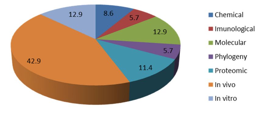
Appl. Sci. 2021, 11, 4355                                                                                                                                    2 of 14

                                    saxitoxins occurrence have been found in Portuguese freshwater systems [7] the lack of
                                    regulations on these cyanotoxins by Portuguese governmental authorities reflects the need
                                    for further research in order to fully assess cyanobacterial impacts, include in monitoring
                                    campaigns the evaluation of other cyanotoxins besides the legislated microcystins [6] to
                                    improve water management, water quality and provide with recommendations to national
                                    authorities in case of an episode of cyanotoxins intoxication occurring.
                                         In toxic cyanobacteria and in cyanotoxins occurrence there can be applied quantitative
                                    and qualitative methods. The first (quantitative) allow the enumeration of the cyanotoxins
                                    applying techniques such as the High Performance Liquid Chromatography (HPLC), Liq-
                                    uid Chromatography–Mass Spectrometry (LC-MS) and Enzyme-Linked Immunosorbent
                                    Assay (ELISA) directly from isolates or environmental samples (water) that will permit after
                                    confronting the measured value with the adopted guideline value the evaluation of the
                                    toxic status of a given isolate or ecosystem. Matrix-Assisted Laser Desorption/Ionization
                                    (MALDI) is another used chemical method that aims to unravel the profile of cyanotoxicity
                                    and cyanobacteria metabolites in a given sample. The second (qualitative) allows the
                                    characterization and identification applying techniques such as the Polymerase Chain
                                    Reaction (PCR) and multiplex Polymerase Chain Reaction (mPCR) of toxic genotypes.
                                    These after DNA sequencing allow inferring of the phylogeny of given taxa. Other meth-
                                    ods such as the quantitative PCR or Real-Time PCR permit the quantification of the toxic
                                    genotypes. Other used assays include the in vivo and in vitro assays based in experiments
                                    that englobe either living organisms (in vivo) or cell lines (in vitro) that after an incubation
                                    period to a given concentration of a cyanotoxin infer on the damage of the tested cyan-
                                    otoxin. The present review summarizes the current research, impacts and future needs
                                    in cyanobacterial studies undertaken in Portugal (Table 1), contributing to redirect the
                                    continuous investigations globally by enumerating current impacts usually associated with
                                    cyanobacterial contamination and to foster the mitigation of toxic CyanoHABs, reinforce
                                    investigations in Portugal and worldwide by establishing research needs consequently
                                    turning the national and international communities and governments more resilient to
                                    this problem.

                     Table 1. Summary of the currents impacts and research needs per type of cyanotoxin in Portugal.

          Cyanotoxin                           Current Impacts                                                    Research Needs
                                                                                                Find MCLR in other genera besides Microcystis sp.
      Microcystins (MC)                  MCLR found in M. aeruginosa
                                                                                          Infer on the phylogeny of MCLR Portuguese DNA sequences
                                                                                                   Enumerate MC variants found in M. aeruginosa
                                                                                    Find and enumerate other MC variants in bloom samples, other genera and
                               Other MC variants found in M. aeruginosa isolates                                  in water samples
                                                                                                  In vivo and in vitro assays for other MC variants
                                                                                        Infer on the phylogeny of MC variants Portuguese DNA sequences
  Cylindrospermopsins (CYN)               Enumerated in Vela Lagoon                  Expand enumeration studies to other ecosystems to assess possible risks
                                                                                         Expand surveillance to the South Region to assess possible risks
                                 Detected genes in North and Center regions                              Isolate the toxin-producing strain
                                                                                           Infer on the phylogeny of CYN Portuguese DNA sequences
                                Found after laboratory cultivation of isolates of
                                                                                                 Find the environmental toxin-producing strain
      Anatoxin-a (ANA)            Chrysosporum, Dolichospermum, Microcystis
                                                                                        Study toxic effects through proteomic, in vivo and in vitro assays
                                                and Oscillatoria
                                                                                         Expand surveillance to the South Region to assess possible risks
                                                                                                     Enumerate through chemical methods.
                                  Enumerated in North and Center Regions
                                                                                                              Find other isoforms
                                                                                          Infer on the phylogeny of Portuguese ANA DNA sequences
       Saxitoxins (SXT)                       Found in C. gracile                       Study toxic effects through proteomic, in vivo and in vitro assays
                                                                                         Expand surveillance to the South Region to assess possible risks
                                                                                                      Enumerate through chemical methods.
                                  Enumerated in North and Center Regions
                                                                                                              Find other isoforms
                                                                                           Infer on the phylogeny of Portuguese SXT DNA sequences
                                 Unidentified toxic metabolites in Portuguese        Isolate C. raciborskii and perform chemical, in vivo and in vitro assays on
            Others
                                                 C. raciborskii                                              strains to identify the toxicity
Appl. Sci. 2021, 11, 4355 3 of 14
1.1. Toxic Cyanobacteria
Although cyanobacterial studies began in Portugal in the 1930s the first identifica-
tion of the first toxic cyanobacterium species in our waters was attributed to Vasconcelos
et al. [8] that with his surveillance and search for microcystins found that this was produced
by the species Microcystis aeruginosa in several amounts and in a diverse range of freshwater
systems of Portugal. In his survey 36 lakes, reservoirs and rivers were monitored. Then
only Microcystis blooms were assessed and 60% of these were found to be toxic. The main
species present in toxic blooms were Microcystis aeruginosa (72%) and Anabaena flos-aquae
(28%) [9]. The main hepatotoxins in Portuguese freshwaters include MCYST-LR, MCYST-
LA, MCYST-YR and [D-Aspl]MCYSTLR [8,10]. Later Pereira et al. [11] found evidence of
saxitoxins production in Montargil Reservoir (South Region) through the chemical analysis
of an isolate of Chrysosporum flos-aquae. Studies persisted and in 2009 Oswald et al. [12]
found anatoxin-a production in strains of Chrysosporum, Dolichospermum, Microcystis and
Oscillatoria with production values ranging from 0.06 µg/g to 24.62 µg/g of cyanobacterial
dry weight [12]. In spite of this finding, the strains were only able to produce anatoxin-a
under laboratory conditions failing so far its environmental detection through chemical
methods. Cylindrospermopsins were found to occur in a lagoon located in the Center
Region (Vela Lagoon) in amounts that reached 12 µg/L in the water [13]. In this study the
cylindrospermopsin-producer identified belonged to Chrysosporum sp. and this was only
achieved after sequencing and blast web search of a positive amplicon that belonged to the
cyrC gene cluster since the isolation of the producing strain failed [13]. Since toxic cyanobac-
terium studies began in Portugal this was the first study to include molecular studies in the
identification of a toxic cyanobacterium producer. More recently Moreira et al. [7] found
evidence of cyanotoxins multiplication in Portugal since most of the previous studies were
referred to the Center and South Regions of the country providing evidence that other
cyanotoxins besides microcystins can be found in the colder North Region of Portugal.
Other results from this study include that anatoxin-a and saxitoxins production were
attributed to the Chrysosporum sp. genus [7]. The invasive and toxic Cylindrospermopsis
raciborskii was found to occur for the first time in the colder North Region of Portugal with-
out forming any blooms and with no cyanotoxicity being attributed so far [14]. Regarding
bloom occurrence, the first intensive surveillance study that searched for all cyanotoxins
in Portuguese freshwater systems belonged to Moreira et al. [14] where it was found that
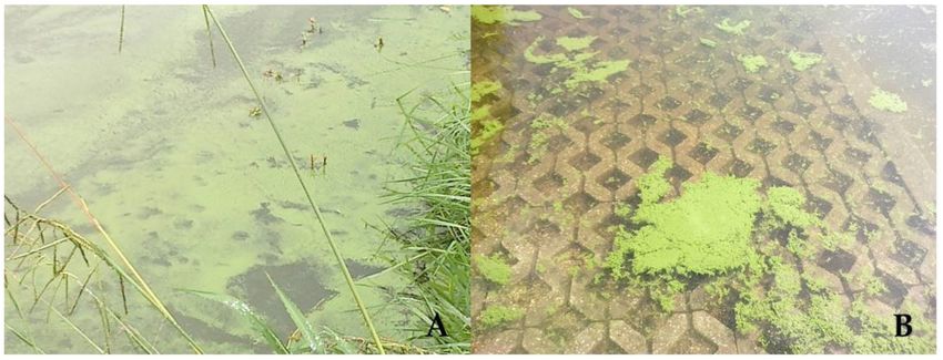
50% of the ecosystems analyzed had blooms (Figure 1). Its composition was comprised of
Microcystis alone or a mixture of Microcystis with Chrysosphorum/Dolichospermum. Though
initial studies by Vasconcelos described that Microcystis was the dominant bloom forming
genus in Portugal recently in a year (2017) with two heat waves during the sampling season
lead to the rise of other types of cyanobacterium species which may have contributed to the
multiplication of cyanotoxicity recommending the deepening of monitoring campaigns as
well as the integration of other cyanotoxins besides the legislated microcystins-LR (MCLR).
In this study only North and Center water systems were sampled; nonetheless, previously
in 2005 in Algarve (South Region) at Beliche reservoir, it was found a bloom of Planktothrix
rubescens that produced microcystins [15]. An extensive study on cyanobacterial blooms in
the south regions of Alentejo and Algarve reservoirs highlighted the occurrence of blooms
not only attributed to M. aeruginosa but to other species belonging to the Nostocales order
in Algarve reservoirs and Microcystis aeruginosa and Anabaena circinalis in reservoirs from
Alentejo region as the predominant bloom-forming species [16].Appl. Sci. 2021, 11, 4355 4 of 14
Figure 1. Bloom formation at Porto City Park Lake 1 (North Region) in 2012 (A) and at Marco de Canaveses (North Region)
in 2012 (B).
1.2. Cyanotoxins Episodes
In worldwide cyanotoxins episodes they resulted in human and animal mortality and
morbidity having a global impact and in Portugal, there is no exception. In fact, in 1993 the
death of 20 patients in a hemodialysis unit in Évora Hospital (South Region) was attributed
to cyanotoxins though none were measured [17]. More recently in 2017 the death of
25 cows in Alentejo (South Region) that ingested water from a nearby stream resulted in the
detection of MCLR in the kidneys of one animal (0.13 µg/L). Phytoplankton examination
revealed that Microcystis was the main genera found and total microcystins were detected
in a concentration of 0.16 µg/L [18]. Though this is the first report of a cyanotoxins animal
poisoning in Portugal the lack of epidemiological data through bloom occurrence in several
ecosystems as described by Moreira et al. [7] may hinder other cyanotoxins outbreaks in
national waters though no human fatality being identified to these metabolites similarly
to the described mortality (Brazil) and morbidity (Australia) episodes [1,2]. Nonetheless
continuous vigilance of these metabolites is essential along with cross-referencing with
health and environmental agencies of Portugal.
2. Cyanobacterial Studies
2.1. Chemical Assays
Cyanotoxins based studies on chemical studies in Portuguese freshwater systems
englobe the screening and enumeration of all the main cyanotoxins and a study on the
peptide diversity of M. aeruginosa strains isolated from several Portuguese freshwater
systems (Figure 2). Microcystins were the first to be chemically unraveled by HPLC where
MCYST-LR, MCYST-LA, MCYST-YR and [D-Aspl] MCYSTLR were found to be present in
both bloom samples and isolated strains of M. aeruginosa gathered from several freshwater
systems with national representation [8,10]. Saxitoxins were the next cyanotoxins to be
characterized this occurring through a HPLC-FLD method followed by LC/MS confirma-
tion from a strain of Chrysosporum flos-aquae collected from a bloom of this cyanobacterium
in Montargil reservoir (South Region) [11]. In this study, five PSP toxins, neoSTX, dcSTX,
STX, GTX6, and GTX5 were found in the same isolate (LMECYA 31) [11]. Screening for
anatoxins was followed later in the mid-2000s but only after laboratory cultivation of
strains belonging to the genera Chrysosporum, Dolichospermum, Microcystis and Oscillatoria
with anatoxin-a production values ranging 0.06 µg/g to 24.62 µg/g of cyanobacterial dry
weight [12]. More recently cylindrospermopsins was found to occur in a lagoon in the
Center Region of Portugal (Vela Lagoon) also through HPLC followed by confirmation
of the mass spectrum through LC/MS in water samples and in the absence of blooms in
concentrations that ranged a minimum of 1.4 µg/L to a maximum of 12 µg/L [13]. Apart
from HPLC and LC/MS methods, a study by Martins et al. [19] used the MALDI-TOFAppl. Sci. 2021, 11, 4355 5 of 14
MS technique to determine the peptide diversity of Portuguese freshwater M. aeruginosa
strains isolated from lakes, rivers and reservoirs. Results from their study include the
finding of aeruginosins, microginins, anabaenopeptins, cyanopeptilins, microcystins, and
microviridins [19]. In microcystins it was found the presence of the variants MCLR, -FR,
-RR, -WR and -YR as the most commonly found [19].
Figure 2. Graphical representation of the percentage of each assay published in Portugal.
2.2. Immunological Assays
Some studies have used the immunological assays (ELISA) marked by Abraxis in
water samples collected from several lakes, rivers and reservoirs with national represen-
tation as well as in food supplements (Figure 2). This method was never used singularly
but as complementary to other methods since it is an enumeration technique. Regarding
water systems, initial studies were based on the search and enumeration of microcystins in
southern reservoirs [20]. Valério et al. [20] in their study showed that microcystins were
detected in 23% of the 53 samples tested and in some of them concentrations exceeded
the WHO guideline of 1 µg/L [20]. Later, a cyanotoxins risk assessment conducted in
eight Center Region reservoirs highlighted again the presence of microcystins in which
two reservoirs resulted in high risk with values above 20 µg/L [21]. Until these studies, no
ELISA immunoassays were conducted on other cyanotoxins besides microcystins. This
situation was surpassed by the study of Moreira et al. [7] where all main cyanotoxins
were enumerated by ELISA. In their study, North Region ecosystems were extensively
studied and monitored for microcystins and resulted that in the five northern ecosystems
analyzed 57% had values above 1 µg/L between April and September. Other cyanotoxins
surveyed included in Vela Lagoon cylindrospermopsins with none of the six samples above
the proposed guideline of 1 µg/L by Humpage and Falconer [22] and in the North and
Center region ecosystems, the enumeration of anatoxin-a showed that 33% were above
the proposed guideline value of 1 µg/L while in saxitoxins only one sample belonging to
a North Region ecosystem exceeded the proposed guideline value of 3 µg/L [13]. Saker
et al. [23] showed the presence of microcystins-LR equivalents with concentrations within
the range of 0.1–4.72 µg/g in food supplements.
2.3. Molecular Studies
Molecular studies were initiated in the mid 2000s (Figure 2) with a first study ap-
plying C. raciborskii Portuguese isolated strains to determine their potential to produce
either cylindrospermopsins or saxitoxins. Results from this analysis were negative for
both cyanotoxins and englobe the application of peptide synthetase (PS) and polyketide
synthase (PKS) genes through simple PCR analysis [24]. Recent studies have shown that
environmental DNA belonging to C. raciborskii can be already found in the north and center
regions of Portugal demonstrating its invasive nature but without forming any blooms
and with no association with toxicity genes [14]. Other studies used the multiplex PCR
technique in uncovering the toxigenic nature of dietary food supplements of cyanobacte-Appl. Sci. 2021, 11, 4355 6 of 14
rial origin. In fact, toxigenic Microcystis were found to be present in all analyzed dietary
supplements produced from the nontoxic cyanobacterium Chrysosporum flos-aquae [25].
PCR was further considered an early warning technique in cyanobacterial investigations
since it correlated well with ELISA assays when applying Portuguese water samples [26].
Valério et al. [27] developed a multiplex PCR that simultaneously amplified mcyA-cd,
mcyAB, and mcyB amplicons of the microcystin gene cluster in 124 cyanobacterial isolates
and in 37 environmental samples. The toxicological status of the isolates was assessed by
high-performance liquid chromatography where the multiplex PCR developed showed
a sensitivity of 92.3% and a specificity of 100%. In environmental samples, ELISA was
used and results for the presence of microcystins in these samples gave a sensitivity of 80%
and a specificity of 100% [27]. Studies applying Real-Time PCR analysis were initiated
by Moreira et al. [28] with the variation of C. raciborskii and cylindrospermopsins in field
samples gathered from Vela Lake with primers previously described characterizing total
cyanobacteria (16S rRNA), C. raciborskii (rpoC1), and cylindrospermopsin synthetase gene
(cyrC). The results report the high abundance of both cyanobacteria and C. raciborskii in
Vela Lake, with C. raciborskii representing 0.4% to 58% of the total cyanobacteria population.
Cylindrospermopsin synthetase gene was detected in one of the samples [28]. In the same
year, Martins et al. [29] quantified Microcystis and MC-producing Microcystis to determine
the genotypic composition of the natural Microcystis population. Results from his study
further highlighted that a negative significant correlation was observed between toxic (with
mcy genes) to non-toxic (without mcy genes) genotypes ratio and the overall Microcystis
density [29]. More recently, Churro et al., [30] developed primers for the Real-Time PCR
amplification of Planktothrix agardhii based on the rpoC1 gene fragment. Cell concentration
determined by the Real-Time PCR method showed a linear correlation with the cell concen-
tration determined from direct microscopic counts and detection limit for cell quantification
was 8 cells/µL. More recently Moreira et al. [7] carried an extensive surveillance study
that included the application of representative genetic markers for the amplification in
water samples of the four main cyanotoxins in the North and Center freshwater systems.
Amplification results include that 79% of the samples had microcystins (mcyA), 40% had
cylindrospermopsins (cyrC), 40% had anatoxin-a (anaC) and finally 50% had saxitoxins
(sxtI) [13].
2.4. Phylogeny Studies
The first study to apply clustering analysis (Figure 2) belongs to Valério et al. [31],
where in their study, M13 PCR fingerprinting, ERIC PCR fingerprinting and amplification
of the internal transcribed spacer (ITS) region were used to characterize cultured strains of
C. raciborskii obtained from several freshwater lakes and rivers of Portugal. Results showed
that when including in this analysis other taxa such as M. aeruginosa, Chrysosporum spp.,
Planktothrix agardhii and Oscillatoria neglecta the potential of the fingerprinting method
demonstrated only the ability to differentiate strains at an intra-specific level failing as
an identification tool. Nonetheless, ITS amplification turned to be a good method for
strain clustering [31]. In 2009 the same authors published a work on the applications of
several molecular techniques (STRR and LTRR PCR fingerprinting, phylogenetic analysis
with the 16S rRNA and rpoC1 genes and M13 and ERIC PCR fingerprints) to evaluate the
level of genetic diversity of 118 cyanobacterial isolates for taxonomic purposes. After the
application of these methodologies in the 118 strains revealed good congruence, indicating
the potential for use in cyanobacterial monitoring, as a quality management control. Later,
Moreira et al. [32] when applying isolates of Portuguese C. raciborskii found that they cluster
together with other European strains and were closely related to Asian and Australian
strains. This study used for the first time the genetic information of four genetic markers
that were concatenated in the analysis (16S rRNA, rpoC1, ITS-L and ITS-S) [32]. Later,
the same authors studied the genetic diversity and population structure of Portuguese
isolates of M. aeruginosa [33]. In their study, a phylogenetic tree was produced resulting
from the concatenation of four distinct genetic markers (16S rRNA, 16S-23S ITS, DNAAppl. Sci. 2021, 11, 4355 7 of 14
gyrase subunit ß and cell division protein ftsZ) and showed that Portuguese M. aeruginosa
population possesses 15 distinct genotypes with the southern strains clustering together.
In this study, toxic strains were also analyzed and showed that they carry no cyanotoxicity
significance appearing intermixed in the phylogenetic tree [33].
2.5. Proteomic Studies
Proteomics research methods have been providing complementary information con-
cerning the toxicology of cyanotoxins and mechanisms of action of these molecules
(Figure 2). It has been an essential research strategy to elucidate ecotoxicological out-
comes in aquatic invertebrates (bivalves) and plants, and also cytotoxic and genotoxic
effects of cyanotoxins in cell models such as Saccharomyces cerevisiae. Methods of global pro-
tein analysis, based on two-dimensional gel electrophoresis (2DE) and MALDI-TOF mass
spectrometry (MS) were used, for example, to characterize the proteome of agricultural
plant species, and describe the adverse effects related to the use of contaminated water
in crop irrigation. This methodology enabled to reveal putative adverse alterations in the
proteome of rice plants (12 and 20-day-old) exposed for 7 days to M. aeruginosa bloom ex-
tract with 0.26–78 µg/L MCLR [34]. Moreover, the same methodology revealed alterations
in ATP synthase, Cytochrome b6-f complex iron-sulfur and oxygen-evolving enhancer
protein markers, and other proteins related to carbohydrate metabolism in tomato plants,
pointing to possible damages in ATP synthesis, energy metabolism and photosynthesis
caused by the toxin MCLR [35]. Yet, a leaf proteome profile in lettuce confirmed some of the
molecular disturbances previously observed and additional alterations in stress/defense
response, protein synthesis and signal transduction, related to MCLR and CYN exposure.
A concomitant decrease in plant growth was also reported [36].
A target proteomics approach, combining affinity chromatography and protein pu-
rification, 2DE and MALDI-TOF mass spectrometry, was used to investigate the role of
glutathione s-transferases (GSTs) in the response of bivalves [37,38]. The studies carried
out revealed protein profiles constituted by multiple GST isoforms in bivalve species.
Alterations in individual isoforms were revealed by this methodology suggesting that
the GST system may play a role in the detoxification processes of cyanotoxins in this
group of aquatic invertebrates [37,38]. A large-scale proteomic profiling, combining liquid
chromatography and high-throughput MS analysis, was used to depict other molecular
processes related to the response of bivalves to cyanotoxins like MCs and CYN. Sub-toxic ef-
fects of cyanotoxins were related for instance to changes in protein folding and stabilization,
cytoskeleton structure, and gene transcription/translation [39].
Proteomic and gene expression studies shed more light concerning the response of
eukaryotic cells to MCs. Using the cell model Saccharomyces cerevisiae, several metabolic
responses were revealed related to exposure to low MCLR concentrations. These studies
revealed dose responses in Base Excision Repair (BER) DNA-repair system, suggesting
genotoxic effects [40,41], and changes in a broad group of proteins with functions linked to
gene translation and DNA replication, oxidative stress, cell cycle regulation and carbohy-
drate metabolism [41]. Alterations in protein markers such as translation initiation factor 1
(RLI1), thioredoxin reductase 1 (TRR1) and UV excision repair protein (RAD23) confirmed
the link between oxidative stress and DNA damage mechanisms [41].
2.6. In Vivo Studies
Initial in vivo research findings on the effects of cyanobacteria and associated toxins
focused mainly on the aquatic environment and their organisms (Figure 2). Zooplankton,
bivalves and fish are among the most studied groups. These works comprehended various
in vivo measurement approaches, using mostly laboratory-exposed organisms. In the early
nineties, Vasconcelos [42] started by studying the impact of toxic and nontoxic cyanobacte-
ria on some freshwater microcrustacean species. Results showed that a toxic Microcystis
aeruginosa strain produced total lethality of three cladocerans (Daphnia longispina, Cerio-
daphnia pulchella and Simocephalus vetulus) in two days. Furthermore, the non-toxic strain ofAppl. Sci. 2021, 11, 4355 8 of 14
the same cyanobacteria species caused total mortality to Daphnia in four days while the
other two species survived longer, suggesting that other compounds apart from MC may
be involved in zooplankton dynamics [42,43]. In the following studies with this group of
organisms, Nogueira et al. [44–46] assessed bioaccumulation and effects promoted by toxic
and non-toxic cyanobacterial strains using D. magna as a biological model. Results sug-
gested that the fitness and growth potential of natural Daphnia populations may be affected
by cyanobacteria, especially when subjected to blooms of toxic strains. Furthermore, CYN
and paralytic shellfish toxins (PSTs) were detected in the tissues of D. magna after expo-
sure to the toxic strains of Cylindrospermopsis raciborskii and Aphanizomenon issatschenkoi,
respectively, indicating that daphnids are potentially capable of passing cyanotoxins to
higher trophic levels. In bivalve mollusks, the accumulation and depuration of cyanotoxins
was reported in several studies where freshwater and intertidal bivalves were exposed
to toxic cyanobacteria. Vasconcelos et al. [47] and Amorim and Vasconcelos [48] reported
the accumulation of high levels of MC by Mytillus galloprovincialis when fed with toxic M.
aeruginosa for several days. The majority of the toxins were contained in digestive tract,
whereas the other organs were barely contaminated. Results also confirmed that mussels
could retain detectable toxins after transfer to a non-toxic phytoplankton food source. Later,
Pereira et al. [49] and Saker et al. [50] reported the accumulation of high levels of PSTs and
CYN by Anodonta cygnea after 2 weeks exposure to the toxic cyanobacteria A. issatschenkoi
and C. raciborskii, respectively. The pattern of PSTs accumulation in the various mussel
organs was similar to the ones obtained for MC in M. galloprovincialis. In its turn, Saker
et al. [50] were the first to report the concentrations of cyanobacterial toxins present in the
haemolymph and found that this fluid contained the highest concentrations of CYN in A.
cygnea, followed by viscera. Following a 2-week depuration period, trace to undetectable
levels of PST were found in A. cygnea, whereas approximately 50% of the CYN remained
in the tissues. Finally, Osswald et al. [51] reported no bioaccumulation of anatoxin-a by
M. galloprovincialis fed with toxic Anabaena sp. (5 × 10⁵ cell mL−1 ) during 2 weeks. The
lack of mortality during all these experiments associated to toxin persistence in the tissues
supports the idea that bivalves are very resistant to cyanotoxins and good toxin vectors.
Thus, it is proposed these mollusks possess detoxification mechanisms. Changes in activity
and expression of GSTs in mussels and clams frequently found in Portuguese waters have
already been linked to MC-producing microalgae or their purified toxins exposure [52–
55]. Working with bivalves of several aquatic habitats, Carneiro et al. [38] showed that
these mollusks presented specific adaptive biotransformation responses to MCs and other
cyanobacteria compounds supported by the modulation of distinct cGST classes. Overall,
results support the relative importance of the GST system as a protective mechanism to
cope with MC-producing cyanobacterial blooms exposure which seems to be different
between the bivalve species [56]. Despite the ability of cyanotoxins uptake and elimination,
physiological alterations along with biochemical and genotoxic effects have been shown
to be induced in bivalves by toxic cyanobacteria [39,57]. Studies with fish are in much
lesser number when compared with bivalves. In one of the first, Vasconcelos [58] showed
that several wild fish species, such as carp, barbel and grey mullet, which were collected
from Portuguese freshwaters, could accumulate MC. However, the amounts of toxins
detected in the edible parts were not very significant in terms of human health. The study
of cyanobacterial effects in fish has been essentially related to the neurotoxin anatoxin-a
(ATX). Results show that ATX producing cyanobacteria (Anabaena sp.) can promote the
acute death of common carp juveniles (107 cell mL−1 ), as well as induce adverse effects in
the early development stages of this fish [59,60]. Furthermore, it has been shown that sub-
lethal doses of ATX could induce bioaccumulation and alterations in relevant biochemical
markers addressing energy metabolism and biotransformation mechanisms in rainbow
trout [61,62].
In the last decade, the effects promoted by cyanobacterial toxins on crop plants attain
growing research interest because of contamination via spray irrigation associated with
agriculture activities. The effects of exposure of edible plants to MC and CYN (extracts andAppl. Sci. 2021, 11, 4355 9 of 14
purified) have been described for rice (Oryza sativa) [34,63], carrots (Daucus carota) [64,65],
lettuce (Lactuca sativa) and spinach (Spinacea oleracea) [66,67]. Reported results include
negative effects on plant growth, oxidative status and nutritional quality. Recently, Llana-
Ruiz-Cabello et al. [68] reported increased sensitivity of spinach to both MC and CYN,
individually and in mixture, in comparison with lettuce. Furthermore, plants exposed
to CYN/MC mixture showed differential accumulation of both toxins, with CYN being
assimilated in a greater amount than MC.
2.7. In Vitro Studies
The first in vitro study (Figure 2) belongs to Dias et al., [69] that evaluated the cyto-
toxicity of MCLR on a kidney cell line (Vero-E6). Results showed that the lowest cytotoxic
MCLR concentration varied between 11 and 100 µM and that cytotoxic effects observed
were attributed to MCLR and not to other bioactive compounds. Results further suggest
that Vero-E6 cell line may constitute a cell model to evaluate the nephrotoxicity of micro-
cystins [69]. In the following year the same authors, Dias et al., [70] studied the impact
of MCLR on tumor promotion at kidney level when applying the same cell line Vero-E6
since MCLR is considered a potent tumor promoter. Results showed that at nanomolar
concentrations MCLR stimulates cell cycle progression in Vero-E6 kidney cell line and
that the analysis of mitogen-activated protein kinase ERK1/2 activity revealed that MCLR
is associated with the activation of this same pathway. Menezes et al. [71] studied the
involvement of the endoplasmic reticulum and autophagy in MCLR toxicity in Vero-E6
and HepG2 cell lines. Results showed that HepG2 cells had an increased sensitivity to
MCLR than Vero cells. Autophagy is triggered in both cell lines as a survival response to
low MCLR concentrations. This study further suggests the involvement of the endoplasmic
reticulum in HepG2 apoptosis triggered by MCLR, while in Vero cells endoplasmic reticu-
lum destructuration could be a consequence of cytoskeleton inflicted damages [71]. Dias
et al. [72] used the comet and/or the micronucleus (MN) assays to study the genotoxicity
of MCLR in kidney- (Vero-E6) and liver-derived (HepG2) cell lines and in blood cells
from MCLR-exposed mice. MCLR treatment caused a significant induction in the MN
frequency in both cell lines. Moreover, they showed that in HepG2 cells, MCLR induces
both chromosome breaks and loss. Alternatively, the comet assay results were negative
in Vero-E6 cells and in mouse leukocytes. In this study Dias et al. [72] evidence MCLR
genotoxicity. In the same year, a study from Valério et al. [73] evaluated the effect of
MCLR on the growth, ROS levels, antioxidant system response and apoptosis induction in
Saccharomyces cerevisiae. After exposure to several concentrations of this cyanotoxin, results
showed that Saccharomyces cerevisiae possesses MCLR toxicity effects known to occur in
higher eukaryotes [73]. Miguens and Valério [74] studied the impacts of microcystins in
the growth of heterotrophic bacteria. In their study MCLR, MCRR and MCYR can reduce
the growth of some heterotrophic bacteria that were initially isolated from freshwater
sources. In the same year, Dias et al. [75] in their study evaluated the susceptibility of four
cyanobacterial isolates (M. aeruginosa, C. gracile, C. bergii, P. agardhii) to distinct antibiotics
(amoxicillin, ceftazidime, ceftriaxone, kanamycine, gentamicine, tetracycline, trimetho-
prim, nalidixic acid and norfloxacin). The overall reduced susceptibility suggests that the
cyanobacteria tested might be naturally non-susceptible to these compounds [75]. Finally,
Salvador et al. [76] studied the effects of light intensity on the levels of expression of mcyA
in two cyanobacterium species: M. aeruginosa and P. agardhii. Results showed that there
were differences in the expression of mcyA between the two species. In M. aeruginosa, the
highest levels of expression occurred at 4 µmol photons/m/s in the adaptation phase,
whereas for P. agardhii it was at 4 µmol photons/m/s in the exponential growth phase [76].
2.8. Culture Collections
Portugal currently harbors three culture collections that possess isolates of cyanobacte-
ria both with marine and freshwater origin. Located in the National Institute of Health Dr.
Ricardo Jorge (INSA) the Estela Sousa e Silva Algal Culture Collection (ESSACC) has nearlyAppl. Sci. 2021, 11, 4355 10 of 14
50 years of existence. The living isolates maintained in the ESSACC contain more than
170 isolates including freshwater cyanobacteria strains isolated from bloom occurrences in
Portugal [77]. Another culture collection is the Coimbra Culture Collection of microalgae
(ACOI) with over 3000 isolates including freshwater cyanobacteria isolates established over
40 years ago (http://acoi.ci.uc.pt/index.php, accessed on 20 February of 2021). The last
and most recently established culture collection in Portugal belongs to the Blue Biotechnol-
ogy and Ecotoxicology Culture Collection (LEGE CC) (http://lege.ciimar.up.pt/, accessed
on 20 February of 2021). LEGE CC holds 386 strains, mainly collected in coastal (48%),
estuarine (11%), and fresh (34%) water bodies, the most part from Portugal (84%) [78].
3. Conclusions
With less than 100 publications since studies began in the 1930s, Portugal is still
at its infancy in comparison to some European countries regarding cyanobacterial risks
in freshwater systems. Much like other European countries studies are mainly circum-
scribed to microcystins LR though other variants have been reported and studied. National
regulations demand since 2007 the surveillance of MCLR in drinking water failing its envi-
ronmental surveillance in lakes and lagoons with recreational impact according to national
authorities. With other cyanotoxins been recently reported such as cylindrospermopsins,
anatoxin-a and saxitoxins, the risks to the national population and the local communities
that inhabit near the sampling sites and use the water for several daily purposes have
increased exponentially endangering their life and health. Given the current impacts, it is
demanded the increase of investigations mainly in the surveillance of the recently reported
cyanotoxins (CYN, ANA and SXT) without neglecting MCLR due to regulations. Inves-
tigations at all aspects with toxin-producing strains (known and unknown metabolites)
isolated from Portuguese freshwater ecosystems and incorporate them into in vivo and
in vitro assays allows to establish the amount of toxicity and permit to determine the level
of exposure to humans and animals. Also retrieve environmental data (continuous surveil-
lance) to support in case of any intoxication by any of the new existing cyanotoxins is a
need since globally there is still a lack of epidemiological studies. Given that cyanotoxins
contamination is a threat to freshwater ecosystems and public health this review summa-
rizes the current impacts and research needs of Portugal, a southern European country, due
to the recent discovery of three harmful cyanotoxins in our waters (CYN, ANA and SXT)
and the existence of an unknown toxic metabolite. This review permits, under the global
scenario of toxic cyanobacteria, the summary of relevant research needs to other countries
that are faced with the uprise of new toxic forms simultaneously redirecting investigations
globally. Finally, it is demanded to expand and increase the frequency of surveillance of
cylindrospermopsins, anatoxins and saxitoxins in Portuguese freshwater systems towards
implementation of national and global future regulations simultaneously contributing
to the knowledge and mitigation on toxic cyanobacteria globally and specifically in the
European continent.
Author Contributions: Conceptualization, C.M. and A.A.; methodology, C.M.; software, C.M.;
validation, C.M.; formal analysis, C.M.; investigation, C.M., A.C. and J.C.M.; resources, C.M.; data
curation, C.M.; writing—original draft preparation, C.M., A.C. and J.C.M.; writing—review and
editing, V.V. and A.A.; visualization, C.M.; supervision, V.V. and A.A.; project administration,
C.M.; funding acquisition, C.M. All authors have read and agreed to the published version of
the manuscript.
Funding: This research was funded by national funds through FCT-Foundation for Science and
Technology within the scope of UIDB/04423/2020 and UIDP/04423/2020 and the FCT projects
PTDC/AAG-GLO/2317/2014 (POCI-01-0145-FEDER-016799) and PTDC/CTA-AMB/31774/2017
(POCI-01-0145-FEDER/031774/2017) and to the Postdoctoral fellowship attributed to Cristiana
Moreira (SFRH/BPD/122909/2016) by the FCT.
Institutional Review Board Statement: Not applicable.
Informed Consent Statement: Not applicable.Appl. Sci. 2021, 11, 4355 11 of 14
Data Availability Statement: Not applicable.
Conflicts of Interest: The authors declare no conflict of interest.
References
1. Bourke, A.; Hawes, R.; Neilson, A.; Stallman, N. An outbreak of hepato-enteritis (the Palm Island mystery disease) possibly
caused by algal intoxication. Toxicon 1983, 21, 45–48. [CrossRef]
2. Jochimsen, E.M.; Carmichael, W.W.; An, J.; Cardo, D.M.; Cookson, S.T.; Holmes, C.E.M.; Antunes, M.B.; Filho, D.A.D.M.; Lyra,
T.M.; Barreto, V.S.T.; et al. Liver Failure and Death after Exposure to Microcystins at a Hemodialysis Center in Brazil. N. Engl. J.
Med. 1998, 338, 873–878. [CrossRef]
3. Gugger, M.; Lenoir, S.; Berger, C.; Ledreux, A.; Druart, J.-C.; Humbert, J.-F.; Guette, C.; Bernard, C. First report in a river in France
of the benthic cyanobacterium Phormidium favosum producing anatoxin-a associated with dog neurotoxicosis. Toxicon 2005, 45,
919–928. [CrossRef] [PubMed]
4. van Apeldoorn, M.E.; van Egmond, H.P.; Speijers, G.J.A.; Bakker, G.J.I. Toxins of cyanobacteria. Mol. Nutr. Food Res. 2007, 51,
7–60. [CrossRef] [PubMed]
5. Sieroslawska, A. Immunotoxic, genotoxic and carcinogenic effects of cyanotoxins. Centr. Eur. J. Immunol. 2010, 35, 105–110.
6. Decreto-Lei, n.º 306/2007 de 27 de Agosto, Diário da República, I Série, n.º 164, 27 de Agosto de 2007. Available online:
https://dre.pt/application/dir/pdf1sdip/2007/08/16400/0574705765.pdf (accessed on 20 February 2021).
7. Moreira, C.; Gomes, C.; Vasconcelos, V.; Antunes, A. Cyanotoxins Occurrence in Portugal: A New Report on Their Recent
Multiplication. Toxins 2020, 12, 154. [CrossRef]
8. Vasconcelos, V.; Sivonen, K.; Evans, W.R.; Carmichael, W.W.; Namikoshi, M. Isolation and characterization of microcystins
(heptapeptide hepatotoxins) from Portuguese strains of Microcystis aeruginosa Kutz. emed Elekin. Arch. Hydrobiol. 1995, 134,
295–305. [CrossRef]
9. Vasconcelos, V. Toxic Cyanobacteria (Blue-green Algae) in Portuguese Freshwaters. Detect. Methods Cynobacterial Toxins 1994,
133–135. [CrossRef]
10. Vasconcelos, V.M.; Sivonen, K.; Evans, W.R.; Carmichael, W.W.; Namikoshi, M. Hepatotoxic microcystin diversity in cyano-
bacterial blooms collected in Portuguese freshwaters. Water Res. 1996, 30, 2377–2384. [CrossRef]
11. Pereira, P.; Onodera, H.; Andrinolo, D.; Franca, S.; Araújo, F.; Lagos, N.; Oshima, Y. Paralytic shellfish poisoning toxins in the
freshwater cyanobacterium Aphanizomenon flos-aquae, isolated from Montargil reservoir, Portugal. Toxicon 2000, 38, 1689–1702.
[CrossRef]
12. Osswald, J.; Rellán, S.; Gago-Martinez, A.; Vasconcelos, V. Production of anatoxin-a by cyanobacterial strains isolated from
Portuguese fresh water systems. Ecotoxicology 2009, 18, 1110–1115. [CrossRef] [PubMed]
13. Moreira, C.; Mendes, R.; Azevedo, J.; Vasconcelos, V.; Antunes, A. First occurrence of cylindrospermopsin in Portugal: A
contribution to its continuous global dispersal. Toxicon 2017, 130, 87–90. [CrossRef] [PubMed]
14. Moreira, C.; Gomes, C.; Vasconcelos, V.; Antunes, A. Monitoring the invasive cyanobacterium Cylindrospermopsis raciborskii —A
case of dispersion into northern Portuguese freshwater systems. In: Harmful Algae 2018—From ecosystems to socioecosystems.
Nantes. International Society for the Study of Harmful Algae 2020. In Proceedings of the 18th Intl. Conf. on Harmful Algae,
Nantes, France, 21–26 October; 2018; pp. 130–134.
15. Paulino, S.; Valério, E.; Faria, N.; Fastner, J.; Welker, M.; Tenreiro, R.; Pereira, P. Detection of Planktothrix rubescens (Cyano-
bacteria) associated with microcystin production in a freshwater reservoir. Hydrobiologia 2009, 621, 207–211. [CrossRef]
16. Galvão, H.M.; Reis, M.P.; Valério, E.; Domingues, R.B.; Costa, C.; Lourenço, D.; Condinho, S.; Miguel, R.; Barbosa, A.B.; Gago, C.;
et al. Cyanobacteria blooms in natural waters in Southern Portugal—A water management perspective. Aquat. Microb. Ecol. 2008,
53, 129–140. [CrossRef]
17. CyanoNews. Toxin Suspect in Mass Killing. Cyanonews 1996, 12, 4. Available online: http://wwwcyanosite.bio.purdue.edu/
cyanonews/cn122/cn122.pdf (accessed on 20 February 2021).
18. Menezes, C.; Nova, R.; Vale, M.; Azevedo, J.; Vasconcelos, V.; Pinto, C. First description of an outbreak of cattle intoxication by
cyanobacteria (blue-green algae) in the South of Portugal. Bov. Pract. 2019, 53, 66–70.
19. Martins, J.; Saker, M.L.; Moreira, C.; Welker, M.; Fastner, J.; Vasconcelos, V.M. Peptide diversity in strains of the cyanobacte-rium
Microcystis aeruginosa isolated from Portuguese water supplies. Appl. Microbiol. Biotechnol. 2009, 82, 951–961. [CrossRef]
20. Valério, E.; Faria, N.; Paulino, S.; Pereira, P. Seasonal variation of phytoplankton and cyanobacteria composition and associated
microcystins in six Portuguese freshwater reservoirs. Ann. De Limnol. Int. J. Limnol. 2008, 44, 189–196. [CrossRef]
21. Menezes, C.; Churro, C.; Dias, E. Risk Levels of Toxic Cyanobacteria in Portuguese Recreational Freshwaters. Toxins 2017, 9, 327.
[CrossRef]
22. Humpage, A.R.; Falconer, I.R. Oral toxicity of the cyanobacterial toxin cylindrospermopsin in male Swiss albino mice: Deter-
mination of no observed adverse effect level for deriving a drinking water guideline value. Environ. Toxicol. 2003, 8, 94–103.
[CrossRef]
23. Saker, M.; Jungblut, A.-D.; Neilan, B.; Rawn, D.; Vasconcelos, V. Detection of microcystin synthetase genes in health food
supplements containing the freshwater cyanobacterium Aphanizomenon flos-aquae. Toxicon 2005, 46, 555–562. [CrossRef]
[PubMed]Appl. Sci. 2021, 11, 4355 12 of 14
24. Valério, E.; Pereira, P.; Saker, M.; Franca, S.; Tenreiro, R. Molecular characterization of Cylindrospermopsis raciborskii strains
isolated from Portuguese freshwaters. Harmful Algae 2005, 4, 1044–1052. [CrossRef]
25. Saker, M.L.; Welker, M.; Vasconcelos, V.M. Multiplex PCR for the detection of toxigenic cyanobacteria in dietary supplements
produced for human consumption. Appl. Microbiol. Biotechnol. 2007, 73, 1136–1142. [CrossRef] [PubMed]
26. Saker, M.L.; Vale, M.; Kramer, D.; Vasconcelos, V.M. Molecular techniques for the early warning of toxic cyanobacteria blooms in
freshwater lakes and rivers. Appl. Microbiol. Biotechnol. 2007, 75, 441–449. [CrossRef] [PubMed]
27. Valério, E.; Chambel, L.; Paulino, S.; Faria, N.; Pereira, P.; Tenreiro, R. Multiplex PCR for detection of microcystins-producing
cyanobacteria from freshwater samples. Environ. Toxicol. 2009, 25, 251–260. [CrossRef]
28. Moreira, C.; Martins, A.; Azevedo, J.; Freitas, M.; Regueiras, A.; Vale, M.; Antunes, A.; Vasconcelos, V. Application of real-time
PCR in the assessment of the toxic cyanobacterium Cylindrospermopsis raciborskii abundance and toxicological potential. Appl.
Microbiol. Biotechnol. 2011, 92, 189–197. [CrossRef] [PubMed]
29. Martins, A.; Moreira, C.; Vale, M.; Freitas, M.; Regueiras, A.; Antunes, A.; Vasconcelos, V. Seasonal Dynamics of Microcystis spp.
and Their Toxigenicity as Assessed by qPCR in a Temperate Reservoir. Mar. Drugs 2011, 9, 1715. [CrossRef]
30. Churro, C.; Pereira, P.; Vasconcelos, V.; Valério, E. Species-specific real-time PCR cell number quantification of the bloom-forming
cyanobacterium Planktothrix agardhii. Arch. Microbiol. 2012, 194, 749–757. [CrossRef] [PubMed]
31. Valério, E.; Chambel, L.; Paulino, S.; Faria, N.; Pereira, P.; Tenreiro, R. Molecular identification, typing and traceability of
cyanobacteria from freshwater reservoirs. Microbiology 2009, 155, 642–656. [CrossRef] [PubMed]
32. Moreira, C.; Fathalli, A.; Vasconcelos, V.; Antunes, A. Genetic Diversity and Structure of the Invasive Toxic Cyanobacterium
Cylindrospermopsis raciborskii. Curr. Microbiol. 2011, 62, 1590–1595. [CrossRef]
33. Moreira, C.; Vasconcelos, V.; Antunes, A. Genetic characterization of Microcystis aeruginosa isolates from Portuguese freshwater
systems. World J. Microbiol. Biotechnol. 2016, 32, 118. [CrossRef] [PubMed]
34. Azevedo, C.C.; Azevedo, J.; Osório, H.; Vasconcelos, V.; Campos, A. Early physiological and biochemical responses of rice
seedlings to low concentration of microcystin-LR. Ecotoxicology 2014, 23, 107–121. [CrossRef]
35. Gutiérrez-Praena, D.; Campos, A.; Azevedo, J.; Neves, J.; Freitas, M.; Guzmán-Guillén, R.; Cameán, A.M.; Renaut, J.; Vasconcelos,
V. Exposure of Lycopersicon Esculentum to Microcystin-LR: Effects in the Leaf Proteome and Toxin Translocation from Water to
Leaves and Fruits. Toxins 2014, 6, 1837. [CrossRef]
36. Freitas, M.; Campos, A.; Azevedo, J.; Barreiro, A.; Planchon, S.; Renaut, J.; Vasconcelos, V. Lettuce (Lactuca sativa L.) leaf-proteome
profiles after exposure to cylindrospermopsin and a microcystin-LR/cylindrospermopsin mixture: A concentra-tion-dependent
response. Phytochemistry 2015, 110, 91–103. [CrossRef] [PubMed]
37. Martins, J.C.; Campos, A.; Osorio, H.; Da Fonseca, R.; Vasconcelos, V. Proteomic Profiling of Cytosolic Glutathione Transferases
from Three Bivalve Species: Corbicula fluminea, Mytilus galloprovincialis and Anodonta cygnea. Int. J. Mol. Sci. 2014, 15, 1887.
[CrossRef] [PubMed]
38. Carneiro, M.; Antas, P.; Reis, B.; Azevedo, J.; Osório, H.; Campos, A.; Vasconcelos, V.; Martins, J. Modulation of hepatic glutathione
transferases isoenzymes in three bivalve species exposed to purified microcystin-LR and Microcystis extracts. Toxicon 2017, 137,
150–157. [CrossRef] [PubMed]
39. Oliveira, F.; Diez-Quijada, L.; Turkina, M.V.; Morais, J.; Felpeto, A.B.; Azevedo, J.; Jos, A.; Camean, A.M.; Vasconcelos, V.; Martins,
J.C.; et al. Physiological and Metabolic Responses of Marine Mussels Exposed to Toxic Cyanobacteria Microcystis aeruginosa and
Chrysosporum ovalisporum. Toxins 2020, 12, 196. [CrossRef] [PubMed]
40. Valério, E.; Campos, A.; Osório, H.; Vasconcelos, V. Proteomic and Real-Time PCR analyses of Saccharomyces cerevisiae VL3
exposed to microcystin-LR reveals a set of protein alterations transversal to several eukaryotic models. Toxicon 2016, 112, 22–28.
[CrossRef]
41. Valério, E.; Barreiros, S.; Rodrigues, S.; Turkina, M.V.; Vasconcelos, V.M.; Campos, A. New Insights in Saccharomyces cerevisiae
Response to the Cyanotoxin Microcystin-LR, Revealed by Proteomics and Gene Expression. Toxins 2020, 12, 667. [CrossRef]
[PubMed]
42. Vasconcelos, V. Preliminary results of a study on the impact of toxic and nontoxic cyanobacteria on some freshwater micro-
crustacean species. Crustaceana 1990, 59, 316–318. [CrossRef]
43. Vasconcelos, V. Cyanobacteria toxins: Diversity and ecological effects. Limnetica 2001, 20, 45–58.
44. Nogueira, I.C.; Pereira, P.; Dias, E.; Pflugmacher, S.; Wiegand, C.; Franca, S.; Vasconcelos, V.M. Accumulation of paralytic shellfish
toxins (PST) from the cyanobacterium Aphanizomenon issatschenkoi by the cladoceran Daphnia magna. Toxicon 2004, 44, 773–780.
[CrossRef]
45. Nogueira, I.C.G.; Saker, M.L.; Pflugmacher, S.; Wiegand, C. Toxicity of the cyanobacteriumCylindrospermopsis raciborskii
toDaphnia magna. Environ. Toxicol. 2004, 19, 453–459. [CrossRef] [PubMed]
46. Nogueira, I.C.; Lobo-Da-Cunha, A.; Vasconcelos, V.M. Effects of Cylindrospermopsis raciborskii and Aphanizomenon ovalispo-
rum (cyanobacteria) ingestion on Daphnia magna midgut and associated diverticula epithelium. Aquat. Toxicol. 2006, 80, 194–203.
[CrossRef] [PubMed]
47. Vasconcelos, V.M. Uptake and depuration of the heptapeptide toxin microcystin-LR in Mytilus galloprovincialis. Aquat. Toxicol.
1995, 32, 227–237. [CrossRef]
48. Amorim, Á.; Vasconcelos, V. Dynamics of microcystins in the mussel Mytilus galloprovincialis. Toxicon 1999, 37, 1041–1052.
[CrossRef]Appl. Sci. 2021, 11, 4355 13 of 14
49. Pereira, P.; Dias, E.; Franca, S.; Carolino, M.; Vasconcelos, V. Accumulation and depuration of cyanobacterial paralytic shellfish
toxins by the freshwater mussel Anodonta cygnea. Aquat. Toxicol. 2004, 68, 339. [CrossRef]
50. Saker, M.L.; Metcalf, J.S.; Codd, G.A.; Vasconcelos, V.M. Accumulation and depuration of the cyanobacterial toxin cylin-
drospermopsin in the freshwater mussel Anodonta cygnea. Toxicon 2004, 43, 185–194. [CrossRef]
51. Osswald, J.; Rellán, S.; Gago, A.; Vasconcelos, V. Uptake and depuration of anatoxin-a by the mussel Mytilus galloprovincialis
(Lamarck, 1819) under laboratory conditions. Chemosphere 2008, 72, 1235–1241. [CrossRef]
52. Vasconcelos, V.M.; Wiegand, C.; Pflugmacher, S. Dynamics of glutathione-Stransferases in Mytilus galloprovincialis exposed to
toxic Microcystis aeruginosa cells, extracts and pure toxins. Toxicon 2007, 50, 740–745. [CrossRef]
53. Fernandes, S.; Welker, M.; Vasconcelos, V.M. Changes in the GST activity of the mussel Mytilus galloprovincialis during exposure
and depuration of microcystins. J. Exp. Zool. A Ecol. Genet. Physiol. 2009, 311, 226–230. [CrossRef]
54. Carneiro, M.; Reis, B.; Azevedo, J.; Campos, A.; Osorio, H.; Vasconcelos, V.; Martins, J.C. Glutathione Transferases Responses
Induced by Microcystin-LR in the Gills and Hepatopancreas of the Clam Venerupis philippinarum. Toxins 2015, 7, 2096. [CrossRef]
[PubMed]
55. Reis, B.; Carneiro, M.; Machado, J.; Azevedo, J.; Vasconcelos, V.; Martins, J.C. Transcriptional responses of glutathione trans-ferase
genes in Ruditapes philippinarum exposed to microcystin-LR. Int. J. Mol. Sci. 2015, 16, 8397. [CrossRef] [PubMed]
56. Antas, P.; Carneiro, M.; Reis, B.; Castelo-Branco, R.; Azevedo, J.; Urbatzka, R.; Campos, A.; Vasconcelos, V.; Martins, J.C. GST
transcriptional changes induced by a toxic Microcystis aeruginosa strain in two bivalve species during exposure and recovery
phases. Ecotoxicology 2018, 27, 1272–1280. [CrossRef]
57. Martins, J.C.; Machado, J.P.; Martins, A.; Azevedo, J.; Teles, L.O.; Vasconcelos, V. Dynamics of Protein Phosphatase Gene
Expression in Corbicula fluminea Exposed to Microcystin-LR and to Toxic Microcystis aeruginosa Cells. Int. J. Mol. Sci. 2011, 12,
9172. [CrossRef] [PubMed]
58. Vasconcelos, V.M. Cyanobacteria toxins in Portugal: Effects on aquatic animals and risk for human health. Braz. J. Med. Biol. Res.
1999, 32, 249–254. [CrossRef] [PubMed]
59. Osswald, J.; Rellán, S.; Carvalho, A.P.; Gago, A.; Vasconcelos, V. Acute effects of an anatoxin-a producing cyanobacterium on
juvenile fish—Cyprinus carpio L. Toxicon 2007, 49, 693–698. [CrossRef] [PubMed]
60. Osswald, J.; Carvalho, A.; Claro, J.; Vasconcelos, V. Effects of cyanobacterial extracts containing anatoxin-a and of pure anatoxin-a
on early developmental stages of carp. Ecotoxicol. Environ. Saf. 2009, 72, 473–478. [CrossRef]
61. Osswald, J.; Azevedo, J.; Vasconcelos, V.; Guilhermino, L. Experimental determination of the bioconcentration factors for
anatoxin-a in juvenile rainbow trout (Oncorhynchus mykiss). Proc. Int. Acad. Ecol. Environ. Sci. 2011, 1, 77–86.
62. Osswald, J.; Carvalho, A.P.; Guimarães, L.; Guilhermino, L. Toxic effects of pure anatoxin-a on biomarkers of rainbow trout,
Oncorhynchus mykiss. Toxicon 2013, 70, 162–169. [CrossRef]
63. Prieto, A.; Campos, A.; Cameán, A.; Vasconcelos, V. Effects on growth and oxidative stress status of rice plants (Oryza sativa)
exposed to two extracts of toxin-producing cyanobacteria (Aphanizomenon ovalisporum and Microcystis aeruginosa). Ecotoxicol.
Environ. Saf. 2011, 74, 1973–1980. [CrossRef]
64. Machado, J.; Azevedo, J.; Freitas, M.; Pinto, E.; Almeida, A.; Vasconcelos, V.; Campos, A. Analysis of the use of microcys-tin-
contaminated water in the growth and nutritional quality of the root-vegetable, Daucus carota. Environ. Sci. Pollut. Res. 2017, 24,
752–764. [CrossRef]
65. Guzmán-Guillén, R.; Campos, A.; Machado, J.; Freitas, M.; Azevedo, J.; Pinto, E.; Almeida, A.; Cameán, A.M.; Vasconcelos, V.
Effects of Chrysosporum (Aphanizomenon) ovalisporum extracts containing cylindrospermopsin on growth, photosynthetic
capacity, and mineral content of carrots (Daucus carota). Ecotoxicology 2016, 26, 22–31. [CrossRef]
66. Pereira, S.; Saker, M.L.; Vale, M.; Vasconcelos, V.M. Comparison of Sensitivity of Grasses (Lolium perenne L. and Festuca rubra L.)
and Lettuce (Lactuca sativa L.) Exposed to Water Contaminated with Microcystins. Bull. Environ. Contam. Toxicol. 2009, 83, 81–84.
[CrossRef]
67. Freitas, M.; Azevedo, J.; Pinto, E.; Neves, J.; Campos, A.; Vasconcelos, V. Effects of microcystin-LR, cylindrospermopsin and a
microcystin-LR/cylindrospermopsin mixture on growth, oxidative stress and mineral content in lettuce plants (Lactuca sativa L.).
Ecotoxicol. Environ. Saf. 2015, 116, 59–67. [CrossRef] [PubMed]
68. Llana-Ruiz-Cabello, M.; Jos, A.; Cameán, A.; Oliveira, F.; Barreiro, A.; Machado, J.; Azevedo, J.; Pinto, E.; Almeida, A.; Campos,
A.; et al. Analysis of the Use of Cylindrospermopsin and/or Microcystin-Contaminated Water in the Growth, Mineral Content,
and Contamination of Spinacia oleracea and Lactuca sativa. Toxins 2019, 11, 624. [CrossRef] [PubMed]
69. Dias, E.; Andrade, M.; Alverca, E.; Pereira, P.; Batoreu, M.C.C.; Jordan, P.; Silva, M.J. Comparative study of the cytotoxic effect of
microcistin-LR and purified extracts from Microcystis aeruginosa on a kidney cell line. Toxicon 2009, 53, 487–495. [CrossRef]
[PubMed]
70. Dias, E.; Matos, P.; Pereira, P.; Batoréu, M.C.; Silva, M.J.; Jordan, P. Microcystin-LR activates the ERK1/2 kinases and stimulates
the proliferation of the monkey kidney-derived cell line Vero-E6. Toxicol. In Vitro 2010, 24, 1689–1695. [CrossRef]
71. Menezes, C.; Alverca, E.; Dias, E.; Sam-Bento, F.; Pereira, P. Involvement of endoplasmic reticulum and autophagy in micro-
cystin-LR toxicity in Vero-E6 and HepG2 cell lines. Toxicol. In Vitro 2013, 27, 138–148. [CrossRef]
72. Dias, E.; Louro, H.; Pinto, M.; Santos, T.; Antunes, S.; Pereira, P.; Silva, M.J. Genotoxicity of microcystin-LR in in vitro and in vivo
experimental models. Biomed. Res. Int. 2014, 2014, 949521. [CrossRef]You can also read


