A New Italian Purple Corn Variety (Moradyn) Byproduct Extract: Antiglycative and Hypoglycemic In Vitro Activities and Preliminary Bioaccessibility ...
←
→
Page content transcription
If your browser does not render page correctly, please read the page content below
molecules
Article
A New Italian Purple Corn Variety (Moradyn)
Byproduct Extract: Antiglycative and Hypoglycemic
In Vitro Activities and Preliminary
Bioaccessibility Studies
Lucia Ferron 1,2 , Raffaella Colombo 1 , Barbara Mannucci 3           and Adele Papetti 1, *
 1   Department of Drug Sciences, University of Pavia, Viale Taramelli 12, 27100 Pavia, Italy;
     lucia.ferron01@universitadipavia.it (L.F.); raffaella.colombo@unipv.it (R.C.)
 2   FlaNat Research Italia Srl, Via Giuseppe di Vittorio 1, 20017 Rho (Milano), Italy
 3   Centro Grandi Strumenti, University of Pavia, Via Bassi 21, 27100 Pavia, Italy; barbara.mannucci@unipv.it
 *   Correspondence: adele.papetti@unipv.it; Tel.: +39-0382-9878-63; Fax: +39-0382-4229-75

 Academic Editor: Teresa Escribano-Bailón

 Received: 19 March 2020; Accepted: 20 April 2020; Published: 23 April 2020

 Abstract: The reuse of byproducts from agricultural and food industries represents the key factor
 in a circular economy, whose interest has grown in the last two decades. Thus, the extraction of
 bioactives from agro-industrial byproducts is a potential source of valuable molecules. The aim of this
 work was to investigate the in vitro capacity of byproducts from a new Italian corn variety, named
 Moradyn, to inhibit the accumulation of advanced glycation end products (AGEs) involved in several
 chronic age-related disorders. In addition, the hypoglycemic effect of Moradyn was tested by in vitro
 enzymatic systems. A Moradyn phytocomplex and its purified anthocyanin fraction were able to
 inhibit fructosamine formation and exhibited antiglycative properties when tested using BSA-sugars
 and BSA-methylglyoxal assays. These properties could be attributed to the polyphenols, mainly
 anthocyanins and flavonols, detected by RP-HPLC-DAD-ESI-MSn . Finally, a Moradyn phytocomplex
 was submitted to a simulated in vitro digestion process to study its bioaccessibility. Moradyn could
 be considered as a promising food ingredient in the context of typical type 2 diabetes risk factors
 and the study will continue in the optimization of the ideal formulation to preserve its bioactivities
 from digestion.

 Keywords: Zea mays L.; purple corn byproduct; circular economy; polyphenols; anthocyanins;
 antiglycative capacity; advanced glycation end products; hypoglycemic effect; in vitro simulated
 digestion process

1. Introduction
     Nowadays, the interest towards purple corn and its phytocomplex composition is rapidly growing.
Pigmented maize varieties are typical Peruvian and Bolivian crops, used to prepare local drinks and
desserts, whose bright color is due to the presence of high anthocyanin levels in their phytocomplex [1].
All maize varieties generally possess genes correlated to pigment synthesis, but Southern American
environmental factors such as temperature, light exposure, and UV radiation elicit biosynthetic
pathways leading to anthocyanin accumulation [2].
     In the literature, research investigating the composition of purple corn extracts obtained from
different tissues, i.e., kernel, cob, husk, and silk, has been reported [1–6]. All extracts share a common
basic composition consisting of six main anthocyanins: Cyanidin-3-O-glucoside, perlagonidin-3-O-
glucoside, peonidin 3-O-glucoside, and their malonylated forms [1,5,7,8]. Besides these major compounds,
other derivatives have been detected: Catechin-(4,8)-pelargonidin-3,5-di-O-glucoside,afzelechin-(4,8)-

Molecules 2020, 25, 1958; doi:10.3390/molecules25081958                         www.mdpi.com/journal/molecules
Molecules 2020, 25, 1958 2 of 21
pelargonidin-3,5-di-O-glucoside [3]; cyanidin-, peonidin-, and perlargonidin-3-O-di-malonylglucoside [9],
cyanidin-3-O-succinylglucoside, cyanidin-3-O-malonylglucoside-5-O-glucoside [5], and cyanidin-3,5-
di-O-glucoside, this one identified for the first time in purple corn by Žilić et al. [10]. Moreover, several
authors have detected ethyl-malonylated anthocyanins that probably originate during the extraction
process, and which are known to stabilize pigments, thus prolonging shelf life [5,8].
Husk and cob extracts are richer in anthocyanins than kernel extracts, ranging from 0.49% to
4.6% (dry weight) for cobs [1,5]; moreover, husks and cobs differ from kernels for the presence of
perlagonidin in their phytocomplex [3].
Nowadays, several food byproducts such as olive leaves and oil mill wastewater, grape skin, fruit
and vegetable seeds, and artichoke bracts and stems have been deeply investigated for their important
role in the inhibition of advanced glycation end products (AGEs) formation [11–15]. AGEs are typical
adducts derived from Maillard reaction or non-enzymatic glycation process. These compounds
are involved in many chronic diseases, such as cardiovascular pathologies, type 2 diabetes,
and neurodegenerative disorders, and their formation increases in association with hyperglycemia and
metabolic dysfunctions [16–19]. Thanks to their high polyphenol content, purple corn byproducts have
been associated with several health properties such as antioxidant, anti-inflammatory, and antiglycative
activity [2,10,20–23]. Moreover, in vitro and in vivo studies demonstrate their capacity in the prevention
of cardiovascular and type 2 diabetes risk factors by exerting a strong anti-hypertensive effect, lowering
cholesterol level, ameliorating insulin resistance, and improving lipid profile in high-fat diet treated
rats [24–28].
However, it is well known that all these health benefits can occur only if the putatively active
compounds are bioavailable. Bioavailability is markedly affected by bioaccessibility, i.e., the release of
compounds after their intake with food, and by the chemical transformations occurring during
gastrointestinal metabolism (digestibility factor) [29,30]; therefore, the evaluation of these two
parameters is mandatory in order to adequately investigate the effectiveness of bioactive molecules
in vitro [31,32].
Recently, FlaNat Research Italia Srl (Rho, MI, Italy) developed a new Italian purple corn variety,
named Moradyn, which is able to sprout and grow in northern Italy, maintaining a highly pigmented
cob. Thus, the aim of the present work is to investigate the health properties of Moradyn phytocomplex
cob extract (CE), compared with its purified anthocyanin fraction (AF). In particular, the capacity to
avoid the accumulation of AGEs was evaluated and hypoglycemic effects were assessed by using
in vitro assays. Furthermore, phytocomplex composition was completely characterized by LC-MS
technique and a preliminary bioaccessibility study was performed by a simulated in vitro static
digestion procedure.
2. Results and Discussion
First of all, Moradyn CE was chemically characterized and tested for its antiglycative and
hypoglycemic activities, as well as its isolated AF. Then, the phytocomplex was submitted to a
simulated in vitro static digestion process for the evaluation of its bioaccessibility.
2.1. Chemical Characterization of Moradyn Cob Extract by RP-HPLC-DAD-ESI-MSn
The qualitative profile of Moradyn CE was investigated by RP-HPLC-DAD-ESI-MSn using a
data-dependent acquisition mode. Figure 1a,b reports the base peak chromatogram registered in
the positive and negative ionization modes, respectively. Twenty compounds were detected and
identified by comparing their selectivity, UV-Vis spectra, information on pseudo-molecular ions,
and fragmentation patterns with reference to pure standard compounds, where commercially available,
or with data reported in the literature. Table 1 summarizes all the compounds identified in Moradyn
cob extract by LC-MSn .Molecules 2020, 25, x 5 of 23
Molecules 2020, 25, 1958 3 of 21
Figure 1. Base peak chromatograms registered in (a) positive and (b) negative ionization mode for
Moradyn CE.
Figure 1. Base peak chromatograms registered in a) positive and b) negative ionization mode for
Table 1. MS and MSn data (negative or positive ionization modes) of the compounds identified in
Moradyn CE.
Moradyn cob extract (CE). Compounds are reported in order of elution; * positive ionization mode;
a compared with standard compounds.
Three anthocyanins were detected: cyanidin-3-O-glucoside (1) (m/z 449 [M+]), pelargonidin-3-O-
glucoside
Compound (2) (m/z 433 [M+Precursor
Rt (min) ]), and peonidin-3-O-glucoside
Ion HPLC-ESI-MS (4)n(m/z 463 [M+]). All these compounds
Compound Identity lost
(m/z) m/z (% of Base Peak)
their hexosyl moiety (162 Da), yielding their corresponding aglycons, and their structures were
1a
confirmed by 5.92
comparison 449 *
with MS2[449]: 287(100)
standard pure compounds tested in cyanidin-3-O-glucoside
the same experimental
2a 6.41 433 * MS2[433]: 271(100) pelargonidin-3-O-glucoside
conditions.
3 a Their
6.53presence has431been previously reported in pericarp-pigmented
MS2[431]: 269 (100), 268 (85) corn lines from the
apigenin-7-O-glucoside
4 a 8.23 463 * MS2[463]: 301(100) peonidin-3-O-glucoside
landrace Apache Red [2], in the pericarp of cvs Arrocillo and Peruano, and in the endosperm of cv
MS2[641]: 479(100), 317(27)
Purepecha
5 [33].8.49
Moreover, the 641same compounds have been generally identified
MS3[479]: 317(100) in purple corn cob
myricetin-3,7-di-O-hexoside
(Zea mays L.), seed (cv Zihei), husk [1,4,8,9,22], and also in the waste derived fromderivative
6 8.70 459 MS2[459]: 235(30), 193(100), 149(30) ferulic acid a commercial
purple 9.77
7 corn colorant 479 MS2[479]: 317(100),
[6]. Cyanidin-3-O-glucoside 316(5), 299(70)
was generally myricetin-7-O-hexoside
the main component, as confirmed
MS2[367]: 191(100), 173(20)
by the8 analysis 10.67
of purple corn367 husk deriving from 295 selected lines 5-O-feruloylquinic
from the 2006 acid
breeding
MS3[191]: 172(40), 127(80), 85(100)
population
9 [4]. 11.00 639
MS2[639]: 477(100), 315(5)
isorhamnetin-3,7-di-O-hexoside
MS3[477]: 315(100), 300(10)
Concerning
10
flavonoids,
15.76
kaempferol
609
and isorhamnetin derivatives
MS2[609]: 463(5), 301(100), 300(50)
differently glycosylated were
quercetin-7-O-p-cumaroylhexoside
the 11
mosta representative.
17.17 Considering
463 the loss of 162 amu and the
MS2[463]: 301(100), 300(30) fact that the relative abundance of
quercetin-7-O-glucoside
12 17.97 609 MS2[609]: 447(100), 285(30) keampferol-3,7-di-O-hexoside
the radical aglycon ion (deriving from a hemolytic cleavage of deprotonated compound) is higher
than the aglycon (deriving from the fragmentation of heterolytic cleavage) [34], compound 17 was
identified as isorhamnetin-3-O-hexoside (m/z 477 [M − H]−). The structure of isorhamentin aglycon
was confirmed by the presence of the intense peculiar ion at m/z 300 in MS3 spectrum, which led us
to discriminate the isobaric compounds rhamnetin and isorhamentin [35]. Two isorhamnetinMolecules 2020, 25, 1958 4 of 21
Table 1. Cont.
Precursor Ion HPLC-ESI-MSn
Compound Rt (min) Compound Identity
(m/z) m/z (% of Base Peak)
MS2[533]: 447(26), 285(100), 284(38)
keampferol-7-
13 19.97 533 MS3[285]: 267(15), 257(100), 241(25),
O-(6”-O-malonyl)-hexoside
199(10), 163(5)
MS2[593]: 447(70), 285(100), 257(15)
14 20.66 593 MS3[285]: 267(15), 257(100), 241(10), keampferol-7-O-rutinoside
199(20)
MS2[623]: 477(20), 315(100)
15 21.85 623 isorhamnetin-7-O-rutinoside
MS3[315]: 300(100)
MS2[447]: 327(20), 285(100), 284(85),
16 22.24 447 kaempferol-7-O-glucoside
257(30), 255(5)
MS2[477]: 315(35), 314(100)
17 23.26 477 isorhamnetin-3-O-hexoside
MS3[315]: 300(100)
MS2[447]: 285(100), 284(95), 151(10),
133(6)
18 a 27.52 447 luteolin-7-O-glucoside
MS3[285]: 267(40), 257(20), 241(40),
199(10), 175(100)
MS2[785]: 609(20), 447(100) kaempferol-3-O-hexosyl-7-
19 33.87 785
MS3[447]: 285(100), 284(80) O-glucuronilhexoside
MS2[815]: 639(60), 477(100) isorhamentin-3-O-hexosyl-7-
20 34.67 815
MS3[477]: 315(100), 300(10) O-glucuronilhexoside
Three anthocyanins were detected: cyanidin-3-O-glucoside (1) (m/z 449 [M+ ]), pelargonidin-3-
O-glucoside (2) (m/z 433 [M+ ]), and peonidin-3-O-glucoside (4) (m/z 463 [M+ ]). All these compounds
lost their hexosyl moiety (162 Da), yielding their corresponding aglycons, and their structures were
confirmed by comparison with standard pure compounds tested in the same experimental conditions.
Their presence has been previously reported in pericarp-pigmented corn lines from the landrace Apache
Red [2], in the pericarp of cvs Arrocillo and Peruano, and in the endosperm of cv Purepecha [33].
Moreover, the same compounds have been generally identified in purple corn cob (Zea mays L.),
seed (cv Zihei), husk [1,4,8,9,22], and also in the waste derived from a commercial purple corn
colorant [6]. Cyanidin-3-O-glucoside was generally the main component, as confirmed by the analysis
of purple corn husk deriving from 295 selected lines from the 2006 breeding population [4].
Concerning flavonoids, kaempferol and isorhamnetin derivatives differently glycosylated were
the most representative. Considering the loss of 162 amu and the fact that the relative abundance of
the radical aglycon ion (deriving from a hemolytic cleavage of deprotonated compound) is higher
than the aglycon (deriving from the fragmentation of heterolytic cleavage) [34], compound 17 was
identified as isorhamnetin-3-O-hexoside (m/z 477 [M − H]− ). The structure of isorhamentin aglycon
was confirmed by the presence of the intense peculiar ion at m/z 300 in MS3 spectrum, which
led us to discriminate the isobaric compounds rhamnetin and isorhamentin [35]. Two isorhamnetin
derivatives differently diglycosylated were identified: isorhamnetin-7-O-rutinoside (compound 15) and
isorhamnetin 3,7-di-O-hexoside (compound 9). The first one (m/z 623 [M − H]− ) fragmented producing
the MS2 base peak at m/z 315 [M − 308]− and a less intense peak ion at m/z 477 [M − 146]− according to
the fragmentation of a disaccharide residue with a 1→2 linkage between the monosaccharides [36].
The second one (m/z 639 [M − H]− ) fragmented showing the sequential loss of two hexose moiety and
was detected as isorhmanetin-3,7-di-O-glucoside. Finally, the last isorhamnetin derivative detected
was compound 20; it was putatively identified as isorhamentin-3-O-hexosyl-7-O-glucuronilhexoside
by the loss of a glucuronilhexosyl moiety (338 amu) followed by the subsequent loss of another
hexoside. The same fragmentation pattern was registered for a kaempferol derivative, indicating
keampferol-3-O-hexosyl-7-O-glucuronilhexoside as the putative chemical structure (compound 19,
m/z 785 [M − H]− ). Using the same approach characterized by the loss of sugar moieties,
kaempferol-7-O-rutinoside (compound 14, m/z 593 [M − H]− ), keampferol-7-O-glucoside (compound 16,
m/z 447 [M − H]− ), kaempferol-7-O-(6”-O-malonyl)-hexoside (compound 13, m/z 533 [M − H]− ),
and kaempferol-3,7-di-O-hexoside (compound 12, m/z 609 [M − H]- ) were detected. Considering aMolecules 2020, 25, 1958 5 of 21
parent ion with m/z 609, another isobaric compound was found in Moradyn extract (compound 10);
its fragmentation gave a base peak at m/z 301 [M – H − 308]− , and secondary peaks at m/z 300
(relative intensity 50%) and m/z 463 (relative intensity 5%), respectively. This fragmentation pattern
could putatively be attributed to the presence of quercetin-7-O-rutinoside or more probably to
quercetin-7-O-p-coumaroylhexoside, considering the very low abundance of m/z 463. Moreover,
quercetin-7-O-glucoside (compound 11, m/z 463 [M − H]− ) was also identified and its chemical structure
was confirmed by the analysis of the pure standard compound. Finally, myricetin-7-O-hexoside
(compound 7, m/z 479 [M − H]− ) and myricetin-3,7-di-O-hexoside (compound 5, m/z 641 [M − H]− )
were detected, together with another compound with molecular mass 448 Da (compound 18) whose
MS2 spectrum gave as base peak an ion at m/z 285 and as secondary peaks the key aglycon fragments
at m/z 241 and 175 [37]. The compound was identified as luteolin-7-O-glucoside (a flavone derivative)
by comparing its fragmentation pattern with that of the reference standard compound. Another
flavone derivative was present: compound 3 (m/z 431 [M − H]− ), that fragmented to yield m/z 269
as the base peak ion and m/z 268 as the radical aglycon ion, thus leading to the identification of
apigenin-7-O-glucoside; the analysis of the reference compound confirmed the chemical structure.
The last two compounds detected in the extract were hydroxycinnamic acid derivatives: compound
8 (m/z 367 [M − H]− ) showed the typical pattern of 5-O-feruloylquinic acid [38], while compound 6
(m/z 459 [M − H]− ) was a feruloyl derivative. Ferulic acid and its derivatives were previously detected
in purple corn flour [39], in colored kernels from ten different genotypes [9], and in a particular
Peruvian purple accession [40].
Anyway, even if the tested Moradyn phytocomplex derived from the morado variety, it is quite
different from the other Peruvian varieties. In fact, no malonylated anthocyanins were detected;
conversely, it is rich in flavonols such as quercetin, myricetin, isorhamnetin, and kaempferol derivatives.
These differences are probably related not only to its genotype but also to environmental factors such
as the photoperiod [1,8,21,39,40].
2.2. Evaluation of Hypoglycemic Activity
Hypoglycemic activity of CE and AF was tested by evaluating their inhibitory activity against
α-glucosidase and α-amylase, two of the main enzymes involved in postprandial hyperglycemia
modulation [41]. AF represents 28% (g/g dry CE) and this recovery agreed with the range of data
reported in literature by Lee et al. [5] and Lao et al. [1] for other purple corn cob varieties. CE showed a
strong dose-dependent inhibitory activity on α-glucosidase (Figure 2) which was completely inhibited
in presence of 1 mg/mL CE, differently from acarbose (used as positive control) that inhibited only
about 78% of the enzyme activity when tested at the same concentration. Regarding the isolated AF,
it completely inhibited the enzyme activity when tested at 0.5 mg/mL. Conversely, neither CE nor AF
were active against α-amylase at the same concentrations tested in the previous assay. The results here
obtained are in agreement with those reported by Flores et al. [42] and Di Sotto et al. [43], indicating low
inhibitory activity against α-amylase for extracts free of ellagitannins, syringic acid or rutin; moreover,
flavonoids have a different effect on such enzymes depending on their conjugation site and class of the
sugar moiety [44]. Even if cyanidin-3-O-glucoside, known to be a potent inhibitor [45], is the main
anthocyanin present in CE, the simultaneous presence of many different polyphenols overall may lead
to the loss of activity. Therefore, these results suggest that Moradyn hypoglycemic activity could be
probably due to a strong synergistic action exerted by all the compounds present in the phytocomplex,
and not only to the anthocyanin fraction.different polyphenols overall may lead to the loss of activity. Therefore, these results suggest that
Moradyn hypoglycemic activity could be probably due to a strong synergistic action exerted by all
the compounds present in the phytocomplex, and not only to the anthocyanin fraction.
Molecules 2020, 25, 1958 6 of 21
Figure 2. Evaluation of hypoglycemic activity of Moradyn CE and anthocyanin fraction (AF) by
α-glucosidase inhibition assay; acarbose was used as positive control.
2.3. Evaluation
Figure 2. of Antiglycative
Evaluation Effect Usingactivity
of hypoglycemic In VitroofBovine Serum
Moradyn CE Albumin-Methylglyoxal,
and anthocyanin fraction -Glucose,
(AF) by α-
-Fructose, and -Ribose Systems
glucosidase inhibition assay; acarbose was used as positive control.
Several authors previously reported the antiglycative activity of different plant extracts mainly
2.3. Evaluation
containing of Antiglycative
polyphenols EffectinUsing
[13,43,46]; In Vitroanthocyanins
particular, Bovine Serumshowed
Albumin-methylglyoxal,
a high effectiveness-Glucose, -
against
Fructose,
protein and -Ribose
glycation Systems
induced by monosaccharides and methylglyoxal (MGO) [47–49].
The antiglycative
Several properties of
authors previously CE and the
reported AF were investigated
antiglycative by evaluating
activity of differentthe inhibition
plant extractsof mainly
AGEs
formation in the middle and last stages of glycation by BSA-MGO and BSA-sugars
containing polyphenols [13,43,46]; in particular, anthocyanins showed a high effectiveness against assays, respectively.
The glycation
protein capacity of the extract
induced to act in the middle
by monosaccharides and stage of protein(MGO)
methylglyoxal glycation was evaluated in the
[47–49].
systemTheconsisting of BSAproperties
antiglycative and MGO;ofthe CEinhibition
and AF were of versperlysine-like
investigated by AGEs formation
evaluating at 0.1, 0.25,of
the inhibition
0.5, and 1 mg/mL after 1, 2, 3, and 7 days of incubation was monitored. A
AGEs formation in the middle and last stages of glycation by BSA-MGO and BSA-sugars assays, dose-response relationship
was registered for CE (Figure 3a). The phytocomplex tested at the highest concentration was able to
respectively.
completely preventofthe
The capacity theAGEs formation
extract to act instarting
the middlefromstage
the beginning
of protein of the monitoring
glycation periodin
was evaluated and
the
its action is always significantly higher than that of aminoguanidine (AG), used
system consisting of BSA and MGO; the inhibition of versperlysine-like AGEs formation at 0.1, 0.25, as positive control;
conversely, the lowest
0.5, and 1 mg/mL afterconcentration was able
1, 2, 3, and 7 days to inhibitwas
of incubation onlymonitored.
40–60% of A AGEs. The samerelationship
dose-response trend was
registered
was registered for CE (Figure 3a). The phytocomplex tested at the highest concentration wasInable
for AF when tested at the same concentrations and monitored at the same times. fact,to
AFcompletely prevent the AGEs formation starting from the beginning of the monitoring period andall
progressively increased its activity until 3 days when values higher than 90% were reached at its
the concentrations with the exception of 0.1 mg/mL for which a decrease in the
action is always significantly higher than that of aminoguanidine (AG), used as positive control; antiglycative capacity
was registeredthe
conversely, at the endconcentration
lowest of the monitoring wasperiod
able to(Figure
inhibit3b).
onlyConsidering
40–60% of that
AGEs.theThe
capacity
sameregistered
trend was
for AF was generally similar to the one registered for the corresponding
registered for AF when tested at the same concentrations and monitored at the same times. concentration of CE,Inand
fact,
Molecules
that AF 2020, 25, x only about 28% (w/w) of the complex, as previously mentioned, it is possible8 to
represented of 23
AF progressively increased its activity until 3 days when values higher than 90% were reached at all
conclude that AF highly
the concentrations with contributed
the exception toofthe
0.1global
mg/mL antiglycative
for which acapacity
decreaseininthe
theBSA-MGO system.
antiglycative capacity
was registered at the end of the monitoring period (Figure 3b). Considering that the capacity
registered for AF was generally similar to the one registered for the corresponding concentration of
CE, and that AF represented only about 28% (w/w) of the complex, as previously mentioned, it is
possible to conclude that AF highly contributed to the global antiglycative capacity in the BSA-MGO
system.
Figure 3. Cont.Molecules 2020, 25, 1958 7 of 21
Figure 3. Antiglycative activity of Moradyn CE (a) and AF (b) on the formation of vesperlysine-like
advanced glycation end products (AGEs) in BSA-MGO assay; AG, aminoguanidine (positive control).
Figure superscript
Different 3. Antiglycative activity
letters withinofeach
Moradyn CE a) time
monitoring and AF b) on significant
indicate the formation (p < 0.05)
of vesperlysine-like
differences
advanced
among AG andglycation
CE or AGend and
products
AF at(AGEs) in BSA-MGO
the different assay; AG, aminoguanidine (positive control).
tested concentrations.
Different superscript letters within each monitoring time indicate significant differences (p < 0.05)
Regarding
among AG BSA-sugar
and CE orsystems,
AG and AF different monitoring
at the different times
tested were selected according to the reactivity
concentrations.
of each sugar: the glycation kinetics is slower for glucose (GLU), followed by fructose (FRU) and then
by ribose (RIB), which
Regarding is the most
BSA-sugar reactive
systems, sugar. monitoring
different Consequently, thewere
times inhibition of AGEs
selected formation
according to the
was monitored until 28 days, 14 days and 24 h for GLU, FRU, and RIB, respectively,
reactivity of each sugar: the glycation kinetics is slower for glucose (GLU), followed by fructose (FRU) according to
theand
literature
then bydata [50].(RIB),
ribose The relationship
which is thebetween extractsugar.
most reactive concentration and activity
Consequently, was evaluated
the inhibition of AGEs
through
formationtesting
wasfour differentuntil
monitored concentrations,
28 days, 14i.e.,
days0.1,and
0.25,
240.5,
h forandGLU,
1 mg/mL,
FRU, in allRIB,
and the BSA-sugar
respectively,
systems (Figures
according to the4–6). AG, known
literature to beThe
data [50]. a potent antiglycative
relationship between agent, wasconcentration
extract also used in these assays as
and activity was
positive
evaluatedcontrol. AGEs
through formation
testing is almost concentrations,
four different completely inhibited by0.25,
i.e., 0.1, 1 mg/mL CE 1inmg/mL,
0.5, and all the systems
in all the
considered
BSA-sugar at systems
all the monitoring times.
(Figures 4–6). AG,Considering
known to CE, be athe highest
potent activity values
antiglycative were
agent, wasregistered
also used in in
thethese
BSA-GLU
assayssystem for all
as positive the tested
control. AGEs concentrations
formation is with
almost inhibitory
completely values similarby
inhibited to 1those
mg/mL registered
CE in all
forthe
AGsystems
(Figureconsidered
4a). In the at BSA-RIB system, a dose-activity
all the monitoring relationship
times. Considering was
CE, the evident
highest and the
activity activity
values were
values were always higher than those registered for AG during the entire monitoring
registered in the BSA-GLU system for all the tested concentrations with inhibitory values similar to period (Figure 5a).
Molecules 2020, 25, x 9 of 23
A those
decrease in the AGEs
registered for AG inhibitory capacity
(Figure 4a). In thewas registered
BSA-RIB when
system, FRU was present,
a dose-activity especially
relationship wasfor the
evident
lowest tested
and the concentration
activity values were (0.1always
mg/mL) (Figure
higher than6a).
those registered for AG during the entire monitoring
period (Figure 5a). A decrease in the AGEs inhibitory capacity was registered when FRU was present,
especially for the lowest tested concentration (0.1 mg/mL) (Figure 6a).
Figure 4. Cont.Molecules 2020, 25, 1958 8 of 21
4. Antiglycative
Figure 2020,
Molecules 25, x activity of Moradyn CE (a) and AF (b) on the formation of vesperlysine-like 10 of 23
AGEs in BSA-GLU assay; AG, aminoguanidine (positive control). Different superscript letters within
each monitoring time indicate significant differences (p < 0.05) among AG and CE or AG and AF at the
different
Figure 4.tested concentrations.
Antiglycative activity of Moradyn CE a) and AF b) on the formation of vesperlysine-like
AGEs in BSA-GLU assay; AG, aminoguanidine (positive control). Different superscript letters within
each monitoring time indicate significant differences (p < 0.05) among AG and CE or AG and AF at
the different tested concentrations.
Figure 5. Antiglycative activity of Moradyn CE (a) and AF (b) on the formation of pentosidine-like
AGEs in BSA-RIB assay; AG, aminoguanidine (positive control). Different superscript letters within
each monitoring time indicate significant differences (p < 0.05) among AG and CE or AG and AF at the
Figure 5. Antiglycative activity of Moradyn CE a) and AF b) on the formation of pentosidine-like
different tested concentrations.
AGEs in BSA-RIB assay; AG, aminoguanidine (positive control). Different superscript letters within
each monitoring time indicate significant differences (p < 0.05) among AG and CE or AG and AF at
the different tested concentrations.Molecules 2020, 25, 1958 9 of 21
Molecules 2020, 25, x 11 of 23
Figure 6. Antiglycative activity of Moradyn CE (a) and AF (b) on the formation of vesperlysine-like
AGEs in BSA-FRU assay; AG, aminoguanidine (positive control). Different superscript letters within
each monitoring time indicate significant differences (p < 0.05) among AG and CE or AG and AF at the
different
Figure tested concentrations.
6. Antiglycative activity of Moradyn CE a) and AF b) on the formation of vesperlysine-like
AGEs in BSA-FRU assay; AG, aminoguanidine (positive control). Different superscript letters within
The anthocyanin fraction had generally a lower capacity to inhibit AGEs formation in the system
each monitoring time indicate significant differences (p < 0.05) among AG and CE or AG and AF at
containing RIB, even if tested at the same concentration of the phytocomplex. In fact, it reached activity
the different tested concentrations.
values of about 38% at 0.1 mg/mL and of 90% at 1 mg/mL (Figure 5b); a decrease in activity was also
registered
Thein the GLU system
anthocyanin fraction(Figure 4b). Conversely,
had generally a lower very hightocapacity
capacity (always
inhibit AGEs higher than
formation 90%
in the for
system
0.25–1 mg/mL)
containing waseven
RIB, detected in the
if tested atsystem
the samecontaining FRU during
concentration of theallphytocomplex.
the incubation In
time (Figure
fact, 6b).
it reached
According to Pearson’s correlation coefficient (R2), the correlation between α-glucosidase
activity values of about 38% at 0.1 mg/mL and of 90% at 1 mg/mL (Figure 5b); a decrease in activity inhibitory
activity andregistered
was also antiglycative
in theactivity tested with
GLU system the 4b).
(Figure different assays was
Conversely, veryalways higher than
high capacity 0.700
(always for
higher
CE over
than time
90% forduring
0.25–1 the monitoring
mg/mL) period, in
was detected with
theexception of the BSA-GLU
system containing system,
FRU during highlighting
all the incubation
that CE polyphenol
time (Figure 6b). composition is strongly related to both α-glucosidase inhibitory and antiglycative
activities. Considering
According AF, higher
to Pearson’s R2 values coefficient
correlation were always registered
(R2), probably due
the correlation to a more
between marked
α-glucosidase
dose-response relationship
inhibitory activity (Table 2). activity tested with the different assays was always higher than
and antiglycative
Our antiglycative results
0.700 for CE over time during are in
theagreement
monitoring with the ones
period, withpreviously
exceptionreported in the literature
of the BSA-GLU system,
for berry extracts
highlighting that rich in polyphenols
CE polyphenol and especially
composition is strongly in related
anthocyanins, which exhibited
to both α-glucosidase a strong
inhibitory and
AGEs
antiglycative activities. Considering AF, higher R2 values were always registered probably due in
inhibitory activity [48]. Moreover, several works also suggested that food byproducts rich to a
polyphenols
more marked aredose-response
positively correlated with (Table
relationship hypoglycemic
2). [51] and antiglycative activities [10,51,52].Molecules 2020, 25, 1958 10 of 21
It is also well known that the different activity values registered for several food matrices and food
byproducts are related to the qualitative and quantitative polyphenolic composition. In addition,
different chemical structures of such compounds could highly affect the antiglycative activity [46],
as confirmed by the results presented here, indicating a higher capacity to prevent AGEs formation for
cob extract, which is rich in flavonoids and anthocyanins, than artichoke stem and outer bract extracts,
which are rich in previously studied hydroxycinnamic acid derivatives [10].
Table 2. Pearson’s correlation coefficients (R2) among α-glucosidase inhibitory activity and antiglycative
activities monitored at different times of Moradyn CE and AF.
Assay CE α-Glucosidase Inhibitory Activity AF α-Glucosidase Inhibitory Activity
BSA-MGO system
1 day 0.908 0.977
2 days 0.840 0.971
3 days 0.813 0.991
7 days 0.800 0.993
BSA-GLU system
7 day 0.802 0.885
14 days 0.337 0.895
21 days 0.488 0.970
28 days 0.021 0.718
BSA-FRU system
1 day 0.728 0.984
4 days 0.745 0.987
7 days 0.803 0.982
14 days 0.915 0.983
BSA-RIB system
1h 0.826 0.918
3h 0.841 0.954
6h 0.827 0.935
24 h 0.880 0.879
2.4. Bioaccessibility Study
CE (1 mg/mL) was digested using an in vitro static approach and the supernatants deriving
from each step (oral, gastric, duodenal, and colon) of the process were analyzed by RP-HPLC-UV in
order to investigate changes in the chromatographic profile. The chromatograms of undigested and
digested CE were recorded both at 520 nm and 370 nm. Based on LC-MSn results, 15 different marker
compounds were selected in the undigested sample and monitored at each digestion step. In Table 3,
the percentage of the relative peak area reduction for each marker was reported. A significant reduction
was registered for all the monitored compounds (Figures 7 and 8) mainly due to the gradual dilution
of CE following the digestion steps (0.5 mg/mL, 0.25 mg/mL, 0.125 mg/mL, and 0.120 mg/mL in oral,
gastric, duodenal, and colon phases, respectively). However, after the oral phase, compounds 4, 6,
and 10 were present in lower amounts than expected, according to the dilution trend; for compounds 6
and 10, this behavior could be due to a probable precipitation caused by a complex formation between
compounds and enzyme [53–55]; for compound 4, as previously reported by Xiao et al., the presence of
a methoxy group on the B ring in peonidin-3-O-glucoside structure could strengthen the link between
α-amylase and anthocyanin [56]. On the contrary, after the gastric step, cyanidin-3-O-glucoside and
perlagonidin-3-O-glucoside relative abundances were higher than expected. This observation agreed
with data already reported in the literature, according to which the strong acidic pH of the gastric
phase and the presence of pepsin caused the anthocyanin bounded fraction release, thus increasing
the concentration of each molecule [57,58]. Significant changes in the profile were registered after the
duodenal step (Figures 7 and 8) for all the peak markers; in particular, the peak area of each anthocyanin
was approximately zero, confirming their high instability at basic pH [57,59]. In the colon phase the
results were quite similar to those obtained in the duodenal phase (as evident in Table 3), and for thisMolecules 2020, 25, x 13 of 23
profile were registered after the duodenal step (Figures 7 and 8) for all the peak markers; in particular,
the peak area of each anthocyanin was approximately zero, confirming their high instability at basic
profile were registered after the duodenal step (Figures 7 and 8) for all the peak markers; in particular,
pH [57,59]. In the colon phase the results were quite similar to those obtained in the duodenal phase
the peak area of each anthocyanin was approximately zero, confirming their high instability at basic
(as evident in Table 3), and for this reason the chromatograms of the colon step were not reported in
pH [57,59]. In
Molecules 25,the
1958colon phase the results were quite similar to those obtained in the duodenal 11 phase
Figures2020,
7 and 8. The above described results agreed with data reported by Chang et al. [60] andof by 21
(as evident in Table 3), and for this reason the chromatograms of the colon step were not reported in
Tavares et al. [61], who assessed the bioaccessibility of cranberry bean and blackberry polyphenols,
Figures 7 and 8. The above described results agreed with data reported by Chang et al. [60] and by
respectively.
reason the In fact, both these
of the works reported
were an notincrease ininthe concentration of the polyphenolic
Tavares et chromatograms
al. [61], who assessed colon step
the bioaccessibility reported
of cranberry Figures 7 and
bean and 8. The
blackberryabove described
polyphenols,
compounds
results agreed after
with the gastric
data phase
reported byand a
Chang subsequent
et al. [60] strong
and by reduction
Tavares etat the
al. end
[61], of
who the intestinal
assessed the
respectively. In fact, both these works reported an increase in the concentration of the polyphenolic
phase, probably
bioaccessibility due
of the to the
cranberry interactions of polyphenols with proteins, generally occurring at neutral
compounds after gastricbean
phaseand
andblackberry polyphenols,
a subsequent respectively.
strong reduction In fact,
at the endboth these
of the works
intestinal
or basic pH
reported an [60].
increase in the concentration of the polyphenolic compounds after the gastric phase and
phase, probably due to the interactions of polyphenols with proteins, generally occurring at neutral
a subsequent
or basic pH [60]. strong reduction at the end of the intestinal phase, probably due to the interactions of
polyphenols with proteins, generally occurring at neutral or basic pH [60].
Figure 7. Chromatographic profile of CE (1 mg/mL) a) undigested; b) after oral phase; c) after gastric
Figure 7. Chromatographic profile of CE (1 mg/mL) (a) undigested; (b) after oral phase; (c) after gastric
phase; d) after duodenal phase, registered at 520 nm.
Figure (d)
phase; 7. Chromatographic profile
after duodenal phase, of CE (1at
registered mg/mL)
520 nm.a) undigested; b) after oral phase; c) after gastric
phase; d) after duodenal phase, registered at 520 nm.
Figure 8. Chromatographic profile of CE (1 mg/mL) (a) undigested; (b) after oral phase; (c) after gastric
phase; (d) after duodenal phase, registered at 370 nm.Molecules 2020, 25, x 14 of 23
Figure 8. Chromatographic profile of CE (1 mg/mL) a) undigested; b) after oral phase; c) after gastric
phase; d) after duodenal phase, registered at 370 nm.
Molecules 2020, 25, 1958 12 of 21
Table 3. Relative peak area reduction (%) of CE selected marker compounds after each simulated
gastrointestinal digestion step (oral, gastric, duodenal, and colon).
Table 3. Relative peak area reduction (%) of CE selected marker compounds after each simulated
Compound Oral Phase
gastrointestinal digestion Gastric
step (oral, gastric, Phase and colon).
duodenal, Duodenal Phase Colon Phase
1 63.96 ± 1.4 69.78 ± 2.60 100 100
2 Compound57.86 ±Oral 0.73Phase Gastric
73.33 Phase
± 1.18 Duodenal100 Phase Colon Phase 100
4 1 74.94 ±63.96
0.78 ± 1.4 84.45 ± 1.94
69.78 ± 2.60 100100 100 100
5 2 61.04 ± 0.48
57.86 ± 0.73 80.03 ± 0.24
73.33 ± 1.18 100100 100 100
6 4 100
74.94 ± 0.78 100
84.45 ± 1.94 100 100 100 100
7 5 54.97 ± 0.69
61.04 ± 0.48 84.54 ± 0.68
80.03 ± 0.24 100100 100 100
8 6 58.96 ± 0.87100 84.95 ±100
0.26 100100 100 100
9 7 ± 5.37± 0.69
48.21 54.97 84.54
72.37 ± 0.68
± 1.12 100 ± 0.15
95.94 100 100
10 8 ± 0.10± 0.87
70.08 58.96 84.95
94.70 ± 0.26
± 0.90 100100 100 100
11 9 48.21
52.85 ± 0.05 ± 5.37 72.37 ±
79.07 ± 0.251.12 95.94 ± 0.15
94.44 ± 0.37 100 98.67 ± 0.32
13 10 ± 0.79± 0.10
56.46 70.08 94.70
82.78 ± 0.90
± 0.69 100 ± 0.44
93.70 100 98.9 ± 0.80
11 52.85 ± 0.05 79.07 ± 0.25 94.44 ± 0.37 98.67 ± 0.32
15 50.64 ± 1.64 80.67 ± 0.22 97.95 ± 0.45 94.99 ± 0.37
13 56.46 ± 0.79 82.78 ± 0.69 93.70 ± 0.44 98.9 ± 0.80
17 66.64 ± 1.30 86.17 ± 0.14 90.54 ± 0.24 95.4 ± 0.24
15 50.64 ± 1.64 80.67 ± 0.22 97.95 ± 0.45 94.99 ± 0.37
18 17 45.78 ± 1.84
66.64 ± 1.30 73.44 ± 0.92
86.17 ± 0.14 83.26 ±
90.54 ± 0.24 0.23 86.46
95.4 ± 0.24 ± 0.20
19 18 60.53 ± 2.02
45.78 ± 1.84 89.44 ± 0.16
73.44 ± 0.92 100
83.26 ± 0.23 86.46 ± 0.20 100
19 60.53 ± 2.02 89.44 ± 0.16 100 100
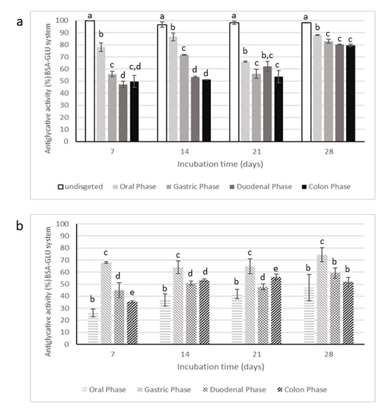
Antiglycative activity of digested samples were evaluated only in BSA-MGO and BSA-GLU
systems, considered as the most representative tests. The data are reported in Figures 9 and 10,
Antiglycative activity of digested samples were evaluated only in BSA-MGO and BSA-GLU systems,
respectively.
considered as the most representative tests. The data are reported in Figures 9 and 10, respectively.
Figure 9. Antiglycative activity of digested Moradyn CE (a) and its negative control (b) on the formation
Figure
of 9. Antiglycative
vesperlysine-like AGEsactivity of digested
in BSA-MGO assay. Moradyn CE a) and letters
Different superscript its negative
withincontrol b) on the
each monitoring
formation
time of significant
indicate vesperlysine-like AGEs
differences in0.05)
(p < BSA-MGO
among assay. Different
undigested and superscript
digested CEletters within each
or undigested CE
monitoring time indicate significant
and digested negative control. differences (p < 0.05) among undigested and digested CE or
undigested CE and digested negative control.Molecules 2020, 25, 1958
x 13 of 23
15 21
Figure 10. Antiglycative activity of digested Moradyn CE (a) and its negative control (b) on the
Figure 10. of
formation Antiglycative activity
vesperlysine-like of digested
AGEs Moradyn
in BSA-GLU assay.CE a) and superscript
Different its negativeletters
controlwithin
b) oneach
the
formation
monitoringoftime
vesperlysine-like AGEs differences
indicate significant in BSA-GLU < 0.05)Different
(p assay. superscriptand
among undigested letters withinCE
digested each
or
monitoring time
undigested CE indicate
and significant
digested differences (p < 0.05) among undigested and digested CE or
negative control.
undigested CE and digested negative control.
Digestion strongly affected the inhibitory activity of CE on AGEs formation, starting from the oral
phase; in fact, the
Digestion supernatants
strongly affectedcollected after this
the inhibitory step had
activity of CEsignificantly lower antiglycative
on AGEs formation, capacity
starting from the
than phase;
oral the undigested
in fact, theCEsupernatants
in BSA-MGOcollectedand BSA-GLU
after this systems.
step had Thesignificantly
gastric step lower
reduced about 50%
antiglycative
of the inhibitory
capacity activity after
than the undigested CE1in day of incubation
BSA-MGO in the BSA-MGO
and BSA-GLU systems. Thesystem andstep
gastric then this activity
reduced about
progressively increased
50% of the inhibitory over after
activity the monitoring time (2, in
1 day of incubation 3, the
andBSA-MGO
7 days). Finally,
systemCE andactivity
then thisappeared
activity
quite totally recovered
progressively increasedafteroverthetheduodenal
monitoring and colon
time (2, phases.
3, and 7 This
days).trend was CE
Finally, alsoactivity
reported for the
appeared
negative control, consisting of an appropriate mixture of digestive fluids
quite totally recovered after the duodenal and colon phases. This trend was also reported for and enzymes for each phase.
the
Thus,
negativeit is possible
control, to speculate
consisting of an that low AGEs
appropriate formation
mixture recorded
of digestive forand
fluids CE enzymes
after the forduodenal and
each phase.
colon phases
Thus, could be
it is possible to due to interactions
speculate that lowoccurring betweenrecorded
AGEs formation MGO andfor digestive
CE afterenzymes, leading
the duodenal to
and
non-fluorescent AGEs formations and, therefore, to false positive results.
colon phases could be due to interactions occurring between MGO and digestive enzymes, leading
In the BSA-GLU
to non-fluorescent AGEssystem, the oral
formations phase
and, did nottohighly
therefore, affect the
false positive antiglycative capacity. On the
results.
contrary,
In the theBSA-GLU
gastric phase caused
system, thea oral
reduction
phaseindid
thenot
inhibitory
highly activity
affect thedown to about 55%;
antiglycative the following
capacity. On the
intestinal the
contrary, steps only slightly
gastric affecteda the
phase caused activity.inNevertheless,
reduction the inhibitoryconsidering
activity down the inhibitory
to about 55%;capacity
the
of the negative
following control,
intestinal steps the antiglycative
only actionthe
slightly affected of CE after both
activity. gastric and
Nevertheless, intestinal the
considering phases could
inhibitory
be mainly
capacity ofassociated
the negative with interaction
control, between GLU
the antiglycative actionand ofdigestive enzymes,
CE after both gastric asand
in the MGO system.
intestinal phases
Therefore,
could be mainlyit is possible to conclude
associated that CE was
with interaction still effective
between GLU and onlydigestive
after the enzymes,
oral phase.as in the MGO
Hypoglycemic
system. Therefore, it activity
is possible of digested
to conclude CEthat
wasCE alsowastested. Inhibitory
still effective only values
after the registered
oral phase.against
α-glucosidase (24.61% ± 1.19%, 5.24% ± 2.43%, 0.97% ± 1.17 % 1.37% ± 2.38%
Hypoglycemic activity of digested CE was also tested. Inhibitory values registered against α- for oral, gastric, duodenal,
and colon phase,
glucosidase (24.61%respectively)
± 1.19%, 5.24% highly decreased
± 2.43%, 0.97%step± 1.17by% step
1.37%until the end
± 2.38% foroforal,
the gastric,
processduodenal,
when CE
had colon
and no activity.
phase,Therefore,
respectively) digestion
highlyseemed to strongly
decreased step by stepcompromise
until the CEendhypoglycemic
of the processactivity
when CE as
demonstrated
had no activity. byTherefore,
the consistent gradual
digestion decrease
seemed in the α-glucosidase
to strongly compromise inhibitory
CE hypoglycemiccapacityactivity
occurringas
during the process.
demonstrated by theThis trend could
consistent gradualnotdecrease
be attributed
in theonly to the reduction
α-glucosidase in anthocyanin
inhibitory content,
capacity occurring
but rather
during thetoprocess.
the decrease in thecould
This trend total polyphenol content.
not be attributed This
only toresult confirmed
the reduction inthat CE hypoglycemic
anthocyanin content,
but rather to the decrease in the total polyphenol content. This result confirmed that CE hypoglycemicMolecules 2020, 25, 1958 14 of 21
action should be ascribed to the synergistic action of all compounds present in the phytocomplex,
as already explained and shown in Figure 2.
3. Materials and Methods
3.1. Reagents
Ethanol, methanol, d-(+)-glucose (GLU), d-(−)-fructose (FRU), disodium hydrogen phosphate
dodecahydrate, sodium carbonate decahydrate and sodium bicarbonate were purchased from Carlo Erba
(Milano, Italy). HPLC-MS-grade formic acid and acetonitrile, hydrochloric acid (37%), Type VI-porcine
pancreatic α-amylase, α-glucosidase from Saccharomices cerevisiae, pepsin from porcine gastric
mucosa (≥400 U mg−1 ), bile extract porcine, pancreatin (8× USP) from porcine pancreas, protease
from Streptomyces griseus type XIV (≥3.5 U mg−1 ), and viscozyme L cellulolytic enzyme mixture,
p-nitrophenyl-alpha-d-glucopyranoside (purity grade ≥ 99%), starch, acarbose (purity grade ≥ 95%),
sodium potassium tartrate tetrahydrate, dinitrosalicylic acid (DNS), methylglyoxal (MGO, 40%
aqueous solution), aminoguanidine hydrochloride (AG, purity grade ≥ 98%), bovine serum albumin
(BSA, purity grade ≥ 98%), d-(−)-ribose (RIB, purity grade ≥ 98%), sodium dihydrogen phosphate
monohydrate, disodium hydrogen phosphate monohydrate, sodium hydroxide pellets, sodium
chloride, sodium azide (purity grade 99.5%), pelargonidin-3-O-glucoside, apigenin-7-O-glucoside,
and quercetin-7-O-glucoside were provided by Merck KGaA (Darmstadt, Germany).
Water was obtained from a Millipore Direct-QTM system (Merck-Millipore, Milan, Italy).
Kuromanin chloride (cyanidin-3-O-glucoside), peonidin-3-O-glucoside, kaempferol-7-O-glucoside,
and luteolin-7-O-glucoside were purchased from Extrasynthese (Genay, Rhone, France).
Sep-Pak C18 cartridges (6 mL, 1 g sorbent) were purchased from Waters Corporation
(Massachussets, MA, USA).
3.2. Moradyn Cob Extract Preparation
The Moradyn population has been developed in Italy (Lombardy region, 2015–2018) starting
from a cross hybrid morado variety (South America) by a pedigree selection of photoperiod, seed,
and cob pigmentation. This variety has been submitted to the Community Plant Variety Office for the
registration (Examination Ref. 4067062) by FlaNat Research Italia Srl. Moradyn cobs were chopped
into small pieces (about 1–2 cm) and extracted by FlaNat Research Italia S.r.l. (Milan, Italy) with
aqueous ethanol for 3 h at 50 ◦ C. The resulting suspension was filtered through 0.45 µm membrane
filters (Merck-Millipore, Milan, Italy) and after removing organic solvent under reduced pressure at
40 ◦ C (Buchi R-II, Buchi, UK), the cob extract (CE) was freeze-dried (Modulyo freeze-drier s/n 5101,
Akribis scientific limited, Cheshire, UK ) and then used in the experiments.
3.3. Anthocyanin Fraction Purification
Anthocyanin fraction (AF) isolation was performed using a Sep-Pak C18 cartridge, following the
procedure reported by Scorrano et al. [62] with some modifications. The cartridge was activated with
10 mL of methanol followed by 20 mL of acidified water (0.01% v/v HCl). Then, 5 mL of ten-fold
concentrated extract was loaded. After having removed sugars and phenolic acids with 10 mL of
acidified water and less polar compounds with 10 mL of ethyl acetate, AF was collected with 10 mL of
acidified methanol (0.01% v/v HCl). The recovered AF was evaporated to remove solvent and then
freeze-dried, obtaining a ready-to-use powder to be opportunely re-suspended for each assay.
3.4. RP-HPLC-DAD-ESI-MSn Analysis
Separation and identification of compounds present in the phytocomplex were performed using a
Thermo Finningan Surveyor Plus HPLC apparatus (Thermo Fischer Scientific, Waltham, MA, USA)
equipped with a quaternary pump, a Surveyor UV-Vis photodiode-array detector, a Surveyor Plus
autosampler, and a vacuum degasser connected to a LCQ Advantage Max ion trap spectrometerMolecules 2020, 25, 1958 15 of 21
through an ESI source. A Gemini® C18 analytical column (150 × 2.0 mm i.d., 5 µm, Phenomenex,
Torrance, CA, USA) operating at a constant flow rate of 0.3 mL/min (injection volume 20 µL) was used
for the separation. The mobile phase consisted of 0.1% formic acid aqueous solution (solvent A) and
of 0.1% formic acid in acetonitrile (solvent B) with the following gradient table: 0–3 min, 2–15% B;
3–45 min, 15–25% B; 45–48 min, 25–35% B; 48–58 min, 2% B, column reconditioning: 10 min. UV-Vis
spectral data of samples were acquired in the range 200–700 nm, and chromatograms were recorded at
280, 325, 370, and 520 nm.
The parameters of the ion mode ESI source had previously been optimized to a ionization voltage
of 5 kV, a capillary voltage of +36 V, a capillary temperature of 300 ◦ C, a sheath gas flow rate of
20 arbitrary units (AU), and an auxiliary gas flow rate of 10 AU. The Thermo Fisher Scientific Excalibur
2.2 software was used for data acquisition and processing. The ion trap operated in data-dependent,
full scan (60–2000 m/z), and MSn mode to obtain fragment ion m/z with a 35% and an isolation width of
3 m/z. Three independent assays were performed. Identification of individual phenolic compounds
was carried out by comparing their retention times, UV-Vis spectra, and MS patterns of fragmentation
with those obtained for original standards, when commercially available.
3.5. Evaluation of Hypoglycemic Activity
Both CE and AF were tested at different concentrations (ranging from 50 µg/mL to 1 mg/mL) to
evaluate their ability to inhibit α-amylase and α-glucosidase activities. CE digestion fractions were
tested only for α-amylase inhibitory activity. Data were expressed as the means ± standard deviation
(SD) of four independent experiments for each tested concentration. They were considered statistically
significant with p valuesMolecules 2020, 25, 1958 16 of 21
Samples without the enzyme solution were used as blank. A mixture containing all reactants was
used as a negative control. Acarbose solutions (0.05–1mg/mL) were used as a positive control.
The results were expressed as α-glucosidase inhibition percentage, calculated using the
following formula:
Inhibition rate (%) = [1 − (Abs 400 nm mixture containing sample – Abs 400 nm blank/Abs 400 nm
negative control)] × 100
3.6. Evaluation of Antiglycative Capacity
The antiglycative properties of both CE and its AF were evaluated by using three different in vitro
BSA-sugars systems (glucose, GLU; ribose, RIB; fructose, FRU) and a BSA-MGO system, following the
methods proposed by Mesìas et al. [11] and by Maietta et al. [63], slightly modified. In all systems,
AG was used as positive control. Conversely, digested CE supernatants were tested only in BSA-GLU
and BSA-MGO assays.
Stock solutions were prepared by dissolving BSA (35 mg/mL), GLU (175 mg/mL), RIB (150 mg/mL),
FRU (175 mg/mL), MGO (0.4 mg/mL), and AG (0.5 mg/mL) in 0.1 M phosphate buffer (pH 7.4) containing
0.02% (w/v) of sodium azide (to avoid microbial contamination). Sample solutions were prepared
by dissolving freeze-dried material in a proper volume of phosphate buffer to obtain 0.1, 0.25, and
1 mg/mL final concentrations in the reaction mixtures. Then, 400 µL of BSA solution, 800 µL of
sugar solution, and 300 µL of sample solution or phosphate buffer (blank) were mixed and the final
reaction mixture was subsequently incubated at 37 ◦ C in a thermostatted bath (Memmert basic WNB,
Schwabach, Germany).
The systems were incubated for different times depending on sugars and MGO reactivity [64],
as follows: GLU systems were incubated for 28 days, monitoring AGEs formation once a week, while
systems containing MGO and FRU for 7 and 14 days, respectively, with analytical determination at 1, 3,
7, and 14 days. Samples containing RIB were incubated for 24 h evaluating the AGEs formation at 1, 3,
6, and 24 h. At the end of each monitored time, the reaction was stopped in an ice bath (15 min) before
the analysis. Vesperlysine-like (λexc 370 nm, λem 440 nm) or Pentosidine-like (λexc 335 nm; λem 420 nm)
fluorescent AGEs (Spectrofluorometer Perkin Elmer L550B) were monitored for BSA-GLU, BSA-FRU,
and BSA-MGO systems, or for BSA-RIB, respectively. CE and AF phosphate buffer solutions were
incubated at the same reaction times to evaluate their intrinsic fluorescence.
The inhibition of AGE formation (%) was calculated using the equation below:
Inhibition % = [1 − (Fluorescence of incubation mixture containing sample – Intrinsic fluorescence
of sample/Fluorescence of incubation mixture without sample)] × 100
The reported data are the means ± standard deviation (SD) of three independent experiments for
each tested concentration, each performed in triplicate. They were considered statistically significant
with p values ofMolecules 2020, 25, 1958 17 of 21
3.8. Bioaccessibility Evaluation
The percentage of soluble polyphenols collected at the end of each digestion step represents the
CE fraction available for absorption [29,58].
To monitor the gastrointestinal effects on CE composition, the oral, gastric, duodenal, and colon
fractions were analyzed using an Agilent Technologies 1260 Infinity II technology series system
(Santa Clara, CA, USA), equipped with quaternary gradient pump, a vial sampler, a degasser,
a thermostatted column oven set at 25.0 ± 0.5 ◦ C, and a variable wavelength detector (VWD).
The HPLC-VWD system was controlled using a personal computer equipped with Agilent OpenLab
CDS ChemStation software on Windows 10. Separation was carried out on the same column and using
the same gradient elution reported for RP-HPLC-DAD-ESI-MSn analysis (Section 3.4). Chromatograms
were recorded at 520 nm and 370 nm.
Furthermore, supernatants obtained by each digestion step were also tested for their antiglycative
capacity using BSA-MGO and BSA-GLU systems and for hypoglycemic activity, assessed as
α-glucosidase inhibitory activity.
3.9. Statistical Analysis
Statistical analysis of the data and Pearson’s correlation coefficients were performed using Microsoft
Office 365. The significant differences (p < 0.05) were evaluated by variance analysis (ANOVA).
4. Conclusions
The reuse of agro-industrial byproducts represents a very important issue in the circular
bio-economy and a potential resource of new products and compounds. Moradyn is a new Italian purple
corn variety rich in polyphenols, including anthocyanin derivatives, such as cyanidin-3-O-glucoside,
perlagonidin-3-O-glucoside, and peonidin-3-O-glucoside. It was demonstrated to possess a good
capacity to inhibit α-glucosidase in an in vitro system and therefore it could act in blocking the intestinal
carbohydrate-digesting enzyme activity.
Moreover, Moradyn phytocomplex could prevent AGEs formation, acting with a strong
antiglycative agent as demonstrated in this work using in vitro model systems. The registered
healthy effects were not only due to anthocyanin fraction, but more probably to a synergistic action of
all polyphenols present in the extract. A further step consisting of the evaluation of bioaccessibility
of Moradyn CE indicated that the in vitro digestion process caused a marked decrease in the tested
bioactivities. These results highlight that such studies are mandatory to obtain preliminary information
useful for proceeding with the selection of suitable coating agents in order to preserve bioactivities.
Moreover, once the carrier has been selected, stability studies on the final ingredient to be marketed
and efficacy on glucose uptake and adipogenesis using cell-based assays will be performed.
Author Contributions: Conceptualization, A.P. and R.C.; methodology, L.F.; formal analysis, L.F. and B.M.;
investigation, A.P., R.C. and L.F.; writing—original draft preparation, A.P., R.C. and L.F.; writing—review
and editing, A.P. and R.C., supervision, A.P. All authors have read and agreed to the published version of
the manuscript.
Funding: This research received no external funding.
Conflicts of Interest: The authors declare no conflict of interest.
References
1. Lao, F.; Giusti, M. Quantification of purple corn (Zea mays L.) anthocyanins using spectrophotometric and
HPLC approaches: Method comparison and correlation. Food Anal. Met. 2016, 9, 1367–1380. [CrossRef]
2. Yonemaru, J.; Miki, K.; Choi, S.; Kiyosawa, A.; Goto, K. A genomic region harboring the Pl1 allele from
the Peruvian cultivar JC072A confers purple cob on Japanese flint corn (Zea mays L.). Breed. Sci. 2018, 68,
582–586. [CrossRef]You can also read