Roles of the fibroblast growth factor signal transduction system in tissue injury repair
←
→
Page content transcription
If your browser does not render page correctly, please read the page content below
Burns & Trauma, 2022, 10, tkac005
                                                                                                         https://doi.org/10.1093/burnst/tkac005
                                                                                                                                        Review

Review

Roles of the fibroblast growth factor signal

                                                                                                                                                                        Downloaded from https://academic.oup.com/burnstrauma/article/doi/10.1093/burnst/tkac005/6552921 by guest on 05 June 2022
transduction system in tissue injury repair
Keyang Chen 1 ,3 ,4 ,† , Zhiheng Rao1 ,3 ,4 ,† , Siyang Dong1 ,2 , Yajing Chen1 ,
Xulan Wang1 , Yongde Luo1 ,2 ,4 ,*, Fanghua Gong1 ,4 ,* and Xiaokun Li1 ,4 ,*
1
 School of Pharmaceutical Sciences, Wenzhou Medical University, Wenzhou, Zhejiang 325000, China, 2 Department
of breast surgery, The First Affiliated Hospital of Wenzhou Medical University, Wenzhou, Zhejiang 325000,
China, 3 Department of neurology, The Second Affiliated Hospital and Yuying Children’s Hospital of Wenzhou Medical
University, Wenzhou, Zhejiang 325000, China and 4 Research Units of Clinical Translation of Cell Growth Factors and
Diseases Research, Chinese Academy of Medical Science, Wenzhou Medical University, Wenzhou, Zhejiang 325000,
China
*Correspondence. Xiaokun Li, Email: xiaokunli@wzmc.edu.cn; Fanghua Gong, Email: gongwenheng@163.com; Yongde Luo,
Email: yongdeluo08@wmu.edu.cn
†
    These authors contributed equally to this work.

Received 23 July 2021; Revised 13 December 2021; Editorial decision 17 January 2022

Abstract
Following injury, tissue autonomously initiates a complex repair process, resulting in either partial
recovery or regeneration of tissue architecture and function in most organisms. Both the repair and
regeneration processes are highly coordinated by a hierarchy of interplay among signal transduc-
tion pathways initiated by different growth factors, cytokines and other signaling molecules under
normal conditions. However, under chronic traumatic or pathological conditions, the reparative
or regenerative process of most tissues in different organs can lose control to different extents,
leading to random, incomplete or even flawed cell and tissue reconstitution and thus often partial
restoration of the original structure and function, accompanied by the development of fibrosis,
scarring or even pathogenesis that could cause organ failure and death of the organism. Ample
evidence suggests that the various combinatorial fibroblast growth factor (FGF) and receptor signal
transduction systems play prominent roles in injury repair and the remodeling of adult tissues
in addition to embryonic development and regulation of metabolic homeostasis. In this review,
we attempt to provide a brief update on our current understanding of the roles, the underlying
mechanisms and clinical application of FGFs in tissue injury repair.

Key words: Tissue injury, Repair, Regeneration, Cell growth, Fibroblast growth factor, Signal transduction

Highlights
•    A total of 18 FGFs in humans activate four prototypes of membrane-spanning receptor tyrosine kinases, FGFRs.
•    FGFs play pleiotropic roles in embryonic development and adult tissue homeostasis including injury repair.
•    Aberrations in FGF signal pathways contribute to an array of diseases.
•    Agonists or antagonists of FGFs are potential agents to treat wounds and injuries.

© The Author(s) 2022. Published by Oxford University Press.
This is an Open Access article distributed under the terms of the Creative Commons Attribution-NonCommercial License (http://creativecommons.org/licenses/by-
                                                                                                                                                                    1
nc/4.0/), which permits non-commercial re-use, distribution, and reproduction in any medium, provided the original work is properly cited. For commercial re-use,
please contact journals.permissions@oup.com
2 Burns & Trauma, 2022, Vol. 10, tkac005
Background master regulators of cell growth and proliferation, organo-
genesis and tissue homeostasis represent a typical class of
In all life forms ranging from a single-cell organism to mul-
factors critical for tissue repair, remodeling and regeneration.
ticellular prokaryotic and eukaryotic species, remodeling,
In this review, we attempt to briefly update current progress
damage or injury always occur at the cellular, tissue and
in our understanding of the role, the therapeutic potential
organ levels in adults as a result of either a normal, intrin-
and the underlying mechanism of the FGF signaling system
sic biological process, a pathological insult or an external
in tissue injury repair.
traumatic incident. The impacts of the damage or injury
are immediately followed by responses at the cellular, tissue
and organismal levels, e.g. the activation and initiation of Review
Downloaded from https://academic.oup.com/burnstrauma/article/doi/10.1093/burnst/tkac005/6552921 by guest on 05 June 2022
the reparative or regenerative processes that antagonize the FGF family
progression of injury and collateral damage, preventing them The FGF family is a group of structurally conserved extra-
from developing into failure or death of the cell, tissue, organ cellular signaling molecules that range in size from 15 to
or organism [1]. It is known that although many organisms 38 kDa and act on a family of transmembrane receptor
have remarkable regenerative ability to restore the original tyrosine kinases, the FGFRs [6–8]. The human FGF family
architecture and function following injury, mammals have is known to contain 22 members, of which 18 polypeptides
rather limited ability or even lose the potential to regenerate [9] are grouped into six subfamilies based on the similarity
their tissues and the associated organs. Instead, they often of their primary sequence structure and receptor binding
adopt a complex wound healing process, resulting in only functionality (Table 1) [10]. Five of the paracrine subfami-
partial restoration to the original structure and function, and lies are the FGF1 subfamily including FGF1 and FGF2, the
more often, with the prominent formation of scar, a non- FGF4 subfamily including FGF4, FGF5 and FGF6, the FGF7
functional or partially functioning mass of fibrotic tissue that subfamily including FGF3, FGF7, FGF10 and FGF22, the
can lead to organ malfunction and even failure [2]. Hence, FGF8 subfamily including FGF8, FGF17 and FGF18, and
effective tissue repair and remodeling are critical for the the FGF9 subfamily including FGF9, FGF16 and FGF20.
survival of all living organisms [3], and practically, restor- The remaining three FGFs including FGF19 (FGF15 in mice),
ing injured tissues and organs is a long-standing aspiration FGF21 and FGF23 constitute the so-called endocrine subfam-
of all humans but a highly challenging goal for clinicians, ily [11–13]. The other four non-signaling FGF-homologous
researchers and engineers. proteins, including FGF11–FGF14 are called intracellular or
In mammals, the repair or regeneration of injured tissues intracrine FGFs, serving as co-factors for the regulation of the
and whole organs is a rather complex biological process that voltage-gated sodium channels important for neuronal and
can be roughly divided into four overlapping phases, includ- myocardial excitability [14].
ing maintenance of homeostasis, an inflammatory response, All FGFs share a core domain of ∼120 amino acids
a proliferative phase and remodeling. In the initial response, with varied homology, which folds into an interleukin 1β (IL-
clotting and isolation of the damaged region(s) occur to 1β)-like β-trefoil barrel structure in three dimensions, while
prevent worsening and to maintain overall tissue and organ both the N-terminus and C-terminus protrude from the barrel
homoeostasis. This is followed by the activation of an inflam- core, being mostly flexible [15,16]. All five subfamilies of
matory response that facilitates the clearance of necrotic autocrine and paracrine FGFs present typical surface domains
debris and prevents infection at the damage site. Then, com- that bind heparin or heparan sulfate (HS) with high yet varied
petent cells or progenitor cells within the damaged area or affinity that can be defined on the basis of the concentration
from adjacent tissues proliferate or migrate to the wound of sodium chloride used to dissociate the binding. The binding
site, giving rise to new cells, from which new tissue with to a HS chain that extends from the transmembrane core pro-
extracellular matrix that supports subsequent tissue repair teins as one type of glycosylation in the extracellular matrix
is laid down. Finally, this newly produced filling tissue is traps the HS-binding FGFs in the vicinity of the secretion cells,
altered or remodeled to resemble the original or the surround- bestowing on these FGFs HS-dependent, enhanced activities
ing, mature functional tissues. These injury-responsive and and autocrine and paracrine modes of action. In contrast, all
reparative processes are multifactorial, tissue-autonomous three endocrine FGFs lose the Arg and Lys-rich composition
and seamlessly cooperative; however, under many conditions, and surface topology compatible with a linear heparin chain
these highly coordinated processes are often interrupted, lead- for high-affinity binding as a result of lacking the β11 strand
ing to chronic wounds, malformation of non-functional tissue structure in the homologous HS-binding domain [9], which
or the development of fibrosis. Most often, an improper ensures their free circulation in blood and to distal tissues or
inflammatory response can lead to the activation of a fibrotic areas of the tissues.
response and scar formation [4]. Except for the four intracrine FGF homologs, all the
The repair and regeneration processes are controlled by autocrine/paracrine and endocrine FGFs take effect by
a variety of cytokines, growth factors, differentiation factors binding to the extracellular domains and activating the
and other molecules with distinct functions that are often in intracellular kinase domain of the transmembrane FGFR
complex association [5]. Fibroblast growth factors (FGFs) as tyrosine kinases. The HS motifs as co-factors are requiredTable 1. The FGF family, tissue expression pattern and functions
FGF subfamily Alternative name Main expression sites Function
FGF1 subfamily
FGF1 aFGF; HBGF1 Brain, pituitary, nerve tissue, retina, adrenal gland, heart Promoting mitosis, wound healing, angiogenesis, hematopoiesis, tumorigenesis and
and bone neurogenesis.
FGF2 bFGF; HBGF2 Various tissues and organs derived from mesoderm, Promoting mitosis, vascular remodeling, bone formation, pulmonary fibrosis, neural
neuroectoderm and tumor tissues development and tumor metabolism.
FGF4 subfamily
FGF4 HST1; HSTF1; K-FGF Posterior part of the limb buds Limb and internal organs development.
FGF5 – Brain Hair follicle development, a brain resident FGF for regulating neuron differentiation
and survival, regulating GFAP expression.
FGF6 HST2 Developing skeletal muscle Myogenesis and muscle regeneration.
FGF7 subfamily
FGF3 Int-2; V-Int-2 Mammary tumors Controlling the inner ear plan.
Burns & Trauma, 2022, Vol. 10, tkac005
FGF7 KGF Fetal lung mesenchymal tissue Preventing lung branch formation and lung inflammation.
FGF10 KGF-2 First observed in the limb bud Lung development, injury and repair.
FGF22 Mammalian brain, skin wound Presynaptic molecule, repairing and stimulating the formation of inhibitory
presynaptic terminal, alleviating depression and vesicle clustering, skin development
FGF8 subfamily
FGF8 AIGF; KAL6 Regulate the growth and differentiation of progenitor AIGF, establishment and maintenance of the midbrain border.
cells, produce ultimate structure of midbrain and
hindbrain
FGF17 – Cortex Similarity with FGF8, neocortex development, an autocrine growth factor in neoplastic
prostate epithelial cells.
FGF18 – Skin and cortical neurons Promoting chondrogenesis, cortical neurons and skin repair, neuroprotector.
FGF9 subfamily
FGF 9 GAF; EKS Neurons in the cortex hippocampus, thalamus, Growth-stimulating effect on glial cells, fetal lung development, enhancing the survival
cerebellum, spinal cord, epithelium and mesothelium of AChE-positive neurons.
FGF16 – Embryonic brown adipose tissue and inner ear Proliferation of embryonic brown adipose tissue, fate decisions of the otic cells.
FGF20 – Brain Enhancing the survival of midbrain dopaminergic neurons, neuro-protective in
Parkinson’s disease.
FGF15/19 subfamily
FGF15 – Absorptive cells of mouse ileum Feedback inhibition of hepatic bile acid synthesis, regulation of glucose and lipid
metabolism.
FGF19 – Absorptive cells of human ileum, can be found in the As a hormone in response to bile acid absorption acting on infarcts, regulation of
brain, skin, retina, gallbladder, small intestine, kidney and glucose and lipid metabolism, non-mitogenic effect.
umbilical cord
FGF21 – Muscle, liver, pancreas, thymus and adipose tissue Playing important role in glucose, lipid and energy metabolism, a cardiovascular
protector of the heart.
FGF23 – Bone, lung, brain, heart, muscle and spleen Regulating phosphate homeostasis in plasma by decreasing reabsorption and
increasing excretion of phosphate in the kidney.
FGF homologous family
FGF11 FHF3 Neuroblastoma, retinoblastoma and brain tumors Induced in endothelial cells by HIF1α and stimulating capillary-like endothelial tube
formation in association with angiogenesis
FGF12 FHF1 Brain, eye, heart and testis Contributing to skeletal growth and development failure of grade II and III KBD.
FGF13 FHF2 Brain and heart Neural differentiation in xenopus early development and controlling proliferation and
differentiation of skeletal muscle.
FGF14 FHF4; Sca27 Adult cerebellum Regulating intrinsic excitability of cerebellum Purkinje neurons.
HBGF heparin binding growth factor, HST heparin-binding secretory transforming, GFAP glial fibrillary acidic protein, KGF keratinocyte growth factor, AIGF androgen-induced growth factor, GAF Glia-activating
factor, EKS elbow–knee synostosis, FHF FGF homologous factor, KBD Kashin-Beck disease, HIF1a hypoxia inducible factor-1alpha, AchE acetylcholinesterase, bFGF basic fibroblast growth factor
3
Downloaded from https://academic.oup.com/burnstrauma/article/doi/10.1093/burnst/tkac005/6552921 by guest on 05 June 20224 Burns & Trauma, 2022, Vol. 10, tkac005
for autocrine and paracrine FGFs to bind with high- exists as a ‘loose’ dimer on the cell surface that is ready to
affinity to and activate FGFRs in almost all tissues, while be fired by the docking of FGF in the presence of a HS motif
transmembrane co-receptors α-klotho (KL) and β-klotho and/or co-receptor KL or KLB. It is therefore possible that
(KLB) are required for endocrine FGFs to bind to and activate other unidentified protein partners impact the interaction of
FGFRs in the endocrine and metabolic tissues. Though HS FGF–FGFR in a similar manner in specific tissues or cells,
is not required for the potentiation of FGFR activation by resulting in tissue-specific biological functions.
endocrine FGFs, it is still important for dimer formation of The conformation changes of the FGFR dimer or
FGFRs on the cell surface. oligomers induced by binding of FGF and cofactor or co-
FGFRs form a family of four highly conserved prototypic receptor are then transmitted to two intracellular kinase
transmembrane receptor tyrosine kinases (FGFR1–4). These domains, ensuring juxtaposition, relief of autoinhibition
Downloaded from https://academic.oup.com/burnstrauma/article/doi/10.1093/burnst/tkac005/6552921 by guest on 05 June 2022
FGFRs are single-pass transmembrane proteins that include and thus activation of autophosphorylation of FGFR kinase
an extracellular domain, a transmembrane domain and an domains at Tyr653 and Tyr654. Subsequent phosphorylation
intracellular tyrosine kinase domain. Three immunoglobulin- on potential tyrosine residues, including Tyr463, Tyr583,
like domains, namely D1 to D3, an acidic amino acids rich Tyr585, Tyr730 and Tyr766, leads to binding or recruitment
region between D1 and D2, a heparin-binding domain on of a number of intracellular adaptors, such as FGF receptor
D2 and an alternatively spliced IIIb or IIIc region on D3 substrate (FRS)2/3, p38, CRK, phospholipase C γ (PLCγ )
comprise the extracellular domain [17]. There are reportedly and signal transducers and activators of transcription
other atypical FGFRs, such as the so-called FGFR5 (also (STATs), which then serve as diversifying signaling hubs
called FGFRL1) that lacks the intracellular kinase domain that typically activate the SOS–Ras/Raf–MAPK–mTOR,
[18]. Alternative splicing generates different isotypes for each GAB1–PI3K–AKT, DAG/IP3-Ca2+ and nuclear STAT signal
prototype of FGFRs, notably the IIIb and IIIc isotypes that pathways [10] with differential cellular growth, survival and
have distinct ligand-binding specificity [19]. metabolic effects, in a spatiotemporal manner and depending
Different FGFs, FGFR isotypes, co-factors and co- on the nature of the tissues and associated organs involved.
receptors are expressed in a more or less tissue-specific FRS2 is a known critical proximal adaptor recruited to
manner; however, together they are present in nearly all phospho-Tyr463 upon FGFR activation, which leads to the
tissues and play a myriad of important roles in embryonic activation of MAPK and AKT pathways that are critical for
development, organogenesis, adult tissue remodeling, injury cell growth, survival and tissue repair [33]. It is also required
and regenerative responses, and metabolic homeostasis [20]. as the downstream products of FGF19-induced FGFR4-KLB
In the adult, both the metabolic and growth-promoting FGFs activation to regulate bile acid synthesis [34]. Whether FRS2
play critical roles in the response to tissue injury, damage and homologs serve the downstream of the activated FGFR1–
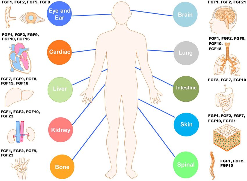
repair and tissue-specific pathologies (Figure 1) [21–23]. FGF KLB and FGFR1–KL by FGF21 and FGF23 that regulate
signaling was shown to elicit cardioprotective effects on the the homeostasis of energy and mineral metabolism, respec-
heart [24, 25] and to be important for epithelial repair in tively, is an interesting subject for future investigation. It is
the lung [26, 27] and wound healing on skin [28]. FGFs are also possible that the specific cellular milieu in metabolic
involved in regulating cerebral injury through promoting tissues, such as white and brown adipose tissues, that contains
neuronal regeneration, neuroprotection and angiogenesis intracellular adaptors different from FRS2 in non-metabolic
[29]. tissues, is important for mediating the effects of FGFR1–KLB
and FGFR1–KL signal pathways. Despite such a distinction,
both the growth-promoting and metabolic pathways initiated
FGF–FGFR signal transduction by FGFRs are important for cell survival and homeostasis and
Like many other types of growth factors, the binding of are a prerequisite for injury repair.
FGF to the ectodomain of FGFR causes dimerization or a
higher-order of oligomerization of FGFRs, followed by con-
formational changes. The binding of the autocrine/paracrine The role of FGF signaling in skeleton
FGF1–10, FGF16–18, FGF20 or FGF22 to the FGFR and muscle repair
ectodomain on the cell surface is dependent on the presence of Skeleton Certain members of FGFs and FGFRs are expressed
cofactor HS chain that extends from the core of a transmem- in characteristic spatiotemporal patterns throughout all
brane glycoprotein, such as glypican or syndecan, resulting in stages of skeletal and muscle development. The FGF signal
the formation of a stable 2:2:2 FGFR–HS–FGFR ternary pathways regulate the development of limb bud and mes-
complex [30, 31]. By contrast, the initial formation of a enchymal condensation, thus playing key roles in chondroge-
stable endocrine FGF–FGFR complex (e.g. 2:2:2 FGF23–KL– nesis, osteogenesis, bone formation and mineral homeostasis
FGFR1) depends on the presence of single transmembrane [35]. Both loss-of-function and gain-of-function mutations in
co-receptor alpha KL or KLB, while the HS chain is only FGFs and FGFRs are associated with dozens of congenital
required for receptor dimerization but not ligand–receptor bone diseases that are broadly classified into chondrodyspla-
interaction [32], resulting in a stable 2:2:2:2 FGF23–KL– sia syndromes and craniosynostosis syndromes. Consistently,
FGFR–HS quaternary complex. It was postulated that FGFR growingBurns & Trauma, 2022, Vol. 10, tkac005 5
Downloaded from https://academic.oup.com/burnstrauma/article/doi/10.1093/burnst/tkac005/6552921 by guest on 05 June 2022
Figure 1. Summary of the known main FGF–FGFR signaling systems in the injury repair of diverse tissues or organs. FGFs and FGFRs participate in the cellular
and metabolic homeostasis of all tissues and associated organs such as the nervous system, lung, heart and cardiovasculature, skeleton, muscle, skin, ear and
eye, to name but a few, and are critical for their remodeling, regeneration and repair of injuries resulting from diverse types of traumatic and pathological insults.
FGF fibroblast growth factor, FGFR fibroblast growth factor receptor
evidence supports important roles of FGFs and FGFRs in the bone repair by inhibiting adipogenic differentiation and
repair of injured or malfunctioning skeleton. As a part of the increasing the number of osteoblasts [42]. A low molecular
skeleton, cartilage and growth plate are types of connective weight isoform of FGF2 promoted bone fracture healing [43].
tissue and are prone to injury [36]. One study found that Local delivery of FGF7 induced bone formation by enhancing
growth-arrest-specific5 (Gas5) regulates the proliferation and osteogenesis and chemoattraction in a rat model of mandible
apoptosis of growth plate by controlling FGF1 expression defects [44]. A novel therapeutic fiber scaffold containing
[37]. Osteochondral defects can potentially progress to FGF2 and FGF18 promoted the repair and regeneration
osteoarthritis, and a recent study showed that FGF2 delivered of calvarium defects [45]. FGF8 functions as a negative
by recombinant adenoviral vector enhances osteochondral regulator of osteogenic fate and was shown to be sufficient
repair [38]. Saw et al. [39] showed that metalloprotease to convert a subset of cranial neural crest cell-derived
regulation of FGF2 is essential in the chondrocyte maturation mesenchymal cells into cartilage in the anterior hard palate
program by promoting growth plate development and [46]. FGF9 from mature osteoblasts was shown to regulate
bone elongation. FGF2 combined with low-intensity pulsed skeletal homeostasis in male mice [47]. Administration of
ultrasound could promote the synthesis and secretion of exogenous FGF9 halted cartilage degradation while aggra-
collagen and thus the differentiation and maturation of vating osteophyte formation in post-traumatic osteoarthritis
chondrocytes [40]. FGF9 promotes chondrocyte hypertrophy [48]. FGF21 acts as a negative regulator of bone density
in the early stage and regulates blood vessels and osteogenesis by enhancing peroxisome proliferator-activated receptor
of growth plate in the late stage of bone development [41]. γ (PPARγ ) activity [49]. FGF23 contributed to wingless-
FGFs play important roles in bone regeneration during integration (Wnt)/β-catenin signaling-mediated osteoarthritis
the fracture healing process. FGF1 was shown to promote in mice [50] and promoted the differentiation of osteoarthritic6 Burns & Trauma, 2022, Vol. 10, tkac005
chondrocytes [51]. Patients with X-linked hypophosphatemic the enlargement of muscle fiber size and protecting muscle
rickets exhibit skeletal or bone deformities including short from atrophy through activation of ERK1/2 and the riboso-
stature, leg deformities, bone pain, dental abscesses and mal protein S6 kinase [69].
radiographic evidence for rickets and osteomalacia, as a Although significant progress has been made in the past
result of elevated FGF23 signaling. Burosumab, a humanized in our understanding of the roles of FGFs and FGFRs in
monoclonal antibody against FGF23, significantly increased the repair and healing of skeletal and muscle system injury
the maximum renal tubular threshold for phosphate and diseases, the precise roles of individual FGFs and FGFRs
reabsorption, serum phosphate and 1,25(OH)2 D with a at different stages and sites of injury, diseases and aging-
favorable safety profile [52]. associated wasting remain to be dissected in detail. Tar-
FGFR1, 2 and 3 were shown to be involved in the geting the FGF system represents a promising avenue for
Downloaded from https://academic.oup.com/burnstrauma/article/doi/10.1093/burnst/tkac005/6552921 by guest on 05 June 2022
FGF-initiated regulation of cartilage and bone formation. treating bone and muscle injury and aging-associated muscle
Although there are some discrepancies, it is generally believed wasting; however, the application dose, timing and dura-
that FGFR3 inhibited the proliferation and differentiation tion of FGFs, the delivery system and the possible combina-
of chondrocytes while promoting the apoptosis of cartilage tion with other modulating signaling molecules need to be
cells. Both FGFR1 and FGFR2 were shown to promote the optimized.
proliferation and differentiation of osteoblasts. FGFR1 gene
polymorphism is associated with fracture non-unions [53],
while FGFR2 polymorphisms are associated with osteogenic Roles of FGF in nerve injury and repair
differentiation [54]. Upon bone marrow ablation, an FGFs play important roles in the development of the nervous
inducible expression of the gain-of-function mutant FGFR2- system by promoting the growth, proliferation, differentia-
P253R at the adult stage resulted in anabolic effects on tion, migration and survival of both neurons and non-neural
trabecular bone via promoting bone formation and inhibiting cells, such as astrocytes, microglia and oligodendrocytes, as
bone resorption in a Wnt/β-catenin-dependent manner [55]. well as in repair, regeneration, demyelination, remyelina-
FGFR3 inhibited the formation of callus and delayed the tion and angiogenesis after damage or injury in the nervous
repair of bone injury by negatively regulating endochondral system.
osteogenesis [56, 57]. Deletion of FGFR3 in osteoclast cell
lineage led to bone mass increase by inhibiting osteoclast
bone resorption in mice [58]. In an osteoarthritis model, a Roles of FGFs in the repair of nerve injury after stroke
competitive FGFR1 inhibitor protected articular cartilage Stroke is an acute cerebrovascular disease attributable to
[59]. By contrast, FGFR3 delayed osteoarthritis progression blockade or sudden rupture of blood vessels in the brain
in mouse knee joints at least in part by down-regulating that prevents blood from effectively flowing into the brain
Indian hedgehog signaling in articular chondrocytes [60, or the nervous tissues [70], leading to reduced availability
61]. FGFR3 deficiency accelerated CXCL12-dependent or loss of supply of nutrients and oxygen and thus death
macrophage chemotaxis, leading to exacerbation of joint of brain cells via necrosis and apoptosis [71]. Studies
destruction while CXCR7 inhibition reversed the damage showed that FGF1 could protect the blood–brain barrier
effect [62]. Taken together, the above studies suggest that (BBB) from dysfunction by upregulating tight junction
FGFR1–2 can exert a deleterious effect on osteoarthritis proteins and inhibiting RhoA through the PI3K–AKT–RAC1
development under certain conditions whereas FGFR3 plays pathway [72]. Intranasal FGF1 administration enhanced
a protective role. angiogenesis via the sphingosine-1-phosphate receptor 1
signaling pathway [73]. FGF2 was found to upregulate
Muscle Adult skeletal muscle retains a remarkable ability platelet-derived growth factor receptor β in cultured pericytes
to rapidly repair the damage caused by exercise, trauma, and in peri-infarct areas in a mouse stroke model [74]
toxins and diseases [63], in which the satellite cells (SCs) and to contribute to the effects of salidroside on dendritic
that are considered the stem cells contribute the most [64]. and synaptic plasticity after cerebral ischemia/reperfusion
FGFs are important mitogens for the self-renewal of SCs (I/R) injury [75]. Intranasal administration of FGF2 in
and thus the repair and regeneration of muscle after injury nanoliposomes designed to bypass the BBB was used for
or upon aging. Satellite cells express FGFR1 and FGFR4 treatment of ischemic stroke injury [76]. Endocrine FGF21 is
at high levels and FGFR3 at low levels, but not FGFR2. known to have no retention in the extracellular matrix and
Studies have demonstrated that FGF1, FGF2, FGF4 and FGF6 potentially a better ability to cross the BBB. Administration
regulate the growth, survival and renewal of SCs by activating of FGF21 alleviated middle cerebral artery occlusion-induced
ERK1/2 and p38α/β MAPKs, PI3 kinase, PLCγ and STATs brain injury via activation of the PI3K/AKT pathway
[65]. FGF21 was found to control muscle mass [66] and [77], protected against Ang II-induced cerebrovascular
alleviate glucocorticoid-induced injury through inhibition of aging and I/R-mediated hippocampal injury [78, 79], and
myostatin expression [67]. Excessive FGF2 removed age- reduced cerebral injury via decreasing endoplasmic reticulum
associated proliferative inhibition of SCs [68]. FGF19 was stress [80]. Under hypoxia conditions, FGF21 protected
also reported to control skeletal muscle mass by stimulaing against injury to cerebral microvascular endothelial cellsBurns & Trauma, 2022, Vol. 10, tkac005 7
and [81] alleviated motor nerve dysfunction by modulating The roles of FGFs in the repair of other types
microglia/macrophage-mediated neuroinflammation [82]. of neural injury Traumatic brain injury (TBI) is a form
Taken together, the potent neurotropic and angiogenic of acquired brain injury occurring as a result of sudden
activities suggested that FGFs are promising therapeutic physical or traumatic damage, resulting in abnormal brain
agents for ischemia stroke. One of the important directions function such as short-term or long-term sensory and motor
of future research is to explore the roles of FGFs and FGFRs deficits [93]. Wang et al. showed that FGF2 enhanced
in different stages of stroke pathogenesis. The safety, efficacy cell proliferation and neuronal survival and protected the
and dose-dependent response of administered FGFs in stroke BBB from breakdown by activating the PI3K/AKT/RAC1
animals and patients also require careful examination. signaling pathway, promoting the expression of tight junction
proteins such as claudin-5, occludin and zonula occludens-1
Downloaded from https://academic.oup.com/burnstrauma/article/doi/10.1093/burnst/tkac005/6552921 by guest on 05 June 2022
FGFs in spinal cord injury and repair Spinal cord injury following TBI [94]. It protected against 1-methyl-4-pheynl-
(SCI) is the physical and psychological damage to any part 1,2,3,6-tetrahydropyridine hydrochloride-induced onset of
of the spinal cord or nerves that change the bodily functions Parkinson’s Disease (PD), preventing dopaminergic neuron
primarily below the site of injury, with many neurological loss by activating the AMPK–PGC1α axis to promote
complications including paraplegia or quadriplegia [83]. The mitochondrial function and reduce inflammation in mouse
pathological process of SCI is a combination of primary brains [95]. Furthermore, Yoshimura et al. [96] suggested
trauma and sequential secondary injuries [83]. Target ther- that FGF-2 could upregulate neurogenesis and protected
apies for improving the clinical outcome of SCI include neurons against degeneration in the adult hippocampus
limiting inflammation, preventing secondary cell death and after TBI. GF21 is an endocrine hormone with effects of
enhancing the recovery, regeneration and plasticity of neu- anti-inflammation, anti-oxidative stress and anti-ER stress,
ronal circuits [84]. A number of studies revealed that FGFs promoting metabolic homeostasis. Activation of the FGFR1–
target the neuropathological cascades associated with sec- KLB signal pathway by FGF21 was shown to preserve BBB
ondary injurious events following SCI [85, 86]. Wang et al. integrity by upregulating PPARγ and increasing proteins
[87] revealed that FGF1 improved the functional recovery of in tight junctions and adhesion junctions, accompanied by
SCI by inducing PRDX1 to modulate autophagy and reduce marked reductions in neurofunctional behavior deficits,
reactive oxygen species in a rat model. Application of novel degree of cerebral edema, brain tissue loss and neuron
FGF1-loaded thermosensitive heparin-poloxamer hydrogel apoptosis in a mouse model of TBI [97]. In an Alzheimer’s
protected spinal cord neuronal and peripheral cells from dete- disease model, administration of FGF21 alleviated mem-
rioration and promoted regeneration upon SCI. A novel scar- ory dysfunction, amyloid plaque pathogenesis and tau
homing delivery system for FGF1 improved neuronal survival hyperphosphorylation in part by modulating the astrocyte–
and plasticity and promoted axon regeneration following SCI neuron lactate shuttle via monocarboxylate transporters
[88]. FGF2 improved the recovery of the blood–spinal cord and correcting brain metabolic defects [98, 99]. FGF20 is
barrier after SCI by increasing junction proteins and Cav-1, highly expressed in the substantia nigra pars compacta of the
inhibiting the expression and activation of MMP-9 involved central nervous system. In a 6-hydroxydopamine-lesioned
in the interaction with FGFR1 [89] and inhibiting ER stress- rat model of PD, administration of an FGF20 variant with
induced cell death [90]. The intracrine FGF13 was shown to enhanced permeability across the BBB prevented the loss of
stabilize microtubules and enhance mitochondrial functions, dopaminergic neurons in the substantia nigra pars compacta
promoting neuronal polarization, axon formation, growth [100]. Increases in oxidative stress contribute to Huntington’s
cone initiation and function recovery following SCI [89, 91]. disease, another neurodegenerative disorder in the brain.
The expression levels of FGF10 in neuron and microglia/- FGF9 was shown to upregulate and activate the ERK–
macrophages increased post SCI, and treatment with FGF10 NRF2 pathway and the downstream glutathione synthesis
inhibited microglia/macrophages activation and proliferation and antioxidant system, attentuating oxidative stress damage
and reduced inflammatory damage via the FGFR2/PI3K/AKT and neuron cell death [101, 102].
and TLR4/NFκB pathways, promoting the recovery process Peripheral nerves relay signals from the brain and spinal
in SCI in animals [92]. cord to the rest of the body. Peripheral nerve injury or
Overall, the recovery of SCI is a complex process as malfunction as a result of a traffic accident, trauma or tumor
it interferes with a range of normal motor, sensory and resection can give rise to the loss of sensory and motor
autonomic functions. The mechanisms underlying patholog- functions, chronic pain and other activity deficits. Although
ical processes of secondary injury upon SCI remain largely surgical techniques are a traditional restoration approach
unclear. Although certain members of the FGF family are [103], exogenous supplement of neurotrophic factors has
present in spinal cord neurons, peripheral cells and canal increasingly become an important strategy for the treatment
structure, how they promote the repair of damaged neurons and recovery of peripheral nerve injury. Heparin-based coac-
and the ligation and regeneration of new axons has yet to ervate or hydrogel delivery of FGF2 facilitated nerve regen-
be determined. Furthermore, clinical evidence for the effi- eration by inhibiting ER stress, accelerating remyelination
cacy of FGF-based agents among patients with SCI is still and axon fiber regeneration, and promoting Schwann cells
lacking. proliferation and the recovery of motor function in models8 Burns & Trauma, 2022, Vol. 10, tkac005
of sciatic nerve crush injury with diabetic neuropathy, mental integrity after bleomycin-induced lung injury in mice [127].
nerve crush injury or digital nerve severing injury [104–107]. FGF2 reduced oxidative stress, inflammation and apoptosis
FGF5 was shown to be an autocrine regulator of Schwann of alveolar epithelial cells and prevented pulmonary capillary
cells and FGF5 administration rapidly promoted Schwann leakage, alleviating acute lung injury [128, 129]. FGF9 is
cell migration and adhesion via upregulation of N-cadherin an antiapoptotic and promigratory factor, maintaining lung
following distal sciatic nerve injury [108]. fibroblasts in an undifferentiated state via activating the
Neonatal hypoxia–ischemia encephalopathy, the most FGFR3 signaling pathway. Both FGF9 and FGF18 are medi-
important cause of morbidity, mortality and neurological ators of epithelial–mesenchymal interactions critical for lung
deficits in term-born infants, is a type of brain damage that development, and promote the survival and migration of lung
occurs often with insufficient reception of oxygen and blood. epithelial cells while inhibiting myofibroblast differentiation
Downloaded from https://academic.oup.com/burnstrauma/article/doi/10.1093/burnst/tkac005/6552921 by guest on 05 June 2022
A study showed that FGF2 gene expression was upregulated in IPF [130].
in the hippocampus of neonatal rats, and intraperitoneal
injection of exogenous FGF2 enhanced cell proliferation in
the hippocampal dentate gyrus region following neonatal FGFs in cardio-vasculature injury repair
hypoxia–ischemia brain damage [109]. A combination of FGF members and associated FGFRs play important roles
neural stem cells and overexpression of FGF2 reduced brain in cardiovascular and lymphatic development, homeostasis
damage and restored sensorimotor function following such and diseases. In heart development, the roles of FGFs range
brain damage [110]. Similarly, a combination of FGF2 with from the formation of outflow tracts to the proliferation of
pluripotent astrocytic stem cells improved cognitive function cardiomyocytes and the formation of heart chambers. FGF8,
in neonatal rats with hypoxic–ischemic brain injury [111]. FGF9, FGF10 and FGF16 were shown to act as paracrine
signals during embryonic heart development, while FGFs 1,
2, 9, 16, 19 and 21 mediate adaptive responses to cardiac
The role of FGFs in lung injury repair regeneration, including restoration of cardiac contraction
FGFs and FGFRs play important roles in lung development, rate after myocardial infarction and reduction of the extent
and aberrant FGF signaling has been implicated in the patho- of myocardial infarcts. Even though FGF15/19, FGF21 and
genesis of pulmonary fibrosis and lung diseases [112]. FGFR3 FGF23 are typical endocrine FGFs, they can function as
and FGFR4 function cooperatively to direct alveogenesis paracrine signals in cardiovascular development or patho-
of mouse lung [113]. FGF10 is considered the main mor- physiology. Note that, although the expression and activation
phogen driving multi-stage lung branching morphogenesis in of FGFs and associated signaling pathways are important for
rodents. It regulates the mobilization and differentiation of cardiovascular repair, they may also contribute to fibrosis,
mesenchymal stem cells and the homeostasis of intrinsic cells remodeling and dysfunction [131]. In heart diseases, serum
of lung structure [114, 115] and plays important roles in lung levels of FGF15/19, FGF21 or FGF23 were shown to decrease
injury repair, while its signaling defects lead to neonatal lung or increase, indicating variable roles of these factors in heart
diseases [116–118]. FGF10 mutations increase the risk of pathophysiology.
chronic airway disease in adulthood [119]. Following injury, Injection of FGF1 coacervate was sufficient to reduce
FGF10 functions to maintain progenitor cell populations the injury and pathologies caused by myocardial infarction
in the airway and promotes alveolar type 2 cell expansion [132]. FGF1 loaded in poly-(lactic-co-glycolic acid) and
and differentiation. Overexpression of FGF10 in bronchial polyethylene glycol microparticles promoted heart regenera-
epithelial stem cells enhanced fibrosis resolution after lung tion in a rat model [133]. A combination of FGF1 and Wnt1
damage [120–122] and promoted the proliferation and trans- agonist/GSK3β antagonist CHIR resulted in substantial
differentiation of lung stem cells, accelerating lung repair reduction in infarct size and improved left ventricular
[123]. chamber function [134–136]. Similarly, FGF2 was shown
Idiopathic pulmonary fibrosis (IPF) is characterized by to be a cardiovascular protector in myocardial infarction
an accumulation of extracellular matrix proteins and fibrob- and I/R injury, by reducing oxidative stress via activating
lasts in the distal airways. In IPF pathogenesis, FGF1 is NRF2-mediated antioxidant defense in conjunction with
upregulated 7.5-fold more than in the normal lung [124]. AKT–GSK3β–FYN pathway activation [137] or by inhibiting
FGF1 counteracted IPF pathogenesis by inhibiting fibroblast apoptosis and promoting angiogenesis via a HIF1α-mediated
collagen production and differentiation into myofibroblasts mechanism [24, 138]. Administration of FGF2 promoted
and reverting epithelial–mesenchymal transition via suppress- angiogenesis and attenuated cardiac remodeling in ischemic
ing TGF-β1 signaling pathways to induce alveolar epithelial heart disease [139–141] or in a rat ischemic cardiomyopathy
cell proliferation [125]. Similarly, FGF2 was shown to be model with surgical ventricular restoration [142]. FGF9 was
antifibrotic in the lung by decreasing collagen deposition shown to inhibit vascular cell apoptosis, activate c-Kit+
and fibroblast to myofibroblast differentiation [126], thus progenitor cells and enhance angiogenesis and neovascular-
exerting a protective or reparative effect following lung injury. ization, improving cardiac function [143]. FGF9 treatment
Endogenous FGF2 was not required for bleomycin-induced of diabetic mice with infarcted myocardium increased
pulmonary fibrosis, but was essential for epithelial repair and anti-inflammatory cytokines and M2 macrophage differ-Burns & Trauma, 2022, Vol. 10, tkac005 9
entiation, resulting in reduced adverse cardiac remodel- reconstitution and stem cell maintenance in the gastrointesti-
ing [144]. Similarly, administration of FGF16 or FGF10 nal tract. FGFR1 and FGFR2 are expressed in the human
coacervate reduced infarct size, interstitial fibrosis, myocar- ileum and throughout adult mouse intestine [158]. FGFR3
dial monocyte infiltration and damage to cell popula- is expressed in the lower half of the intestinal crypts while
tions [145, 146], preventing myocardial infarction-induced FGFR4 is restricted to the epithelium of the embryonic gut
injury. [159]. FGF1, FGF7, FGF8, FGF9, FGF10, FGF15/19 and
A recent study showed that FGFR signaling is a critical FGF18 are reportedly expressed in the intestine in a spa-
regulator of vascular development, which is achieved by tiotemporal manner [158, 160].
FGF-dependent control of c-MYC expression that, in turn, In experimental models of intestinal I/R injury, both FGF1
regulates expression of the glycolytic enzyme hexokinase and FGF2 were shown to be protective [161, 162]. FGF2
Downloaded from https://academic.oup.com/burnstrauma/article/doi/10.1093/burnst/tkac005/6552921 by guest on 05 June 2022
2. FGFR1 and FGFR3 double-mutant mice exhibited improved healing of colonic anastomoses through activating
blood and/or lymphatic vascular defects, while hexokinase2 fibroblasts, collagen deposition and angiogenesis in rats [163]
overexpression partly rescued such defects [147]. Mice or cooperated with IL-17 to repair damaged epithelium in
with endothelial cell-specific double knockout of FGFR1 intestine [164]. FGF7 also promoted healing of colonic anas-
and FGFR2 showed significantly decreased vessel density, tomoses by increasing cell proliferation and mucus produc-
increased endothelial cell apoptosis and worsened tissue tion and reducing inflammation [165]. Similarly, FGF7 atten-
hypoxia in the peri-infarct areas following reperfusion, uated I/R and radiation-induced injuries by reducing intesti-
demonstrating an essential role of endothelial FGFR1 nal epithelial cell apoptosis and the disruption of tight junc-
and FGFR2 in cardiac functional recovery and vascular tions via an AhR–E2F1–FGFR2IIIb signaling pathway [166,
remodeling during cardiac injury [25]. 167]. FGF7 and FGF10 promoted the repair of the resected
small bowel via activating intestinal epithelial FGFR2IIIb
[168, 169]. FGF2 and IL-17 in synergy promoted the repair of
FGFs in kidney injury repair the damaged intestinal epithelium through GRB2-inhibiting
FGFs and FGFRs play important roles in kidney development Act1-mediated signal cross-talk [164]. In tissue reconstitu-
and defects of the FGF signal pathways contribute to renal tion, patterning of the endoderm could be accomplished by
pathologies. Evidence has shown that many FGF members, the combined activities of Wnt, Bone morphogenetic protein
particularly those signaling through FGFR1 and FGFR2, and FGF. Palifermin, a truncated from of recombinant FGF7,
such as FGF1, FGF2, FGF7 and FGF10, are mitogenic has been clinically used to treat oral mucositis resulting from
and antiapoptotic for various kidney cell types, such as radio- or chemo-therapy [170]. Taken together, current stud-
collective, tubular and glomerular cells, promoting the ies revealed important roles of FGFs in intestinal development
survival and outgrowth of the associated renal tissues [148, and adult tissue injury repair.
149]. FGF-stimulated FGFR2 signaling played important
roles in protecting against tubular cell death and acute
kidney injury through ERK1/2 activation [150]. FGF1 Advances in the roles of FGFs in liver repair
was reported to suppress oxidative stress, inflammation The liver is a vital organ and the hub of multiple bio-
and diabetic nephropathy via activating the PI3K/AKT- logical processes including the various forms of nutrition
mediated pathway [151]. FGF2 is abundant in tissues such handling and metabolism, endocrine and immune regulation
as brain, kidney and cartilage. It was shown to protect and detoxification. It has a unique capacity for regeneration
against renal I/R injury by inhibiting the High-mobility group and injury repair. The liver tissue is a mass of cells tun-
box 1-mediated inflammatory response and attenuating neled through with bile ducts and blood vessels, with the
mitochondrial damage [152]. FGF7 was shown to modulate parenchymal hepatocytes making up ∼60% of the liver and
ureteric bud growth and nephron number in the developing performing more metabolic functions than any other group
kidney and contribute to tubular cell growth and repair upon of cells in any other organ. By contrast, the non-parenchymal
kidney damage [23, 153]. FGF10 treatment improved renal cells, including sinusoidal endothelial cells, Kupffer cells and
function and histological integrity and suppressed excessive stellate cells, comprise the rest of the liver tissue to assist
autophagy and ER stress in models of renal I/R injury [154, the metabolic functions. Several FGFs and FGFRs have been
155]. FGF23 levels were reported to be higher upon acute shown to play important roles in liver development, health
kidney injury than in normal situations [156], due in part to and disease. FGF8 and FGF10 as morphogens contribute sig-
the increased production of FGF23 in osteoblasts. Elevated nificantly to embryonic liver development [171, 172]. FGF7
serum FGF23 levels are both an indicator and a mediator of produced in Thy1(+) mesenchymal cells in close proximity
poor outcome in chronic kidney disease [157]. to liver progenitor cells is a critical regulator of PLCs in
response to liver injury [173]. Similarly, FGF9 is also a liver
repair factor, providing a paracrine mitogenic signal from
Roles of FGFs in intestinal injury and repair stellate cells to hepatocytes during acute liver injury [174].
All four FGF receptors and several FGF ligands are implicated FGF5 knockout mice fed a high-fat diet had higher levels of
in controlling cell proliferation, differentiation, epithelial cell serum alanine transaminase and aspartate amino transferase10 Burns & Trauma, 2022, Vol. 10, tkac005
with nonalcoholic steatohepatitis (NASH)-like pathologies, in a double-paracrine manner [198]. A lack of FGF7 could
including marked inflammation, focal necrosis, fat deposition further delay cutaneous wound healing in diabetic mice. In
and fibrosis [175]. diabetic rats, FGF10 enhanced wound repair of scalded skin
FGFR3 and FGFR4 are the main FGFRs expressed in the together with FGF21 [199]. With novel delivery strategies
liver and are involved in the development of hepatocellular that improve skin penetration, FGF10 was shown to inhibit
carcinoma (HCC) [176, 177]. Ectopically gained FGFR1 and ER stress and promote keratinocyte proliferation, accelerat-
FGFR2 in hepatocytes have also been shown to play roles ing wound healing and hair growth [200, 201]. The approval
in HCC development [178]. FGF5, FGF8, FGF9, FGF17 of parlifermin for accelerating the healing of severe oral
and FGF18 act as paracrine signals while FGF19 acts as mucositis resulting from cancer chemoradiotherapy attests to
an endocrine signal in HCC development [179–182]. The the role and efficacy of FGF7 in the repair and regeneration
Downloaded from https://academic.oup.com/burnstrauma/article/doi/10.1093/burnst/tkac005/6552921 by guest on 05 June 2022
endocrine FGF19 is produced in the ileum but acts as a nega- of wounded skin or mucus [170, 202].
tive regulator of hepatic bile acid metabolism and a stimulator FGF2 treatment promoted epithelium–mesenchyme tran-
of gallbladder filling [183]. It also functions as a postprandial, sition in skin wounds, accelerating wound closure [203],
insulin-independent activator of hepatic protein and glycogen possibly through a feedback regulatory loop involving the
synthesis [184]. Mouse FGF15 was shown to protect against Wnt/β-catenin signal pathway [204] or NFκB/JNKs pathway,
fibrosis through increased bile acid activation of farnesoid X independent of the PI3K/JNKs pathway, in fibroblasts and
receptor in hepatic stellate cells [185]. FGF21 is a hepatocyte blood vessel endothelial cells [205]. In addition to metabolic
secreted stress-responsive hormone and regulates glucose and correction, FGF21 encapsulated in a thermosensitive hep-
lipid metabolism by targeting white adipose tissue [186– arin–poloxamer hydrogel accelerated wound healing in dia-
188]. Serum FGF21 levels were elevated in non-alcoholic betic animals [206]. FGFs were also tested for the repair
fatty liver, and pharmacological FGF21 protected against and remodeling of dermis as a potential anti-aging cosmetic
non-alcoholic fatty liver diseases including hepatosteatosis utility. Recombinant FGF1 strongly stimulated fibroblast and
and NASH [189]. Taken together, current findings reveal keratinocyte proliferation, suggesting a high potential for
important roles of different composite members of the FGF repairing skin conditions [207]. It increased type 1 procol-
signal transduction system in liver tissue homeostasis, func- lagen synthesis and reduced the generation of reactive oxy-
tional performance, regeneration and injury repair, aiding in gen species, protecting ultraviolet B ray (UVB)-induced skin
the potential design of novel therapeutic strategies for liver damage and photoageing [208]. Similarly, FGF2 contained in
function recovery upon injury and in disease. dalteparin and protamine nanoparticles inhibited ultraviolet
B ray irradiation-induced apoptosis of dermal fibroblasts and
epidermal keratinocytes and alleviated the decline of elasticity
FGF signaling in skin repair and acanthosis [209]. A combination of platelet-rich plasma
The skin as the largest superficial organ of our body consists and FGF2 was effective in treating wrinkles and the depressed
of two main sections: the epidermis made of keratinocytes areas of the skin [210].
and epithelial cells and the dermis made of dense, irregu- In summary, current studies show the potential of FGFs in
lar connective tissue housing blood vessels, fibroblasts, hair promoting the repair of skin from damage or injury of varied
follicles, sweat glands and other structures. The hypodermis etiologies. Future studies should focus on improving wound-
beneath the dermis is mainly composed of loose connective healing efficacy while reducing the risk of scar formation and
and fat tissues. Upon traumatic injury, the skin as the first side effects, improving formulation and application conve-
and foremost outside defense system to any injury sets into nience, and lowering treatment cost when used for cosmetic
motion an autonomous cascade of complex healing events purposes.
that can be roughly divided into four overlapping phases,
including hemostasis, inflammatory reactions, cellular pro-
liferation and tissue remodeling, resembling that of many FGFs in eye and ear damage repair
other tissues [190]. Among many important factors, mem- FGF signaling is critically required during several steps of
bers of the FGF family play diverse roles in these highly vertebrate lens and optic nerve development, including induc-
orchestrated biological processes [191–193]. FGF7 and its tion of the lens vesicle, proliferation of lens epithelial cells,
homologue FGF10 are known to be expressed in the mes- differentiation of lens fiber cells and elongation of ganglion
enchymal fibroblasts in the dermis or hypodermis but act nerve axon [211]. Genetic deficiencies of FGF receptors dis-
specifically on various types of epithelial cells including ker- rupted the expression of lens-specific genes Cdh1, Crystallins,
atinocytes of the skin by activating the resident FGFR2IIIb Maf , Pax6 and Prox1, affecting the survival and proliferation
[194–196]. Both FGF7 and FGF10 are effective for promoting of lens epithelial cells and elongation of fiber cells [212,
wound healing, wound closure and better scar formation on 213]. Transgenic overexpression of FGF1 or FGF3 resulted
skin wounded from physical trauma, burns and pathologies in premature differentiation of lens epithelial cells [212, 214],
such as diabetic ulcers. FGF7 increased cell migration ability, whereas over-activation of FGF signaling as a result of NF1
improved antibacterial effect and promoted skin repair [197] and SPR1/2 deletion abrogated lens induction and fiber cell
or fibroblast contraction, and accelerated wound contraction differentiation, respectively [215, 216]. Regeneration of theBurns & Trauma, 2022, Vol. 10, tkac005 11
adult mammalian optic nerve upon injury is often very limited (repifermin), FGF18 (sprifermin), FGF19 (e.g. NGM282)
and a recent study showed that the speed of regeneration of and FGF21 (e.g. LY2405319 and PF-05231023), have been
retinal ganglion cell axonal could be accelerated by a single developed as pro-FGF signaling therapeutics, which activate
application of FGF2 [217], which increased the number of FGFRs to enhance the effects of both proliferation-promoting
M2-like macrophages that is beneficial for axonal regrowth and metabolic FGFs (Table 2). Trafermin as a recombinant
in adult Rana pipiens [218]. In a diabetic retinopathy model, form of FGF2 was approved in 2001 in Japan for the
FGF5 promoted retinal ganglion cell survival, delaying dia- treatment of patients with skin ulcers [228, 229]. Palifermin, a
betic retinopathy [219]. recombinant, truncated form of human FGF7, was approved
Corneal neovascularization is a pathological change as a in 2004 in the USA for the treatment of cancer patients with
result of invasion of new blood vessels into the cornea from oral mucositis [230]. Burosumab, neutralizing antibodies
Downloaded from https://academic.oup.com/burnstrauma/article/doi/10.1093/burnst/tkac005/6552921 by guest on 05 June 2022
the limbus, which can lead to inflammation, edema, scarring for FGF23, was approved as a first-in-class treatment
and poor corneal transparency and visual acuity. It was for X-linked hypophosphotemia, relieving pathologically
shown that FGFs, in particular FGF2, played a role in corneal low serum phosphate-caused damage to the bone and
neovascularization, and anti-FGF agents could be used to kidney.
treat this disease [220]. FGF2 also contributed to the devel- Several clinical trials have been undertaken for some FGF-
opment of posterior capsule opacification after lens extrac- based agents for human diseases related to tissue injury repair.
tion surgery, partly by promoting epithelium to mesenchyme The phase II/III safety and efficacy trials of trafermin showed
transition [221]. A human FGF1 derivative TTHX1114 ame- that FGF2 could be given safely to acute ischemic stroke
liorated short-term nitrogen mustard damage to cultured patients, and the ideal effective time window might exceed 5 h
rabbit corneas and improved corneal endothelial dystrophies [231, 232]; however, it could cause adverse neurological out-
by stimulating the proliferation, survival and regeneration comes, such as fever, leucocytes, vomiting and hypokalemia.
of corneal endothelial cells [222, 223]. The teleost retina FGF2 was also assessed for efficacies of repairing large trau-
can grow throughout the lifetime with a robust regenerative matic and sub-acute tympanic membrane perforation [233–
response following injury, in which the Muller glial cells play 237], and of regenerating aged atrophic vocal fold [238] in
important roles in producing progenitors that feed into retinal human clinical trials. In patients with critical limb ischemia
growth and repair. It was found that FGF8a might serve as a having high rates of amputation and mortality, FGF1, deliv-
niche factor for Muller glial cells, acting through Notch sig- ered via expression from a non-viral naked DNA plasmid,
naling to regulate spontaneous and injury-dependent Muller improved pain and skin ulcers in Phase I and II clinical
glia (MG) proliferation or quiescence [224]. trials, but failed in a Phase III clinical trial for reduction of
Tympanic membrane or eardrum is a layer of cartilaginous amputation or death [239]. The use of FGF1 for spinal cord
connective tissue with skin on the outer surface and mucosa injury was shown to be safe and feasible in a small sample
covering the inner surface between the external auditory canal trial [240]. In patients with symptomatic knee osteoarthritis,
and the middle ear and ossicles, which functions to sense intra-articular application of sprifermin, a recombinant form
sound waves and convert them into nerve impulses for hear- of human FGF18, showed benefits of increasing cartilage
ing. Studies showed that FGF2 was induced upon tympanic thickness and reducing cartilage loss without any local or
membrane perforation as a result of traumatic injury or infec- systemic safety concerns in a phase I trial [241, 242]. In a
tion damage, and facilitated perforation closure by promoting phase II randomized, controlled trial in patients with more
the mitotic phases of fibroblast and endothelial cells, inducing symptomatic knee osteoarthritis, administration of sprifermin
neovascularization and arrangement of collagenous fibers improved total femorotibial joint cartilage thickness after
and preventing eardrum atrophy [225]. Hydrogel or collagen 2 years with statistical significance but uncertain clinical
membrane impregnated with FGF2 promoted the repair or importance [243]. FGF-21 or FGF-19 analogs were used as
regeneration of the pierced or ruptured tympanic membrane a new approach to alleviate hepatic fat accumulation and the
[226, 227]. resultant metabolic stress in non-alcoholic fatty liver disease
[244]. Furthermore, non-FGF based FGFR agonists were also
proposed as useful alternatives to FGFs in the treatment of
Progress in clinical application of FGF analogs ischemic vascular disease [245].
As mentioned previously, FGF signal transduction systems A major consideration in the clinical application of FGF
play many key roles in the genesis of various tissues and asso- analogs for injury repair is the likelihood of development of
ciated organs during embryonic development by serving as hypertrophy, benign tissue mass, hyperplasia or even cancer,
mitogens and morphogens. In adults, these systems are impor- due to their potent activity in promoting cell proliferation
tant for maintaining both metabolic and cellular homeostasis that is difficult to predict and control. Amplification
and are viable targets for repair or regeneration of injured and overexpression of FGFs are associated with different
tissues or organs. The FGF-based agents can be roughly cate- types of cancers [20]. Muscle-specific overexpression of
gorized into three classes, FGF signal-enhancing therapeutics, FGF19 in mice promoted the development of hepatocellular
FGF signal-blocking therapeutics and FGF gene-related carcinoma [182]. In 2000, recombinant human basic
therapy. As of today, recombinant FGFs or FGF analogs, fibroblast growth factor was approved by the Chinese Food
such as FGF1, FGF2 (trafermin), FGF7 (palifermin), FGF10 and Drug Administration for treating chronic wounds,You can also read


