Sharing Pain: Using Pain Domain Transfer for Video Recognition of Low Grade Orthopedic Pain in Horses
←
→
Page content transcription
If your browser does not render page correctly, please read the page content below
Sharing Pain: Using Pain Domain Transfer for
                                                      Video Recognition of Low Grade Orthopedic Pain in Horses
                                                      Sofia Broomé 1* , Katrina Ask 2 , Maheen Rashid   4,5
                                                                                                               , Pia Haubro Andersen 3 , Hedvig
                                                      Kjellström 1,6

                                                      1 Division of Robotics, Perception and Learning, KTH Royal Institute of Technology,
                                                      Stockholm, Sweden
                                                      2 Department of Anatomy, Physiology and Biochemistry, Swedish University of
arXiv:2105.10313v3 [cs.CV] 7 Jan 2022

                                                      Agricultural Sciences, Uppsala, Sweden
                                                      3 Department of Clinical Sciences, Swedish University of Agricultural Sciences, Uppsala,
                                                      Sweden
                                                      4 Department of Computer Science, University of California, Davis, USA
                                                      5 Univrses, Stockholm, Sweden
                                                      6 Silo AI, Stockholm, Sweden

                                                      * sbroome@kth.se

                                                      Abstract
                                                      Orthopedic disorders are common among horses, often leading to euthanasia, which
                                                      often could have been avoided with earlier detection. These conditions often create
                                                      varying degrees of subtle long-term pain. It is challenging to train a visual pain
                                                      recognition method with video data depicting such pain, since the resulting pain
                                                      behavior also is subtle, sparsely appearing, and varying, making it challenging for even
                                                      an expert human labeller to provide accurate ground-truth for the data. We show that
                                                      a model trained solely on a dataset of horses with acute experimental pain (where
                                                      labeling is less ambiguous) can aid recognition of the more subtle displays of orthopedic
                                                      pain. Moreover, we present a human expert baseline for the problem, as well as an
                                                      extensive empirical study of various domain transfer methods and of what is detected by
                                                      the pain recognition method trained on clean experimental pain in the orthopedic
                                                      dataset. Finally, this is accompanied with a discussion around the challenges posed by
                                                      real-world animal behavior datasets and how best practices can be established for
                                                      similar fine-grained action recognition tasks. Our code is available at
                                                      https://github.com/sofiabroome/painface-recognition.

                                                      1    Introduction
                                                      Equids are prey animals by nature, showing as few signs of pain as possible to avoid
                                                      predators [1]. In domesticated horses, the instinct to hide pain is still present and the
                                                      presence of humans may disrupt ongoing pain behavior [2]. Further, recognizing pain is
                                                      inherently subjective and time consuming, and is therefore currently challenging for
                                                      both horse owners and equine veterinarian experts. An accurate automatic pain
                                                      detection method therefore has large potential to increase animal welfare.
                                                         Orthopedic disorders are frequent in horses and are, although treatable if detected
                                                      early, one of the most common causes for euthanasia [3–5]. The pain displayed by the
                                                      horse may be subtle and infrequent, which may leave the injury undetected.

                                   January 10, 2022                                                                                         1/27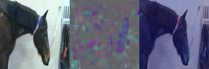
Pain is a complex multidimensional experience with sensory and affective
                   components. The affective component is associated with changes of behaviour, to avoid
                   pain or to protect the painful area [6]. While some of these behaviours may be directly
                   related to the location of the painful area, such as lameness in orthopedic disorders and
                   rolling in abdominal disorders [7], other pain behaviours, such as facial expressions, are
                   thought to be universal as means of communication of pain with conspecifics. Acute
                   pain has a sudden onset and a distinct cause, such as inflammation, trauma, or
                   ischemia [8, 9] and all these elements may be present in orthopedic pain in horses.
                       Recognizing horse pain automatically from video requires a method for fine-grained
                   action recognition, which can pick up subtle behavioral signals over long time, and
                   further, the method should be possible to train using a small dataset. In many widely
                   used datasets for action recognition [10–12], specific objects and scenery may add class
                   information. This is not the case in our scenario, since the only valid evidence present
                   in the video are poses, movements and facial expressions of the horse.
                       The Something-Something dataset [13] was indeed collected for fine-grained
                   recognition of action templates, but its action classes are short and atomic. Although
                   the classes in the Diving48 and FineGym datasets [14, 15] are complex and require
                   temporal modeling, the movements that constitute their classes are densely appearing in
                   a continuous sequence during the video, contrary to video data showing horses under
                   orthopedic pain with sparse expressions thereof.
                       A further important complication is that the labels in the present scenario are
                   inherently noisy, since the horse’s subjective experience of pain can not be observed.
                   Instead, pain induction and/or human pain ratings are used as proxy when labeling
                   video recordings. To further complicate matters, the behavioral patterns that we are
                   searching for might appear in both pain and non-pain data, although with different
                   frequency.
                       Expert pain assessment in horses is mainly performed by evaluating predetermined
                   body behaviors and facial expressions displayed by the horse during a short observation
                   period, usually two minutes. The observer can either stand outside the box stall or
                   observe the horse in a video. Equine pain research has focused on identifying certain
                   combinations of behaviors and facial expressions for general pain and specific types of
                   pain, such as orthopedic pain [16–20]. It is important to understand that all behaviors
                   and facial expressions are part of the non-verbal communication system of healthy
                   horses as well, and that it is their combinations and frequency which can indicate if pain
                   is present. Being an easier-to-observe special case, recordings of acute pain (applied for
                   short duration and completely reversibly, under ethically controlled conditions) have
                   been used to investigate pain-related facial expressions [21] and for automatic equine
                   pain recognition from video [22]. Until now, it has not been studied how this generalizes
                   to the more clinically relevant orthopedic pain.
                       This article investigates machine learning recognition of equine orthopedic pain
                   characterized by sparse visual expressions. To tackle the problem, we use domain
                   transfer from the recognition of clean, experimental acute pain, to detect the sparsely
                   appearing visible bursts of pain behavior within low grade orthopedic pain data (Fig. 1).
                   We compare the performance of our approach to a human baseline, which we
                   outperform on this specific task (Fig 2).
                       Our contributions are as follows:

                      • We are the first to investigate domain transfer between the recognition of different
                        types of pain in animals.
                      • We present extensive empirical results using two real-world datasets, and highlight
                        challenges arising when moving outside of clean, controlled benchmarking datasets
                        when it comes to deep learning for video.

January 10, 2022                                                                                       2/27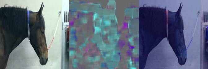
Fig 1. We present a study of domain transfer in the context of different types of pain
                   in horses. Horses with low grade orthopedic pain only show sporadic visual signs of
                   pain, and these signs may overlap with spontaneus expressions of the non-pain class – it
                   is therefore difficult to train a system solely on this data.

                       • We compare domain transfer from a horse pain dataset to standard transferred
                         video features from a general large-scale action recognition dataset, and analyze
                         whether these can complement each other.
                       • We present an explainability study of orthopedic pain detection in 25 video clips,
                         firstly for a human expert baseline consisting of 27 equine veterinarians, secondly
                         for one of our neural networks trained to recognize acute pain. We compare which
                         signs of pain veterinarians typically look for when assessing horse pain with what
                         the model finds important for pain classification.

                      Next, we present related work in Section 2, followed by methodology along with
                   dataset descriptions in Section 3. The emphasis lies on the experiments in Section 4 and
                   the discussion thereof, presented in Section 5. Finally, we conclude and outline future
                   directions in Section 6.

                   2     Related work
                   Although many methods are relevant to our problem, our setting in terms of data is
                   unique and requires a tailored approach. Weakly supervised action recognition is
                   relevant in that we share the same goal: extracting pertinent information from weakly
                   labeled video data. However, these methods typically rely on training with a large
                   number of video clips, which is not accessible in our setting. Our orthopedic pain
                   dataset consists of few (less than 100), although long (of several minutes) samples.

                   Weakly supervised action recognition and localization. Multiple-instance
                   learning (MIL) has been used extensively within deep learning for the task of weakly
                   supervised action localization (WSAL), where video level class labels alone are used to
                   determine the temporal extent of class occurrences in videos [23–27]. However, training
                   deep models within the MIL-scenario can be challenging. Error propagation from the
                   instances may lead to vanishing gradients [28–30] and too quick convergence to local
                   optima [31]. This is especially true for the low-sample setting, which is our case.

January 10, 2022                                                                                       3/27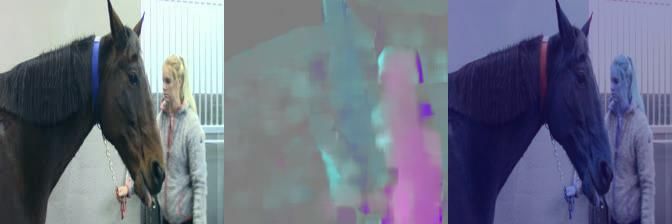
Typically, videos are split into shorter clips whose predictions are collated to obtain
                   the video level predictions. Multiple methods use features extracted from a pre-trained
                   two-stream model, I3D [32], as input to their weakly supervised model [23–25]. In
                   addition to MIL, supervision on feature similarity or difference between clips in
                   videos [23, 25, 33] and adversarial erasing of clip predictions [34, 35] are also used to
                   encourage localization predictions that are temporally complete. In early stages of our
                   study, we mainly attempted a MIL approach, but the noisy data, low number of
                   samples and similar appearance of videos from the two classes were prohibitive for such
                   a model to learn informative patterns.
                       Arnab et al. [36] cast the MIL-problem for video in a probabilistic setting, to tackle
                   its inherent uncertainty. However, they rely on pre-trained detection of humans in the
                   videos, which aids the action recognition. This is not applicable to our scenario since
                   horses are always present in our frames (i.e., detecting a horse would not help us to
                   temporally localize a specific behavior), and the behaviors we are searching for are more
                   fine-grained than the human actions present in datasets such as [10] or [12] (e.g., fencing
                   or eating). Another difference of WSAL compared to our setting is that we are agnostic
                   as to what types of behavior we are looking for in the videos. For this reason, it is not
                   possible for us to use a localization-by-temporal-alignment method such as the one by
                   Yang et al. [37]. Moreover, their work relies on a small number of labeled and trimmed
                   instances, which we do not have in this study.

                   Automatic pain recognition in animals. In [38], a Support Vector Machine
                   (SVM) cascade framework is used to recognize facial action units in sheep from single
                   images, to then assess pain according to pre-defined thresholds. A similar method is
                   applied to horses and donkeys in [39], presenting per pain-related facial action unit
                   classification results.
                       In [40], the work in [38] is continued, using automatically recognized sheep facial
                   landmarks to assess pain on a single-frame basis. In this work, disease progression is
                   monitored from video data, by applying the same pipeline on every 10th frame, and
                   averaging their pain scores. Improving on [38], they use subject-exclusive
                   (leave-one-animal-out) testing for the video part of their experiments.
                       Using a deep learning approach, Tuttle et al. [41] recognize induced inflammatory
                   joint pain in albino mice from single images. While the classification task is binary, the
                   pain labels are set according to human scorings based on facial expressions, on a scale
                   from 1 to 10 (five action units associated with pain that each can have a confidence score
                   of 0-2). It is not clear from the article how they go from the ten-class scale to binary
                   labels. Andresen et al. [42] apply this method to black-furred mice moving more freely
                   in their cages compared to [41], still in a single-frame setting. Both methods [41, 42] use
                   Imagenet [43] pre-trained, standard CNN networks [44, 45] as kept-fixed back-bones,
                   while training a fully-connected classification head, on their datasets. Further, both can
                   improve their classification accuracy by averaging the network confidence over images
                   taken within a narrow time span. Andresen et al. [42] furthermore point to the difficulty
                   of generalizing between different types of pain, which we investigate closer in this article.
                       Lencioni et al. [46] train light-weight CNN models (two convolutional layers) from
                   scratch to recognize facial pain expressions automatically in horses from single images.
                   Separate models are trained for the eye, ear and mouth regions, to recognize three levels
                   of pain (0-2). The labels are entirely based on the Horse Grimace Scale (HGS), scored
                   by humans, although the data was recorded before and after routine surgical castration.
                   It is not clear if pain images were selected only post castration, or if the selection was
                   made only based on visibility of the pain cues. Similarly, Li et al. [47] train separate
                   CNN-based classifiers on small crops of different facial regions to recognize EquiFACS
                   units [48], which can be used for pain evaluation.

January 10, 2022                                                                                          4/27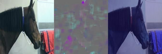
In a previous work [22], we were the first to perform pain recognition in animals
                   with models learning patterns from sequences rather than single frames, showing large
                   improvement from training on single images. We used deep recurrent two-stream
                   models, and trained with labels set according to clean experimental acute induced pain.
                   The presented system uses no pre-defined behaviors or facial expressions, but learns
                   spatio-temporal features based on raw videos and their pain labels only. In this article,
                   we build on the same approach (with slight modifications listed in the Appendix), and
                   use it in this empirical study of how well different models can handle a domain shift in
                   the test data.
                       In a more recent work of ours [49], we perform equine pain recognition on 3D pose
                   representations extracted from multi-view surveillance data, on the same low-grade
                   orthopedic pain trial as in this paper. Although the horses and pain trial are the same
                   as in the current work, the crucial difference is that the data used in [49] is different
                   (surveillance data in the box, whereas here, we use videos recorded with a tripod outside
                   the box, where the facial expression is visible), and that only the pose representation is
                   used for classification. This is advantageous to reduce the amount of extraneous
                   information. However, the potential disadvantage is that any facial expressions are not
                   possible to take into account. As a result, it is perhaps the adjustment of pose as a
                   result of previous pain that is recognized in [49], rather than whether a pain experience
                   is ongoing. Similarly to the present work, it is found that low-grade orthopedic pain is
                   difficult to detect, compared to the less noisy pain trial used in [22].

                   Pain in horses. The definition of animal pain includes a change in motivation, where
                   the animal develops behaviors to avoid pain or to protect the painful area [6].
                   Depending on the origin of pain, the animal may perform different behaviors. Horses
                   with abdominal pain may stretch, roll and kick at the abdomen, while horses with
                   orthopedic pain may be reluctant to move and have an abnormal weight distribution or
                   movement pattern [7]. Therefore, pain assessment tools such as pain scales often target
                   pain of a specific origin.
                       Facial expressions, on the other hand, seem to be universal for pain within a species.
                   Grimace scales have been successfully applied to pain from different origins, such as
                   post-surgical pain or laminitis in horses [17, 18, 50]. It also seems like pain-related facial
                   expressions are present during both acute and chronic pain, and may be shown by the
                   animal during several weeks post-injury, but not consistently and perhaps tailing
                   away [51].
                       Acute pain has a sudden onset and a distinct cause, such as inflammation, while
                   chronic pain is more complex, sometimes without a distinct cause, and by definition
                   lasts for more than three months. Acute (and sometimes chronic) pain arises from the
                   process of encoding an actually or potentially tissue-damaging event, so called
                   nociception, and may therefore be referred to as nociceptive pain. When pain is
                   associated to decreased blood supply and tissue hypoxia, it is instead termed ischemic
                   pain [8, 9].
                       Orthopedic pain in horses can be of both acute and chronic character where a very
                   common diagnosis is osteoarthritis, with related inflammatory pain of the affected
                   joint [52]. In humans, the disease is known to initially result in nociceptive pain localized
                   to the affected joint, but when chronic pain develops, central sensitization occurs with a
                   more widespread pain [53]. How pain-related facial expressions and other behaviors vary
                   between horses with acute and chronic orthopedic pain is yet to be described, and so is
                   the relation between pain intensity and alterations in facial expressions.

January 10, 2022                                                                                           5/27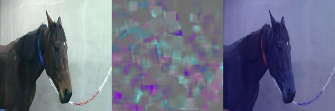
Dataset   # horses   # videos      # clips     Pain         No pain      Total       Labeling
           PF        6          60            8784        03:41:25     06:03:44     09:45:09    Video-level. Induced clean
                                                                                                experimental acute pain,
                                                                                                on/off (binary)

           EOP(j)    7          90            6710        03:37:36     05:03:25     08:41:01    Video-level. Induced
                                                                                                orthopedic pain,
                                                                                                varying number of hours
                                                                                                prior to the recording.
                                                                                                Binarized human CPS pain
                                                                                                scoring before/after
                                                                                                recording
                                                                                                (3 independent raters)

                                Table 1. Overview of the datasets. Frames are extracted at 2 fps, and clips consist of
                                10 frames. Duration shown in hh:mm:ss.

                                3       Method
                                Central to this study are two datasets depicting pain of different origin in horses (Table
                                1). The datasets are similar in that they both show one horse, trained to stand still,
                                either under pain induction or under baseline conditions (Section 3.1). In our
                                experiments, we investigate the feasibility of knowledge transfer between different pain
                                domains (Section 3.3). We use the macro average F1-score as metric, which is more
                                conservative than accuracy when there is class imbalance – it does not favor the
                                majority class.

                                3.1     Datasets
                                In Table 1, we show an overview of the two datasets used in this article. It can be noted
                                that neither of the two show a full view of the legs of the horses. This could otherwise
                                be an indicator of orthopedic pain. Both datasets mainly depict the face and upper
                                body of the horses, see, e.g., Fig 3.

                                3.1.1    The Pain Face dataset (PF)
                                The experimental setup and video recording of the PF dataset have been described in
                                detail in [21, 22]. Briefly, the dataset consists of video recordings of six clinically healthy
                                horses with and without acute pain. The pain is either ischemic (from a pressure cuff)
                                or inflammatory (from a capsaicin substance on the skin) and was applied for short
                                durations of time under ethically controlled conditions.

                                Labels. The induced pain is acute and takes place during 20 minutes, during which
                                the horse shows signs of pain almost continuously. Thus, the video-level positive pain
                                label is largely valid for all clips extracted from it. The labels are binary; any video clip
                                extracted from this period is labelled as positive (1), and any video clip from a baseline
                                recording is labelled as negative (0).

                                3.1.2    The EquineOrthoPain(joint) dataset (EOP(j))
                                The experimental setup for the EOP(j) dataset is described in detail in previously
                                published work [16]. Mild to moderate orthopedic pain was induced in eight clinically

January 10, 2022                                                                                                             6/27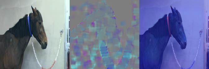
healthy horses by injecting lipopolysaccharides (LPS) into the tarsocrural joint (hock).
                   This is a well-known and ethically approved method for orthopedic pain induction in
                   horses, resulting in a fully reversible acute inflammatory response in the joint [54].
                   Before, and during the 22-52 hour period after induction, several five minute videos of
                   each horse were recorded regularly. A video camera, attached to a tripod at
                   approximately 1.5 metres height and with 1.5 metres distance from the horse, recorded
                   each horse when standing calmly in the stables outside the box stall.

                   Labels. The dataset contains 90 different videos associated with one pain label each
                   (Table 1). Notably, these labels are set immediately before or after the recording of the
                   video when the horse is in the box stall, and not simultaneously to the video recording.
                   Three independent raters observed the horse, using the Composite Orthopedic Pain
                   Scale (CPS) [55] to assign each horse a total pain score ranging from 0 to 39. The pain
                   label is the average pain score of these three ratings. In this study, the lowest pain
                   rating made was 0 and the highest was 10.
                       For the binary classification used in this study (following prior work [22]), the CPS
                   score is thresholded so that any value larger than zero post-induction is labeled as pain,
                   and values equal to zero are labeled as non-pain. This means that we consider possibly
                   very weak pain signals (e.g., 0.33) as painful, adding to the challenging nature of the
                   problem. One of the horses was excluded from our experiments, because it did not have
                   CPS scores > 0 after the pain induction.

                   3.2    Cross-validation within one domain
                   When running cross-validation training, we train and test within the same domain. We
                   train with leave-one-subject-out cross-validation. This means that one horse is used as
                   validation set for model selection, one horse as held-out test set, and the rest of the
                   horses are used for training. The intention with these experiments is to establish
                   baselines and investigate the treatment of weak labels as dense labels for the two
                   datasets.

                   Treating weak labels as dense labels. We distinguish between clips and videos,
                   where clips are five second long windows extracted from the videos (several minutes
                   long) (Table 1). For both datasets, the pain labels have been set weakly on video level.
                   In practice, treating these labels as dense means giving the extracted clips the same
                   label as the video.

                   Architectures. We use two models in our experiments: the two-stream I3D,
                   pre-trained on Kinetics (kept fixed until the ‘Mixed5c’ layer), and the recurrent
                   convolutional two-stream model (hereon, C-LSTM-2) from [22]. Each stream (RGB and
                   optical flow) of C-LSTM-2 consists of four blocks of convolutional LSTM-layers, with
                   max pooling and batch normalization in each. The convolutional LSTM layer was first
                   introduced by Shi et al. [56], and replaces the matrix multiplication transforms of the
                   classical LSTM equations (cf., [57]) with convolutions. This allows the layer to ingest
                   data with a spatial grid structure, and to maintain a spatial structure for its output as
                   well. The classical LSTM requires flattening all input data to 1D vectors, which is
                   suboptimal for image data, where the grid structure matters. The two streams are fused
                   by addition after the last layer, flattened and input to a two-class classification head.
                   The classification head provided with the I3D implementation is kept and retrained to
                   two classes.
                       The output of the models are binary pain predictions. We follow the supervised
                   training protocol of [22] with minor modifications; more details can be found in the

January 10, 2022                                                                                       7/27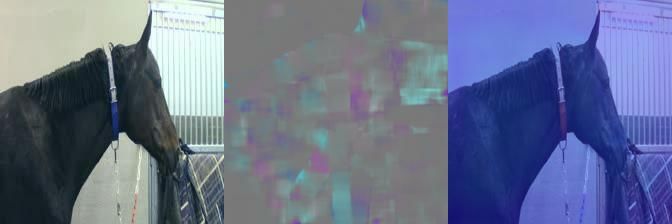
Appendix (Section C.1). The main difference is that we resample the minor class clips
                   with a different window stride, to reduce the class imbalance. We also run on a higher
                   frame resolution, 224x224 instead of 128x128. Further implementation details can be
                   found in [22], in the Appendix and in the public code repository.

                   3.3    Domain transfer
                   When running domain transfer experiments, we use two different methods. The first is
                   to train a model on the entire dataset from one domain for a fixed number of epochs
                   without validation, and test the trained model on another domain. This means that the
                   model has never observed the test data domain. We also run experiments where we first
                   pre-train a model on the source domain, and fine-tune its classification layer on the
                   target domain. In this way, the model has acquainted itself with the target domain, but
                   not seen the specific test subject.
                       To choose the number of epochs for model selection when training on the entire
                   dataset, we use the average best number of epochs from when running intra-domain
                   cross-validation (Section 3.2) and multiply this with a factor of how much larger the
                   dataset becomes when including test and validation set (1.5 when going from 4 to 6
                   horse subjects). This was 77 ∗ 1.5 = 115 epochs when training C-LSTM on PF, and
                   42 ∗ 1.5 = 63 epochs when training I3D on PF. This takes around 80h on a GeForce
                   RTX 2080 Ti GPU for the C-LSTM-2, which is trained from scratch, and around 4h for
                   the I3D where only the classification head is trained. Except for the number of epochs,
                   the model is trained with the same settings as during intra-domain cross-validation.

                   3.4    Veterinary expert baseline experiment
                   As a baseline for orthopedic pain recognition, we engaged 27 Swedish equine
                   veterinarians in rating 25 clips from the EOP(j) dataset. In veterinary practice, the
                   decision of whether pain is present or not is often made quickly based on the
                   veterinarian’s subjective experience The veterinarians were instructed to perform a
                   rating of pain intensity of the horses in the clips using their preferred way of assessment.
                   We asked for the intensity to be scored subjectively from no-pain (0) to a maximum of
                   10 (maximal pain intensity). The maximum allowed time to spend in total was 30
                   minutes. The average time spent was 18 minutes (43 seconds per clip). The participants
                   were carefully instructed that there were clips of horses without and with pain, and that
                   only 0 represented a pain-free state.
                       There is no gold standard for assessment of pain in horses. Veterinary methods rely
                   on subjective evaluation of information collected on the history of the animal, its social
                   interaction and attitude, owners’ complaints and an evaluation of both physical
                   examination and behavioral parameters (REF y). In this case only the behavioral
                   changes could be seen. The assessments in practice are rarely blinded, but influenced by
                   knowledge of the history and physiological state of the animal or the observation of an
                   obvious pain behaviours. To simulate the short time span for pain estimation and avoid
                   expectation bias, each clip was blinded for all external information, and only five
                   seconds long, i.e., the same temporal footprint as the inputs to the computer models.
                   The intention with this was to keep the comparison to a computer system more
                   pragmatic – a diagnostic system is helpful to the extent that it is on par with or more
                   accurate than human performance, reliable and saves time. Another motivation to use
                   short clips was for the feasibility of the study, and to avoid rater fatigue. It is extremely
                   demanding for a human to maintain subtle behavioral cues in the working memory for
                   longer than a few seconds at a time. In effect, this fact in itself pinpoints the need for
                   an automatic pain recognition method.

January 10, 2022                                                                                          8/27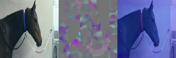
The clips selected for this study were sampled from a random point in time from 25
                   of the videos of the EOP(j) dataset, 13 pain and 12 non-pain. Only pain videos with a
                   CPS pain label ≥ 1 were included in order to have a clearer margin between the two
                   classes, making the task slightly easier than on the entire dataset. We first extracted
                   five such clips from random starting points in the video, and used the first of those
                   where the horse was standing reasonably still without any obstruction of the view.
                       Behaviors in the 25 clips were manually identified and listed in Table 7. Those
                   related to the face were identified by two other veterinary experts in consensus
                   according to the Horse Grimace Scale [17]. The behaviors we were attentive to for each
                   clip are listed in Table 2.

                                  Behavior                                           Symbol
                                  From the Horse Grimace Scale [17]
                                  Backwards ears, moderately present                       e1
                                  Backwards ears, obviously present                        e2
                                  Orbital tightening, moderately present                   o1
                                  Orbital tightening, obviously present                    o2
                                  Tension above the eye area,
                                    moderately present                                     t1
                                  Tension above the eye area,
                                    obviously present                                      t2
                                  Mouth strained and pronounced chin,
                                    moderately present                                     c1
                                  Mouth strained and pronounced chin,
                                    obviously present                                      c2
                                  Strained nostrils and flattening of the profile,
                                    moderately present                                     n1
                                  Strained nostrils and flattening of the profile,
                                    obviously present                                      n2
                                  Other
                                  Large movement                                            m
                                  Mouth play                                                p
                                  Lowered head                                               l
                                  Clearly upright head                                      u
                                  Human in clip                                             h

                   Table 2. Explanation of the listed behavior symbols appearing in Table 7.

                   4     Experiments
                   In this section, we describe our results from intra-domain cross-validation training (4.1),
                   domain transfer (4.2) and from the human expert baseline study on EOP(j) and its
                   comparison to the best performing model, which was trained only on acute pain (4.3).

                   4.1    Cross-validation within one domain
                   The results from training with cross-validation within the same dataset are presented in
                   Table 3 for both datasets. When training solely on EOP(j), C-LSTM-2 could not
                   achieve a higher result than random performance (49.5% F1-score), and I3D was just
                   above random (52.2). Aiming to improve the performance, we combined the two

January 10, 2022                                                                                        9/27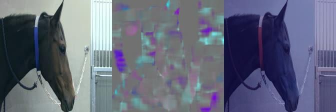
Dataset          # horse folds      F1-score      Accuracy
                                  C-LSTM-2
                                  PF               6                  73.5   ±7.1   75.2   ±7.4
                                  EOP(j)           7                  49.5   ±3.6   51.2   ±2.8
                                  PF + EOP(j)      13*                60.2   ±2.6   61.8   ±3.2
                                   (*PF                               69.1   ±4.9   71.1   ±3.9 )
                                   (*EOP(j)                           53.4   ±3.0   53.9   ±2.5 )

                                  I3D
                                  PF               6                  76.1   ±1.5   76.6   ±1.1
                                  EOP(j)           7                  52.2   ±2.3   52.6   ±2.2
                                  PF + EOP(j)      13*                59.5   ±4.3   62.2   ±2.7
                                   (*PF                               71.3   ±3.5   73.1   ±1.4 )
                                   (*EOP(j)                           49.4   ±5.3   52.9   ±3.7 )

                   Table 3. Results (% F1-score) for intra-domain cross-validation for the respective
                   datasets and models. The results are averages of five repetitions of a full
                   cross-validation and the average of the per-subject-across-runs standard deviations.

                   datasets in a large 13-fold training rotation scheme. After mixing the datasets, and
                   thereby almost doubling the training set size and number of horses, the total results on
                   13-fold cross-validation for the two models were 60.2 and 59.5 on average, but where the
                   PF folds on average obtained 69.1 and 71.3 and the EOP(j) folds obtained 53.4 and 49.4.
                   Thus, the performance on PF deteriorated for both models, as well as on EOP(j) for
                   I3D (49.4) and only slightly improved on EOP(j) (53.4) for C-LSTM-2. This indicates
                   that the weak labels of EOP(j) and general domain differences between the datasets
                   hindered standard supervised training with a larger, combined dataset.

                   4.2    Domain transfer to EOP(j)
                   Table 4 compared to Table 3 shows the importance of domain transfer for the task of
                   recognizing pain in EOP(j). One trained instance of the C-LSTM-2, which has never
                   seen the EOP(j) dataset (hereon, C-LSTM-2-PF †), achieves 58.2% F1-score on it –
                   higher than any of the other approaches. I3D, which achieved higher overall score when
                   running cross-validation on PF, did not generalize as well to the unseen EOP(j) dataset
                   (52.7). For I3D, trials with models trained during a varying number of epochs are
                   included in the Appendix, although none performed better than 52.7% F1-score.
                       Fine-tuning (designated by FT in Table 4) these PF-trained instances on EOP(j)
                   decreased the result (54.0 and 51.8, respectively), presumably due to the lesser amount
                   of clearly discriminative visual cues in the EOP(j) data. This goes in line with results in
                   Table 3; the data and labels of the EOP(j) data do not seem to be suited for supervised
                   training.
                       Table 5 shows that the results for individual horses may increase when applying a
                   multiple-instance learning filter during inference to the predictions across a video and
                   base the classification only on the top 1%/5% confident predictions (significantly for
                   subjects A, H, and I, and slightly for J and K); however, for other subjects, the results
                   decreased (B, N). As described in Section 4.3, there may be large variations among
                   individuals for this type of pain induction.

January 10, 2022                                                                                       10/27
Model A B H I J K N Global
I3D [32], Kinetics
PF, EOP(j)-FT 48.7±4.1 56.8±0.9 47.0±3.4 43.6±4.7 53.6±1.7 54.5±0.9 58.2±0.6 51.8±2.3
PF, EOP(j) unseen 54.96 45.05 53.42 56.52 55.69 41.66 46.59 52.70
3 repeated runs 49.9 ±2.8 48.0 ±2.1 47.2 ±2.6 52.2 ±1.8 59.0 ±1.0 40.7 ±0.8 46.1 ±2.2 52.6 ±0.4
C-LSTM-2, scratch
PF, EOP(j)-FT 50.4±2.7 55.1±0.8 44.9±1.4 45.7±0.7 69.3±1.7 51.2 ±0.7 60.8±0.8 54.0±1.3
PF, EOP(j) unseen † 61.55 56.34 55.55 51.51 64.76 45.11 57.84 58.17
3 repeated runs 59.4 ±4.6 56.7 ±2.7 60.5 ±5.7 52.1 ±1.8 53.2 ±14.0 49.6 ±3.9 54.2 ±4.2 56.3 ±2.8
Table 4. F1-scores on EOP(j), when varying the source of domain transfer, for models
trained according to Section 3.3. FT means fine-tuned (three repetitions of full
cross-validation runs). Column letters indicate different test subjects. † represents a
specific model instance, reoccurring in Tables 5, 6 and 7.
Model instance MIL-filter A B H I J K N
C-LSTM-2-PF † - 61.55 56.34 55.55 51.51 64.76 45.11 57.84
C-LSTM-2-PF † Top 5% 88.31 51.13 59.06 59.06 65.37 31.58 36.36
C-LSTM-2-PF † Top 1% 88.31 51.13 53.33 59.06 65.37 45.83 45.0
Table 5. Results on video-level for EOP(j), when applying a multiple-instance learning
(MIL) filter during inference on the clip-level predictions. The column letters designate
different test subjects. The model has never trained on EOP(j), and is the same model
instance as in Tables 4, 6 and 7.
4.3 Veterinary expert baseline experiment
The method of the expert baseline study is described in Section 3.4. Next, we compare
and interpret the decisions of the human experts and the C-LSTM-2-PF † on the 25
clips of the study.
Comparison between the human experts and the C-LSTM-2-PF †. Table 7
gives an overview of the ratings of the 25 clips given by the experts and by the model.
First, we note that the C-LSTM-2-PF † instance outperforms the humans on these clips,
achieving 76.0% F1-score, compared to 47.6 ± 5.5 for the experts (Table 6, Fig 2). The
experts mainly had difficulties identifying non-pain-sequences. Similarly, however, when
the model was tested on the entire EOP(j) dataset, its non-pain results were lower than
Rater No pain Pain Total
Human expert 34.7 ±10.0 60.6 ±4.4 47.6 ±5.5
C-LSTM-2-PF † 75.0 76.9 76.0
C-LSTM-2-PF †* 54.47 61.86 58.17
Table 6. F1-scores (%) on the 25 clips of the expert baseline. The C-LSTM-2-PF †
instance was trained on PF but never on EOP(j). Asterisk: results on the entire EOP(j)
dataset for comparison.
January 10, 2022 11/27Fig 2. Pain predictions on the 25 clips included in the baseline study (Table 7), by the
human experts (left), and by the C-LSTM-2-PF † (right).
its pain results as well (Table 6), indicating the difficulty of recognizing the non-pain
category.
Most of the clips rated as painful by the experts contain behaviors that are
classically associated with pain, for example as described in the Horse Grimace
Scale [58]. Among the pain clips, clip 6 is the only one without any listed typically
pain-related behaviors. The veterinarians score the clip very low (0.96) and seem to
agree that the horse does not look painful. The model interestingly scores the clip as
painful, but with a very low confidence (0.52).
There are three clips with clear movement of the horse (8, 12, 18), where 8 and 12
are wrongly predicted by the model as being non-pain. Clip 18 is correctly predicted as
being non-pain with high confidence (0.9991), suggesting that the model associates
movement with non-pain. On clip 18, the human raters mostly agree (17) with the
model that this horse does not look painful and the average rating is low (0.96). It can
further be noted that the three incorrect pain predictions (17, 20, 24) made by the
model occurred when there was either e1 (moderately backwards ears, pointing to the
sides) or l (lowered head), or both. Also, 24 is the only clip with a human present,
which might have confused the model further (Fig 3).
The four most confident and correct non-pain predictions (>0.99) made by the
model are the ones where the head is held in a clear, upright (u) position. Similarly, the
three most confident and correct pain predictions (>0.99) by the model all contain ear
behavior (e1 or e2).
5 Discussion
Why is the expert performance so low? Tables 6, 8 and Fig 2 show a low
performance for the human experts in general and especially for non-pain.
Increasing the threshold to 1 and 2 reduced the accuracy for pain, which may be due
to false inclusion of scores of 0 if the raters scored 1 or 2 for non-pain, contrary to the
instructions. Vice versa, the accuracy for non-pain increased when the threshold was
extended, which may be due to inclusion of scores of 1 and 2, used as non-pain (even
though they were informed that only 0 is used for non-pain). A reluctance to assess zero
pain is difficult for clinicians who are taught that signs of pain may be subtle.
The results point to the difficulty of observing pain expressions at a random point in
time for orthopedic pain, and without context. The LPS-induced orthopedic pain may
further have complicated the rating process, since it varies in intensity among
January 10, 2022 12/2727 Experts C-LSTM-2-PF †
Clip Behaviors CPS Label Avg. rating # correct Pred. Conf.
1 e1 2 1 2.7 23 1 0.9999
2 o1 l 2 1 1.1 10 1 0.9241
3 e2 t1 c1 n1 p 4.33 1 3.4 22 1 0.9997
4 e2 o1 l 4.67 1 3.7 21 1 0.6036
5 t1 c1 3.67 1 0.37 4 1 0.9853
6 1.33 1 0.96 13 1 0.5200
7 u 4.33 1 1.3 12 1 0.9063
8 c2 n2 m u p 6.33 1 4.7 25 0 0.8623
9 e1 t1 c1 n1 p 3 1 4.6 25 0 0.5504
10 e2 t1 c1 n2 l p 2 1 4.4 25 1 0.9999
11 e2 t1 c1 n2 l 1 1 6.8 27 1 0.8231
12 e1 t1 c1 n1 m 4.33 1 2.5 22 0 0.8046
13 e2 t1 c1 n2 1.67 1 4.4 26 1 0.9840
14 e2 o1 t1 c1 n1 u 0 0 5.5 0 0 0.9993
15 e2 t1 0 0 4.1 1 0 0.5848
16 e2 o1 t1 c1 n1 0 0 3.9 4 0 0.8439
17 e1 o1 t1 n1 l p 0 0 4.4 5 1 0.6456
18 t1 m u p 0 0 0.96 17 0 0.9991
19 0 0 0.48 21 0 0.9568
20 t1 l 0 0 2.9 9 1 1.00
21 u 0 0 1.3 13 0 0.9999
22 0 0 2.7 4 0 0.6128
23 c1 n1 u 0 0 1.6 8 0 0.9989
24 e1 t1 n1 h p 0 0 2.4 9 1 1.00
25 e2 o1 t1 n1 0 0 5.9 0 0 0.6135
Table 7. Overview of the predictions on 25 EOP(j) clips made by the human
veterinarian experts and by one C-LSTM-2 instance, trained only on PF. The labels for
the C-LSTM-2 were thresholded above 0 (same threshold as for the experts). The
behavior symbols in the Behavior column are explained in Table 2.
individuals, despite administration of the same dose. This results in different levels of
pain expressions [59], sometimes occurring intermittently. Hence, there will be ‘windows’
during the observed time where the horse expresses pain clearly [60]. The other parts of
the observed time will then contain combinations of facial expressions that some raters
interpret as non-pain, and some raters interpret as pain. If a ‘window’ is not included in
the five second clip, it is difficult for the rater to assign a score, decreasing their
accuracy.
Threshold No pain Pain Total
0 28.1 ±11.2 72.7 ±9.4 51.3 ±4.6
1 37.4 ±14.1 65.2 ±9.7 51.9 ±5.7
2 48.5 ±18.7 52.7 ±13.8 50.7 ±7.5
Table 8. Accuracies (%) from the expert baseline, varying with the chosen pain
threshold.
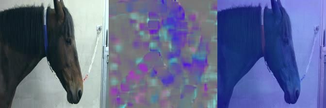
January 10, 2022 13/27Fig 3. The figures can be displayed on click as videos in Adobe Reader.
RGB, optical flow, and Grad-CAM [61] saliency maps of the C-LSTM-2-PF †
predictions on clips 10 and 24 (Table 7). Clip 10 (left) is a correct prediction of pain.
Clip 24 (right) is a failure case, showing an incorrect pain prediction, and we observe
that the model partly focuses on the human bystander. The remaining 23 clips with
saliency maps can be found in the Appendix.
Significance of results. Having trained the C-LSTM-2 on a cleaner source domain
(PF), without ever seeing the target domain (EOP(j)) before, gave better results than
all other attempts, including fine-tuning (58.2% F1-score for the best instance, and
56.3 ± 2.8% for three repeated runs). Despite being higher than human performance,
these F1-scores on the overall dataset are still modest and significantly lower than the
recognition of acute pain in [22]. However, the results are promising, especially since
they were better for the clips used for the human study (with higher pain-scores) (76%,
vs. 48% for the human experts). This may mean that the noise in the labels on the
overall dataset – both inherent to pain labelling and specific for the sparse pain
behavior related to low-graded orthopedic pain, obscures the system’s true performance
to some extent.
The human expert baseline for classification on clip-level of the EOP(j) dataset,
together with the intra-domain results (Table 3), shows the difficulty in detecting
orthopedic pain for humans and standard machine learning systems trained in a
supervised manner, within one domain. Poor performance of raters in assessing low
grade pain is the case generally, and points to the necessity of this study. The lack of
consensus is troubling since veterinary decision-making regarding pain recognition is
critical for the care of animals in terms of prescribing alleviating treatments and in
animal welfare assessments [62]. As an example of this, veterinarians can score assumed
pain in horses associated with a particular condition on a range from ‘non-painful’ to
‘very painful’ [63]. One important advantage of an automatic pain recognition system
would be its ability to store information over time, and produce reliable predictions
according to what has been learned previously. Humans are not able to remember more
than a few cues at the time when performing pain evaluation. This creates the need for
automated methods and prolonged observation periods, where automated recognition
can indicate possible pain episodes for further scrutiny. In equine veterinarian clinics,
such a system would be of great value. In summary, even a system with a
less-than-perfect accuracy would be useful in conjunction with experts on site.
Expected generalization of results. This study has been performed on a cohort
of, in total, n = 13 horses. It is therefore, as always, important to bear in mind the
possible bias in these results. Nevertheless, we want to emphasize that the paper was
dedicated to investigating generalizability, and that there already is a domain gap
between the two groups of n = 6 and n = 7 horses. The recordings of the two groups
(datasets) were made four years apart, in different countries, and, naturally, on entirely
different horse subjects. In addition to this, whenever we evaluated our system in the
intra-domain setting, the test set consisted only of data from a previously unseen
individual (leave-one-subject-out testing). Considering this, our findings do indicate
that the method would generalize to new individuals – in particular if the system could
January 10, 2022 14/27be trained on an increased amount of clean base-domain data.
Differences in pain biology and display of pain in PF and EOP(j). Both
video sets were recorded of horses under short term acute pain after a base line period.
However, the noxious stimuli and the anatomical location of the pain differed widely.
The PF dataset was created by application of two well-known experimental noxious
stimuli of only little clinical relevance (capsaicin (refa) and ischemia (refb)).s.Both
stimuli are used in in pain research in human volunteers, induce pain lasting for 10 - 30
minute and pain levels corresponding to 4 or 5 on a 10 point scale, where 0 is no pain
and 10 is worst imaginable pain. Due to this short time span, the controlled course of
pain intensity,the controlled experimental conditions and the predictability of the model,
these data present the most noise-less display of possible behavioural changes due to the
pain experienced. Further, because the pain is of such short duration, the horse will not
be able to compensate or modify its behaviours. However, such data are less useful for
clinical situations. During clinical conditions, pain intensity is unpredictable,
intermittent and of longer duration, allowing the horse to adapt to the pain, according
to its previous experience and temperament. In real clinical situations, there is no
ground truth of the presence or intensity of pain. The LPS model represents an acute,
joint pain caused by inflammation of the synovia, resulting in orthopedic pain which
ceases within 24 hours. The degree and onset of inflammation, and thus the resulting
pain is known to be individual, depending on a range of factors which can not be
accounted for in horses, including immunological status and earlier experiences with
pain (ref c). Because the horse has time to to adapt to and compensate the pain, by for
example unloading the painful limb, pain will be intermittent or of low grade presenting
in unpredictable epochs (ref d). The low-noise data set therefore showed to be feasible to
learn from, even if the pain kinds were different. Whether a low noise dataset also can
improve recognition of chronic or neuropathic pain types, remains to be investigated.
Pain intensity and binarization of labels. As noted above, the labels in the PF
dataset were set as binary from the beginning, according to whether the pain induction
was ongoing or not, while the binary labels in the EOP(j) dataset were assigned
afterwards, based on thresholding of the raters’ CPS scores. The videos in EOP(j) were
recorded during pain progression and regression. Hence, they contain different pain
intensities, ranging from very mild to moderate pain. Introducing more classes in the
labeling may mirror the varying intensities more accurately than binary labels, but the
low number of samples in EOP(j) (90) restricts us to binary classification. Increasing
the number of classes would not be sound in this low-sample scenario, when using
supervised deep learning for classification, a methodology which relies on having many
samples per class, in order to learn patterns statistically.
Furthermore, deciding pain intensity labels in animals is difficult. More accurate
human pain recognition has been found for higher grimace pain scores [64], underlining
that mild pain intensity is challenging to assess. This is in agreement with studies in
human patients, where raters assessing pain-related facial expressions struggled when
the patients reported a mild pain experience [65]. Grimace scores seem to follow the
regression of pain after analgesic administration [66] and may therefore aid in defining
pain intensity. However, the relation between pain intensity and level of expression is
known to be complex in humans and may be so in animals. Pain intensity estimation on
a Visual Analogue Scale was not accurate enough in humans, and the estimation seemed
to benefit from adding pain scores assessing pain catastrophizing, life quality and
physical functioning [67]. As discussed by [62], pain scores may instead be used to
define the likelihood of pain, where a high pain score increases the likelihood that the
animal experiences pain. In addition, when pain-related behaviors were studied in
January 10, 2022 15/27horses after castration, no behaviors were associated to pain intensity [68]. This leaves
us with no generally accepted way to estimate pain intensity in animals, supporting our
choice of using binary labels in this study.
Labels in the real world. None of the equine pain datasets were recorded with the
intention to run machine learning on the videos. This presents noise, in both data and
labels. We point to how one can navigate a fine-grained classification problem, on a
real-world dataset in the low-data regime, and show empirically that knowledge could
be transferred from a different domain (for the C-LSTM-2 model), and that this was
more viable than training on the weak labels themselves.
Domain transfer: Why does the C-LSTM-2 generalize better than I3D?
Despite performing better on PF during intra-domain cross-validation, I3D does worse
upon domain transfer to a new dataset (Table 4) compared to the C-LSTM-2. It is
furthermore visible in Table 3 that the I3D performance on EOP(j) deteriorates when
combining the two datasets, perhaps indicating a proneness to learning dataset-specific
spurious correlations which do not generalize. In contrast, the C-LSTM-2 slightly
improves its performance on EOP(j) when merging the two training sets.
We hypothesize that this is because I3D is an over-parameterized model (25M
parameters), compared to the C-LSTM-2 (1.5M parameters). An I3D pre-trained on
Kinetics with its large number of trainable parameters is excellent when a model needs
to memorize many, predominantly spatial, features of a large-scale dataset with cleanly
separated classes, in an efficient way. When it comes to fine-grained classification of a
lower number of classes, which can generalize to a slightly different domain, and
moreover requires more temporal modeling than when the task is to separate ‘playing
trumpet’ from ‘playing violin’ (or at Kinetics’ most challenging: ‘dribbling’ from
‘dunking’), it seems, from our experiments, that it is not a suitable architecture.
Another reason could be the fact that the C-LSTM-2 is trained solely on horse data,
from the bottom up, while the I3D has its back-bone unchanged in our experiments. In
that light, the C-LSTM-2 can be considered more specialized to the problem. Although
Kinetics-400 does contain two classes related to horses: ‘grooming horse’ and ‘riding or
walking with horse’, the C-LSTM-2 undoubtedly has seen more up-close footage of
horses. In fact, somewhat ironically, the ‘riding or walking with horse’ coupled with
‘riding mule’ is listed in [11] as the top confused class of the dataset, using the
two-stream I3D.
How does I3D do if trained solely on the PF dataset? This is where the model size
becomes a problem. I3D requires large amounts of training data to converge properly;
Epoch Scratch Pre-trained Pre-trained,
freeze back-bone
25 46.8 ±8.9 46.5 ±4.4 51.7 ±1.4
63 46.3 ±9.3 46.4 ±7.5 52.6 ±0.4
115 46.6 ±10.4 47.4 ±1.4 52.6 ±0.05
200 46.3 ±7.2 43.2 ±3.8 52.4 ±0.5
# trainable parameters 24,542,116 24,542,116 4,100
Table 9. Global average F1-scores for domain transfer experiments for I3D, using
varying pre-training and fine-tuning schemes. The model is trained on the PF dataset
and tested on the EOP(j) dataset. Only the pre-trained model, fine-tuned with a frozen
back-bone could achieve results slightly above random performance on EOP(j).
January 10, 2022 16/27the duration of Kinetics-400 is around 450h. It is, for ethical reasons, difficult to collect
a 450h video dataset (>40 times larger than PF) with controlled pain labels. Table 9
shows additional results when training I3D either completely from scratch (random
initialisation) on the PF data, or from a pre-trained initialisation, compared to when
only training the classification head (freezing the back-bone). The results point to the
difficulty of training such a large network in the low data regime.
Weakly supervised training on EOP(j). During the course of this study, we
performed a large number of experiments in a weakly supervised training regime on
EOP(j). Our approach was to extract features from pre-trained networks and combine
these into full video-length, to then run multiple-instance learning training on the
feature sequences (the assumption being that a pain video would contain many negative
instances as well). The training was attempted using both simple fully-connected
networks, LSTM models and attention-based Transformer models. Training on the full
video-length is computationally feasible since the features are low-dimensional compared
to the raw video input. The predictions from the pre-trained networks were also used in
this training scheme, both as attention within a video-level model or as pseudo-labels
when computing the various loss functions we experimented with.
The results were generally not higher than random, even when re-using the features
from the best performing model instance (C-LSTM-2-PF †). Our main obstacle, we
hypothesize, was the low statistical sample size (90) on video-level. To run weakly
supervised action or behavior recognition, a large number of samples, simply a lot of
data, is needed – otherwise the training is not stable. This was visible from the
significant variance across repeated runs in this type of setting. Controlled video data of
the same horse subjects, in pain and not, does not (and, for ethical reasons, should not)
exist in abundance. For this reason, we resorted to domain transfer from clean,
experimental acute pain as the better option for our conditions.
6 Conclusions
We have shown that domain transfer is possible between different pain types in horses.
This was achieved through experiments on two real-world datasets presenting significant
challenges from noisy labels, low number of samples and subtle distinction in behavior
between the two classes.
We furthermore described the challenges arising when attempting to move out of the
cleaner bench-marking dataset realm, which is still under-explored in action recognition.
Our study indicated that a deep state-of-the-art 3D convolutional model, pre-trained on
Kinetics, was less suited for fine-grained action classification of this kind than a smaller
convolutional recurrent model which could be trained from scratch on a clean source
domain of equine acute pain data.
A comparison between 27 equine veterinarians and a neural network trained on
acute pain was conducted on which behaviors were preferred by the two, respectively.
The comparison indicated that the neural network prioritized other behaviors than
humans during pain-non-pain classification. We thus demonstrated that the domain
transfer may function better for low grade pain recognition than human expert raters,
when the classification is pain-no pain.
We presented the first attempt at recognizing low grade orthopedic pain from raw
video data and hope that our work can serve as a stepping stone toward further
recognition and analysis of horse pain behavior in video.
January 10, 2022 17/276.1 Future work
Directions for future work include processing data showing the horse in a more natural
environment, such as in its box or outdoors, among other horses, though it might be
challenging to collect data in these circumstances. This would require a more robust
tracking of the horse in the video, for instance using animal pose estimation methods
such as [69, 70]. Learning to discriminate between other affective states, such as stress
and pain, or the opposite, recognizing when an animal is free of pain, is another
important but difficult avenue to consider [62, 71].
7 Acknowledgments
The authors would like to thank Elin Hernlund and Marie Rhodin for valuable
discussions. This work has been funded by Vetenskapsrådet and FORMAS.
References
1. Taylor PM, Pascoe PJ, Mama KR. Diagnosing and treating pain in the horse. Where
are we today? Veterinary Clinics of North America - Equine Practice. 2002;18(1):1–19.
doi:10.1016/S0749-0739(02)00009-3.
2. Torcivia C, McDonnell S. In-Person Caretaker Visits Disrupt Ongoing Discomfort
Behavior in Hospitalized Equine Orthopedic Surgical Patients. Animals. 2020;10(2).
doi:10.3390/ani10020210.
3. Penell JC, Egenvall A, Bonnett BN, Olson P, Pringle J. Morbidity of Swedish Horses
Insured for Veterinary Care between 1997 and 2000: Variations with Age, Sex, Breed
and Location. Veterinary Record. 2000;157:470–477.
4. Pollard D, Wylie CE, Newton JR, Verheyen KLP. Factors Associated with Euthanasia
in Horses and Ponies Enrolled in a Laminitis Cohort Study in Great Britain. Preventive
Veterinary Medicine. 2020;174(November 2019):104833.
doi:10.1016/j.prevetmed.2019.104833.
5. Slayter J, Taylor G. National Equine Health Survey (NEHS) 2018; 2018. Available from:
www.bluecross.org.uk/80135/national-equine-health-survey.htmlhttp:
//dx.doi.org/10.1136/vr.f4967.
6. Sneddon LU, Elwood RW, Adamo SA, Leach MC. Defining and assessing animal pain.
Animal Behaviour;97:201–212. doi:10.1016/j.anbehav.2014.09.007.
7. Ashley FH, Waterman-Pearson AE, Whay HR. Behavioural assessment of pain in horses
and donkeys: application to clinical practice and future studies. Equine Veterinary
Journal. 2005;37(6):565–575. doi:10.2746/042516405775314826.
8. Loeser JD, Treede RD. The Kyoto protocol of IASP Basic Pain Terminology. Pain.
2008;137(3):473–477. doi:10.1016/j.pain.2008.04.025.
9. Romero R, Souzdalnitski D, Banack T. Ischemic and Visceral Pain. Vadivelu N, Urman
RD, Hines RL, editors. Springer Verlag New York; 2011.
10. Gu C, Sun C, Ross DA, Vondrick C, Pantofaru C, Li Y, et al. AVA: A Video Dataset of
Spatio-Temporally Localized Atomic Visual Actions. In: Proceedings of the IEEE
Conference on Computer Vision and Pattern Recognition (CVPR); 2018.
11. Kay W, Carreira J, Simonyan K, Zhang B, Hillier C, Vijayanarasimhan S, et al. The
Kinetics Human Action Video Dataset. CoRR. 2017;abs/1705.06950.
12. Soomro K, Zamir AR, Shah M. UCF101: A Dataset of 101 Human Actions Classes
From Videos in The Wild. CoRR. 2012;abs/1212.0402.
13. Mahdisoltani F, Berger G, Gharbieh W, Fleet DJ, Memisevic R. Fine-grained Video
Classification and Captioning. CoRR. 2018;abs/1804.09235.
January 10, 2022 18/27You can also read