Shedding light on the dark matter of the biomolecular structural universe: Progress in RNA 3D structure prediction
←
→
Page content transcription
If your browser does not render page correctly, please read the page content below
Shedding light on the dark matter of the
                                                                      biomolecular structural universe:
                                                                  Progress in RNA 3D structure prediction
                                                                             Fabrizio Puccia , Alexander Schuga,∗
                                                          a
                                                              John von Neumann Institute for Computing, Jülich Supercomputer Centre,
                                                                        Forschungszentrum Jülich, 52428 Jülich, Germany
arXiv:1904.06514v1 [q-bio.MN] 13 Apr 2019

                                            Abstract
                                            Structured RNA plays many functionally relevant roles in molecular life. Structural in-
                                            formation, while required to understand the functional cycles in detail, is challenging to
                                            gather. Computational methods promise to complement experimental efforts by predicting
                                            three-dimensional RNA models. Here, we provide a concise view of the state of the art
                                            methodologies with a focus on the strengths and the weaknesses of the different approaches.
                                            Furthermore, we analyzed the recent developments regarding the use of coevolutionary in-
                                            formation and how it can boost the prediction performances. We finally discuss some open
                                            perspectives and challenges for the near future in the RNA structural stability field.
                                            Keywords: RNA structure prediction, Computational modeling, Direct Coupling
                                            Analysis, Coevolution

                                            1. Introduction
                                                In the last two decades growing attention has been dedicated to the understanding of
                                            RNA. As for proteins, RNA structure and function are closely tied and play a determin-
                                            ing role in many biomolecular processes such as the splicing process, transcriptional and
                                            translational machineries, and RNA localization and decay [1]. Despite this importance,
                                            the number of available experimental RNA structures at an atomic level stored in public
                                            databases such as the Protein Data Bank (PDB) [2] or the Nucleic Acids Database (NDB) [3]
                                            remains limited due to challenging experimental problems related to the preparation and/or
                                            crystallization of RNAs that are usually more flexible and dynamic with respect to proteins
                                            [4]. Currently, more than 90% of structures stored in the PDB database [2] are proteins,
                                            while less than 5% of the human genome encodes for proteins. This discrepancy has stirred
                                            the curiosity of scientists and lead to the remaining 95% of the human genome sometimes
                                            being referred to as the dark matter of the genome [5, 6].

                                              ∗
                                                corresponding author
                                                Email addresses: f.pucci@fz-juelich.de (Fabrizio Pucci), al.schug@fz-juelich.de (Alexander
                                            Schug)
                                            Preprint submitted to Methods                                                               April 16, 2019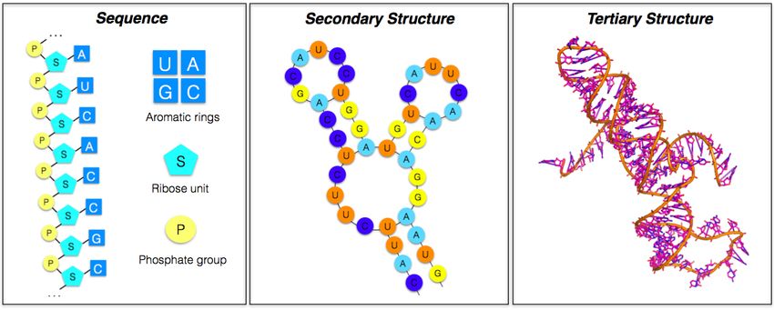
To overcome the lack of structurally resolved RNA, computational methods have com-
plemented experimental efforts to get more insight into how RNA structure and dynamics
determine its functions [7, 8, 9]. Significant efforts have been devoted to the construction of
methods to predict the RNA secondary structure mainly employing thermodynamics-based
models [10]. These methods have been recently achieved significant improvements by the
incorporation of auxiliary structural information from high-throughput chemical probing
technologies [11, 12].
    However, even if the knowledge of the RNA secondary structure provides important
information, it is not sufficient to fully explain RNA function or interactions with other
biomolecules [13]. During the last years lot of attention has been focused on the construction
of RNA 3D structure prediction tools of increasing accuracy and speed [14, 15, 16, 17, 18,
19, 20, 21, 22, 23, 24, 25, 26].
    In this review we provide a concise overview of these methodologies, present their strengths
and limitations, and highlight the open challenges in RNA structure prediction. We will par-
ticularly underline the recent development related to the use of coevolutionary information
to improve the accuracy of the RNA 3D structure prediction methods. The structural in-
formation remains, however, static and provided one piece to the puzzle of RNA function.
Another important component is the dynamics of RNA, for example while undergoing large
conformational rearrangements [27, 28], which is exhaustively covered in the excellent review
[29].

2. From the RNA sequence to its 3D structure
    The basic unit of RNA is the nucleotide that is formed by planar aromatic rings linked
to a ribose unit that in turn is attached to a phospate group (see fig 1). The sequence
of the different constituent nucleotides (adenine, guanine, cytosine, uracil) of a given RNA
molecule is defined as its primary structure.
    Nucleotides typically complement each other by forming the canonical base pairs A-U
and C-G, which maximizes inter-nucleotide hydrogen bonding. This leads to short chains
of nucleotides folding in anti parallel double helices. The nucleotides that do not form
Watson-Crick base pairs can remain unpaired or establish less stable non-canonical base
pairs forming internal and bulge loops, hairpins and junctions. The secondary structure
is thus essentially defined as the set of base pairs occurring in the RNA molecules.
    The tertiary structure is the complete set of three-dimensional coordinates of all atoms
of the RNA structure. This includes formation of a plethora of tertiary motifs such as pseu-
doknots, stacking of helices, multiple base pairing, ribose zipper and loop-loop interactions
that determine the molecular shape in the physical space.
    An accurate computational prediction of the RNA tertiary structure starting from its
sequence is particularly challenging as the 3D structure depends not only on the sequence
but also on the environmental conditions such as the ion concentrations and temperature.

                                              2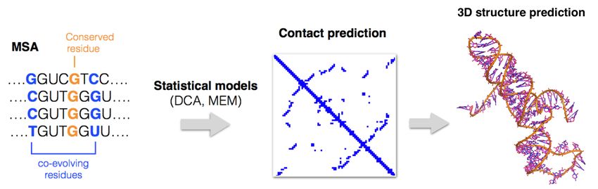
Figure 1: From primary (sequence), to secondary and to tertiary RNA structures.

3. Computation modeling of RNA 3D structure
    Here, we review and compare some widely known methods for the prediction of the three-
dimensional structure of RNA. The available approaches can be roughly divided in three
different types: fragement-based, physics-based and comparative modeling. To compare the
state of the art prediction methods and assess their performance, a blind experiment for the
RNA 3D structure prediction has been established in the last years [30, 31, 32] with the
last round focused on the challenging prediction of six RNA structures of riboswitches and
ribozymes [32].

3.1. Fragment-based homology methods
    The main idea behind this approach is to assemble the 3D prediction of target molecules
using small fragments from libraries with similar sub-sequences. The theoretical justification
of such a procedure comes from assuming that the distribution of the different conformations
observed in known RNA structures for given fragment sequences is a good approximation
for the conformation of similar or identical sub-sequences.
    The basics steps of these methods consist first in the fragmentation of the secondary
structure used as input. As a second step a search algorithm is employed to match these ele-
ments from fragment libraries constructed from databases of known RNA structures. Finally,
all the elements are assembled together using different algorithms (see below) and, usually,
a final refinement stage using atomic force field or coarse-grained potentials is performed.
    One advantage of these methods is their computational efficiency as the fragmentation
assembly drastically reduces the conformational search space. As the structural diversity
of the fragment library directly limits the accuracy of the composed assembly, good results
require a large and diverse library as well as a good scoring function. Here, we list methods
belonging to this class and some of their characteristics.
                                                3
• RNAComposer [15]: after the fragmentation step, the predicted secondary structure
elements (stem, loops and single strands) constitute the input pattern for a search in
the FRABASE 3D fragment data-set developed by the authors. From the matched
elements a 3D structure is constructed by first superimposing and then merging them.
Finally, an energy minimization is performed in the CHARMM force field [33].
• Vfold3D [18] uses a coarse-grained representation of the RNA. First, it utilizes VFold2D,
a free energy-based model, to predict the secondary structure from which it extracts
motifs (helices, hairpin loops, internal loops,...). From these motifs it searches the best
template in the VFoldMTF database. After assembling the 3D structural motifs and
the addition of all atoms to the coarse-grained structure (according to the template)
it performs an all-atom structure refinement.
• 3dRNA [21] uses a two-steps procedure where first the smallest secondary elements
(SSEs) are assembled in hairpins and duplexes one by one following the 5’ to 3’ end
direction. Then, these structures are further assembled into a complete tertiary struc-
ture by selecting the junction component from a junction database. Finally, to assure
the chain connectivity, the assembled model is energy minimized in the AMBER 98
force field [34].
• FARNA [22] (Fragment Assembly of RNA) also uses a coarse-grained representation
of the RNA structure and a fragment assembly strategy employing a Monte Carlo
process that is guided by a low-resolution knowledge-based energy function. The
authors developed knowledge-based base-pairing and base-stacking potentials to which
they add several other terms such as the penalty for steric clashes. The structural
model undergoes a second refining step in an all-atom potential to improve the accuracy
and to better discriminate competing structural models. The two-step protocol is
called FARFAR (Fragment Assembly of RNA with Full Atom Refinement) and is
part of the ROSETTA package.
• MC-Fold/MC-Sym [25] this pipeline uses the combination of small motifs called
nuclotide cyclic motifs (NCMs). The NCM-3D fragments are assigned to the given
sequence by choosing the structure with higher probability of occurencies. Then the
structural NCMs are concatenated using a Las Vegas probabilistic algorithm.
3.2. Physics-based methods
In contrast to the previous methods, the physics-based models do not use template
structures in the assembly of RNA fragment/motifs but derive and parameterize energy
functions depending on specific conformations, similar to approaches applied for proteins
[35, 36]. These methods can be further separated in ab-initio approaches or knowledge-
based approaches. In the latter methods, the energetic functions are derived using the inverse
Boltzmann law from the probability of occurrences of certain sequence-structure elements in
a dataset of known structures. In constrast, the ab-initio methods are based on force fields
adopting usually harmonic potentials for bond lengths and angles, Lennard-Jones potentials
4for Van der Waals interactions, and electrostatic potentials that get reparameterized based
on RNA structure and thermodynamics data.
Such energetic functions are then used in Molecular Dynamics (MD) simulations or
Monte Carlo (MC) minimization often associated with enhanced sampling techniques such
as temperature replica exchange or discrete molecular dynamics simulations in which the
energy function is substituted with discrete step function potentials that drastically reduce
the computational cost of the method.
The strength of the physics-based methods is that they are applicable to sequences with
no known similar sequences or even sub-sequences. Their disadvantage is their need to
explore a large conformational search space which increases computational demands and
decreases their computational efficiency in comparison to fragment-based methods.
Here in the following the list of the computational tools that use this approaches.
• iFoldRNA [20] uses a simplified ”three-bead per nucleotide” representation of the
RNA structure, and it is based on a replica-exchange discrete molecular dynamic
(DMD) simulations protocol to span the conformational space. DMD incorporates
base-pairing and base-stacking interactions into an energy function where in addition
an entropic estimation of the loop formation is also considered.
• NAST [23] uses coarse-grained representation of RNA considering one quasi-atom
per RNA nucleotide base. A simplified knowledge-based energy function, derived from
the observed RNA geometries at the nucleotide level, is used to predict the target
structure by global energy minimization. NAST necessitates as input the (known or
predicted) secondary structure information and accepts also tertiary contacts to guide
the folding.
• SimRNA [17] uses a coarse-grained representation of the RNA structures reducing the
number of explicitly represented atoms per residue from about thirty to only five. It
is based on dedicated RNA statistical potentials to compute the structure free energy
and identify the native structure via Monte Carlo sampling.
3.3. Comparative homology-based modeling
Another type of methods uses homology modeling approaches by identifying structurally
related template and geometrically aligning residues from the target onto corresponding
residues in the template. Examples of this type of methods are RNABuilder [37] and
ModeRNA [16]. The latter makes also extensively use of the evolutionary information
by using multiple RNA sequence alignments to better reveal patterns of conservation that
improve the accuracy of the prediction starting from the 3D template.
In order to improve the accuracy of homology-based methods it has been shown that the
addition of multiple templates can be successfully employed. Another characteristic that
is common to this type of methods is that they model (short) regions with no template
by employing fragment-based insertion approaches. Finally the methods perform usually a
geometry optimization using a force field in order to obtain physically reasonable conforma-
tions. The RNABuilder method for example uses a multi-resolution approach that handles
5at different level of resolution the forces, rigidifying certain bonds, residues or molecules part
while keeping flexible the others.
The major drawback of this class of methods resides in the difficulty of having the
template structure for the given sequence and an informative multiple sequence alignment.
Indeed for the RNA structure templates there is the limitation of the number of structure
deposited in the widely known database [3]. Regarding the alignments one can use those
available for many RNA families in the Rfam database [38] or perform alignment via com-
monly used multiple RNA sequence alignment packages such as R-Coffe [39], Muscle [40] or
Infernal [41].
The strength of these methods is their high accuracy with modest computational costs
when good structural templates can be found. Their performance drops in the absence of
such templates.
3.4. Performance assessment and RNA-Puzzles prediction
Most of the analyzed RNA structure prediction methods participated in the RNA-Puzzle
competitions [30, 31, 32] in which a set of experimentally resolved RNA 3D structures had
to be blindly predicted. To assess the performance of the predictors and rank the models,
different metrics have been used such as the root mean square deviation (RMSD) between
the predicted the experimental crystal structures that gives a more global information about
the model’s accuracy or the deformation index and the complete deformation profile matrix
that instead capture the ”local” accuracy at the nucleotide interaction level.
In the first RNA-Puzzle round [30] in addition to two simple small targets that were
relatively well predicted, the more challenging riboswitch structure were not accurately re-
produced with a mean RMSD accuracy of about 15 Å. Moreover while most methods achieve
good performance on Watson-Crick base pairs, non-Watson-Crick interactions remain diffi-
cult to predict and clash score remains generally quite high.
In the second RNA-Puzzle round [31] the best RMSDs for a long nucleotide sequence
range between 6.8 and 11.7 Å indicating a global improvement of methods’ performance.
A substantial amelioration for non-Watson-Crick interactions prediction is also observed.
Finally in the last RNA-Puzzle competition [32], the predictions achieved a consistently
high level of accuracy especially when a high-homology template can be identified. For
example in the case of the SAM-I riboswitch aptamer prediction that has as template (PDB
code 3QIR), the average RMSD over all predicted models is about 4.3 Å, with a standard
deviation of less then 2 Å.
Unfortunately, when the homology with the template is not high enough, the accuracy
of methods is still not satisfactory and depends on the length of the RNA sequence. Small
RNA sequences can be predicted with good accuracy as exemplified in the case of the ZTP
riboswitch predicted with an averaged RMSD of about 6 Å and as also shown in the previous
RNA-Puzzle round. For long sequences such as the ydaO riboswitch no method is capable
of reliably predicting the native three dimensional conformation with an average RMSD of
about 16 Å.
In order to improve the structure prediction of these challenging targets, there is a need
for new and more performing algorithms. In the next section we will thus present recent
6progress in this direction and more in detail we will show how the coevolutionary information
can been used to improve significantly the methods’ accuracy.
Figure 2: 3D RNA structure prediction methods and their principal characteristics
4. Including evolutionary information to improve 3D structure prediction
4.1. Residue co-evolution and contact prediction
A significant amount of data obtained from high-throughput sequencing technologies
provides us an invaluable source of evolutionary information that can be used in order to
improve the protein [44, 45, 42, 50, 46] and RNA structure prediction [24, 49]. The basic
idea behind these approaches is tracing co-variation of amino acid or nucleic acid pairs in
proteins and RNA belonging to homologous families. Such co-variation indicates structural
7proximity of the involved residues and is hence related to biomolecular structural and sta-
bility properties. Compensatory mutations occur when a mutation with a detrimental effect
at a given site, interact with a secondary mutations at another site to restore the molec-
ular fitness [47] thus indicating the tendency of co-evolving residues to represent physical
interactions that are important for the stability and function of biomolecules.
In the last decade many statistical methods have been developed to identify co-evolving
residue pairs in a multiple sequence alignment (MSA) [48]. One can assume that such
correlation occurs due to the spatial proximity of the two residues even if it can also arise
from indirect effects related to the transitivity of the interaction between pairs and tertiary
residues.
The use of the statistical methods such as maximum entropy models (MEM) or direct
coupling analysis [24, 44, 46, 50] allows to unravel the transitive effects in the network
of constrained residue-residue interactions and thus they give more efficient and robust
contact-prediction. Using these statistical-based approaches one can detect long-range ter-
tiary contacts from sequence covariation whose prediction difficulty has been one of the main
limitation to the advancement of the computational RNA 3D structure prediction methods.
Figure 3: Statistical-based contact prediction from coevolutionary data improved the 3D RNA structure
prediction
4.2. Direct coupling analysis (DCA)
The basic assumption of this method is to associate the probability of observation P (σ)
of a given sequence σ = (a1 , a2 ...aL ) of length L in a MSA to the Hamiltonian energetic
function H(σ) using the Boltzmann law
1 −βH(σ)
P (σ) =
e (1)
Z
where β is the temperature and Z is the partition function of the system, and where the
Hamiltonian is assumed to have the following simplified form
8L
X L−1 X
X L
H(σ) = − hi (ai ) − Jij (ai , aj ) (2)
i i j=i+1
consisting only of single site terms, i.e. hi (ai ), and residue pair interactions Jij (ai , aj ).
These parameters can be inferred from the MSA using a plethora of different approaches.
For example in [49] a pseudo-maximum likelihood (pmlDCA) approximation have been
employed, while a computational intensive message-passing algorithm (mpDCA) is used in
[44] and a more efficient mean field algorithm (mfDCA) in [50]. The list of other type of
popular algorithm used in the inverse inference step can be found in [24].
There are also pitfalls. Frequently, some species are over-represented in the MSA, e.g.
because of their medical importance or the ease of handling them experimentally. Thus,
these sequences need to be re-weighted. In addition, the quality of the MSA such as the
proper placement of gap regions influences contact prediction accuracy. This loss of contact
prediction precision directly leads to a decreased quality of 3D prediction. Another drawback
is that the DCA prediction of the tertiary contacts is far from being perfect with only a
modest overall true positive (TP) predicted contacts; it should be noted, however, that only
relatively few (O(10)) higher ranking pairs that show higher TP rate are already sufficient
to boost the performance of the structural modeling. Still, these methods significantly boost
performance without too much computational effort.
4.3. Contact guided 3D RNA-structure prediction
While the use of coevolutionary data has been already fruitfully applied to protein struc-
ture determination during the last decade [42, 43, 44, 45, 46, 50, 51, 52, 53], the contact
guided prediction of the three-dimensional RNA structure is relatively new. Indeed, the
previous mutual information (local) approach to the extraction of coevolution signals from
MSA was not sufficiently accurate [56] to provide reliable tertiary contact predictions.
Recent investigations [24, 49, 55] instead show that the use of a global approach to extract
the top-ranked site-pairs with stronger co-evolutionary signals can be efficiently employed
as distance constraints in modeling tools.
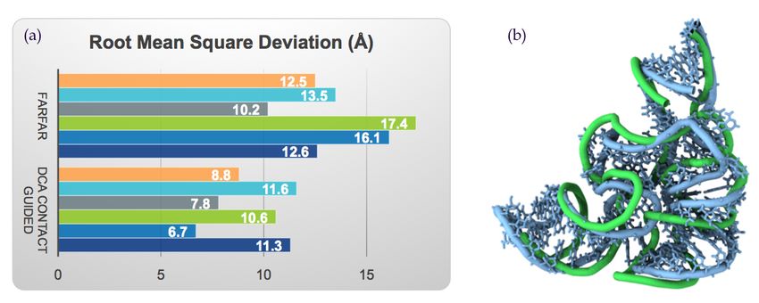
In [24] the authors show that in the prediction of the structure of six representative
riboswitches with the Rosetta-based method FARFAR, the use of predicted tertiary contacts
by mfDCA improves the RMSD in average by about 30% with respect to the case in which
only secondary structure information (SSI) is provided. In figure 4 we report this explicit
comparison for all the six structure considered.
These results have been confirmed in [42] where the authors show a significant improve-
ment of prediction quality when the evolutionary based contact prediction computed via
the pmlDCA approach. In this work, contacts are used as spatial constraints in the NAST
coarse-grained structure prediction method. A further confirmation in [55] highlights that
prediction RMSDs for the same structures as analyzed in [24, 42] are lowered by about 30%
when using the tertiary contacts predicted via mfDCA in the 3dRNA method [21] com-
pared to not using such tertiary contact constraints. [42] and [57] also demonstrated how
9Figure 4: (a) DCA contact-guided RNA structure prediction improvement with respect to the state of the
art (Rosetta-based) method for the six riboswitches from [24]. (b) Overlay of the DCA-contact guided
predicted (blue) and the experimental structure (green) for the thiamine pyrophosphate-specific (TPP)
riboswitch (PDB code 2gdi). In the prediction, the first 100 top contacts as computed via mean field DCA
from the MSA of the RF00059 family have been used as constraints in the FARFAR method.
DCA-based methods show good accuracy in the prediction of intermolecular RNA-protein
contacts.
5. Future challenges and outlook
Even if in the last decade tremendous advances has been achieved in RNA structure
prediction, its accuracy still is not as high as for protein structure prediction. Moreover
there are open and intriguing challenges in the field that will hopefully be tackled in the
close future:
• The role played by the environmental conditions such as ions that strongly influence
the RNA structure has to be fully investigated and clarified [58, 59, 60]. Since in vivo
RNA can adapt different conformations with respect to in vitro ones, this will be also
important to understand such differences and give important information for RNA
biology.
• In the next years, thanks to the advancement of the next generation sequencing tech-
nologies, the amount of sequence information will continue to increase exponentially.
Currently coevolutionary methods focus on the prediction of two-site interactions (con-
tacts), but this increased amount of information promises to also allow to predict higher
order correlations that could further boost structure prediction methods.
• Further improvements of RNA force fields will continue to increase the accuracy of
predictions. These can help to better understand the role of the different RNA confor-
mations, their stability and to gain new insights about the RNA structural dynamics.
10• Combining structure prediction methods or simulations with experimental data such
as Selective 2-hydroxyl acylation analyzed by primer extension (SHAPE) [61], Flu-
orescence Resonance Energy Transfer (FRET) [62] or small angle X-Ray scattering
(SAXS) [63, 64, 65] will allow to probe RNA structures where a single method fails
[66].
• Inter-molecular protein interactions and contacts can be predicted via DCA and related
methods [67]. This could be transferred to RNA.
• Finally, it becomes more and more clear that base modifications such as the methyla-
tion or deamination play an important role in RNA biology by modifying the structure
as well as the function of RNA. It could be thus of great interest in the next future
to address and investigate these (epi)transcriptomics data to better understand all
biological processes in which the RNA is involved.
References
[1] Y. Wan, M. Kertesz, R.C. Spitale, E. Segal, H. Chang, Understanding the transcriptome through RNA
structure, Nat. Rev. Genet. 12, 10.1038 (2011).
[2] H.M. Berman, J. Westbrook, Z. Feng, G. Gilliland, T.N. Bhat, H. Weissig, Shindyalov I.N., Bourne
P.E.. The Protein Data Bank H.M., Nucl. Ac. Res., 28: 235-242 (2002).
[3] B.C. Narayanan at al., The Nucleic Acid Database: new features and capabilities, Nucl. Ac. Res. 42,
D114-D122 (2013).
[4] R. Schroeder, A. Barta, K. Semrad, Strategies for RNA folding and assembly. Nat. Rev. Mol. Cell.
Biol. 5, 908-919 (2004).
[5] P. Kapranov, G. St Laurent, Dark Matter RNA: Existence, Function, and Controversy. Front. Genet.
23, 10.3389/fgene.2012.00060 (2012).
[6] K.R. Chi, The dark side of the human genome. Nature 538, 10.1038/538275a (2016).
[7] M. Zuker, P.O. Stiegler, Optimal computer folding of large RNA sequences using thermodynamics and
auxiliary information, Nucleic Acids Res. 9, 133-48 (1981).
[8] K. Aigner, F. Dressen, G. Stege, Methods for Predicting RNA Secondary Structure, RNA 3D Structure
Analysis and Prediction, 19-41, Springer (2012).
[9] S.A. Mortimer, M.A. Kidwell, J.A. Doudna, Insights into RNA structure and function from genome-
wide studies, Nat Rev Genet. 15, 469-79 (2014).
[10] J.S. Reuter and D.H. Mathew, RNAstructure: software for RNA secondary structure prediction and
analysis, BMC Bioinformatics 11, 129 (2010).
[11] P. Ge, S. Zhang, Computational analysis of RNA structures with chemical probing data, Methods.
79-80,60-6 (2015).
[12] R. Lorenz, M.T. Wolfinger, A. Tanzer, I.L. Hofacker, Predicting RNA Secondary Structures from
Sequence and Probing Data, Methods 103, 86-98 (2016).
[13] C. Laing, T.J. Schlick, Computational approaches to 3D modeling of RNA, Phys Condens Matter. 22,
283101 (2010).
[14] M. Rother, K. Rother, T. Puton, J.M. Bujnicki, ModeRNA: a tool for comparative modeling of RNA
3D structure, Nucl. Ac. Res. 39, 4007-22 (2011).
[15] M. Popenda et al., Automated 3D structure composition for large RNAs, Nucl. Ac. Res. 40, e112
(2012). M. Biesiada, K.J. Purzycka, M. Szachniuk, J. Blazewicz, R.W. Adamiak, Automated RNA 3D
Structure Prediction with RNAComposer, Methods Mol. Biol. 1490, 199-215 (2016).
[16] M. Rother, K. Rother, T. Puton, J.M. Bujnicki, ModeRNA: a tool for comparative modeling of RNA
3D structure, Nucl. Ac. Res. 39, 4007-22 (2011).
11[17] M.J. Boniecki et al., SimRNA: a coarse-grained method for RNA folding simulations and 3D structure
prediction, Nucl. Ac. Res. 20, 44 (2016).
[18] C. Zhao, X. Xu, S.J. Chen, Predicting RNA Structure with Vfold, Methods Mol. Biol. 1654, 3-15
(2017).
[19] C.Y. Cheng, F.C. Chou, R. Das, Modeling complex RNA tertiary folds with Rosetta, Methods Enzym.
553, 35-64 (2015).
[20] F. Ding, S. Sharma, P. Chalasani, V.V. Demidov, N.E. Broude, N.V. Dokholyan, Ab initio RNA folding
by discrete molecular dynamics: from structure prediction to folding mechanisms, RNA 14, 1164-73
(2008).
A. Krokhotin, K. Houlihan, N.V. Dokholyan, iFoldRNA v2: folding RNA with constraints, Bioinfor-
matics 31, 2891-3 (2015).
[21] Y. Zhao, Y. Huang, Z. Gong, Y. Wang, J. Man, Y. Xiao, Automated and fast building of three-
dimensional RNA structures, Sci Rep. 2,734 (2012).
[22] R. Das, D. Baker, Automated de novo prediction of native-like RNA tertiary structures, Proc. Natl.
Acad. Sci. U. S. A. 11, 104 (2007).
[23] C. Flores, R.B. Altman, Coarse-grained modeling of large RNA molecules with knowledge-based po-
tentials and structural filters, RNA 15, 1769-1778 (2010).
[24] E. De Leonardis et al., Direct-coupling analysis of nucleotide coevolution facilitates RNA secondary
and tertiary structure prediction, Nucl. Ac. Res. 43, 10444-10455.
[25] M. Parisien, F. Major, The MC-Fold and MC-Sym pipeline infers RNA structure from sequence data,
Nature 452, 5155 (2008).
[26] T. Cragnolini, P Derreumaux and S. Pasquali, Ab initio RNA folding, J. Phys.: Condens. Matter 27,
233102 (2017).
[27] P.C. Whitford, et al., Nonlocal helix formation is key to understanding S-adenosylmethionine-1 ri-
boswitch function, Biophys. J. 96:2, 2-9 (2009).
[28] B. Lutz, et al., Differences between cotranscriptional and free riboswitch folding, Nucl. Ac. Res. 42,
26872696 (2014).
[29] J. Sponer et al., RNA Structural Dynamics As Captured by Molecular Simulations: A Comprehensive
Overview, Chem. Rev., 118, 4177-4338 (2018).
[30] J.A. Cruz et al., RNA-Puzzles: a CASP-like evaluation of RNA three-dimensional structure prediction,
RNA 18, 610-625 (2012).
[31] Z. Miao et al., RNA-Puzzles Round II: assessment of RNA structure prediction programs applied to
three large RNA structures, RNA 21, 1066-1084 (2015).
[32] Z. Miao et al.,RNA-Puzzles Round III: 3D RNA structure prediction of five riboswitches and one
ribozyme, RNA. 23, 655-672 (2017).
[33] B.R. Brooks et al., CHARMM: The Biomolecular Simulation Program, J Comput Chem. 30, 1545-1614
(2009).
[34] D.A. Case et al.,AMBER 2018, University of California, San Francisco (2018).
[35] A. Schug, T. Herges, W. Wenzel, Reproducible protein folding with the stochastic tunneling method,
Phys. Rev. Lett. 91(15), 158102 (2003).
[36] A. Schug, et al., Comparison of stochastic optimization methods for allatom folding of the trpcage
protein, ChemPhysChem 6(12), 2640-2646 (2005).
[37] S.C. Flores, Y. Wan, R. Russel, S.B. Altman, Predicting RNA structure by multiple template homology
modeling, Pac. Symp. Biocomput. 216-227 (2010).
[38] S. Griffiths-Jones, A. Bateman, M. Marshall, A. Khanna, S.R. Eddy, Rfam: an RNA family database,
Nucl. Ac. Res. 31, 439-441 (2003).
[39] P. Di Tommaso et al., T-Coffee: a web server for the multiple sequence alignment of protein and RNA
sequences using structural information and homology extension, Nucl. Ac. Res. 39, W13-W17 (2011).
[40] R.C. Edgar, MUSCLE: multiple sequence alignment with high accuracy and high throughput, Nucl.
Ac. Res. 32, 1792-1797 (2004).
[41] E. P. Nawrocki and S. R. Eddy, Infernal 1.1: 100-fold faster RNA homology searches, Bioinformatics
1229, 2933-2935 (2013)
[42] D.S. Marks et al., Protein 3D structure computed from evolutionary sequence variation, PLoS ONE 6
(2011).
[43] T.A. Hopf, L.J. Colwell, R. SheridaN, B. Rost, C. Sander, D.S. Marks, Three-dimensional structures
of membrane proteins from genomic sequencing Cell. 149, 1607-1621 (2012).
[44] M. Weigt, R.A. White, H. Szurmant, J.A. Hoch, T. Hwa Identification of direct residue contacts in
protein-protein interaction by message passing. Proc. Natl. Acad. Sci. USA. 106, 67-72 (2009).
[45] A. Schug, M. Weigt, J.N. Onuchic, T. Hwa, H. Szurmant, High-resolution protein complexes from
integrating genomic information with molecular simulation, Proc. Natl. Acad. Sci USA 106(52): 22124-
22129 (2009).
[46] A.E. Dago et al., Structural basis of histidine kinase autophosphorylation deduced by integrating ge-
nomics, molecular dynamics, and mutagenesis, Proc. Natl. Acad. Sci. USA 109, E1733-E1742 (2012).
[47] D.N. Ivankov, A.V. Finkelstein, F.A. Kondrashov, A structural perspective of compensatory evolution,
Curr. Opin. Struct. Biol. 26, 104-112 (2014).
[48] D. de Juan, F. Pazos, A. Valencia, Emerging methods in protein co-evolution, Nat Rev Genet. 14,
249-61 (2013).
[49] C. Weinreb, A. Riesselman, J.B. Ingraham, T. Gross, C. Sander, D.S. Marks, 3D RNA and functional
interactions from evolutionary couplings, Cell. 165, 963-975 (2016).
[50] Morcos F, Pagnani A, Lunt B, Bertolino A, Marks DS, Sander C, Zecchina R, Onuchic JN, Hwa T,
Weigt M, Direct-coupling analysis of residue coevolution captures native contacts across many protein
families, Proc. Natl. Acad. Sci. USA 108, E1293-E1301 (2011).
[51] S. Ovchinnikov et al., Protein structure determination using metagenome sequence data, Science 355,
294-298 (2017).
[52] J.I. Sulkowska, F. Morcos, M. Weigt, T. Hwa, J.N. Onuchic, Genomics-aided structure prediction Proc.
Natl. Acad. Sci USA 109, 10340-10345 (2012).
[53] D.T. Jones, T. Singh, T. Kosciolek, S. Tetchner, MetaPSICOV: combining coevolution methods for
accurate prediction of contacts and long range hydrogen bonding in proteins. Bioinformatics 31, 9991006
(2015).
[54] Zerihun MB, Schug A, Biomolecular coevolution and its applications: Going from structure prediction
toward signaling, epistasis, and function, Biochem Soc. Trans. 45, 1253-1261 (2017).
[55] J. Wang, K. Mao, Y. Zhao, C. Zeng, J. Xiang, Y. Zhang, and Y. Xiao, Optimization of RNA 3D struc-
ture prediction using evolutionary restraints of nucleotidenucleotide interactions from direct coupling
analysis, Nucl. Ac. Res. 45, 6299-6309 (2017).
[56] J.Y. Dutheil , F. Jossinet, E. Westhof, Base pairing constraints drive structural epistasis in ribosomal
RNA sequences. Mol. Biol. Evol. 27, 18681876 (2010).
[57] Q. Zhou et al.,Global pairwise RNA interaction landscapes reveal core features of protein recognition,
Nat. Commun. 9, 2511 (2018).
[58] D.E. Draper, A guide to ions and RNA structure, RNA 10, 335-43 (2004).
[59] Y. Zhu, Z. He, S.-J. Chen, TBI Server: A Web Server for Predicting Ion Effects in RNA Folding, PLoS
ONE 10, e0119705 (2015).
[60] S. Roy, S.P. Hennelly, H. Lammert, J. N. Onuchic, K.Y. Sanbonmatsu, Magnesium controls aptamer-
expression platform switching in the SAM-I riboswitch, Nucleic Acids Research, gky1311 (2019).
[61] S. Kirmizialtin, S.P. Hennelly, A. Schug, J.N. Onuchic, K.Y. Sanbonmatsu, Integrating molecular
dynamics simulations with chemical probing experiments using SHAPE-FIT, Meth. Enzym. 553: 215-
234 (2015).
[62] I. Reinartz et al., Simulation of FRET dyes allows quantitative comparison against experimental data,
J. Chem. Phys. 148(12): 123321-6 (2018).
[63] Z. Xia, D.R. Bell, Y. Shi, P. Ren, RNA 3D Structure Prediction by Using a Coarse-Grained Model and
Experimental Data, J. Phys. Chem. B 117, 31353144 (2013).
[64] Weiel M, Reinartz M., and Schug, A., Rapid interpretation of small-angle X-ray scattering data, PloS
Comp. Biol., 3, e1006900 (2019).
13[65] A. Gupta et al., Formation of a Secretion-Competent Protein Complex by a Dynamic Wrap-around
Binding Mechanism, J. Mol. Bio., 430:3157-3169 (2018).
[66] A. Ponce-Salvatierra et al., Computational modeling of RNA 3D structure based on experimental data,
Biosci. Rep. 39, BSR20180430 (2019).
[67] H. Szurmant and M. Weigt, Inter-residue, inter-protein and inter-family coevolution: bridging the
scales, Curr. Op. Struct. Biol. 50, 26-32 (2018).
14You can also read