SKINSCAN: LOW-COST 3D-SCANNING FOR DERMATOLOGIC DIAGNOSIS AND DOCUMENTATION - IVPL
←
→
Page content transcription
If your browser does not render page correctly, please read the page content below
SKINSCAN: LOW-COST 3D-SCANNING FOR DERMATOLOGIC DIAGNOSIS AND
DOCUMENTATION
Merlin A. Nau1,2∗ , Florian Schiffers1 , Yunhao Li1 , Bingjie Xu1 , Andreas Maier2
Jack Tumblin1 , Marc Walton4 , Aggelos K. Katsaggelos1,3,5 , Florian Willomitzer3 , Oliver Cossairt1,3
1
Department of Computer Science, Northwestern University, Evanston, USA
2
Pattern Recognition Lab, Friedrich-Alexander-Universität Erlangen-Nürnberg, Germany
3
Department of Electrical and Computer Engineering, Northwestern University, Evanston, USA
4
Center for Scientific Studies in the Arts, Northwestern University, Evanston, USA
arXiv:2102.00508v1 [eess.IV] 31 Jan 2021
5
Department of Radiology, Northwestern University, Chicago, USA
∗
merlin.nau@fau.de
ABSTRACT
The utilization of computational photography becomes
increasingly essential in the medical field. Today, imag-
ing techniques for dermatology range from two-dimensional
(2D) color imagery with a mobile device to professional
clinical imaging systems measuring additional detailed three-
dimensional (3D) data. The latter are commonly expensive
and not accessible to a broad audience. In this work, we pro-
pose a novel system and software framework that relies only
on low-cost (and even mobile) commodity devices present in
every household to measure detailed 3D information of the
human skin with a 3D-gradient-illumination-based method. /
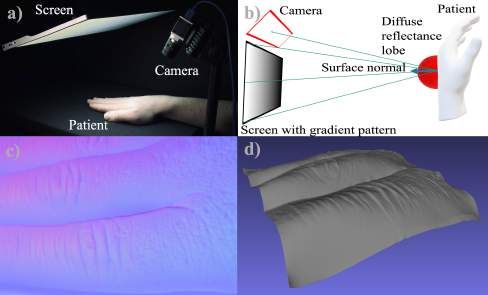
We believe that our system has great potential for early-stage Fig. 1: a) Experimental setup with a vision camera and a
diagnosis and monitoring of skin diseases, especially in vastly lightweight screen. b) Illustration of gradient illumination.
populated or underdeveloped areas. c) The recovered normal map. d) 3D reconstruction
Index Terms— Topographic Imaging, Three-Dimensional
Imaging, Photometric Stereo, Dermatologic Imaging
assessing asymmetries of the lesion shape, border irregulari-
ties, color variegation, the lesion’s diameter and the evolution
1. INTRODUCTION
of shape and color. Currently, it is most common to eval-
uate the above criteria solely based on 2D image informa-
3D scanning provides access to a plethora of useful features
tion. However, since lesions can also grow in height, this
in the diagnosis and documentation of skin diseases [1].
evaluation can be significantly improved with additional ac-
For example, skin cancer is the most common cancer in the
cess to 3D topographic information of the skin [5] (in fact,
United States developed by every fifth person throughout
melanomas at a young age are misdiagnosed by up to 40%, in
their lifetime [2]. While skin cancer is treatable, early detec-
comparison with pigmented lesions [6]). More to the point,
tion is essential for a successful prognosis. Therefore, routine
surface orientation on skin cancer is a feature that can support
screenings are vital to ensure an early-stage diagnosis and
the distinction between malign and benign tissue [7]. Al-
therapy [3].
though increasingly used by professional physicians in their
Generally, the most important evaluation for a correct di-
diagnosis and documentation, clinical 3D imaging techniques
agnosis of skin cancer is based on the ABCDE [4] guided rule
to measure human skin like optical coherence tomography [8]
Thanks to Northwestern Alumnae for funding. or laser-based scanners [9], are tied to expensive hardware,
©2021 IEEE. Personal use of this material is permitted. Permission from are not portable, and hence are not accessible to a broad audi-
IEEE must be obtained for all other uses, in any current or future media, ence.
including reprinting/republishing this material for advertising or promotional
purposes, creating new collective works, for resale or redistribution to servers In this work, we ask the question: Can routine examina-
or lists, or reuse of any copyrighted component of this work in other works. tions of skin disease be augmented with low-cost but precise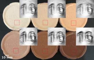
3D imaging techniques to improve assessment, monitoring,
and diagnosis effectiveness? Mobile applications for phones
or tablets that allow the exchange of images between the med-
ical doctor and patient are broadly available [10]. However,
these apps only allow for patient-driven image capture of 2D
RGB images. Currently patients cannot capture and transfer
3D information of their skin abnormalities to a medical doctor
for further evaluation. This feature becomes even more crit-
ical during the time of the COVID pandemic. The main ad-
vantages of 3D over 2D imaging are that the data is invariant        Fig. 2: Illustration of the gradient illumination procedure. Im-
to the translation and rotation of the object, surface texture,      ages are acquired under each gradient pattern in horizontal
and external illumination. These advantages can significantly        and vertical direction. From there the directional images are
improve the repeatability of patient-driven measurements, en-        subtracted, forming x and y component of the normal map.
abling a better dermatological evaluation across different skin
tones, pigmentation variations, as well as, skin conditions or
tattoos.                                                             [17] are not able to resolve fine details in 3D and are not fea-
     While some success has been made in developing state-           sible for our proposed task.
of-the-art artificial intelligence classification systems for skin        Active techniques, such as time-of-flight [18] or active
diseases from 2D images alone, the initial attempts also             triangulation (“structured-light”) [19], employ an additional
leave room for significant improvement [11]. In particular,          light-source or projector to encode depth-information into
additional depth information could prove of benefit for Deep-        the measurement. While active triangulation techniques
Learning (DL) techniques by enriching the feature space.             would provide the required measurement precision on human
     Previously, several approaches using structure-from-            skin [19], the hardware necessary to perform the respective
motion or triangulation (“structured ligh”) have been explored       measurement is still rarely found in consumer devices. An-
for mobile wound documentation and measurement [12].                 other active method able to measure small surface details is
However, most open source proposed methods turned out                Photometric Stereo (PS) [20].
to be too imprecise to provide detailed structural images of              Here, multiple light sources are placed around the diffuse
skin topography [12]. Active triangulation techniques per-           object, capturing images with each light source switched on
form significantly better but require a suitable and expensive       and the remaining lights off. Compared to 3D estimations
sensor [5].                                                          from shading in a single frame, the illumination from various
     In this paper, we propose to use mobile gradient illumina-      directions supports the reliability of results. To be concise,
tion [13] to build a low-cost measurement system for derma-          the amount of reflected light is dependent on the light source
tological evaluation of skin. In particular, our main contribu-      positions relative to the object. Thus, one can resolve the sur-
tions are                                                            face orientation. Debevec et al. [21] extended PS by the idea
    1. A method for inexpensive 3D capture of skin using             of placing multiple light sources and cameras in a Light stage
        commodity hardware (like screens and webcams, or             surrounding the object of interest. However, the complicated
        tablets)                                                     and bulky setup makes it difficult to apply it in a mobile set-
    2. Preliminary results of 3D skin capture using the pro-         ting.
        posed method                                                      Ma et al. [22] showed that a screen displaying intensity
    3. Development of an open-source framework for gradient          gradients could replace multiple light sources. The resulting
        illumination-based 3D capture of skin.                       Gradient Illumination (GI) approach can recover accurate 3D
While this paper’s focus is on the analysis and documentation        information by using a single camera with a polarization lens
in medical 3D imaging, the method we have developed could            and a half-spherical-shaped light source. In 2016, Riviere et
also have much broader applications. For instance, industrial        al. [13] proposed a mobile extension by using gradients dis-
object inspection or conservation in cultural heritage poses         played on the tablet screen to recover the surface normals for
exciting scenarios for a mobile and low-cost 3D method [14].         specular and diffuse reflection by placing a polarization lens
                                                                     on the front camera. All these so-called “reflectance-based”
           2. 3D IMAGING OF HUMAN SKIN                               methods are highly dependent on the reflective properties of
                                                                     the objects of interest. For specular objects, Willomitzer et
3D imaging poses a set of challenges with no universal solu-         al. [14] proposed the idea of utilizing a tablet to perform de-
tion. Achieving reliable measurements for different materials        flectometry measurements using the screen and front camera.
at arbitrary spatial and temporal resolutions is not feasible.       However, this system is not tailored to measure rather diffuse
Passive methods, such as Passive stereo [15], Structure from         objects like human skin. This paper’s method offers substan-
Motion [16], or even single-frame DL-based depth estimation          tially improved results on human skin.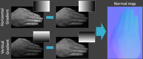
components of the normal by:

                                                                                            nx,y = rx,y − r̂x,y                (4)

                                                                    Finally, we estimate the z-component assuming a normal by:
                                                                                               q
                                                                                        nz = 1 − n2x − n2y                  (5)

                                                                    Open source software framework
    (a) Colorchecker                  (b) Calibration Curve
                                                                    The methods presented in this paper are part of an open-
     Fig. 3: Radiometric calibration using a color chart.           source software framework that can be downloaded from
                                                                    1
                                                                      . The framework is implemented in Python and has been
                                                                    tested on Ubuntu, macOS, and Windows. To encourage indi-
                       3. METHODS                                   vidual adjustments and extensions, we subdivide our project
For our desired application of measuring human skin, we             in an object-oriented manner with base classes for compo-
identify gradient illumination as a suitable measurement            nents necessary in any structured light setup. These include
technique. A depth map can be obtained from the mea-                a camera, projector, image reconstruction, and calibration
sured surface normal data by integration algorithms, such           class. Consequently, the classes for the specific cameras,
as, the Frankot-Chellapa algorithm [23]. Gradient illumina-         projectors, and reconstruction and radiometric, intrinsic, and
tion employs an electronic display where each pixel can be          extrinsic calibration are inherited from these base classes,
considered as an individual light-source.                           allowing specific adjustments. Namely, we have adapted
    Our experimental setup consists of only a programmable          camera classes for Basler cameras, the Pi-Cam and Web-
screen and a camera (see Fig. 1) and, in principle, can be im-      cams. The projection unit provides functionality for any
plemented with just a single mobile device (e.g., smartphone        display or projector.
or tablet) according to the solution presented in [14]. When            Radiometric calibration is accomplished by placing a
imaging objects with Lambertian reflectance, one can define         color chart next to the sample, as displayed in Fig. 3a. Con-
the observed radiance r from a viewing position ~v (e.g., a         sequently, the camera’s intensity values are linearized by
camera ray in Fig. 2h) as an integration over a product of the      applying a gamma correction, see Fig. 3b. The last pattern
incident illumination direction vector ω
                                       ~ created by the gradi-      displayed is always a constant illumination at maximum in-
ent patterns P (~
                ω )x,y programmed on the screen, and the bidi-      tensity, which allows for normalization across the frames.
rectional reflectance distribution function (BRDF). For dif-        Intrinsic calibration is implemented using the OpenCV tool
fuse objects the BRDF R is approximated as the product of           kit [24]. Extrinsic calibration is accomplished by placing
the diffuse albedo ρd and the foreshortening factor, dependent      a mirror with ChArUco markers [25] on the edges under
   ~ and the surface normal direction ~n = (nx , ny , nz )T :
on ω                                                                the screen. The pose of the mirror is retrieved from the
                                                                    ChArUco markers. Further, a checkerboard pattern is dis-
                  ω , ~n) = ρd · max(~
                R(~                  ω · ~n, 0)               (1)   played on the screen. Consequently, the camera captures
                                                                    the reflection of the pattern in the mirror. Thus rotation and
                            Z                                       translation between camera, object, and screen are retrieved.
                 r(~v ) =       Pi (~
                                    ω )R(~
                                         ω , ~n)d~
                                                 ω            (2)   The components are then used in dedicated calibration and
                            Ω
                                                                    capture sessions, yielding the calibration data, normal maps,
     Because a screen can only project gradients along two          integrated depth maps, point clouds, and color images. The
 directions, e.g., x and y, we assume ~n to be aligned with         results are bundled and saved in a folder.
~z = [0, 0, 1], which coincides with the centroid of a diffuse
 reflectance lobe of a flat surface pointing towards the screen.                               4. RESULTS
 By integrating Eq. 2 the radiance is resolved according to [22]
 as                                                                 In this section, we present reconstructed surface normal maps
                                       2πρd                         from different objects. Our experiments’ setup consists of
                       rx (~v ) = nx (      )                (3)
                                        3                           a 15 inch LCD screen and a Basler ace 2 grayscale surface
     Compared to Ma et al. [22], we do not integrate over the       camera, both steadily mounted on a monitor arm and tripod,
entire hemisphere. Instead, the integration is only performed       facing the object of interest. A radiometric calibration has to
in the x and y directions. Displaying the gradient patterns in      be employed to ensure consistency of the computed normal
both x and y directions for both [0, 1] and [1, 0], we calculate    orientations over a surface with color variance. Therefore a
rx , ry and r̂x r̂y , respectively. We can resolve the x and y        1 https://github.com/merlzbert/SkinScan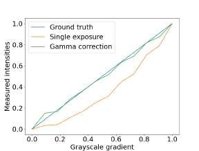
a)                           b)                                the rule may rely on color images only but can be significantly
                                                                     supported by shape estimation. For example, color is not di-
                                                                     rectly coupled to the depth of a mole. Furthermore, shape and
                                                                     color boundaries do not always coincide. Finally, the color
                                                                     of an image is dependent on ambient lighting. Secondly, we

d)
 d)   c)                           d)
                                                                     imaged a banana peel where the skin has darkened over sev-
                                                                     eral areas of the peel. We have further inserted small razor
                                                                     cuts as well as dents pushed in with a finger. Specifically, it
                                                                     is essential to point out that the region around the cuts and
                                                                     dents has darkened. Color variations can make the assess-
                                                                     ment of 3D structures from (2D) images hard. As expected,
      e)             f)
                                                                     the color variation is not visible in the normal map as the sur-
                                                                     face orientation remains unchanged. This shows that gradient
                                                                     illumination is independent of the surface texture albedo and
                                                                     indeed provides structural information. In particular, the cuts
                                                                     and dents are visible. While the reflectance properties of ba-
                                                                     nanas are quite different from human skin, the example still
                                                                     shows that our method can factor the reflectance information
                                                                     from 3D shape features similar to what dermatological appli-
                                                                     cations require.
  Fig. 4: a) Banana peel with cuts and dents. b) Surface normal           To showcase another example of varying colors, we im-
  map the banana peel. c) Skin section containing two small          itated different human skin tones with silicone used in the
  moles. d) Surface normal map of skin section. e) 3D re-            special effects industry. Consequently, silicone tones were
  construction of the cuts in the banana peel. f) Silicone skin      molded in a plastic cup to recreate six human skin tones. The
  replicas and their 3D reconstruction of the number ‘16’.           skin replicas and their corresponding 3D reconstructions of
                                                                     the embedded number ‘16’ are shown in Fig. 4. Although
                                                                     there is a slight variance between the samples, the number
  matte Color Gauge Micro chart, containing linearly reflec-         is resolved for all frames. It is essential to mention that the
  tive grayscale tiles, is used to correct the captured intensity    arising number has a height of fewer than 0.5 millimeters.
  values. The intensity correction function to linearise the ob-
  served grayscale values is shown in Fig. 3b.                                   5. DISCUSSION AND OUTLOOK
      We present the acquired normal maps in a typical red,
  green and blue (RGB) fashion resembling normalized x, y,           We have proposed a method for mobile 3D imaging with low-
  and z components. The normal maps alongside the grayscale          cost components using gradient illumination for dermatolog-
  albedo images are displayed in Fig. 4. Moreover, the integra-      ical imaging. The framework requires only a screen (e.g., a
  tion of the acquired surface normals yields a depth map. Point     tablet or external monitor) and a camera (e.g. the Pi-CAM) to
  clouds are created from the depth maps, which are saved in a       provide high-quality 3D measurements of human skin or sim-
  mesh for visualization, see Fig. 4 and Fig. 1. The texture on      ilar surfaces. We have also developed an open-source frame-
  the point cloud is obtained by averaging the images captured       work to provide the software programming necessary for each
  under each gradient illumination. As can be seen from the          component required by our method, including cameras, dis-
  3D rendering, in Fig. 1, an accurate 3D representation of the      plays, calibration techniques, and image reconstruction algo-
  folds and wrinkles of the skin has been captured, and the re-      rithms. Although we have focused on using our method for
  sult is independent of environmental factors (e.g., ambient il-    dermatological applications, its use may be much broader,
  lumination, viewing direction, skin texture) so that the results   e.g., in digital cultural heritage or biological imaging on-site.
  are highly repeatable. As discussed, we postulate that such a           While our method assumes diffuse reflectance, the reflec-
  method can significantly increase the reliability of assessment    tion properties of skin across the entire human body and for
  and monitoring of skin disease in remote health applications.      various skin tones and ethnicities is much more complicated.
      In addition to the results of a 3D-scan of a human hand        As the next steps for our framework, we will incorporate ap-
  shown in Fig. 2, we provide three more reconstructions show-       proximate BRDF models and wavelength-dependent illumi-
  casing that our proposed setup is a viable option for dermato-     nation to improve 3D reconstruction accuracy for arbitrary
  logic diagnosis. Firstly, we captured two moles arising from       skin types.
  the surrounding tissue. The change of surface orientation, and          Challenges that remain are the separation of diffuse and
  therefore depth, is visible throughout the moles. This advan-      specular components on surfaces with varying reflective fea-
  tage can be directly tied to the ABCDE rule. Parameters of         tures without using a polarization lens. Specifically, this poses
challenges for very shiny wounds with high fat or water con- Varshney, “Fairness of classifiers across skin tones in dermatol-
tent. ogy,” in International Conference on Medical Image Comput-
In future studies, we will evaluate the clinical use-case of ing and Computer-Assisted Intervention. Springer, 2020, pp.
320–329.
our method with our clinical partners. One aspect will be to
develop machine-learning algorithms that improve the accu- [12] Syamantak Kumar, Dhruv Jaglan, Nagarajan Ganapathy, and
racy of the classical 2D-RGB skin disease classification al- Thomas M Deserno, “A comparison of open source libraries
ready for 3d reconstruction of wounds,” in Medical Imag-
gorithms by incorporating the structural 3D information pro- ing 2019: Imaging Informatics for Healthcare, Research, and
vided by our method. Applications. International Society for Optics and Photonics,
2019, vol. 10954, p. 109540A.
6. REFERENCES [13] J. Riviere, P. Peers, and A. Ghosh, “Mobile surface reflec-
tometry,” in Computer Graphics Forum. Wiley Online Library,
[1] A. Rahman, A. K. Rahman, and B. Rao, “Early detection
2016, vol. 35, pp. 191–202.
of skin cancer via terahertz spectral profiling and 3d imag-
ing,” Biosensors and Bioelectronics, vol. 82, pp. 64–70, Au- [14] F. Willomitzer, C. K. Yeh, V. Gupta, W. Spies, F. Schiffers,
gust 2016. A. Katsaggelos, M. Walton, and O. Cossairt, “Hand-guided
qualitative deflectometry with a mobile device,” Optics Ex-
[2] R. S. Stern, “Prevalence of a history of skin cancer in 2007:
press, vol. 28, pp. 9027–9038, March 2020.
results of an incidence-based model,” Archives of dermatology,
vol. 146, pp. 279–282, March 2010. [15] N. Lazaros, G. C. Sirakoulis, and A. Gasteratos, “Review of
stereo vision algorithms: from software to hardware,” Inter-
[3] S. V. Mohan and A. L. S. Chang, “Advanced basal cell car-
national Journal of Optomechatronics, vol. 2, pp. 435–462,
cinoma: epidemiology and therapeutic innovations,” Current
November 2008.
dermatology reports, vol. 3, pp. 40–45, March 2014.
[16] J. L. Schonberger and J. M. Frahm, “Structure-from-motion re-
[4] D. S. Rigel, R. J. Friedman, A. W. Kopf, and D. Pol-
visited,” in Proceedings of the IEEE Conference on Computer
sky, “Abcde—an evolving concept in the early detection of
Vision and Pattern Recognition. IEEE, 2016, pp. 4104–4113.
melanoma,” Archives of dermatology, vol. 141, pp. 1032–
1034, August 2005. [17] F. Liu, C. Shen, and G. Lin, “Deep convolutional neural fields
for depth estimation from a single image,” in Proceedings of
[5] M. Ares Rodrı́guez, S. Royo Royo, M. Vilaseca Ricart, J. A.
the IEEE conference on computer vision and pattern recogni-
Herrera Ramı́rez, X. Delpueyo Español, and F. Sanàbria Or-
tion. IEEE, 2015, pp. 5162–5170.
tega, “Handheld 3d scanning system for in-vivo imaging of
skin cancer,” in 3DBST 2014-5th International Conference [18] A. Kolb, E. Barth, R. Koch, and R. Larsen, “Time-of-flight
and Exhibition on 3D Body Scanning Technologies. Hometrica cameras in computer graphics,” in Computer Graphics Forum.
Consulting, 2014, pp. 231–236. Wiley Online Library, 2010, vol. 29, pp. 141–159.
[6] A. Ferrari, A. Bono, M. Baldi, P. Collini, M. Casanova, E. Pen- [19] F. Willomitzer and G. Häusler, “Single-shot 3d motion picture
nacchioli, M. Terenziani, I. Marcon, M. Santinami, and C. Bar- camera with a dense point cloud,” Optics express, vol. 25, pp.
toli, “Does melanoma behave differently in younger children 23451–23464, September 2017.
than in adults? a retrospective study of 33 cases of childhood [20] A. Sohaib, A. R. Farooq, G. A. Atkinson, L. N. Smith, M. L.
melanoma from a single institution,” Pediatrics, vol. 115, pp. Smith, and R. Warr, “In vivo measurement of skin microrelief
649–654, March 2005. using photometric stereo in the presence of interreflections,”
[7] Z. She and P. S. Excell, “Lesion classification using 3d skin JOSA A, vol. 30, pp. 278–286, March 2013.
surface tilt orientation,” Skin Research and Technology, vol. [21] P. Debevec, “The light stages and their applications to photo-
19, pp. e305–e311, 2013. real digital actors,” SIGGRAPH Asia, vol. 2, November 2012.
[8] M. Mogensen, L. Thrane, T. M. Jørgensen, P.E. Andersen, and [22] W. C. Ma, T. Hawkins, P. Peers, C. F. Chabert, M. Weiss, and
G. B. E. Jemec, “Oct imaging of skin cancer and other derma- P. Debevec, “Rapid acquisition of specular and diffuse normal
tological diseases,” Journal of biophotonics, vol. 2, pp. 442– maps from polarized spherical gradient illumination,” Render-
451, July 2009. ing Techniques, vol. 2007, pp. 10, June 2007.
[9] O. Zenteno, E. González, S. Treuillet, B. Castañeda, B. Valen- [23] R. T. Frankot and R. Chellappa, “A method for enforcing in-
cia, A. Llanos, and Y. Lucas, “Volume estimation of skin ul- tegrability in shape from shading algorithms,” IEEE Transac-
cers: Can cameras be as accurate as laser scanners?,” in Euro- tions on pattern analysis and machine intelligence, vol. 10, pp.
pean Congress on Computational Methods in Applied Sciences 439–451, July 1988.
and Engineering. Springer, 2017, pp. 735–744.
[24] YM Wang, Y Li, and JB Zheng, “A camera calibration tech-
[10] A. Trettel, L. Eissing, and M. Augustin, “Telemedicine in der- nique based on opencv,” in The 3rd International Conference
matology: findings and experiences worldwide–a systematic on Information Sciences and Interaction Sciences. IEEE, 2010,
literature review,” Journal of the European Academy of Derma- pp. 403–406.
tology and Venereology, vol. 32, pp. 215–224, February 2018.
[25] Gwon Hwan An, Siyeong Lee, Min-Woo Seo, Kugjin Yun,
[11] Newton M Kinyanjui, Timothy Odonga, Celia Cintas, Noel CF Won-Sik Cheong, and Suk-Ju Kang, “Charuco board-based
Codella, Rameswar Panda, Prasanna Sattigeri, and Kush R omnidirectional camera calibration method,” Electronics, vol.
7, no. 12, pp. 421, 2018.You can also read