Supercontinuum radiation in fluorescence microscopy and biomedical imaging applications
←
→
Page content transcription
If your browser does not render page correctly, please read the page content below
Research Article                                                                                      Journal of the Optical Society of America B   1

Supercontinuum radiation in fluorescence microscopy
and biomedical imaging applications
C HETAN P OUDEL1             AND    C LEMENS F. K AMINSKI1,*
1 Department   of Chemical Engineering and Biotechnology, University of Cambridge, United Kingdom.
* Corresponding   author: cfk23@cam.ac.uk

Compiled December 24, 2018

      Compact, high brightness supercontinuum sources have already made a big impact in the fields of fluores-
      cence microscopy and other biomedical imaging techniques, such as OCT and CARS. In this review, we
      provide a brief overview on the generation and properties of supercontinuum radiation for imaging appli-
      cations. We review specific uses of supercontinuum sources and their potential for imaging, but also their
      limitations and caveats. We conclude with a review of recent advances in UV supercontinuum generation,
      near-IR microscopy, exciting new potentials for the use of hollow core PCFs, on-chip supercontinuum
      generation and technologies to improve supercontinuum stability for certain applications.
      © 2018 Optical Society of America

      http://dx.doi.org/10.1364/ao.XX.XXXXXX

1. INTRODUCTION                                                                most solid core fibers, the photonic bandgap effect in hollow core
                                                                               fibers [12], and anti-resonance in some recently reported hollow
Fluorescence microscopy and biomedical imaging technologies
                                                                               fiber designs [13, 14]. SC sources are often (erroneously) referred
have made enormous progress over the past two decades and
                                                                               to as ’white light lasers’. Temporally coherent and laser-like
an entirely new set of tools are available in the life sciences
                                                                               SC can be generated through short pulse pumping, but many
that permit targeted structural and functional information to be
                                                                               of the most useful SC sources for biological applications come
gained from biological systems. Exciting developments include
                                                                               from high power, incoherent supercontinua, that are driven by
super-resolution methods [1–4] that go beyond the diffraction
                                                                               longer pump pulses. Both types of SC radiation can provide
limit in optical imaging as well as methods that allow one to
                                                                               high spatial coherence as they are usually generated by the fun-
measure spectroscopic parameters, such as wavelength spec-
                                                                               damental mode of optical fibers. Average output powers of
trum and fluorescence lifetime [5]. These techniques provide
                                                                               multiple Watts are routinely achieved in commercial devices,
information not only on the location of sub-cellular structures
                                                                               making them suitable for many applications. SC sources have
but also on their function and environment. The field has been
                                                                               been widely employed in applications ranging from studies of
massively boosted by huge technological developments in de-
                                                                               fundamental processes [11] in physics, biology and chemistry
tectors and light sources, but also molecular labeling methods to
                                                                               through absorption and excitation spectroscopy [15–18]; gener-
permit specific staining of many different targets. The increasing
                                                                               ating ultrashort femtosecond pulses to create optical clocks and
sophistication of bioimaging experiments has placed an ever
                                                                               frequency combs [19] for precision frequency metrology (leading
increasing demand on the enabling photonics technologies. One
                                                                               to a Nobel prize in 2005); optical communication; atmospheric
of the most groundbreaking developments over the past two
                                                                               science, and light detection and ranging (LIDAR) [20]; biomed-
decades in this context is the compact fiber-based supercontin-
                                                                               ical imaging using optical coherence tomography (OCT) [21]
uum (SC) source.
                                                                               and coherent anti-Stokes Raman scattering (CARS) [22]; and live
   Exhaustive reviews on the topic of generating SC and its
                                                                               cell imaging using various microscopy techniques [23]. In this
subsequent properties already exist in the literature, for which
                                                                               short review, we present aspects of SC sources most relevant for
we refer the reader to [6–11]. The freedom in design afforded
                                                                               the demands in microscopy (mostly fluorescence based modal-
by photonic crystal fibers (in tuning hole diameter, periodicity,
                                                                               ities) and for biomedical imaging applications. We begin with
core size and material) has allowed scientists to study and use
                                                                               the properties of SC that are critical in the context of imaging.
the interplay between dispersion, nonlinearity, optical losses
                                                                               We then provide an overview of methodologies used to select
and polarization effects [9] to generate tailored SC properties for
                                                                               desired wavelengths, and go on to review the wide variety of
various applications. Additionally, a number of waveguiding
                                                                               SC-enabled microscopies reported so far, which include wide-
mechanisms can be exploited – using total internal reflection in
Research Article                                                                                 Journal of the Optical Society of America B   2

field and laser scanning techniques, spectral imaging, lifetime          leads to temporally incoherent SC light with uneven spectra and
imaging, two-photon absorption, super-resolution and OCT and             large differences in spectral density profiles from shot-to-shot
CARS microscopies. We conclude with an outlook of current                (see Figure 1). SC instability does not necessarily pose a prob-
challenges in the field, and recent developments that may enable         lem in general fluorescence microscopy applications since the
new applications in the future, including the use of gas-filled          timescale of experiments is much longer than the instantaneous
hollow-core fibers to generate tunable UV radiation with high            fluctuations, causing them to be averaged over. Temporally
pulse energies and peak powers.                                          incoherent light is, in fact, often favored for imaging because
                                                                         coherent waves can introduce unwanted speckle patterns in the
                                                                         image. On the other hand, applications such as CARS need a
2. PROPERTIES OF SUPERCONTINUUM TAILORED
                                                                         high degree of coherence, for which fs pumping of all-NDi fibers
   FOR IMAGING
                                                                         is more suitable since modulation instability and soliton-related
Ranka’s[25] use of solid core PCF and a mode-locked Ti:sapphire          effects do not occur in this case. This generates stable, flat and
laser, and Birks’s report [26] on tapered optical fibers were the        coherent SC with much better signal-to-noise [24, 35], albeit with
first demonstrations to herald the promise of efficient, cost-           smaller spectral widths at comparable peak power [36].
effective and routine generation of octave-spanning SC in the                A big limitation in using fs pumps is the maximum obtainable
laboratory. The creation of endlessly single-mode PCFs support-          SC power. The peak intensity damage threshold in microstruc-
ing all generated wavelengths within one fundamental guided              tured fibers usually limits the maximal spectral power density
mode of the fiber [27] complemented these developments. Today,           to∼0.5mW/nm. For applications requiring higher power densi-
after over forty years of theoretical and experimental research          ties, using ps-ns input pulses increases it to several mW/nm in
into SC generation, the spectral broadening mechanisms are               the visible range [37]. Of course, when longer pulses are used
understood and have been identified to be soliton dynamics,              as input pump sources, the instability problem is greater (both
self-phase modulation (SPM), four-wave mixing (FWM), mod-                in the NDi and ADi regimes): larger fluctuations are seen, and
ulation instability (MI), Raman self-frequency shift (RSFS) and          the coherence of the SC is compromised. Despite the stability
dispersive wave generation (DW) [11, 28–30]. SC can today be             issue, the massive development of fiber laser sources over the
generated routinely using a range of fiber types, input pump             last few decades has provided cost-effective, reliable means of
sources, pulse energies, and input pulse durations ranging from          providing ps-ns input pulses for SC generation with low main-
femtoseconds (fs) to continuous wave sources [7]. SC generated           tenance and smaller fingerprint than fs sources. Therefore, for
by pumping PCFs can be broadly categorized according to the              microscopy and bioimaging applications, generation of SC has
injected pulse durations, as A: ultrafast fs pulses and B: long          mostly switched over to using these ps-ns fiber laser pump
pulses (ps-ns pulses or continuous wave), summarized in Table            sources. The technology has also been commercialized by a few
1. This can be further subdivided based on whether the SC gen-           companies making SC generation widely accessible through a
erating medium is pumped in the normal dispersion (NDi) or               variety of compact turnkey sources, usable by general biology
anomalous dispersion (ADi) regimes. The dispersion regimes               laboratories [22]. Further efforts over the last decade have re-
dictate propagation dynamics and SC generation mechanisms                sulted in SC sources with higher power, faster pulses and greater
inside the nonlinear fibers. Therefore, choosing the right regime        range of wavelengths. As an example of commercial technology,
for desired output characteristics is critical, affecting not just the   one of the widely successful incoherent SC source is based on
obtainable bandwidth and pulse durations but also properties             coupling ps pulses from a mode-locked (typically 40MHz) yt-
such as the SC dynamics like coherence and shot-to-shot stability.       terbium fiber laser into an engineered PCF with a ZDW around
In what follows, we discuss how these properties (spectral band-         1050nm, yielding high average SC output powers at up to ∼20W
width, brightness, pulse energy, pulse duration, average power,          with 6ps pulse widths, few hundred nJ maximum pulse en-
coherence, stability) can be controlled by selecting appropriate         ergy and single-mode operation in the 400-2400nm range [38].
input pulses and dispersion regimes for specific applications,           Groups working on higher average output from ps sources have
such as fluorescence microscopy.                                         reached powers up to 39W, with 31.7mW/nm spectral power
    SC generation over the last few decades was driven primarily         density and good uniformity across the full visible spectral range
by a push to increase spectral bandwidth and create octave-              [39].
spanning SC encompassing the full visible range and NIR. An                  SC has also been generated using continuous wave sources
efficient way of generating such broadband SC is by using fs             like fiber lasers using simple, cost-effective setups yielding
pulses from Ti:Sapphire (800nm) lasers to pump tailored PCFs.            broad spectral profiles. Massive output power densities (10s of
These PCFs are fabricated specifically to place the input source         mW/nm) can be generated from 5-50W pump sources and used
wavelengths in the ADi regime, as in Ranka’s experiment [25].            in applications where high average brightness is more important
Owing to its enormous potential in optical imaging, this method          than short-pulse characteristics, e.g. peak power. However, SC
was picked up already in 2004 by various biophotonics labo-              generated from CW input undergoes significant intensity fluctu-
ratories, reporting either the use of custom-built PCFs [31–33]          ations and has negligible temporal coherence due to modulation
or tapered silica fibers [34] in the ADi regime with hundreds            instability [7]. The spectral coverage usually lies in the NIR with
of mW input power from ∼80MHz Ti:Sapphire lasers. SC cov-                bandwidth not large enough to extend into the visible range.
ered a broad wavelength spectrum from ∼400nm to near IR                  This is one of the biggest limitations of CW input for SC gen-
wavelengths of ∼1000-1500nm because of efficient broadening              eration as obtaining output in the visible spectrum is of prime
via soliton dynamics. The spectral power densities were fairly           importance in applications like fluorescence microscopy. Some
low (less than 0.5mW/nm) but usable for general fluorescence             attempts have been made to increase bandwidth and simultane-
microscopy applications.                                                 ously push it towards the visible range going down to 600nm
    However, pumping in the ADi regime also leads to high in-            [40] but this required industrial class fiber lasers requiring enor-
stability arising from shot noise in the input laser pulses, which       mous pump powers of 400W, which is impractical for routine
gets amplified stochastically by modulation instabilities [7]. This      use. Another concept for extending to shorter wavelengths in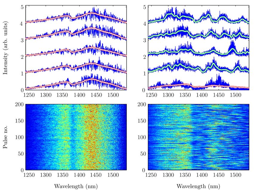
Research Article                                                                             Journal of the Optical Society of America B   3

Fig. 1. Comparison of shot-to-shot differences in SC spectra obtained with femtosecond pumping in the normal dispersion (NDi
- left panels) and anomalous dispersion regimes (ADi - right panels). The red/white lines (top left) represent a long term average
(10,000 shots) of the all-NDi spectrum, where each individual shot trace (blue) mimics the shape of the average long-term spectrum.
This stability in the NDi regime can also be observed in the 200 consecutive single-shot spectra (bottom left). The green/white
lines (top right) are low pass filtered versions of individual shot-to-shot spectra, all of which show significant deviations from their
long term average (red/white line in the bottom) in the ADi regime SC. Consecutive single shot spectra also show large differences
(bottom right) in the ADi regime. Clearly, the dynamics of SC in the NDi regime are in stark contrast with dynamics in the ADi
regime. Adapted from [24].

        Pulse duration     Femtosecond pulses                      Pico-nanosecond pulses and continuous waves

        Spectral width     Broad spectrum (visible-NIR)            Broad spectrum when using pulsed input, some covering
                                                                   400-2400nm; Mostly only IR spectrum for CW sources
        Coherence and      Coherent and stable (NDi regime);       Usually incoherent, unstable but can be coherent with ps
        stability          Incoherent, unstable (ADi regime)       sources (NDi regime); Large fluctuations, incoherent for CW
        Spectral power     Low, around 0.5mW/nm                    Moderate values for ps-ns pulses, around few mW/nm;
        density                                                    High power for CW: 10s of mW/nm
        Pump        peak   fs input: few kW                        ps-ns input: Usually 100s of W; CW input: Few W
        power
        Convenience        Usually very expensive, complex,        Cost-effective and low maintenance. Widespread applica-
                           and difficult to maintain               tions for pulsed output.

Table 1. General characteristics of different SC generated by altering pump sources.

CW SC was demonstrated through fiber tapering, causing dis-            trum. Traditionally, fluorescence studies of cellular processes
persive waves to be further blue-shifted. Wavelength extending         have used mercury and xenon arc lamps for broadband illumina-
to 650nm can thus be reached even with moderate pump pow-              tion but suffer from low illumination efficiency and low spatial
ers of around 35W [41]. Finally, through Ge-doping and fiber           coherence, and therefore cannot be used to improve resolution
tapering, the first pure CW white-light SC was generated in 2012       in scanning-microscopy applications. Diodes and monochro-
with a spectrum spanning 470nm to more than 1750nm at 9.3W             matic lasers provide bright illumination without the necessary
power [42].                                                            broad bandwidth. The handful of monochromatic lasers that are
                                                                       commonly available pose an unnecessary restriction on the vast
3. WAVELENGTH SELECTION SCHEMES                                        available toolbox of excitable fluorophores, rendering only a few
                                                                       fluorophores usable whose excitation wavelengths match the
Perhaps the most useful property of a SC source in fluorescence        fixed laser wavelengths. Even then, the most efficient excitation
microscopy lies in its massively broadband wavelength spec-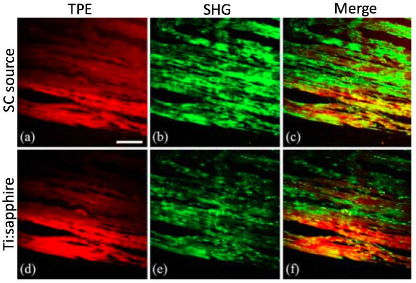
Research Article                                                                              Journal of the Optical Society of America B   4

Fig. 2. Different schemes to select desired excitation bands from SC sources: (a) filter wheel (b) prism [32] (c) axial monochromator
[43] (d) LVTF [44] (e) AOTF [45] and (f) AOBS [46]. Their individual characteristics are summarized in Table 2.

                    Characteristics            Filter wheel   Prism   Axial monochromator      LVTF        AOTF        AOBS

                    High transmission               +          −                +                 +           −           +
                    Tuning speed                    −          −                −                 −           +           +
                    Wide wavelength range           +          +                +                 +           +           +
                    Tunable bandwidth               −          +                −                 +           −           −
                    Simultaneous multi-color        −          −                −                 −           +           +
                    Out-of-band suppression         +          +                +                 −           −           −
                    Steep spectral edge             +          −                −                 −           −           −
                    Polarization insensitive        +          −                −                 +           −           +

Table 2. Wavelength selection schemes with their pros(+) and cons(-).

wavelength can lie between available laser wavelengths [23].           put (∼1mW/nm from commercial sources) is still sufficient for
    The increasing complexity of bioimaging experiments and            most imaging applications [32]. In conjunction with versatile
rising demand to simultaneously image in multiple colors ne-           wavelength selection schemes, a SC source can simultaneously
cessitate flexible use of excitation wavelengths across the entire     excite a number of fluorophores, each at their optimal absorp-
visible range to avoid crosstalk. Previously, dye laser systems        tion wavelengths. This provides high specificity with minimal
with tunable wavelengths have been used [47] to excite multiple        cross-excitation and permits studies of various structures and
fluorophores, but the gain curves of these dyes limit the tuning       phenomena at the same time.
range to 50-100nm and do not cover a significant portion of the            Good wavelength selection schemes should provide full flex-
visible spectrum. Tunable Ti:Sapphire lasers provide another           ibility in selecting wavelengths and bandwidth over a large
alternative to access the full visible spectrum but they can only      spectrum at high speed with potentials for multiplexed imag-
achieve this through processes like multi-photon absorption,           ing. Many such technologies have been proposed but the most
harmonic generation and optical parametric oscillators (OPO),          noteworthy ones (see Figure 2) include bandpass filter wheels,
making the process expensive and difficult to operate, and re-         prism-based spectrometers [32], axial monochromators [43], mo-
quiring specialist maintenance. SC sources overcome most of            torized linear variable tunable filters (LVTF) [44], acousto-optic
these spectral restrictions with a large bandwidth spanning the        tunable filters and beam splitters (AOTF and AOBS) [45, 46]. In
visible and NIR without any gaps, and thus lifting the restric-        Table 2, we summarize and compare the characteristics of these
tions of matching fluorophores to available laser lines. The full      technologies.
SC spectrum is rarely used simultaneously for imaging. Most                Using a filter wheel with 6-12 bandpass filters can accommo-
fluorescence applications pick out desired excitation wavelength       date a wide range of wavelengths. The high-precision multi-
bands from the output. While this means that most of the power         layer coatings on these filters provide excellent out-of-band sup-
is discarded, the average power of the spectrally selected out-        pression (usually over OD 5) and high edge steepness. How-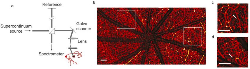
Research Article                                                                             Journal of the Optical Society of America B    5

ever, changing wavelengths requires physical movement of the         4. MICROSCOPY   MODALITIES                         USING          SUPER-
filter wheel in the beam path over 50-200ms timescale (when             CONTINUUM RADIATION
motorized), precluding the possibility of simultaneous multi-
wavelength selective excitation. Dual, triple or quad band fil-      SC sources have revolutionized microscopy for biological and
ters are now available for performing simultaneous excitation        medical imaging applications, offering significant advantages
but these are not tunable for arbitrary selection. A different       over monochromatic lasers in spectral flexibility and over tradi-
tunable implementation uses prisms to spatially disperse the         tional lamp sources in terms of fast pulsed nature, high bright-
beam, part of which goes through a translatable aperture to se-      ness, spatial coherence, deep tissue penetration and contrast [50],
lect a central wavelength and bandwidth [32], but this leads to      low-maintenance, and cost-effectiveness. In what follows, we
large power losses and does not allow simultaneous excitation        discuss how SC properties are exploited in different microscopy
of multiple wavelength bands. Another excitation technique           applications. We review the use of SC spectral flexibility, par-
uses an on-axis monochromator based on a custom-designed             ticularly in hyperspectral imaging; SC spatial coherence used
lens to intentionally maximize the beam’s longitudinal chro-         for point scanning microscopies; the fast pulsed nature of SC
matic aberration while keeping other aberrations low [43]. This      for spectroscopy and fluorescence lifetime imaging (FLIM); high
wavelength-dependent longitudinal dispersion of foci along the       peak power applications in multi-photon excitation and sec-
optical axis allows coupling desired part of the focused beam        ond harmonic generation; simplification of super-resolution
into a finite aperture fiber and discarding all other out-of-focus   microscopy instrumentation using SC sources; and finally, the
wavelengths.                                                         bright, coherent light applications of SC for OCT and CARS
                                                                     microscopies.

    To continuously tune the central wavelength along with band-     A. Widefield and confocal scanning                   techniques,       and
width, one can use LVTFs whose cutoff wavelengths vary lin-             wavelength-resolved imaging
early along their length. A useful configuration involves the SC     SC sources provide a wider spectrum of wavelengths and much
beam going through two LVTFs (one as shortpass, one as long-         greater brightness than thermal sources or LEDs for widefield
pass) translated independently to make a variable bandwidth          fluorescence microscopy techniques. In particular, incoherent
filter[44]. Circular LVTFs use the same concept but work by          SC is best for widefield imaging since it provides flat-field illu-
rotating the filters. LVTFs work well for tuning excitation when     mination without generation of speckle artefacts or aberrations.
the optical beam width is small in cross section as finite beam      These artefacts are seen with traditional coherent laser sources
widths deteriorate the edge steepness. The need for mechanical       due to interference between light waves and degrade image qual-
translation slows their tuning speed (∼hundreds of ms) and           ity [51, 52]. Incoherent SC with large spectral bandwidth and
they cannot be used for simultaneous wavelength selection.           excellent beam profiles are marketed commercially, permitting
                                                                     easy integration into traditional setups for widefield microscopy.
    In contrast to previous technologies, AOTFs provide very         The SC output can additionally be launched into a multimode
fast, programmable wavelength tuning (µs) with simultaneous          fiber to impose a strong spatial incoherence and uniform il-
output possible for up to eight different wavelengths over a         lumination field. SC sources have found a greater market in
large spectral range (hundreds of nanometers) with no mov-           confocal laser scanning fluorescence microscopy, which was the
ing parts, demonstrated [48] for spectrally-resolved imaging.        first bioimaging application to adopt SC radiation [31, 32]. This
Acousto-optic technologies effectively produce a phase grating       is because fiber-generated SC radiation can easily be focused
to diffract a specific part of the incident light with very narrow   onto a diffraction-limited spot and point-scanned through the
passband (∼1nm) under phase-matching conditions of optical           sample. It is conceivable that high power commercial SC sources
and acoustic waves. However, AOTFs do suffer from low out-of-        will replace monochromatic lasers in all future confocal micro-
band suppression, low transmission due to polarization selectiv-     scopes, simplifying the setup, reducing costs and enhancing
ity, and sometimes from wavelength sidebands that introduce          versatility [37]. It is important to consider the effect of longitu-
a wavelength dependent angular spread in their output [48],          dinal chromatic aberration and chromatic variations in beam
which degrades image quality. Additional compensation optics         divergence potentially affecting the spatial resolution. A study
like prism elements can correct for polarization selectivity to      quantifying and comparing 3D point spread functions (PSF) in
restore high transmission and remove angular spread, and have        the blue and red spectral regions in a confocal setup utilizing a
been commercialized as acousto-optic beam splitters (AOBS)           commercially available SC source found that the displacement
[34, 46], making them a single versatile instrument handling         of the focal spot along the z-axis was comparable in extent to the
both excitation and emission selection efficiently.                  full-width-half-maximum of the PSF [48]. Therefore, chromatic
                                                                     aberrations caused by using a SC source do not pose a signifi-
                                                                     cant limitation for performing high-resolution confocal imaging
    No single wavelength filtering scheme fulfills the needs of      in multi-color throughout the visible spectrum. In other tech-
all imaging experiments, although acousto-optic technologies         niques like volumetric confocal reflectance microscopy [53], this
provide more versatility than others. It is important to evaluate    chromatic aberration feature has been maximized using aspheric
and choose wisely between them by considering the criteria (eg.      lenses because it permits one to encode depth information spec-
speed, tunability, out-of-band suppression to minimize crosstalk,    trally and to be read out by a spectrum analyzer or spectrometer.
simultaneous multi-color) most relevant for the experiment in        This way, multiple depths in biological specimen can be probed
question. Once spectral selection is performed appropriately,        simultaneously and rapidly, with one group demonstrating a
the output light can be coupled to a microscope directly for         157µm axial range with micrometer resolution captured in a
widefield imaging, to a scan unit for point-scanning, or modified    single shot from epithelial tissue [54]. This technique eliminates
in other ways (eg. beam shaping, pulse compression) for desired      the need for mechanical axial scanning, making it useful even
applications.                                                        for endoscopic imaging systems.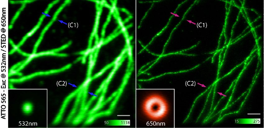
Research Article                                                                              Journal of the Optical Society of America B   6

Fig. 3. Hyperspectral imaging reveals that different regions of Convallaria majalis have different emission properties at (a) 620nm
and (b) 530nm excitations. (c) Excitation-emission plots for four regions in (a). Adapted from [49].

    Combining the flexible excitation from a SC with appropri-         times at their optimal absorption wavelengths. SC sources can
ately multiplexed emission detectors enables the method of             replace fast-pulsed flash lamps in spectrofluorometers [62], pro-
hyperspectral imaging (wavelength-resolved imaging over a              viding higher brightness to improve signal-to-noise ratios for
continuous spectral range, see Figure 3). Hyperspectral imag-          sensitive measurements. Merging the capabilities of spectrofluo-
ing allows capturing and completely characterizing the entire          rometry with spatial imaging leads to a powerful quantitative
excitation and emission profile of known and unknown fluo-             imaging technique called fluorescence lifetime imaging (FLIM).
rophores and biological samples at all available wavelengths for       Several implementations of FLIM using SC radiation are in use,
each pixel of the image [23, 49, 55, 56]. This information can be      including widefield- and spinning-disk-based systems (time-
used in post-acquisition spectral unmixing methods to reliably         gated FLIM [63, 64] and frequency domain FLIM [65, 66]), and
discriminate fluorophores with overlapping spectra in biological       the more popular point-scanning-based time-correlated single-
studies [57, 58]. The same technique can be used to remove             photon counting (TCSPC) FLIM systems [32]. All these FLIM
unwanted autofluorescence in the emission channel that might           implementations can provide the spatial distribution of fluo-
be affecting the data. In other cases, tissue autofluorescence may     rophores, and their individual percentage contribution to fluo-
contain useful information. Tissues undergoing disease progres-        rescence in each image pixel.
sion can show structural and metabolic changes affecting their             Like hyperspectral imaging, unmixing multiple fluorophores
absorption, scattering and fluorescence characteristics [59] and       in an image can be done using fluorescence lifetime as a param-
these subtle changes can be picked up by SC-based hyperspec-           eter, even with minimal a priori information [67]. FLIM can also
tral imaging techniques. This has been particularly successful         be applied to reveal the intrinsic lifetime contrast (see Figure
in label-free imaging of biological specimens. Probing for more        4) to investigate health states of tissues. The autofluorescence
spectral information using SC at extended wavelengths and              lifetime characteristics can be studied by simply exciting endoge-
increased acquisition rates will greatly enhance non-invasive          nous fluorophores without labels [5, 61] at desired wavelengths
disease diagnostics [60].                                              using a SC source. SC sources have opened up a more practical
                                                                       way to study Forster resonance energy transfer (FRET) using
B. Fluorescence lifetime microscopy (FLIM) and multi-                  FLIM to observe complex protein interactions, protein structures
   parameter imaging                                                   and molecular conformations at high-speeds in live cells [68].
The inherent pulsed nature of most SC sources is useful for time-      By minimizing cross-excitation of acceptors, SC sources enable
resolved fluorescence spectroscopy. This technique can measure         multi-channel FLIM-FRET [69] from the same laser source. Some
the excitation and emission spectra of fluorophores, along with        advanced FLIM setups using SC can capture multi-parametric
the average lifetime of their fluorescence decay with a high-          and hyperspectral 6D [56] and 7D [55] data including not just the
temporal (sub-ns) resolution. The fluorescence decay lifetime of       spatial profile and absorption-emission spectra but also simul-
fluorophores is exquisitely sensitive to their micro-environment       taneously the fluorescence lifetime and polarization anisotropy
and can provide a great deal of quantitative information about         profiles (x,y,z,τ,λex ,λem , r) for each pixel. This presents great
the pH, local viscosity, temperature, ion concentration, protein       opportunities to improve studies of multiplexed signaling path-
aggregation, protein binding, and kinetics of chemical reactions       ways, tissue properties, and functional aspects at the molecular
around the fluorophores [5, 61]. SC sources provide the high-          scale.
frequency (∼MHz) pulsed excitation required for lifetime mea-             While the temporal instability of incoherent SC sources may
surements and now some commercial sources can also offer easy          be an issue in certain applications, fluorescence lifetime mea-
tuning of this repetition rate (80, 40, 20, 10, 5, etc. in MHz) for    surements are mostly unaffected by the shot-to-shot variations
capturing shorter or longer lifetimes. The short pulse width (few      because this noise is small compared to the photon-shot noise
ps) of the SC output helps reduce errors in temporal measure-          in imaging systems. In the most widely used TCSPC imple-
ments. Selecting narrow bands from the SC spectrum also allows         mentation, only a small fraction of excitation pulses generate
specificity in exciting fluorophores with distinct spectra and life-   a detectable photon to avoid photon pileup effects. On the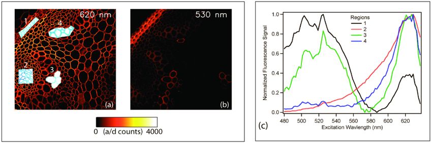
Research Article Journal of the Optical Society of America B 7
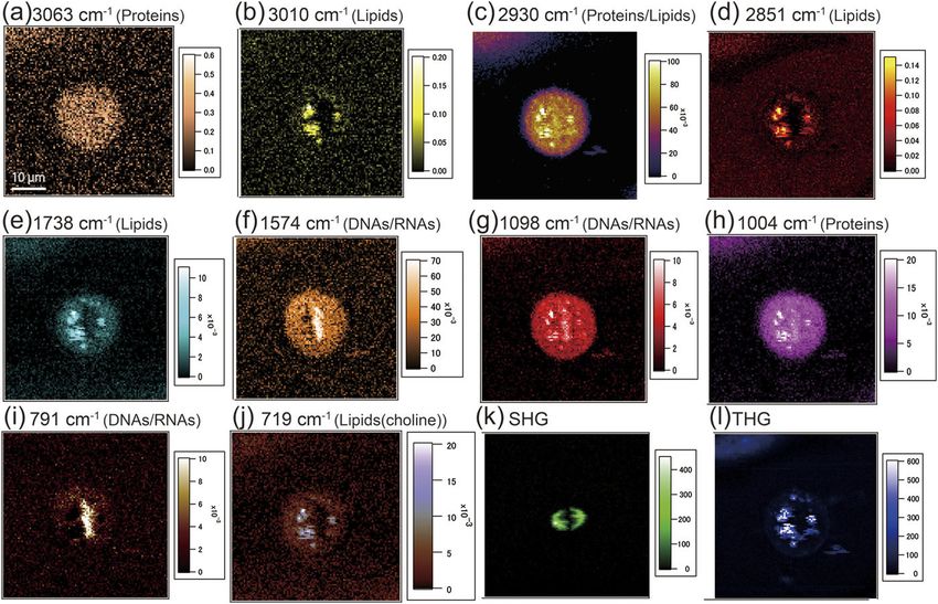
Fig. 4. Intensity and intensity-merged FLIM images of autofluorescence from a section of unstained human pancreas showing clear
lifetime differences between tissue types: A-artery, I-islets of Langerhans and C-collagen connective tissue. (a)-(c), λex =440-450nm,
λem >470nm. (d)-(f), λex =520-530nm, λem >570nm. Adapted from [32].
timescales of TCSPC FLIM image acquisition (seconds to min- common pulsed laser sources[72], opening the possibility of us-
utes), the average power of the SC is fairly stable [32]. How- ing an increased range of dyes [73]. The SC also provides both
ever, other techniques like fluorescence correlation spectroscopy the excitation and depletion wavelength bands from the same
(FCS) require much more stable lasers and cannot make use of source, eliminating the second laser entirely. Easy and stable tem-
the SC radiation used for most imaging applications, produced poral alignment can be achieved without the need for synchro-
by pumping PCFs in the ADi regime. FCS as a technique relies nization of multiple excitation and depletion sources. SC sources
on detecting fluctuations in fluorescence intensity from a small also readily provide ps pulse width output, which are most ef-
sample volume and therefore, any fluctuations stemming from ficient for STED, obviating the necessity for pulse-stretching of
varying laser illumination will introduce errors in the acquired fs range pulses given by Ti:Sapphire lasers. Thus, SC sources
FCS curve, negatively affecting the experimental outcome [70]. allow making major simplifications to the very complicated in-
Despite this caveat of instability, other SC features like spectral strumentation, which is one of the primary drawbacks of STED
tunability, simplicity and cost-effectiveness has made the fiber- microscopy. In an example from a few years ago, simultane-
laser-pumped SC a more attractive option than mode-locked ous two-color STED was demonstrated for the co-localization of
Ti:Sapphire lasers in many FLIM and multi-parametric imaging two different proteins at super-resolution (35nm lateral, 90nm
applications. axial resolution) in 3 dimensions with a single commercial SC
source (SC-450-PP-HE) instead of four very-expensive pulsed
C. Super-resolution microscopy: Stimulated Emission Deple- laser sources that would normally be required [74]. This SC
tion (STED) source provided pulse energies (20nJ for 20nm bandwidth at
The ability to arbitrarily choose and rapidly change excitation 1MHz) comparable to that of lasers commonly used in STED
wavelengths using a SC source greatly enhances the number of but the acquisition speed was limited by the low repetition rate.
usable photo-activated dyes and fluorophores [71]. These fluo- Improving resolution at greater speeds with STED depends on
rophores can be turned on and off with different wavelengths very efficient depletion with high pulse powers and therefore
and form the basis of many super-resolution techniques like will benefit from improved SC sources providing greater pulse
photo-activated localization microscopy (PALM), stochastic op- powers even at higher repetition rates. However, the main draw-
tical reconstruction microscopy (STORM) and stimulated emis- back in using SC for STED is the large fluctuation in the pulse
sion depletion (STED), among others. energy which can vary from 10% to 80% and can induce large
The high illumination densities (∼5kW/cm2 ) necessary for optical noise and hinder certain applications[7]. Balanced de-
PALM and STORM [3, 4] require very high laser powers (∼50- tection schemes might be able to reduce the noise down to shot
100mW) at specific wavelengths not attained easily using SC noise levels in these cases.
sources. However, STED instrumentation greatly benefits from
the simplicity, easy maintenance and affordable cost of a SC D. Two-photon excitation (TPE) microscopy and second har-
source. STED uses one laser pulse focused onto a diffraction- monic generation (SHG)
limited spot for fluorophore excitation. Simultaneously or a few Achieving greater peak powers in custom-built SC sources has
ps later, the excitation pulse is followed by a donut shaped red- permitted two-photon excitation (TPE) microscopy with these
shifted laser pulse to de-excite fluorophores through stimulated sources. TPE is caused via the absorption of two photons, each
emission (see Fig.5). Only a few fluorophores confined in a sub- around twice the wavelength of a single photon excitation event
diffraction-sized donut center are allowed to fluoresce, improv- [77, 78]. This allows probing into deeper tissue by using red-
ing the imaging resolution beyond the diffraction limit [1] and shifted wavelengths to increase penetration depth and minimize
reaching resolution below 50nm. A pulsed tunable SC source scattering and photodamage. This technique has been valu-
used for STED can include wavelength ranges not covered by able in neuroscience and in medical studies involving imaging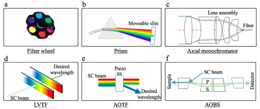
Research Article                                                                           Journal of the Optical Society of America B   8

Fig. 5. STED of immunolabeled tubulin fibers with 532nm excitation and 650nm depletion donut beam. Comparing between confo-
cal image (left) and STED (right) shows the resolution enhancement with STED. Adapted from [72].

Fig. 6. Two-photon excitation of fluorescence, second harmonic generation and merge images of mouse muscle comparing the
performance of a nanosecond SC excitation (a-c) against femtosecond Ti:sapphire excitation (d-f). Adapted from [75].

in-vivo or in thick tissues. However, TPE is a nonlinear pro-        TPE was also demonstrated using fs pulse SC output and 8mW
cess requiring very high photon fluxes in a small confined focal     power at the sample [80]. However, in all these cases, generating
volume (using large peak powers at the sample and typically          SC with the appropriate characteristics for TPE required pump-
at least 0.01-0.1nJ energies for laser-scanning) in a very short     ing PCFs with the fs Ti:Sapphire lasers, making them impractical
timeframe. It has traditionally only been possible using mode-       in most general biology laboratories. The challenge, then, is for
locked femtosecond lasers that compress the high laser power         longer pulse SC sources to be able to perform TPE efficiently by
into ultrashort fs pulse packets. By giving out one intense pulse    generating large enough peak powers to achieve the required
every 10-25ns, these lasers manage to excite two-photon pro-         power densities. In 2016, Lefort et al. [75] demonstrated that
cesses while keeping average power into the sample relatively        both TPE and second harmonic generation (SHG) could be pos-
low to avoid photobleaching and phototoxicity. When femtosec-        sible from a compact broadband SC source with ns pulses and
ond input pulses are pumped through microstructured fibers, in       sufficient peak power. Image quality (see Figure 6) using the SC
addition to wavelength broadening, they also exhibit dramatic        source (1ns pulses with 370nJ energy, 370W peak power, 92mW
temporal broadening to yield output pulse durations of a few         average power) was comparable to the Ti:Sapphire (150fs pulses
ps, but the pulse energies and peak intensities can still be large   with 70pJ energy, 460W peak power, 5.5mW average power).
enough for TPE.                                                      Over the years, as novel fibers and ns pump source technologies
                                                                     become mature, TPE should be widely accessible using com-
   Two-photon excitation using SC was achieved more than a           pact and stable turn-key SC sources that provide large spectral
decade ago by using the near IR part of the spectrum (17W peak       densities with higher peak powers and intensities.
power and 1.7GW/cm2 at the sample, sufficient for TPE even
with pulse lengths of 1-5ns) [34]. Others used the visible range
of the SC (500-600nm) [33] and used pulse compression tech-
niques to increase peak intensities [79]. Simultaneous three-color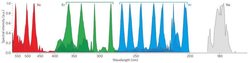
Research Article                                                                            Journal of the Optical Society of America B   9

Fig. 7. (a) Schematic of an OCT setup using visible light (520-630nm) from a commercial SC source for in vivo retinal imaging to
measure oxygen metabolism. (b) Wide-field view of retinal micro-vasculature imaged using this setup. (c-d) Magnified view of
highlighted areas in (b), with arrows showing the smallest capillary vessels. Scale bar: 200µm. Adapted from [76].

E. Optical coherence tomography (OCT)                                age echoes hiding weak object structures and reducing the reso-
                                                                     lution and sensitivity. Pulse shaping implementations have been
Optical coherence tomography (OCT) is a non-invasive opti-
                                                                     used to create Gaussian spectra but this comes with large losses
cal analogue of ultrasound techniques, enabling depth-resolved
                                                                     in output power. A noteworthy attempt to reduce noise while
imaging of biological tissues (see Figure 7) at µm scale resolu-
                                                                     simultaneously shaping the pulse without much loss has used
tion by using interferometric detection of photon time of flight
                                                                     appropriate tapering of PCFs to manipulate fiber dispersion to
[37]. Nowadays, OCT is routinely used in the diagnosis of reti-
                                                                     be in the NDi regime for all wavelengths, eliminating modula-
nal diseases [81]. OCT ideally requires a stable and noiseless,
                                                                     tion instability and soliton effects [84]. Going forward, improv-
spatially coherent source with low temporal coherence, single
                                                                     ing the stability of SC generated from longer pump pulses is the
transverse mode and high brightness (power of few mW/nm).
                                                                     key to compact, cost-effective yet high-performance OCT.
The axial resolution of fine microstructures in OCT scales as
λ2 /∆λ, depending inversely on the bandwidth [82], and thus
benefits greatly from a source with long range NIR bandwidth.        F. Coherent Anti-Stokes Raman Scattering (CARS) mi-
These requirements for OCT are very difficult to fulfill and in         croscopy
the past, super-luminescent diodes and thermal sources were          Other useful biological imaging applications of SC sources are
used for their bandwidth to provide 10-15µm axial resolution.        CARS microscopy and CARS microspectroscopy - techniques
However, the low brightness of these sources limits their use in     allowing visualization of chemical composition and structures
clinical OCT, as higher brightness is necessary to counter signal    of samples, based on their characteristic intrinsic vibrational
attenuation in strongly scattering biological tissue and to reduce   contrast. CARS is label-free and has a non-deteriorating signal,
the integration time in real-time imaging of in-vivo tissues [60].   unlike in fluorescence microscopy where photobleaching even-
    SC sources fulfill most of the necessary criteria listed above   tually causes a large reduction in the SNR. CARS microscopy
and have been shown to improve resolution by an order of mag-        requires two beams (a pump pulse and a synchronous broad-
nitude, while providing higher brightness and faster acquisition     band Stokes pulse) and produces a signal when the difference
in state-of-the-art high-resolution OCT measurements. While          between pump and Stokes frequencies matches the Raman reso-
resolution can also be improved by using highly specialized          nance frequency of the sample [86]. These synchronous beams
broad-bandwidth, ultrashort-pulsed, mode-locked Ti:Sapphire          are difficult to produce without using two synchronized lasers or
lasers, the expense and expertise these require are limiting. OCT    complex setups involving OPA and OPOs, or four-wave-mixing-
researchers began to show an interest in SC generation after         based sources which are the current state-of-the-art methodolo-
2001 when ultrahigh resolution OCT (resolution of 2.5µm) at          gies. However, these beams are also readily produced through
1.3µm central wavelength was demonstrated from a SC source           SC generation [87] using fs lasers pumping PCFs tailored to
built using microstructured fibers pumped with a mode-locked         produce appropriate wavelengths. Temporally coherent SC radi-
Ti:sapphire laser [21]. Nowadays, SC radiation optimized for         ation has so far only provided low peak powers and low spectral
OCT is available even from compact commercial sources using          power density. For a reasonably high-resolution CARS image,
fiber and microchip lasers which can inexpensively provide a         this poses a severe limitation of long acquisition times [88].
broader spectral coverage than Ti:Sapphire lasers. These new            However, other techniques like multiplex CARS do not re-
SC sources cover several interesting spectral bands like the visi-   quire high temporal coherence. Incoherent and ultra-broadband
ble range for maximum biological chromophore absorption and          SC generated in the ADi regime using longer pulses and greater
sub-micrometer ultra-high resolution OCT, the 1.05µm range for       peak powers is ideally suited for multiplex CARS, especially
ophthalmic OCT with enhanced penetration depth and reduced           when multiple signals can be generated and detected hyperspec-
scattering, and the 1.3-1.7µm range for in vivo non-invasive biop-   trally [89]. Multiplex CARS microscopy with sub-micrometer
sies [83]. Additionally, the broad bandwidth can be exploited        resolution has been demonstrated [90] from broadband SC
to perform spectroscopic OCT. However, ultra-broad SC gener-         (>2000cm−1 ), combined with TPE of fluorescence [86], and with
ated from an interplay of various nonlinear effects in the ADi       second harmonic generation (SHG) and third harmonic genera-
regime still suffer from instability (shot-to-shot variations) and   tion (THG) signals [85] to study proteins and lipids in cells (see
uneven (non-Gaussian) spectral profile, leading to multiple im-      Figure 8). This multiplexed nonlinear approach permits differ-
Research Article Journal of the Optical Society of America B 10
Fig. 8. CARS, second and third harmonic generation images of a HeLa cell in the mitotic phase. Chromosomes appear in a line in
the center of the cell in the DNA/RNA images and spindle fibers are seen in the SHG images. CARS images at (a-j) 3063, 3010, 2930,
2851, 1738, 1574, 1098, 1004, 791, and 719 cm−1 , and the (k) SHG and (l) THG images. Adapted from [85].
ent types of contrast to be achieved simultaneously to study have an intrinsic fluorescence signature when excited by UV
dynamics of living cellular structures. Even compact, turn-key light. One scheme to reach UV uses generation of second-, third-
solutions are commercially available now that use IR SC [22] for and fourth- harmonic of mode-locked Ti:Al2 O3 laser to go down
such multi-modal imaging. to 205nm but has very complex and limited tuning capability
[93]. If an optical parametric oscillator is used in conjunction,
5. CHALLENGES AND RECENT ADVANCEMENTS the tunability can be expanded from 250-355nm [94] but the
very high system complexity makes it impractical for general
It is clear from the various applications mentioned above that
microscopy use. Another technique uses SC generation assisted
the useful features of compact SC sources like spectral flexibil-
by cascaded four-wave mixing in a PCF to cover the entire UV-A
ity, moderately high spectral power density, fast pulses, low
range (300-400nm) [22]. The issue with SC generation in the
cost, low maintenance and small footprint have had a signifi-
UV using popular silica based PCFs is that they undergo UV
cant impact in biological and clinical imaging research. Despite
damage (solarization) with wavelengths under 380nm. There
their growing popularity, making SC more reliable and universal
have been a few noteworthy advances in the last few years to
in imaging applications demands overcoming some important
replace silica in PCFs with exotic glasses like ZBLAN [95] to
technological challenges. We briefly discuss these below. PCF
increase long term viability. This allows generating SC with
parameters largely determine the underlying spectral broaden-
shorter UV wavelengths while still being ultra-broadband, span-
ing and SC stability so better PCF design and simulation, and
ning more than three octaves in the 200-2500nm range. Even
their reproducible manufacturing with good fiber quality is at
relatively low power and compact laser pumps can be used for
the heart of tackling many of these challenges.
this. Similarly, exciting advances in gas-filled hollow-core (HC)
fibers, which use photonic bandgaps for trapping and guiding
A. UV generation
light within a central hollow core [12], have allowed harnessing
There is great demand to expand the coverage of SC sources to the UV-transparency and low UV damage susceptibility of gases.
the UV range from 200-400nm and a number of recent efforts While these HC fibers usually suffer from higher propagation
have been made in this context [92]. UV coverage would find losses, certain microstructured designs allow low loss transmis-
broad applications in fluorescence microscopy, spectroscopy and sion, higher optical damage thresholds and also exquisite control
biomedical photonics since many molecules of interest impor- of fiber nonlinearity and group-velocity dispersion (GVD), with
tant in cancer and metabolism research, like NADH and FAD,Research Article Journal of the Optical Society of America B 11
Fig. 9. Coherent ultrashort pulses of tunable bandwidths in the UV range generated in gas-filled hollow-core PCFs, by adjusting
pressure and gas mixtures (Ne, Ar, Kr, Xe gases). Adapted from [91] and [12].
a smooth and broadband ADi profile simply by varying pressure will surely open new vistas for in-vivo deep tissue imaging in
and gas mixtures [96, 97]. The normal GVD of a chosen noble animals. Longer NIR wavelengths have so far been overlooked
gas can be balanced against the anomalous GVD of the kagome for imaging due to the lack of appropriate laser sources and
PCF to tune the ZDW across NIR, visible, and even in the UV. suitable detectors. However, with a SC source and the arrival of
With this level of control, the tunable wavelength can be pushed new IR-CCD photodetectors such as indium gallium arsenide
into the deep-UV and also just as easily into the IR up to 2500nm, (InGaAs, λ upto 1700nm) and indium antimonide (InSb, for
using a relatively low power and compact laser pump, with λ >1700) based detectors, novel imaging opportunities can be
wide-ranging applications in sensing and photochemistry stud- realized. Recent research[50] utilizing SC has identified new
ies [98]. The generation of a coherent, ultrafast, deep-ultraviolet optical windows (II: 1100-1350nm, III: 1600-1870nm and IV: cen-
signal, optimized to be emitted in a narrow wavelength band tered at 2200nm) with better transmission, minimal absorption
tunable from 176-550nm has been demonstrated [12] by varying and reduced scattering than the optical window I (650-900nm)
pressure in inert gases like Ne, Ar, Kr and Xe (see Fig. 9). The for deep brain imaging [104].
same group also demonstrated the shortest optical wavelength
in a SC generated to date, from 124nm-1200nm using Raman C. On-chip supercontinuum generation
active hydrogen-filled HC PCF [99]. However, all these results SC generation has already been demonstrated in various chip-
from HC PCFs so far have used fs input pulses with µJ pulse based waveguides. These can become important elements to
energies (with the exception of a recent paper [100] using 500nJ expand the functionality of photonic integrated circuits or be
pulse energy), which sets the source requirements quite high and useful in integrated chip-based imaging applications in the fu-
with very low repetition rates in the kHz making time-resolved ture. SC has been generated in silicon photonic nanowires [105],
imaging very slow. Less stringent source requirements would in silica [106] and chalcogenide [107] waveguides although only
open the door for widespread use, especially if MHz repetition silicon nitride [108, 109] waveguides have allowed pushing the
rates and high average powers could be achieved in the deep spectrum to broaden towards visible wavelengths for potential
UV to IR range. Simple, compact sources of spatially coherent on-chip imaging applications.
and ultrafast UV light with long-term stable operation would
have excellent application potential in fluorescence microscopy D. Stability with greater output powers
and in the fields of UV absorption spectroscopy, UV-resonant Increasing SC stability while at the same time increasing spectral
Raman spectroscopy and laser lithography. power density with watt-level total output and broad bandwidth
has always been one of the big technological challenges in the SC
B. Near-infrared microscopy field. While each of these are easily possible separately (former
On the other end of the visible spectrum is the near infra-red in fs NDi regime and latter using continuous waves), combining
optical window (650-900nm), easily accessible using a SC source these two output characteristics using the same pump source
and silicon based detectors (for λResearch Article Journal of the Optical Society of America B 12
would undergo optical damage [99]. Recent advances in gas- 12. P. S. J. Russell, P. Hölzer, W. Chang, A. Abdolvand, and J. C. Travers,
filled HC-PCFs with versatile control of dispersion and non- “Hollow-core photonic crystal fibres for gas-based nonlinear optics,” Nat.
linearity can allow generating more stable SC [100]. Numerical Photonics 8, 278–286 (2014).
simulations have shown that the generated dispersive wave 13. W. Belardi and J. C. Knight, “Hollow antiresonant fibers with reduced
attenuation,” Opt. Lett. 39, 1853–1856 (2014).
pulses in these fibers are highly coherent and independent of
14. R. Sollapur, D. Kartashov, M. Zürch, A. Hoffmann, T. Grigorova,
input pulse energy when the pulse durations are tens of fs [91].
G. Sauer, A. Hartung, A. Schwuchow, J. Bierlich, J. Kobelke, M. Chem-
However, the bigger problem of achieving higher stability with nitz, M. A. Schmidt, and C. Spielmann, “Resonance-enhanced multi-
longer pump pulses still exists. Future developments to resolve octave supercontinuum generation in antiresonant hollow-core fibers,”
this issue will enable a huge range of applications. Light. Sci. & Appl. 6, e17124 (2017).
15. R. S. Watt, C. F. Kaminski, and J. Hult, “Generation of supercontinuum
radiation in conventional single-mode fibre and its application to broad-
6. CONCLUSIONS band absorption spectroscopy,” Appl. Phys. B: Lasers Opt. 90, 47–53
SC sources provide spectral versatility and spatially coherent, (2008).
16. J. Hult, R. S. Watt, and C. F. Kaminski, “High bandwidth absorption
high brightness radiation often at affordable costs and smaller
spectroscopy with a dispersed supercontinuum source,” Opt. Express
footprint than established tunable laser technologies. The fea-
15, 11385 (2007).
tures offered by the compact pulsed SC laser make it an ideal 17. J. M. Langridge, T. Laurila, R. S. Watt, R. L. Jones, C. F. Kaminski, and
source for a wide range of biophotonics applications in fluores- J. Hult, “Cavity enhanced absorption spectroscopy of multiple trace
cence microscopy and biomedical imaging. Recent advances gas species using a supercontinuum radiation source.” Opt. express
in novel PCF-based technologies and hollow-core fibers offer 16, 10178–10188 (2008).
promise that current challenges will be surmounted in the near 18. C. F. Kaminski, J. Hult, R. S. Watt, and T. Laurila, “Cavity Enhanced
future, and that the use of SC sources will become common- Spectroscopy of High-Temperature H2O in the Near-
place, replacing many monochromatic and complex tunable Infrared Using a Supercontinuum Light Source,” Appl. Spectrosc. Vol.
laser sources in research laboratories and in the clinic. 63, Issue 12, pp. 1389-1395 63, 1389–1395 (2009).
19. T. Udem, R. Holzwarth, and T. W. Hänsch, “Optical frequency metrol-
ogy,” Nature 416, 233–237 (2002).
7. FUNDING 20. J. Kasparian, M. Rodriguez, G. Mejean, J. Yu, E. Salmon, H. Wille,
R. Bourayou, S. Frey, Y. B. Andre, A. Mysyrowicz, R. Sauerbrey, J. P.
This project has received funding from the European Union’s Wolf, and L. Woste, “White-light filaments for atmospheric analysis,”
H2020-MSCA-ITN-2016 research and innovation programme un- Science 301, 61–64 (2003).
der the Marie Sklodowska-Curie Grant Agreement No. 722380 21. I. Hartl, X. D. Li, C. Chudoba, R. K. Ghanta, T. H. Ko, J. G. Fujimoto, J. K.
(SUPUVIR). CFK acknowledges funding from the UK Engineer- Ranka, and R. S. Windeler, “Ultrahigh-resolution optical coherence
ing and Physical Sciences Research Council, EPSRC (grants tomography using continuum generation in an air–silica microstructure
EP/L015889/1 and EP/H018301/1), the Wellcome Trust (grants optical fiber,” Opt. Lett. 26, 608–610 (2001).
3-3249/Z/16/Z and 089703/Z/09/Z), the UK Medical Research 22. A. Labruyère, A. Tonello, V. Couderc, G. Huss, and P. Leproux, “Com-
pact supercontinuum sources and their biomedical applications,” Opt.
Council, MRC (grants MR/K015850/1 and MR/K02292X/1),
Fiber Technol. 18, 375–378 (2012).
MedImmune, and Infinitus (China), Ltd. 23. C. F. Kaminski, R. S. Watt, A. D. Elder, J. H. Frank, and J. Hult, “Su-
percontinuum radiation for applications in chemical sensing and mi-
croscopy,” Appl. Phys. B: Lasers Opt. 92, 367–378 (2008).
REFERENCES
24. S. Dupont, Z. Qu, S. S. Kiwanuka, L. E. Hooper, J. C. Knight, S. R.
1. S. W. Hell and J. Wichmann, “Breaking the Diffraction Resolution Limit Keiding, and C. F. Kaminski, “Ultra-high repetition rate absorption
By Stimulated-Emission - Stimulated-Emission-Depletion Fluorescence spectroscopy with low noise supercontinuum radiation generated in an
Microscopy,” Opt. Lett. 19, 780–782 (1994). all-normal dispersion fibre,” Laser Phys. Lett. 11, 75601 (2014).
2. M. G. L. Gustafsson, “Surpassing the lateral resolution limit by a factor 25. J. K. Ranka, R. S. Windeler, and A. J. Stentz, “Visible continuum
of two using structured illumination microscopy,” J. Microsc. 198, 82–87 generation in air–silica microstructure optical fibers with anomalous
(2000). dispersion at 800 nm,” Opt. Lett. 25, 25 (2000).
3. E. Betzig, G. H. Patterson, R. Sougrat, O. W. Lindwasser, S. Olenych, 26. T. A. Birks, W. J. Wadsworth, and P. S. J. Russell, “Supercontinuum
J. S. Bonifacino, M. W. Davidson, J. Lippincott-schwartz, and H. F. Hess, generation in tapered fibers,” Opt. Lett. 25, 1415 (2000).
“Imaging Intracellular Fluorescent Proteins at Nanometer Resolution,” 27. W. Wadsworth, N. Joly, J. Knight, T. Birks, F. Biancalana, and P. Russell,
Science. 313, 1642–1646 (2006). “Supercontinuum and four-wave mixing with Q-switched pulses in end-
4. M. Rust, M. Bates, and X. Zhuang, “Stochastic optical reconstruction lessly single-mode photonic crystal fibres.” Opt. express 12, 299–309
miscroscopy (STORM) provides sub-diffraction-limit image resolution,” (2004).
Nat. Methods 3, 793–795 (2006). 28. G. P. Agrawal, Nonlinear fiber optics (Academic Press, 2013).
5. W. Becker, “Fluorescence lifetime imaging - techniques and applica- 29. A. V. Husakou and J. Herrmann, “Supercontinuum generation of higher-
tions,” J. Microsc. 247, 119–136 (2012). order solitons by fission in photonic crystal fibers.” Phys. Rev. Lett. 87,
6. G. Genty, S. Coen, and J. M. Dudley, “Fiber supercontinuum sources 203901 (2001).
(Invited),” J. Opt. Soc. Am. B 24, 1771 (2007). 30. J. Dudley and J. Taylor, Supercontinuum Generation in Optical Fibers
7. J. M. Dudley, G. Genty, and S. Coen, “Supercontinuum generation in (Cambridge University Press, 2010).
photonic crystal fiber,” Rev. Mod. Phys. 78, 1135–1184 (2006). 31. G. Mcconnell, “Confocal laser scanning fluorescence microscopy with
8. P. S. J. Russell, “Photonic crystal fibers.” Science. 299, 358–362 a visible continuum source,” Opt. Express 12, 2844–2850 (2004).
(2003). 32. C. Dunsby, P. M. P. Lanigan, J. McGinty, D. S. Elson, J. Requejo-Isidro,
9. J. Knight, “Photonic crystal fibres,” Nature. 424, 847–851 (2003). I. Munro, N. Galletly, F. McCann, B. Treanor, B. Önfelt, D. M. Davis,
10. J. R. Taylor, “Introduction and history,” in Supercontinuum Generation M. a. a. Neil, and P. M. W. French, “An electronically tunable ultrafast
in Optical Fibers, J. M. Dudley and J. R. Taylor, eds. (Cambridge laser source applied to fluorescence imaging and fluorescence lifetime
University Press, Cambridge, 2010), pp. 1–29. imaging microscopy,” J. Phys. D: Appl. Phys. 37, 3296–3303 (2004).
11. R. R. Alfano, ed., The Supercontinuum Laser Source (Springer-Verlag, 33. J. A. Palero, V. O. Boer, J. C. Vijverberg, H. C. Gerritsen, and H. J. C. M.
New York, 2006).You can also read