Targeted immune epitope prediction to HHLA2 and MAGEB5 protein variants as therapeutic approach to related viral diseases
←
→
Page content transcription
If your browser does not render page correctly, please read the page content below
Achinko et al. BMC Immunology       (2021) 22:49
https://doi.org/10.1186/s12865-021-00440-w

 RESEARCH                                                                                                                                           Open Access

Targeted immune epitope prediction to
HHLA2 and MAGEB5 protein variants as
therapeutic approach to related viral
diseases
Daniel A. Achinko*, Anton Dormer, Mahesh Narayanan and Elton F. Norman

  Abstract
  Background: Targeted immunotherapy is mostly associated with cancer treatment wherein designed molecules
  engage signaling pathways and mutant proteins critical to the survival of the cell. One of several genetic
  approaches is the use of in silico methods to develop immune epitopes targeting specific antigenic regions on
  related mutant proteins. In a recent study we showed a functional association between the gamma retrovirus
  HERV-H Long Terminal Associating (HHLA1, HHLA2 and HHLA3) proteins and melanoma associated antigen of the B
  class proteins (MAGEB5), with a resultant decrease in expression of HLA class I and II immune variants. HLA-C and
  HLA-DRB5 were the main HLA class I and II Immune variants, respectively, that showed expression changes across
  viral samples of interest. Specific immune variants for HLA-C and HLA-DRB5 were filtered for the top ten based on
  their relative frequency of counts across the samples.
  Results: Protein variants for HHLA1, HHLA2, HHLA3 and MAGEB5 were used to predict antigenic epitope peptides
  to immune peptide-MHC class I and II binding using artificial neural networks. For IC50 peptide scores (PS) ≥ 0.5
  with a transformed binding ability between 0 and 1, the top 5 epitopes identified for all targeted genes HHLA1,2 &
  3 and MAGEB5 were qualified as strong or weak binders according to the threshold. Domain analysis using NCBI
  Conserved Domain Database (CDD) identified HHLA2 with immunoglobulin-like domains (Ig_C1-set) and MAGEB5
  with the MAGE Homology Domain (MHD). Linear regression showed a statistical correlation (P < 0.001) for HHLA2
  and MAGEB5 predicted epitope peptides to HLA-C but not HLA-DRB5. The prediction model identified HLA-C
  variant 9 (HLA-C9, BAA08825.1 HLA-B*1511) at 1.1% as the most valuable immune target for clinical considerations.
  Identification of the 9-mer epitope peptide within the domain showed for HHLA2: YANRTSLFY (PS = 0.5837) and
  VLAYYLSSSQNTIIN (PS = 0.77) for HLA-C and HLA-DRB5, respectively and for MAGEB5, peptides: FVRLTYLEY (PS =
  0.5293) and YPAHYQFLWGPRAYT (PS = 0.62) for HLA-C and HLA-DRB5, respectively.
  Conclusion: Specific immune responses to targeted epitope peptides and their prediction models, suggested co-
  expression and co-evolution for HHLA2 and MAGEB5 in viral related diseases. HHLA2 and MAGEB5 could be
  considered markers for virus related tumors and targeted therapy for oncogenic diseases.
  Keywords: MAGEB5, HLA, HHLA2, Immunotherapy, Epitopes, Viral-oncology

* Correspondence: da4815@gmail.com; daniel@pepvax.co;
achdany@yahoo.co.uk
PepVax, Inc., 0411 Motor City Drive, Suite #750, Bethesda, MD 20817, USA

                                        © The Author(s). 2021 Open Access This article is licensed under a Creative Commons Attribution 4.0 International License,
                                        which permits use, sharing, adaptation, distribution and reproduction in any medium or format, as long as you give
                                        appropriate credit to the original author(s) and the source, provide a link to the Creative Commons licence, and indicate if
                                        changes were made. The images or other third party material in this article are included in the article's Creative Commons
                                        licence, unless indicated otherwise in a credit line to the material. If material is not included in the article's Creative Commons
                                        licence and your intended use is not permitted by statutory regulation or exceeds the permitted use, you will need to obtain
                                        permission directly from the copyright holder. To view a copy of this licence, visit http://creativecommons.org/licenses/by/4.0/.
                                        The Creative Commons Public Domain Dedication waiver (http://creativecommons.org/publicdomain/zero/1.0/) applies to the
                                        data made available in this article, unless otherwise stated in a credit line to the data.
Achinko et al. BMC Immunology (2021) 22:49 Page 2 of 14 Introduction expresses 3 immunoglobulin (Ig) domains in the extra- Epitopes are molecular structures recognized by immune cellular region of the protein. HHLA2 specifically binds receptors as targets [1]. Binding epitopes are presented CD28H and co-stimulates T cells [16]. In the immune to CD8+ and CD4+ T cells by class I and class II MHC system, HHLA2 protein expresses in a combined fashion molecules, respectively. Binding affinity of epitopes to on monocytes and dendritic cells and upregulated by in- the different MHC molecules is very important in deter- flammatory signals such as lipopolysaccharide and IFNy. mining immunogenicity [2]. Every MHC molecule has a HHLA2 has not been shown to express on resting T or potential uniqueness specific to its binding ability to a B cells but upregulated when T cells are activated [17]. distinct set of antigenic peptides [3]. There has been a HHLA2 is expressed in about 20 to 70% of large number great advancement in the development of algorithms of human cancers including lung, thyroid, breast, pan- that identify peptide regions in targeted genes against creas, melanoma, bladder, colon just to name a few [18]. immune cells and they focus on binding affinity predic- This work further showed that HHLA2 was highly tion of known peptides [4]. MHC peptide epitopes with expressed in about 50% of tumors related triple-negative high binding affinity have been associated with strong breast cancer (TNBC) with a high risk of spreading. The immune responses and though the necessity of high expression of Melanoma associated antigens (MAGE) binding affinity of the peptide, it is not sufficient to qual- have also been shown to be highly expressed in TNBC ify immunogenicity [5]. The NetMHCpan method [6] but co-expression of HHLA2 and MAGE related genes uses Artificial Neural Networks (ANN) as a method for especially MAGEB5 have not been demonstrated in any peptide prediction. ANN is ideally the most recognized cancer type. method to identify non-linear patterns believed to be The family of MAGE genes comprises 19 members part of HLA-I interactions [7]. NetMHCpan exploits and located on chromosome X. These genes further sub- both peptide and the primary HLA sequence as the in- divide into four families (A to D) which is based on their formation inputted to drive ANN predictions, which in- chromosomal location and observed similarities between corporates all known and available HLA variant data encoded proteins. There are four known MAGEB genes and the method output was presented in units of pre- located on Xp21.3 [19]. MAGE A, B and C genes have dicted affinity (IC50 nM) [8]. not been shown to express in normal tissues except in The Human Endogenous Retrovirus-H Long Terminal the testes. MAGEB5 and MAGEB6 have been identified Repeat (HERV-H LTR) are retroviral sequences that are in different tumor types and the activation of these integrated into the genome in the course of evolution genes stems from the demethylation of their relative and are known to cover 8% of the human genome [9]. promoter regions [20]. Work done by [20, 21] didn’t lo- HERVs are also known as retrotransposons due to ab- cate MAGEB5 in the testes but identified it in a semi- sence of the envelope gene. Their condition for func- noma, which is a malignant neoplasm of the testes with tional transcription is that HERV sequences should a 95% treatment success when discovered early. The ex- maintain an LTR that is functional or controlled by a pression pattern of MAGEB5 given its absence in the different promoter not affected by nucleotide substitu- testes may suggest its specificity of co-expression with tions or deletions interrupting the open reading frames HHLA2 in viral cancer related diseases and tumors. (ORFs) [10]. Retroviruses integrated primate genome via Given the plethora of MHC polymorphisms which exogenous infection, which could affect somatic and at specifically identify a subset of peptides facilitating cellu- times germline cells resulting in their vertical transmis- lar immunity and also providing broad coverage of al- sion to the offspring in a Mendelian fashion with a fix in leles, makes it an essential task yet complicated for the human population [11, 12]. Envelope retroviruses, vaccine discovery [22]. Modeling immune variants and HERV-W and HERV-K are shown to be involved in genetic interactions related to cancer has not been done Multiple Sclerosis (MS) and Amyotropic Lateral Scler- before and our work shows a prediction model in which osis (ALS), respectively and are diseases of the nervous all immune variants predicting epitope peptides for co- system [13, 14]. HERV-H are non-envelope viruses and expressed genes could be considered for identifying im- derive three genes known as: HHLA1 with cytogenic lo- mune variants with potential clinical outcomes for vac- cation 8q24.22, HHLA2 with genomic location 3q13.13 cine development. and HHLA3 with genomic location 1p31.1. Work done HHLA2 and MAGEB5 have been individually shown by [15] identified from the EST database, 2 genes to be expressed in various tumor types and the former (HHLA2 and HHLA3) which had transcripts poly adeny- has been associated with viral related diseases. No data lated with an LTR of the HERV-H family. online showed an investigated analysis of co-expression HHLA2 is part of the B7 family and a co-stimulatory pattern between HHLA genes with focus on HHLA2 molecule with a role in activating and downregulating T and MAGE genes with focus on MAGEB5. The observed lymphocytes. It has distinct genomic properties in that it co-expression pattern between HHLA and MAGEB5 as
Achinko et al. BMC Immunology             (2021) 22:49                                                                               Page 3 of 14

demonstrated by our previous article [23] and this one,                           (AF110315, NM_001145095) (Dataset 3) of the same
are the first to show co-expression and co-regulated pat-                         abundance and HHLA2 identified with 6 variants (NM_
terns which can occur independently in viral and cancer                           001031693, AF126164, BC010922, NM_001036646, NR_
diseases and also in viral related cancer diseases.                               027404) (Dataset 4) with the most abundant being NM_
                                                                                  001036645. Two immune gene variants, HLA-C and
Materials and methods                                                             HLA-DBR5 for HLA class I and HLA class II, re-
Data used                                                                         spectively showed a considerable expression level
This work is based on further analysis of data obtained                           across samples and were considered for further ana-
in our last article [23]. The datasets generated and ana-                         lysis in this study. Majority of the variants for HLA-C
lyzed during the current study were obtained from the                             (Dataset 5) and HLA-DBR5 (Dataset 6) had high
GEO (RRID: SCR_005012) repository, and specifically                               abundance in the data but only the top 10 variants
G E O Pr o f i l e s d a t a b a s e (w w w . n c b i . n l m . n i h . g o v /   for HLA-C (AF026218, AF130734, AF170577,
geoprofiles/) with the Accession numbers: GDS5093,                                AF418978, D50290, D50291, D50292, D50293,
GDS4424, GDS5614, GDS5613, GDS2606, GDS4238,                                      D50294, D50295) and HLA-DRB5 (NM_002124,
GDS2023, GDS3489, GDS2676, GDS4669. The data                                      AY961072,        AY961073,     BC033827,       BC108922,
showed that, variants of melanoma associated antigens                             HM067861,         HM067862,      HM067863,       L02545,
(MAGE) of the B class (MAGEB5) protein, genetically                               M20430), were considered. The graphical distribution
coevolved with the Human Endogenous Retrovirus-H                                  of all the gene related variants for this study is seen
Long Terminal Repeat-Associating 2 (HERV-H LTR-A2)                                on Fig. 1.
gene (HHLA2). HHLA2 protein variant encodes a pro-
tein found on B-cells (monocytes) and known to regulate                           HLA class I epitope prediction for MAGEB5 and HHLA 1,
cell immunity by binding to a particular site on T-                               2, & 3 variants
lymphocytes with aim to inhibit their proliferation.                              The protein sequences in FASTA format were obtained
MAGEB5 identified with one nucleotide variant (NM_                                for all identified HLA-C, HHLA1, HHLA2, HHLA3 and
001271752) (Dataset 1) and HHLA2 identified with 7                                MAGEB5 variants from the National Center of Biotech-
nucleotide variants (AF126162, BC035971, NM_                                      nology Information (NCBI) Protein database. The
001282556, NM_001282557, NM_001282558, NM_                                        NetMHCpan 4.0 Server [24] based on prediction of
001282559) (Dataset 2) but the most abundant was,                                 peptide-MHC class I binding using artificial neural net-
NM_007072. Given that HHLA1 and HHLA3 are mem-                                    works (ANN) was used to predict binding peptide epi-
bers of the HHLA gene cluster and were shown to co-                               topes to MHC. The method was trained on ligands
express together, they were also considered for epitope                           naturally eluted alongside binding affinity data. The ser-
analysis in this article. HHLA1 identified with 2 variants                        ver uses neural networks to predict the binding to any

  Fig. 1 Frequency Distribution of Immune and Protein Related Gene Variants: This figure shows the frequency distribution of the various protein
  variants used in the study. The top ten immune variants per immune gene (HLA-C and HLA-DRB5) were considered for this study and all peptide
  variants identified for HHLA1 (n = 2), HHLA2 (n = 7), HHLA3 (n = 5) and MAGEB5 (n = 1) were all considered for this study. HHLA3 had protein
  variants with the lowest frequency
Achinko et al. BMC Immunology (2021) 22:49 Page 4 of 14
MHC molecule of a known targeted peptide of interest. Statistical analyses
Our targets of interest were protein variants for HHLA Selection of MHC HLA class I (HLA-C) and II (HLA-
1, 2 &3 and MAGEB5. Each targeted sequence was indi- DRB5) immune variants for epitope prediction analysis
vidually matched to each HLA-C protein variant and was based on all identified variants per sample from previ-
identified epitope peptide targets of 9 amino acids se- ous work [23]. The variants with highest abundance in the
quence in length were classified as strong binders at a data were based on the proportion of each variant per im-
%Rank < 0.5 and weak binders at %Rank > 2. Screening mune gene across all samples divided by the sum of total
for immune peptides was based on strong binders and variants for the given gene (ni/nT, I = individual variant,
given that no strong binders were identified, the top 5 T = total gene variants) across all samples in the dataset.
predicted peptide epitopes for all HLA-C protein variant The selection of top 5 peptides per protein variant for tar-
were considered for further analysis using the IC50 value geted genes (HHLA 1, 2 & 3 and MAGEB5) was based on
also known as the binding affinity value of the MHC to highest peptide score (≥ 0.5), known as log transformed
the peptide at a given concentration. The range between binding affinity (aff) values measured as IC50 in nM with
0 and 1 defined a negative binder to the MHC = 0 and a formula 1- log50k(aff) in the range of 0–1 (Jensen et al.,
positive binder = 1. 2018). Two peptide regions were detected within the data-
set as core and icore peptides. The core peptides were 9
HLA class II epitope prediction for MAGEB5 and HHLA 1, AA regions predicted in direct contact with MHC and
2, & 3 variants suggested mutation sites while the icore defined immune
Protein sequence variants for HLA-DR5, HHLA1, protein interaction of peptide core of actual protein se-
HHLA2, HHLA3 and MAGEB5 in FASTA format were quence subject to insertions and deletions. This makes the
obtained from the National Center of Biotechnology In- targeted region of great clinical importance when detect-
formation (NCBI) Protein database. The NetMHCIIpan ing for genetic variance within a said population and pos-
3.0 Server [25] based on prediction of binding peptides sible resistance to designed medication. Linear regression
for MHC class I was used to predict epitopes to targeted analysis between targeted genes (HHLA2 and MAGEB5)
proteins of interest which were protein variants for was done using Wizard statistical software version 1.9.34
HHLA 1,2 & 3 and MAGEB5. Each targeted protein se- [27] with a 95% confidence interval and data modeling of
quence was individually matched to each HLA-DR5 pro- each immune gene, HLA-C and HLA-DRB5, and its vari-
tein immune variant and identified epitope peptide of 15 ants as outcome variable against HHLA2 and MAGEB5 as
amino acids in sequence length were classified as strong dependent variables was also performed using formula:
binders at a %Rank < 2 and weak binders at %Rank > 10. Immune gene = HHLA2 + MAGEB5 + C (constant). The
Screening for immune peptides was based on strong null hypothesis was considered at β ≤ 0 for statistical sig-
binders and given that no strong binders were identified, nificance consideration which defined the coefficients of
the top 5 predicted peptide epitopes for each HLA-DR5 the dependent variables in the model. The post analysis of
protein variant were considered for further analysis predicted peptide epitopes data considered three parame-
using the IC50 value also known as the binding affinity ters: i) the protein variant type, ii) the epitope peptide vari-
value of the MHC to the peptide on the targeted protein ant per protein and iii) the peptide score.
at a given concentration. The range between 0 and 1 de-
fined a negative binder to the MHC = 0 and a positive A) The prediction of a single but different
binder = 1. Characteristic graphical representations of antigenic epitope peptides with different scores for
each amino acid on the predicted epitope was obtained different protein variants of the same gene: This
based on binding score of each amino acid on the se- indicates the prediction by the same immune
quence. This acted as a guide to core peptides on the variant to different antigenic regions of protein
string. variants of the same gene. The prediction of several
but different singular epitope peptides for the same
Protein variant and domain analysis or different antigenic protein variants of the same
Protein conformation is highly dependent on domain gene. This indicates the peptide prediction by the
type and critical to the prediction of antigenic sites for same or different immune variants to the protein
immune detection and destruction. Domain prediction variants of the same gene.
on the targeted protein variants of related genes (HHLA
1,2 & 3 and MAGEB5) was done through the Conserved
Domain Database (CDD) which is a protein annotation
source consisting of multiple sequences aligned using Protein variant Peptide epitope score
models related to ancient domains and proteins of full Variant A PHDSTAGKG 0.70
length [26]. FGIHKLMQR 0.69Achinko et al. BMC Immunology (2021) 22:49 Page 5 of 14
Statistical analyses (Continued)
Protein variant Peptide epitope score
E) The prediction of a similar antigenic epitope
Variant B RFKHYDEDY 0.80
ADELIRNGG 0.75 peptide region with single nuclear variation for a
gene having a different score: This indicates that
the same or different immune protein variants
predicted at the same immunogenic site,
different variants for the same antigenic
B) The prediction of a single but the same antigenic epitope peptide in the targeted protein variant of a
epitope peptide with the same score for different given gene. This mutant peptide could be favored
protein variants of the same gene: This indicates by translocation which is a property of virus-related
the peptide prediction by the same immune variant proteins.
to different protein variants of the same gene.
Protein variant Peptide epitope score
Protein variant Peptide epitope score
Variant A PHDSTAGKG 0.70
Variant A PHDSTCGK 0.7 PHDSTCGKG 0.65
Variant B PHDSTCGK 0.7 Variant B RFKHYDEDY 0.60
RFGHYDEDY 0.58
Results
III) The prediction of the same antigenic epitope
MHC HLA class I predicted binding peptide epitopes to
peptide for different protein variants of the same
HHLA 1, 2 & 3 and MAGEB5
gene presenting with different scores: This indicates
The top 5 predicted peptide epitopes to HLA-C obtained
the prediction by different immune variants to the
for each targeted gene variant showed a differential se-
same peptide region of different protein variants of
quence pattern to the various immune variants used for
the same gene.
their prediction. For HHLA1, two predicted epitope pep-
tide variants were common across (KTLPSTSHW &
RRVARTQWL) with the same peptide score meanwhile
another epitope peptide showed mutational changes
Protein variant Peptide epitope score
(SQASTLGAF, SQASTSGAF, QASPTSGAF, blue = un-
Variant A PHDSTCGKG 0.70
changed nucleotides, red = mutations) with different
Variant B PHDSTCGKG 0.60 scores across protein variants of the gene (Fig. 2A). For
HHLA2 (Fig. 2B), one predicted epitope peptide (GRWT
MKDGL) was common for all 7 protein variants with the
same peptide score and 6 peptide epitopes (IQNGNASLF,
IV) The prediction of the same antigenic epitope YANRTSLFY, AQTALSFFL, RGSEVVIHW) were com-
peptide repeatedly for a given protein variant of the mon for 6 protein variants with the same score. For
same gene having different scores: This indicates HHLA3 (Fig. 2C), one epitope peptide (IISPVTCMY) was
that the predicted peptide region was identified by predicted 3 times each for two protein variants. The pep-
different immune epitopes for the same protein tide scores were different per protein variant but the same
variant of a given gene. for each peptide for the different protein variants. The
peptide VLSTERGPY was predicted twice for two protein
variants. The peptide scores were different per protein
variant but the same for each peptide for the different pro-
Protein variant Peptide epitope score tein variants. The peptides QRILSQPTF and RRIHRVSLV
Variant A PHDSTCGKG 0.70 were predicted 3 and 2 times for three and two protein
PHDSTCGKG 0.65 variants, respectively. The peptide scores were different
Variant B RFGHYDEDY 0.60 per protein variant but the same for each peptide for the
RFGHYDEDY 0.58 different protein variants. For MAGEB5 (Fig. 2D), FVRL
TYLEY was the only predicted peptide that occurred twice
with different scores, for the only identified protein variantAchinko et al. BMC Immunology (2021) 22:49 Page 6 of 14
Fig. 2 Binding Affinity of Predicted Epitope Peptides to Target
a Genes (HHLA1,2 & 3 and MAGEB5) by HLA-C Immune variants: Each
Immune variant was used at the NetMHCpan 4.0 Server to predict
epitope peptides for each protein variant of the target genes. The
plotted graphs showed gene-protein variants on the x-axis against
and their different predicted epitope peptides represented by their
peptide binding score on the y-axis and measured by their binding
affinity; 1-log (IC50). A This figure shows the number of protein
variants for HHLA1 (n = 2) and number of predicted unique epitope
peptides (n = 8). Two epitope peptides [KTLPSTSHW (check) & RRVA
RTQWL (dot)] were common to both protein variants and one
epitope peptide (QASPTSGAF) was predicted for amino acid
variations. B This figure shows the number of protein variants for
HHLA2 (n = 7) and number of unique predicted epitope peptides
(n = 9) with 5 of them (AQTALSFFL, GRWTMKDGL, IQNGNASLF,
RGSEVVIHW, YANRTSLFY) repeating in almost all protein variants. C
b This figure shows the number of protein variants for HHLA3 (n = 5)
and number of unique predicted epitope peptides (n = 11) with 7 of
them (IISPVTCMY, QRILSQPTF, RRIHRVSLV, TEHLLRAVL, VLSTERGPY,
VTCMYTSRW, YPDPKRAFL) repeating in at least two protein variants.
D This figure shows the number of protein variants for MAGEB5
(n = 1) and number of unique predicted epitope peptides (n = 4)
with one (FVRLTYLEY) repeating due to different binding scores
for this gene. All other peptides were different and with
different scores.
c
MHC HLA class II predicted binding peptide epitopes to
HHLA 1, 2 & 3 and MAGEB5
HLA-DRB5 was the targeted MHC class II immune
variant used to predict 15 mer epitopes to HHLA (1,2 &
3) and MAGEB5 genes. For HHLA1 (Fig. 3A), two
different peptides TFQFFYAQSVKHVNV and SKKFFS
LLSVTSYSS were identified for the two protein variants
with three different peptide scores. For HHLA2 (Fig.
3B), one peptide (VLAYYLSSSQNTIIN) was predicted
with the highest score for all protein variants, then
d epitope peptide NKGLWILVPSAILAA was predicted
with next highest score across all protein variants, then
epitope peptide KGLWILVPSAILAAF was predicted
three times for each protein variant with the same score
across all protein variants for the gene. For HHLA3 (Fig.
3C), epitope peptide EMQRILSQPTFTEHL was
predicted with the same score for 3 different protein
variants, then VMCVRPLSPSKAIIS was predicted with
the same peptide score for 2 protein variants, then
epitope peptide ARKNLRRIHRVSLVM was predicted
four times two different scores per protein variant and
this pattern was the same for another protein variant.
For MAGEB5 (Fig. 3D), epitope peptide QFLLYKFKMK
QRILK and FLVVIFLKGNCANKE were predicted two
times for the same protein variant but the former had
different peptide scores while the latter had the same
peptide score.Achinko et al. BMC Immunology (2021) 22:49 Page 7 of 14
Fig. 3 Binding Affinity of Predicted Epitope Peptides to Target
a Genes (HHLA1,2 & 3 and MAGEB5) by HLA-DRB5 Immune variants:
Each Immune variant was used at the NetMHCIIpan 3.0 to predict
epitope peptides for each protein variant of the target genes. The
plotted graphs showed gene-protein variants on the x-axis against
and their different predicted epitope peptides represented by their
peptide binding score on the y-axis and measured by their binding
affinity; 1-log (IC50). A This figure shows the number of protein
variants for HHLA1 (n = 2) and number of predicted unique epitope
peptides (n = 3). The three epitope peptides were common to both
protein variants. B This figure shows the number of protein variants
for HHLA2 (n = 7) and number of unique predicted epitope peptides
(n = 3) repeating in all protein variants. C This figure shows the
number of protein variants for HHLA3 (n = 5) and number of unique
predicted epitope peptides (n = 6) repeating in at least two protein
b variants. D This figure shows the number of protein variants for
MAGEB5 (n = 1) and number of unique predicted epitope peptides
(n = 3) with two of them (FLVVIFLKGNCANKE & QFLLYKFKMKQRILK)
repeating due to different binding scores
Predicted peptide domain analysis
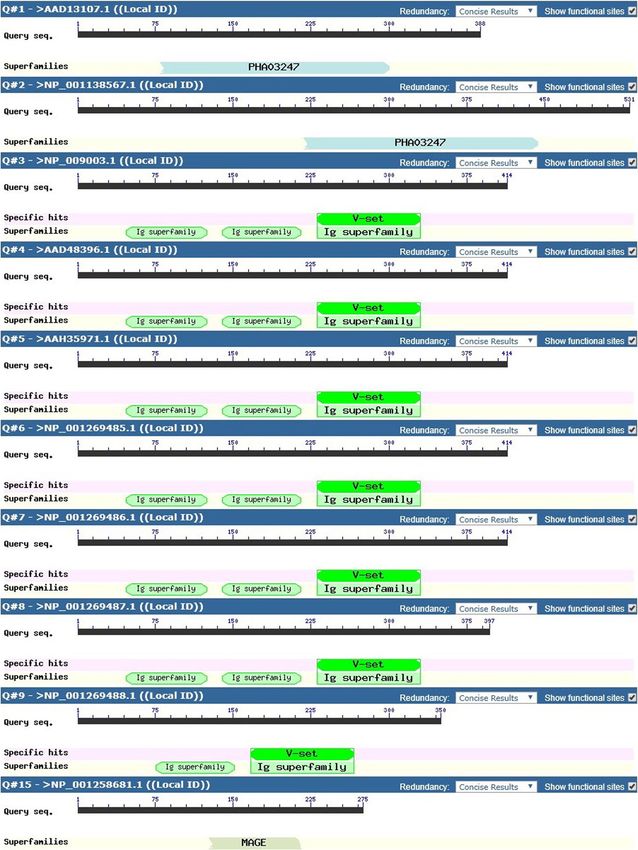
The four targeted genes (HHLA 1, 2 & 3, MAGEB5) and
related protein variants were subjected to domain
analysis and out of the 15 total variants, HHLA3 protein
variants identified with no known domains (Fig. 4). For
HHLA1, the superfamily Polyhydroxyalkanoates domain
(PHA03247) was identified for the two protein variants
though at different amino acid locations. For HHLA2,
six out of the 7 protein variants identified with one
c Immunoglobulin V-set domain (pfam07686) found on
antibodies and three superfamily Immunoglobulin C1-
set domains (pfam07654) and superfamily Immuno-
globulin domains (cl11960) in HERV-H LTR-
associating 2, per variant was identified. One HHLA2
variant identified with one Immunoglobulin V-set,
one superfamily Immunoglobulin C1-set and one
superfamily Immunoglobulin domains. For MAGEB5,
MAGE Homology Domain (pfam01454) was identified
on the only identified protein variant. The derived
epitope peptides for MHC HLA class I and II ob-
tained for the various targeted genes were evaluated
for their domain presence. For HHLA1 protein vari-
d ants, two HLA Class I epitope peptides (QASPTSGAF
and RRVARTQWL) and no epitope peptide for HLA
class II were identified in the domain. For HHLA2
protein variants, 5 HLA class I epitope peptides
(RGSEVVIHW, YANRTSLFY, IQNGNASLF, LICSVL
SVY and IINESRFSW) and one epitope peptide
(VLAYYLSSSQNTIIN) for HLA class II were identi-
fied in the domain. No domains were identified for
HHLA3 protein variant sequences used for peptide
prediction while for MAGEB5 protein variants, two
HLA class I epitope peptides (FVRLTYLEY and CSYP
AHYQF) and two HLA class II epitope peptides
(FLVVIFLKGNCANKE and YPAHYQFLWGPRAYT)
were identified in the domain.Achinko et al. BMC Immunology (2021) 22:49 Page 8 of 14 Fig. 4 Domain Predictions for Protein Variants of Targeted Genes (HHLA1,2 & 3 and MAGEB5). This figure shows the different domains identified for the targeted protein variants of related genes (HHLA1,2 & 3 and MAGEB5). The superfamily Polyhydroxyalkanoates domain (PHA03247) was identified for the two protein variants for HHLA1 though at different amino acid positions. Six out of the 7 protein variants for HHLA2 demonstrated Immunoglobulin V-set domain (pfam07686) common to antibodies and three superfamily Immunoglobulin C1-set domains (pfam07654) and superfamily Immunoglobulin domains (cl11960) found in HERV-H LTR-associating 2. One HHLA2 variant identified with one Immunoglobulin V-set, one superfamily Immunoglobulin C1-set and one superfamily Immunoglobulin domains. For MAGEB5, MAGE Homology Domain (MHD) (pfam01454) was identified on the only protein variant Linear regression and modeling of immune data equation post analysis was HLA-C = 0.21 (HHLA2) – For analysis on HLA-C predicted epitope peptides for 2.194 (MAGEB5) + 1.218. Only the variation of HHLA2 targeted genes (HHLA 1, 2 & 3) (Table 1), there was a and the constant were statistically significant in the positive linear correlation (Pearson correlation, p < model. The predicted impact of the various HLA-C im- 0.001) observed only between HHLA2 & MAGEB5 mune variants used in the model were as follows: HLA- (Fig. 5A) and for the predicted model (HLA-C = C1 = 11.2%, HLA-C2 = 15%, HLA-C3 = 13.5%, HLA- HHLA2 + MAGEB5 + C), β = 0.21 (< 0), standard error, C4 = 13.5%, HLA-C5 = 10.9%, HLA-C6 = 8.7%, HLA- SE = 0.111 and p = 0.029 (Fig. 5B). The resultant C7 = 6.8%, HLA-C8 = 2.7%, HLA-C9 = 1.1% and HLA-
Achinko et al. BMC Immunology   (2021) 22:49                                                                    Page 9 of 14

Table 1 HLA-C Immune Epitopes Count for HHLA and MAGEB5:       epitopes using protein variants for HLA-C and HLA-
This table presents the various counts of unique epitope       DRB5 immune genes identified several epitope peptides
peptides predicted by each HLA-C immune variant (C1–10) to     for targeted genes; HHLA1, HHLA2, HHLA3 and
protein variants for HHLA1, HHLA2, HHLA3 and MAGEB5. HHLA2     MAGEB5. The wide variation in predicted epitope pep-
demonstrated the highest epitope peptide counts due to more    tides for targeted genes to HLA-C immune variants sug-
protein variants (n = 7) while MAGEB5 had fewer epitope
                                                               gest a possible immune selection pressure dominant for
peptide counts due one protein variants (n = 1). Some immune
variants never predicted any epitope peptides for any of the
                                                               HLA class I immune variants which may result from the
protein variants for the targeted proteins (HHLA 1,2 & 3 and   detection of smaller antigenic regions (9 mer) compared
MAGEB5)                                                        to a larger antigenic region (15 mer) for HLA-DRB5.
HLA I Immune allele    HHLA1    HHLA2      HHLA3    MAGEB5     From previous work done by [28], it was shown that the
                                                               most efficient peptide size detected by HLA class I im-
HLA-C1                 2        7          7        1
                                                               mune molecules is 9 amino acids. Work done on peptide
HLA-C2                 3        13         2        1
                                                               binding groove for HLA class I and II molecules showed
HLA-C3                 1        0          2        0          that, the latter had a wider groove permitting it to cleave
HLA-C4                 0        0          0        0          a longer antigenic peptide than the former [6]. The dif-
HLA-C5                 1        6          0        0          ference in peptide binding groove structure could ac-
HLA-C6                 1        0          0        0          count for organism and antigenic specificity and it has
                                                               also been documented that HLA class I immune mole-
HLA-C7                 6        26         4        2
                                                               cules are more specific to viral peptide detection [29].
HLA-C8                 2        33         4        2
                                                               The functional specificity attached to HLA class I and II
HLA-C9                 1        13         0        1          immune molecules could suggest that the latter will
HLA-C10                0        0          0        0          focus on more conserved regions across different patho-
                                                               gens making it less focus on evolutionary spots when de-
C10 = 16.7% (Fig. 5C). For analysis on HLA-DRB5 pre-           tecting peptide regions unlike the former which is more
dicted epitope peptides for targeted genes (HHLA 1, 2 &        focused on most antigenic regions which could be sub-
3) (Table 2), no positive linear correlation was observed      ject to high antigenic variation. Our data showed a con-
(p = 0.724) and especially between HHLA2 & MAGEB5.             served detection of epitope peptides for HLA-DRB5
The      predicted    model      (HLA-DRB5 = HHLA2 +           immune variants across targeted genes as seen in Table
MAGEB5 + C) had the following parameters; β = 0.104,           2 which was not the case HLA class I epitope peptides
SE = 0.125 and p = 0.202 (Fig. 5D) with equation: HLA-         as seen by a wide variation for HLA-C immune variants
DRB5 = 0.104 (HHLA2) – 0.711 + 1.13. The variation for         with most of them not able to identify any peptide. The
each of the targeted genes was not significantly import-       most diverse epitope peptides were identified for
ant in impacting the model. The predicted impact of the        HHLA2 which was also associated with immune-like do-
various HLA-DRB5 immune variants used in the model             mains. We are suggesting that the specificity of HLA
were as follows: HLA-DRB5–1 = 6.2%, HLA-DRB5–2 =               class I immune variants could be the driving force for
9.2%, HLA-DRB5–3 = 11.8%, HLA-DRB5–4 = 15.2%,                  evolutionary diversity in this immune class which was
HLA-DRB5–5 = 13.5%, HLA-DRB5–6 = 11.9%, HLA-                   observed through the predicted synonymous and non-
DRB5–7 = 10.4%, HLA-DRB5–8 = 7.8%, HLA-DRB5–9 =                synonymous amino acid changes for epitope peptides of
6% and HLA-DRB5–10 = 8% (Fig. 5C).                             HHLA1. The most diversity and number of predicted
                                                               epitope peptides also occurred for the HLA class I im-
Discussion                                                     mune class differentially detecting at least 9 peptides
This study is a follow up from a previously published          across the protein variants. For HLA class II immune
work [23] on expression patterns of MAGEB5 and                 molecules, their predicted epitope peptides were mostly
HHLA2 transcripts across viral and cancer associated           the same across all the protein variants for each gene
samples, whose genetic co-expression patterns suggests         and for each HLA-DRB5 immune variants. MHC pep-
viral disease and cancer causation. The up-regulation of       tide epitopes with high binding affinity have been associ-
HHLA2 and MAGEB5 (≥ 4 folds) saw a down-regulation             ated with strong immune responses and though the
(≥ 3 folds) of MHC HLA class I and II immune variants          necessity of high binding affinity of the peptide, it is not
with a one to two-fold up-regulation of HLA-C and              sufficient to qualify immunogenicity [5]. In the same
HLA-DRB5 for HLA class I and II immune variants, re-           study, the quest to know if predicted epitope peptides
spectively. The genetic association between MAGEB5             for different selected HLA class I immune alleles had
and HHLA2 genes formed the basis for further investi-          binding affinities at the threshold of 500 nM (IC50 ≤ 500
gating their roles for therapeutic intervention in virus       nM) using binding assays, showed that predicted epitope
and cancer related diseases. Prediction of peptide             peptides varied relative to each immune variant with
Achinko et al. BMC Immunology (2021) 22:49 Page 10 of 14
a
b
c
d
Fig. 5 (See legend on next page.)Achinko et al. BMC Immunology (2021) 22:49 Page 11 of 14
(See figure on previous page.)
Fig. 5 Statistical Analysis Representation of Genetic Association Between HHLA2 and MAGEB5: These figures are based on epitope prediction
counts by HLA-C and HLA-DRB5 on protein variants from HHLA1, HHLA2, HHLA3 and MAGEB5. Analysis demonstrated a suggested genetic
relationship between HHLA2 and MAGEB5, possible co-expression and co-evolution patterns in a given biological system. A This figure shows a
regression analysis plot of positive correlation (Pearson correlation, p < 0.001) between HHLA2 and MAGEB5 epitope peptides predicted by HLA-C.
B This figure shows the predicted model (HLA-C = HHLA2 + MAGEB5 + C), β = 0.21 (< 0), standard error, SE = 0.111 and p = 0.029. Only the
variation of HHLA2 and the constant were statistically significant in the model. C) The predicted impact frequency of the various HLA-C immune
variants used in the model were compared to those predicted HLA-DRB5. Though opposing effects were observed for relative immune variants,
they weren’t comparable because they are different immune genes. D) The predicted model (HLA-DRB5 = HHLA2 + MAGEB5 + C) had the
following parameters: β = 0.104, SE = 0.125 and p = 0.202, with equation: HLA-DRB5 = 0.104 (HHLA2) – 0.711 + 1.13. The variation for each of the
targeted genes was not significantly important in impacting the model
similar results for assay experiments at binding scores of and the LTR site of HERV-H is also known to provide
0.66 to 1.5. This information supports our selection of polyadenylation signals to HHLA2 and HHLA3 [31].
the epitope peptides with highest binding scores per pro- This variable expression pattern in the HHLA 1, 2 & 3
tein variant for each gene. The prediction model deter- genes could be seen in the differential domain expres-
mined peptide sites known as the core, which were sion patterns observed. Only HHLA2 showed immune
prone to synonymous or nonsynonymous amino acid related domains with a potential to interact with im-
changes and this was observed only for HHLA1 gene. mune proteins and considered an immune checkpoint
Core predicted epitope peptides were prone to high pep- protein target for therapeutic development. The protein
tide binding scores which varied based on amino acid variants for HHLA3 didn’t identify with any known do-
changes relative to the immune variant which suggested mains while HHLA1 protein variants showed differential
that immunogenicity of a peptide depended on the locations of the tegument protein domain common to
amino acid type making it critical for immune response herpes simplex virus (HSV). MAGEB5 identified with
and vaccine design [30]. HHLA 1, 2 & 3 are considered MAGE Homology domain (MHD) which has no define
to be derived from Human endogenous retroviruses function yet. For all the epitope peptides predicted for
(HERV-H) made of repetitive genomic elements result- HLA-C immune variants, majority were identified in the
ing from ancient retroviral and germline infections due domain while those for HLA-DRB5 immune variants
to multiple viral infections. HHLA1 is considered a were identified outside the domain. The domain is con-
spliced transcript from the promoter region of HERV-H sidered independent functional regions of a protein with
high restriction to evolutionary changes [32] which
Table 2 HLA-DRB5 Immune Epitopes Count for HHLA and could be the preferred target site for HLA class I im-
MAGEB5: This table presents the various counts of unique mune variants. The prediction of a HHLA1 epitope pep-
epitope peptides predicted by each HLA-DRB5 immune variant tide within its identified domain and which is subject to
(DRB5–1-10) to protein variants for HHLA1, HHLA2, HHLA3 and
amino acid changes suggest that amino acid variations
MAGEB5. HHLA1 showed unique epitopes for all protein
variants as predicted by all immune variants. HHLA2
could result from immune pressure which is used as a
demonstrated the highest epitope peptide counts due to more route to immune escape. Work done on T-cell immune
protein variants (n = 7) while MAGEB5 had fewer epitope targeting of HIV 1 virus showed that selective immune
peptide counts due one protein variants (n = 1). Some immune pressure could lead to viral escape of mutations within
variants never predicted any epitope peptides for any of the targeted epitopes during an acute infection [33] or in the
protein variants for the targeted proteins (HHLA 1,2 & 3 and course of a chronic infection [34]. Coevolution of genes
MAGEB5) has always been a functionally related concept and given
HLA II Immune allele HHLA1 HHLA2 HHLA3 MAGEB5 the domain diversity for the targeted proteins, this con-
HLA-DRB5–1 3 21 5 3 cept was considered for HHLA2 and MAGEB5, previ-
HLA-DRB5–2 3 14 17 2 ously shown to genetically interact [23]. MAGE genes
HLA-DRB5–3 3 14 2 3
are known to be naturally expressed only in the testis
and differentially expressed in other tissues in relation to
HLA-DRB5–4 3 21 5 3
carcinogenesis [28] and just like HERV-H genes, it was
HLA-DRB5–5 3 21 0 1 shown that they are epigenetically regulated for them to
HLA-DRB5–6 3 21 5 3 be expressed. Retroviral genes have been documented to
HLA-DRB5–7 3 21 0 1 be highly methylated in their natural tissues of expres-
HLA-DRB5–8 3 21 3 2 sion but an increase in transcript levels is observed in
HLA-DRB5–9 3 21 2 2
tumors due to hypomethylation [35] likewise for MAGE
genes their promoter regions are highly methylated for
HLA-DRB5–10 3 21 5 3
epigenetic regulation but the expression of theirAchinko et al. BMC Immunology (2021) 22:49 Page 12 of 14 transcripts in tumors are due to unmethylation [36]. one another. The relationship type observed for MHC This epigenetic regulation pattern could implicate simi- class I (HLA-C) immune variants targeting HHLA2 epi- lar transcription factors to the activation of these genes tope peptides for the control of MAGEB5 brings into at differential locations in the genome leading them to mind gene drivers in protein interaction biological net- co-express in related cancer types. From the previous works wherein targeting the driver controls the network work on virus and cancer data we observed the expres- and prevents the biological driven process. LCK gene is sion pattern for HHLA2 and MAGEB5 across all sam- a lymphoid-specific phosphor tyrosine kinase (PTK) with ples suggesting that they could co-express and co- role in the maturation of T-cells and also transduction function together. The modeling of our data considered of signals from the T-cell receptor upon binding to an HHLA2 and MAGEB5 as dependent variables to HLA-C antigen. Genetic targeting of LCK gene in mice showed and HLA-DRB5 immune variants in gene regulated and improper thymocyte maturation to CD4 and CD8 T-cell prediction outcome analysis. This type of analysis was phenotypes and also compromising the TCR signaling done for its first time for MHC immune molecules and pathway [37]. From these studies HHLA2 can be likened targeted epitope peptides. The relationship between to the role of LCK in a functional network involving HHLA2 and MAGEB5 showed a positive correlation and MAGEB5 and other related proteins, wherein disrupting their modelled relationship with HLA-C was statistically the function of HHLA2, inhibits the successful expres- significant based on HLA-C and HHLA2 relative peptide sion of MAGEB5, hence preventing any viral related variation. The modelled relationship for HLA-DRB5 manifestations resulting from HHLA2 and MAGEB5 co- showed no positive correlation between HHLA2 and expression. These results demonstrate a genetic disease MAGEB5 and the relationship of HLA-DRB5 = interaction network which could be driven by HHLA2 HHLA2 + MAGEB5 + C showed no statistical signifi- and epitope peptide targeting with the right immune cance based on the variation of any of the dependent variant could be a good clinical solution to be variables (HHLA2 and MAGEB5). This observation sug- considered. gest that the differential selection of epitope peptides is a critical relationship between the immune variants for Conclusion HLA class I (HLA-C) immune variants and the related This study focused on understanding how key expressed gene of interest which is HHLA2 for this study. The genes in a cancer related viral disease could be statistical significance showed a percentage prediction of considered for immune targeted studies against epitope the immune variant whose interaction with HHLA2 will peptides of targeted genes. We suggest that Laboratory inhibit the expression of MAGEB5 which had a non- testing of these predicted variants are needed for significant variation per the relationship model. Based molecular affirmation. HHLA 1, 2 & 3 and MAGEB5 on the null hypothesis, the lesser the prediction percent- were the most expressed in a previous study and we age per immune variant the more effective it is against could see specific positive correlation patterns between targeted epitope peptides predicted for the protein vari- HHLA2 and MAGEB5. Both genes are expressed in case ants of HHLA2 and MAGEB5 genes. The variant predic- of a tumor and linking them to viral data associates their tion frequency for HLA-C2 was higher than that of co-expression to viral related cancers. This study is the HLA-C9 though they showed a similar epitope peptide first to model targeted specific immune variants and variation for HHLA2 and MAGEB5, suggesting that, the their related antigenic epitopes with aim to understand possible contribution of HHLA1 and HHLA3 could how to control gene expression by targeting important affect immune variant efficiency. Therefore, the lowest genetic players in a protein biological disease network. prediction percentage in the data should be considered a The variation of HHLA2 epitope peptides happened to better target for immune variant immunotherapy design. be statistically significant with HLA class I (HLA-C) im- The modelled relationship of HLA-DRB5 = HHLA2 + mune variants considered in this study and their per- MAGEB5 + C showed no statistically significant value centage prediction scores are a great way of evaluating for any dependent variable and the immune variants in their effect on the model. This analysis translates into a consideration, which should suggest that the immune targeted immune-antigenic application study within a impact by HLA class II molecules works independently population wherein the immune variant distribution of the interaction between HHLA2 and MAGEB5. This within the community needs to be deciphered and used was further confirmed by the fact that, there was no as a basis to target disease related genes. Viruses are in- positive correlation between HHLA2 and MAGEB5. volved in several cancer types and drive several kinds of Therefore, in designing target immune epitopes for ther- downstream genomic expressions based on their portal apy using MHC HLA class II molecules, epitope pep- of entry within the cell which leads to chronic viral dis- tides for HHLA2 and MAGEB5 should be equally eases like HIV and hepatitis C and or cancers like Hodg- considered as potential targets and independent from kin lymphoma. Additional analysis and understanding of
Achinko et al. BMC Immunology            (2021) 22:49                                                                                            Page 13 of 14

these downstream reactions is a key to properly inhibit-                       Received: 2 January 2021 Accepted: 20 July 2021
ing viral disease effects within humans and populations.
Further analysis of the expression and genomic inter-
action of identified epitope peptides between HHLA2                            References
and MAGEB5 in relation to specific immune variants                             1. Murphy K, Weaver C, Janeway's immunobiology. Garland science. 2016.
provides a good basis of therapeutic approaches to in-                         2. Sette A, et al. The relationship between class I binding affinity and
                                                                                   immunogenicity of potential cytotoxic T cell epitopes. J Immunol. 1994;
hibit this reaction and prevent viral effects related to                           153(12):5586–92.
chronic diseases and cancers.                                                  3. Falk K, Rötzschke O, Stevanovié S, Jung G, Rammensee HG. Allele-specific
                                                                                   motifs revealed by sequencing of self-peptides eluted from MHC molecules.
                                                                                   Nature. 1991;351(6324):290–6. https://doi.org/10.1038/351290a0.
Supplementary Information                                                      4. Kringelum JV, Nielsen M, Padkjær SB, Lund O. Structural analysis of B-cell
The online version contains supplementary material available at https://doi.       epitopes in antibody:protein complexes. Mol Immunol. 2013;53(1–2):24–34.
org/10.1186/s12865-021-00440-w.                                                    https://doi.org/10.1016/j.molimm.2012.06.001.
                                                                               5. Paul S, Weiskopf D, Angelo MA, Sidney J, Peters B, Sette A. HLA class I
 Additional file 1.                                                                alleles are associated with peptide-binding repertoires of different size,
                                                                                   affinity, and immunogenicity. J Immunol. 2013;191(12):5831–9. https://doi.
                                                                                   org/10.4049/jimmunol.1302101.
Acknowledgements                                                               6. Barra C, Alvarez B, Paul S, Sette A, Peters B, Andreatta M, et al. Footprints of
Not Applicable.                                                                    antigen processing boost MHC class II natural ligand predictions. Genome
                                                                                   Med. 2018;10(1):84. https://doi.org/10.1186/s13073-018-0594-6.
                                                                               7. Nielsen M, Lundegaard C, Worning P, Lauemøller SL, Lamberth K, Buus S,
Authors’ contributions                                                             et al. Reliable prediction of T-cell epitopes using neural networks with novel
Achinko DA conceived the work and generated all the dataset used in this           sequence representations. Protein Sci. 2003;12(5):1007–17. https://doi.org/1
write up. Achinko DA retrieved the dataset and did the write up while              0.1110/ps.0239403.
Narayanan M, Anton D and Norman EF reviewed the write up. All authors          8. Nielsen M, Lundegaard C, Blicher T, Lamberth K, Harndahl M, Justesen S,
reviewed the manuscript. The author(s) read and approved the final                 et al. NetMHCpan, a method for quantitative predictions of peptide binding
manuscript.                                                                        to any HLA-A and -B locus protein of known sequence. PLoS One. 2007;2(8):
                                                                                   e796. https://doi.org/10.1371/journal.pone.0000796.
Funding                                                                        9. Balada E, Vilardell-Tarrés M, Ordi-Ros J. Implication of human endogenous
Not Applicable.                                                                    retroviruses in the development of autoimmune diseases. Int Rev Immunol.
                                                                                   2010;29(4):351–70. https://doi.org/10.3109/08830185.2010.485333.
                                                                               10. Küry P, Nath A, Créange A, Dolei A, Marche P, Gold J, et al. Human
Availability of data and materials                                                 endogenous retroviruses in neurological diseases. Trends Mol Med. 2018;
Dataset 1 MAGE-virus samples identified (n = 10) on GEO profile database;          24(4):379–94. https://doi.org/10.1016/j.molmed.2018.02.007.
http://article.scholarena.com/datasets/Dataset_1.xls                           11. Young GR, Stoye JP, Kassiotis G. Are human endogenous retroviruses
The datasets generated and/or analyzed during the current study are                pathogenic? An approach to testing the hypothesis. Bioessays. 2013;35(9):
available in [23].                                                                 794–803. https://doi.org/10.1002/bies.201300049.
The datasets generated and analyzed during the current study were              12. Christensen T. HERVs in neuropathogenesis. J NeuroImmune Pharmacol.
obtained from the GEO (RRID: SCR_005012) repository, and specifically GEO          2010;5(3):326–35. https://doi.org/10.1007/s11481-010-9214-y.
Profiles database (www.ncbi.nlm.nih.gov/geoprofiles/) with the Accession       13. Viola MV, Frazier M, White L, Brody J, Spiegelman S. RNA-instructed DNA
numbers: GDS5093, GDS4424, GDS5614, GDS5613, GDS2606, GDS4238,                     polymerase activity in a cytoplasmic particulate fraction in brains from
GDS2023, GDS3489, GDS2676, GDS4669.                                                Guamanian patients. J Exp Med. 1975;142(2):483–94. https://doi.org/10.1084/
The following datasets below were generated and analyzed in a previous             jem.142.2.483.
study [23] and also used during the current study, but are not publicly        14. Dendrou CA, Fugger L, Friese MA. Immunopathology of multiple sclerosis.
available because they have not yet been deposited in a public repository          Nat Rev Immunol. 2015;15(9):545–58. https://doi.org/10.1038/nri3871.
but are available from the corresponding author or the referenced article      15. Mager DL, Hunter DG, Schertzer M, Freeman JD. Endogenous retroviruses
above [23] as indicated.                                                           provide the primary polyadenylation signal for two new human genes
Dataset 2 MAGE-virus gene values per sample retrieved (n = 10) from GEO            (HHLA2 and HHLA3). Genomics. 1999;59(3):255–63. https://doi.org/10.1006/
profile database; http://article.scholarena.com/datasets/Dataset_2.xls             geno.1999.5877.
Dataset 3 MAGE-virus normalized gene RMEAN values (n = 19,688) across          16. Zhu Y, Yao S, Iliopoulou BP, Han X, Augustine MM, Xu H, et al. B7-H5
samples; http://article.scholarena.com/datasets/Dataset_3.xls                      costimulates human T cells via CD28H. Nat Commun. 2013;4(1):2043.
Dataset 4 MAGE-virus immune related genes (n = 69) across samples; http://         https://doi.org/10.1038/ncomms3043.
article.scholarena.com/datasets/Dataset_4.xls                                  17. Zhao R, Chinai JM, Buhl S, Scandiuzzi L, Ray A, Jeon H, et al. HHLA2 is a
Dataset 5 MAGE-virus most variable genes (n = 200) across samples; http://         member of the B7 family and inhibits human CD4 and CD8 T-cell function.
article.scholarena.com/datasets/Dataset_5.xls                                      Proc Natl Acad Sci U S A. 2013;110(24):9879–84. https://doi.org/10.1073/pna
Dataset 6 MAGE-virus Gene Ontology protein interaction biological process          s.1303524110.
data; http://article.scholarena.com/datasets/Dataset_6.xls                     18. Janakiram M, Chinai JM, Fineberg S, Fiser A, Montagna C, Medavarapu R,
                                                                                   et al. Expression, clinical significance, and receptor identification of the
Declarations                                                                       newest B7 family member HHLA2 protein. Clin Cancer Res. 2015;21(10):
                                                                                   2359–66. https://doi.org/10.1158/1078-0432.CCR-14-1495.
Ethics approval and consent to participate                                     19. Lurquin C, de Smet C, Brasseur F, Muscatelli F, Martelange V, de Plaen E,
Not Applicable.                                                                    et al. Two members of the human MAGEB gene family located in Xp21.3
                                                                                   are expressed in tumors of various histological origins. Genomics. 1997;
                                                                                   46(3):397–408. https://doi.org/10.1006/geno.1997.5052.
Consent for publication
                                                                               20. De Smet C, et al. DNA methylation is the primary silencing mechanism for a
Not Applicable.
                                                                                   set of germ line- and tumor-specific genes with a CpG-rich promoter. Mol
                                                                                   Cell Biol. 1999;19(11):7327–35. https://doi.org/10.1128/MCB.19.11.7327.
Competing interests                                                            21. Lucas S, De Plaen E, Boon T. MAGE-B5, MAGE-B6, MAGE-C2, and MAGE-C3:
The authors declare that they have no competing interests.                         four new members of the MAGE family with tumor-specific expression. Int J
Achinko et al. BMC Immunology (2021) 22:49 Page 14 of 14
Cancer. 2000;87(1):55–60. https://doi.org/10.1002/1097-0215(20000701)87:1<
55::AID-IJC8>3.0.CO;2-J.
22. Watkins DI, Burton DR, Kallas EG, Moore JP, Koff WC. Nonhuman primate
models and the failure of the Merck HIV-1 vaccine in humans. Nat Med.
2008;14(6):617–21. https://doi.org/10.1038/nm.f.1759.
23. Achinko DA, et al., Genetic Association between HERV-H LTR Associating 2
(HHLA2) protein and MAGE-B5 Variant in Viral Related Diseases J Hum Genet
Genomic Med. 2020;(1):105.
24. Jurtz V, et al., NetMHCpan-4.0: Improved Peptide-MHC Class I Interaction
Predictions Integrating Eluted Ligand and Peptide Binding Affinity Data. J
Immunol. 2017;199(9):3360–8.
25. Jensen KK, Andreatta M, Marcatili P, Buus S, Greenbaum JA, Yan Z, et al.
Improved methods for predicting peptide binding affinity to MHC class II
molecules. Immunology. 2018;154(3):394–406. https://doi.org/10.1111/
imm.12889.
26. Marchler-Bauer A, Bo Y, Han L, He J, Lanczycki CJ, Lu S, et al. CDD/SPARCLE:
functional classification of proteins via subfamily domain architectures.
Nucleic Acids Res. 2017;45(D1):D200–3. https://doi.org/10.1093/nar/gkw1129.
27. West J. MacStats. 1996.
28. Ojwang EMA, et al. In silico identification of universal HLA stimulating B and
T-cell restricted mage epitopes for vaccine development. Online J
Bioinformatics. 2014;15(1):114–32.
29. Croft NP, Smith SA, Pickering J, Sidney J, Peters B, Faridi P, et al. Most viral
peptides displayed by class I MHC on infected cells are immunogenic. Proc
Natl Acad Sci U S A. 2019;116(8):3112–7. https://doi.org/10.1073/pnas.181
5239116.
30. Ogishi M and Yotsuyanagi H. Quantitative Prediction of the Landscape of T
Cell Epitope Immunogenicity in Sequence Space. Front Immunol. 2019;10:
827. https://doi.org/10.3389/fimmu.2019.00827.
31. Mager DL, Freeman JD. HERV-H endogenous retroviruses: presence in the
New World branch but amplification in the Old World primate lineage.
Virology. 1995;213(2):395–404. https://doi.org/10.1006/viro.1995.0012.
32. Finn RD, et al. Pfam: the protein families database. Nucleic Acids Res. 2013;
42(D1):D222–30.
33. Cao J, McNevin J, Malhotra U, McElrath MJ. Evolution of CD8+ T cell
immunity and viral escape following acute HIV-1 infection. J Immunol. 2003;
171(7):3837–46. https://doi.org/10.4049/jimmunol.171.7.3837.
34. Price DA, Goulder PJR, Klenerman P, Sewell AK, Easterbrook PJ, Troop M,
et al. Positive selection of HIV-1 cytotoxic T lymphocyte escape variants
during primary infection. Proc Natl Acad Sci U S A. 1997;94(5):1890–5.
https://doi.org/10.1073/pnas.94.5.1890.
35. Cegolon L, Salata C, Weiderpass E, Vineis P, Palù G, Mastrangelo G. Human
endogenous retroviruses and cancer prevention: evidence and prospects.
BMC Cancer. 2013;13(1):4. https://doi.org/10.1186/1471-2407-13-4.
36. Karpf AR, Bai S, James SR, Mohler JL, Wilson EM. Increased expression of
androgen receptor coregulator MAGE-11 in prostate cancer by DNA
hypomethylation and cyclic AMP. Mol Cancer Res. 2009;7(4):523–35. https://
doi.org/10.1158/1541-7786.MCR-08-0400.
37. Molina TJ, Kishihara K, Siderovskid DP, van Ewijk W, Narendran A, Timms E,
et al. Profound block in thymocyte development in mice lacking p56lck.
Nature. 1992;357(6374):161–4. https://doi.org/10.1038/357161a0.
Publisher’s Note
Springer Nature remains neutral with regard to jurisdictional claims in
published maps and institutional affiliations.You can also read