Tear Film Dynamics between Low and High Contact Lens Dry Eye Disease Questionnaire (CLDEQ-8) Score with a Lehfilcon A Silicone Hydrogel Water ...
←
→
Page content transcription
If your browser does not render page correctly, please read the page content below
diagnostics

Article
Tear Film Dynamics between Low and High Contact Lens Dry
Eye Disease Questionnaire (CLDEQ-8) Score with a Lehfilcon A
Silicone Hydrogel Water Gradient Contact Lens: A
Non-Invasive Methodology Approach
Raúl Capote-Puente, María-José Bautista-Llamas                       and José-María Sánchez-González *

                                         Department of Physics of Condensed Matter, Optica Area, Vision Research Group (CIVIUS), Faculty of Pharmacy,
                                         University of Seville, 41012 Seville, Spain
                                         * Correspondence: jsanchez80@us.es

                                         Abstract: The purpose of this study is to evaluate the tear film dynamics between individuals with
                                         low and high Contact Lens Dry Eye Disease Questionnaire (CLDEQ-8) scores when wearing Lehfilcon
                                         A silicone hydrogel water gradient contact lenses. In this study, we implemented a longitudinal,
                                         single-location, self-comparison investigation. Variables measured included conjunctival redness,
                                         lipid layer thickness, tear meniscus height, first and mean non-invasive break-up time, CLDEQ-8,
                                         and standard patient evaluation of eye dryness (SPEED). In the second phase, participants were
                                         re-evaluated after 30 days of wearing the contact lenses to assess the tear film wearing the lenses.
                                         In a longitudinal comparison by group, we found that lipid layer thickness decreased 1.52 ± 1.38
                                         (p < 0.01) and 0.70 ± 1.30 (p = 0.01) Guillon patterns degrees in the low and high CLDEQ-8 group,
                                         respectively. MNIBUT increased in 11.93 ± 17.93 (p < 0.01) and 7.06 ± 12.07 (p < 0.01) seconds.
                                         Finally, LOT increased in 22.19 ± 27.57 (p < 0.01) and 16.87 ± 25.09 (p < 0.01). In conclusion, this
Citation: Capote-Puente, R.;             study demonstrates the effectiveness of Lehfilcon A silicone hydrogel water gradient contact lenses
Bautista-Llamas, M.-J.;                  in improving tear film stability and reducing subjective dry eye symptoms in individuals with low
Sánchez-González, J.-M. Tear Film        and high CLDEQ-8 scores. However, it also led to an increase in conjunctival redness and a decrease
Dynamics between Low and High            in tear meniscus height.
Contact Lens Dry Eye Disease
Questionnaire (CLDEQ-8) Score with       Keywords: tear film dynamics; contact lens dry eye questionnaire; CLDEQ-8; prelens tear film; lipid
a Lehfilcon A Silicone Hydrogel          pattern; noninvasive break-up time; contact lens
Water Gradient Contact Lens: A
Non-Invasive Methodology
Approach. Diagnostics 2023, 13, 939.
https://doi.org/10.3390/
                                         1. Introduction
diagnostics13050939
                                               The tear film is a thin layer of fluid that covers the preocular surface formed by cornea,
Academic Editor: Takashi Kojima
                                         bulbar, and palpebral conjunctiva, providing a smooth and clear surface for light to pass
Received: 30 January 2023                through [1]. The tear film is composed of three layers: the inner mucin layer, the middle
Revised: 16 February 2023                aqueous layer, and the outer lipid layer. The inner mucin layer is produced by the goblet
Accepted: 27 February 2023               cells in the conjunctiva, a thin layer of tissue that lines the eyelids and covers the white part
Published: 1 March 2023                  of the eye [2]. The mucin layer is secreted by goblet cells, Henle crypts, and Manz glands,
                                         being located in the deepest stratum of the lacrimal film containing glycoproteins [3]. The
                                         middle aqueous layer with a seromucosal composition is produced by the main lacrimal
                                         gland, located above the outer corner of each eye and the Krause and Wolfring accessory [4].
Copyright: © 2023 by the authors.
                                               This layer contains water, electrolytes, urea, glucose, and several other molecules that
Licensee MDPI, Basel, Switzerland.
                                         supply the cornea with nutrients, including antimicrobials antibodies such as lysozyme
This article is an open access article
                                         or lactoferrin and enzymes that help to protect the eye from infection [5,6]. The aqueous
distributed under the terms and
                                         layer also provides most of the tear film’s volume and is responsible for maintaining the
conditions of the Creative Commons
Attribution (CC BY) license (https://
                                         tear film’s thickness. The outer lipid layer is produced by the meibomian glands, located
creativecommons.org/licenses/by/
                                         in the eyelids. This layer helps to prevent the tear film from evaporating too quickly by
4.0/).
                                         creating a barrier on the surface of the eye [7]. The lipid layer also helps to spread the tear

Diagnostics 2023, 13, 939. https://doi.org/10.3390/diagnostics13050939                                  https://www.mdpi.com/journal/diagnostics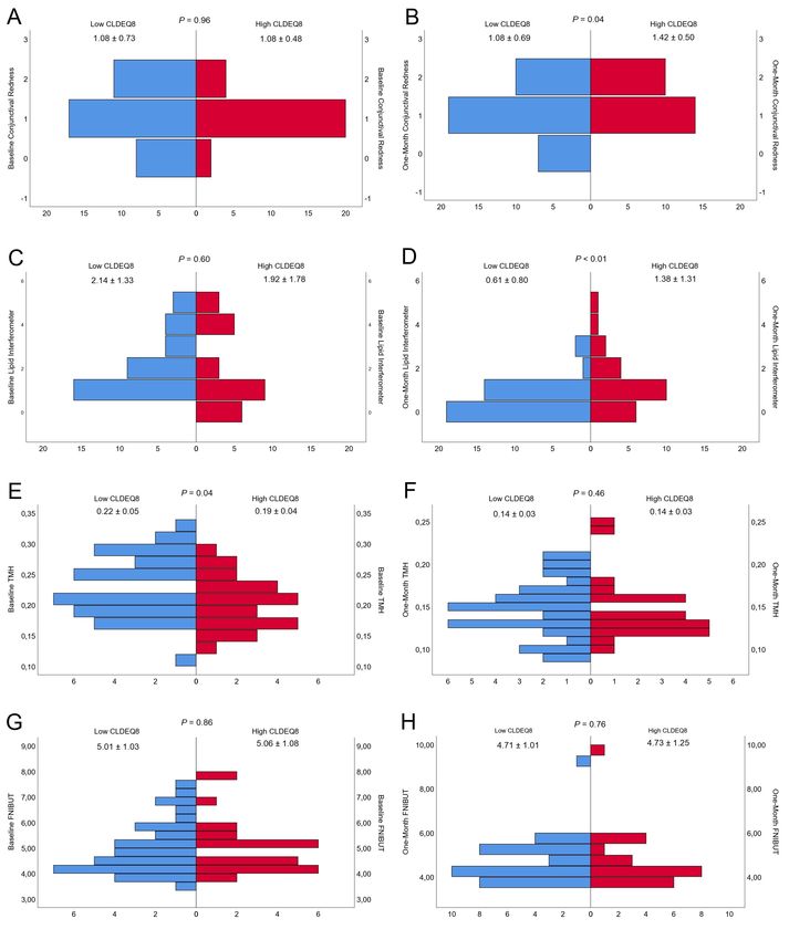
Diagnostics 2023, 13, 939                                                                                            2 of 12

                            film evenly across the surface of the eye, which is important for maintaining a clear and
                            smooth surface for light to pass through [1].
                                  The tear film is constantly being replenished and refreshed, with new fluid being
                            produced and old fluid being drained away through the tear ducts [8]. This constant
                            turnover of fluid helps to ensure that the tear film remains fresh and healthy. The dynamics
                            of the tear film are critical for maintaining the health and function of the eye. A disruption
                            in the balance of any of the three layers is a hallmark about tear film instability that can
                            lead to a variety of eye problems, such as dry eye syndrome, blepharitis, and other forms
                            of ocular surface disorders [5]. Dry eye syndrome is a common condition characterized
                            by a lack of sufficient tears to keep the eye lubricated. This can cause discomfort and a
                            feeling of grittiness or burning in the eye [9]. Dry eye syndrome can be caused by a variety
                            of factors, including age, certain medical conditions, and certain medications. Blepharitis is
                            an inflammation of the eyelids that can cause redness, itching, and a feeling of grittiness or
                            burning in the eye [8]. It can also cause the eyelids to become swollen and crusted. These
                            disorders can lead to dry eye symptoms, tear film instability, and visual disturbances. The
                            tear film is a complex and dynamic structure that plays a critical role in maintaining the
                            health and function of the eye.
                                  Contact lenses are a popular form of vision correction that are worn directly on the
                            surface of the eye. They provide a convenient and discreet alternative to glasses and can
                            be worn for extended periods of time [10]. However, wearing contact lenses can also lead
                            to a variety of symptoms related to the health and comfort of the eye. One of the most
                            common symptoms experienced by contact lens wearers is dry eye [11]. This can cause
                            discomfort and a feeling of grittiness or burning in the eye [12]. Dry eye is particularly
                            prevalent among contact lens wearers, with estimates suggesting that up to 30% of contact
                            lens wearers experience dry eye symptoms [11].
                                  To evaluate the symptoms of dry eye among contact lens wearers, a variety of question-
                            naires have been developed. One commonly used questionnaire is the Contact Lens Dry
                            Eye Questionnaire (CLDEQ-8) [13]. This questionnaire is a validated tool that is designed to
                            assess the symptoms of dry eye in contact lens wearers [13,14]. It consists of eight questions
                            that assess symptoms such as dryness, burning, and discomfort. The questionnaire also
                            includes a visual analogue scale (VAS) to rate the severity of symptoms [15]. The CLDEQ-8
                            questionnaire is a quick and easy tool that can be used to evaluate dry eye symptoms in
                            contact lens wearers. It has been validated in multiple studies and has been shown to have
                            good reliability and validity [16,17]. The questionnaire can be used to monitor the progres-
                            sion of dry eye symptoms over time, and to evaluate the effectiveness of interventions to
                            improve symptoms [13]. In addition to dry eye symptoms, contact lens wearers may also
                            experience other symptoms such as redness, itching, and foreign body sensation. These
                            symptoms can be evaluated using questionnaires such as the Ocular Surface Disease Index
                            (OSDI) and the Contact Lens Symptom Survey (CLSS) [18]. The use of questionnaires can
                            provide valuable information on the prevalence, severity, and progression of symptoms
                            among contact lens wearers, and can be used to evaluate the effectiveness of interventions
                            to improve symptoms.
                                  The dynamics of the tear film, such as its thickness and stability, can be affected by
                            various factors, such as dry eye syndrome, contact lens wear, and aging [1]. Therefore, it is
                            important to have accurate and non-invasive methods to measure the tear film dynamics
                            in order to diagnose and monitor these conditions. One of the most commonly used non-
                            invasive techniques for measuring the tear film dynamics is the non-invasive break-up time
                            (NIBUT) test [19]. This test measures the time it takes for the tear film to break up after a
                            blink, which reflects the stability of the tear film. The longer the NIBUT, the more stable the
                            tear film. Another non-invasive method is the measurement of the tear film thickness using
                            optical coherence tomography (OCT) [20]. This technique uses light waves to produce
                            detailed images of the ocular surface, including the tear film. These images can be used to
                            measure the thickness of the tear film and to detect any abnormalities.
Diagnostics 2023, 13, 939                                                                                             3 of 12

                                 Other non-invasive methods for measuring the tear film dynamics include the mea-
                            surement of the tear meniscus height (TMH) [21]. The TMH test uses a digital camera to
                            photograph the eye and measure the height of the tear meniscus, which reflects the volume
                            of the tear film [21]. One of the most recent non-invasive methods for measuring the tear
                            film dynamics is the use of interferometry [19]. This technique uses light waves to measure
                            the thickness and stability of the tear film. The interferometer creates an interference pattern
                            of the light reflecting off the tear film, which can be used to measure the thickness and
                            stability of the tear film [19].
                                 The purpose of this study is to evaluate the tear film dynamics between individuals
                            with low and high Contact Lens Dry Eye Disease Questionnaire (CLDEQ-8) scores when
                            wearing Lehfilcon A silicone hydrogel water gradient contact lenses. This study aims to
                            use a non-invasive methodology approach to investigate the potential correlation between
                            CLDEQ-8 scores and tear film dynamics. This research will provide insight into the
                            relationship between dry eye symptoms and tear film dynamics in contact lens wearers,
                            and may aid in the development of more effective interventions for managing dry eye
                            symptoms in this population. Additionally, it will also provide information about the
                            performance of Lehfilcon A silicone hydrogel water gradient contact lens in managing
                            subjective dry eye symptoms by CLDEQ-8 questionnaire. The findings of this study may
                            be useful for both clinicians and researchers in the field of ocular surface disorders and
                            contact lens wear.

                            2. Materials and Methods
                            2.1. Design
                                 In this study, we implemented a longitudinal, single-location, self-comparison investi-
                            gation. It took place in the Optics and Optometry departments of the University of Seville’s
                            Pharmacy School. The research was carried out in accordance with the guidelines set forth
                            in the Helsinki Declaration and received approval from the University of Seville’s Ethical
                            Committee Board (0384-N-22).

                            2.2. Subjects
                                 All participants in the final analysis provided their informed consent and were pro-
                            vided with a detailed explanation of the study’s procedures. To be included in the study,
                            participants had to meet the following criteria: (I) be in good ocular health and not currently
                            undergoing any eye treatment, (II) be between the ages of 18 and 35, (III) score above 0 on
                            the Contact Lens Dry Eye Questionnaire 8 (CLDEQ8), (IV) be daily or monthly silicone
                            hydrogel contact lens wearers, and (V) have a spherical equivalent refraction of ≤5.50
                            diopters or less and refractive astigmatism of ≤1.50 diopters or less. Participants were
                            excluded from the study if they had any of the following: (I) an active ocular infection or
                            inflammation, or a history of ocular surgery, (II) were taking any medications that could
                            affect the tear film or ocular surface, (III) had Sjogren syndrome, Rheumatoid arthritis,
                            diabetes, or thyroid disorders, or (IV) were pregnant or breastfeeding.

                            2.3. Materials
                                 All materials were described in a previous study [22]. Only listed below: Clinical Plat-
                            form (ICP) Ocular Surface Analyzer (OSA) from SBM System® (Orbassano, Torino, Italy),
                            Nonmydriatic infrared meibography digital fundus camera Cobra® HD (Construzione
                            Strumenti Oftalmici CSO® , Firenze, Italy), Contact Lens Dry Eye Questionnaire 8 (CLDEQ-
                            8) [13,14] and the Standard Patient Evaluation of Eye Dryness (SPEED). Silicone hydrogel
                            contact lens (TOTAL 30® , Alcon Inc., Fort Worth, TX, USA) and a multipurpose solution
                            (MPS) (Lens 55® Care Hyaluropolimer Plus 360 mL, Servilens Fit and Cover® , Granada,
                            Spain) for all subjects.
Diagnostics 2023, 13, 939 4 of 12
2.4. Procedure
In the initial phase, participants were selected based on specific criteria and their
samples were collected from the non-optometry field. After this period, surveys and
non-invasive tests were conducted to assess tear film fluctuations. Variables measured
included conjunctival redness, lipid layer thickness, tear meniscus height, and meibomian
gland dysfunction. In the second phase, participants were re-evaluated with the same
variables measured in phase one after 30 days of wearing the contact lenses to assess the
tear film in front of the lenses. Participants were instructed to follow a specific lens care
regimen and avoid eye drops or lubricants. The test conditions were consistent, and the
measurements were alternated between eyes. Participants were also instructed to blink
normally between measurements.
2.5. Statistical Analysis
Data analysis was conducted using IBM Corp’s SPSS software (version 26.0). De-
scriptive statistics such as mean and standard deviation were used. The normality of the
data was determined using the Shapiro-Wilk test. Differences in categorical variables were
assessed using the chi-square test, and differences in numerical variables between different
time points were evaluated using the Wilcoxon test. The group were separate with the
CLDEQ-8 diagnostic criteria established by Chalmers et al. [13,14] of 12 score points. Low
CLDEQ-8 group were patients with a baseline CLDEQ-8 ≤ 12 and high CLDEQ-8 group
were patients with a baseline CLDEQ-8 > 12 score points. All tests were set at a signifi-
cance level of 95% (p value < 0.05). The sample size was calculated using the GRANMO®
calculator, with a two-tailed test, alpha and beta risks of 5% and 20%, respectively, and an
estimated standard deviation of 0.45. The recommended sample size was 28 subjects.
3. Results
A group of thirty-one subjects with low levels of astigmatism and myopia were fitted
with silicone hydrogel contact lenses (Lehfilcon A). From the sample, 7 (22.6%) were male
and 24 (77.4%) were female. Twenty-one subjects were from Italy (67.75%), and the rest
of the patients were from different countries including Spain (12.90%), Mexico (6.46%),
Slovenia (3.22%), Poland (3.22%), Germany (3.22%), and Austria (3.22%). Mean age of the
subjects was 22.23 ± 1.39 (19 to 25) years old. The refraction of the subjects was sphere
(diopters) −2.64 ± 1.15 (−5.50 to −0.50), cylinder (diopters) −0.44 ± 0.37 (−1.50 to 0.00),
and axis (degrees, ◦ ) 111.44 ± 70.08 (5.00 to 180.00).
Regarding visual acuity (Log MAR): −0.03 ± 0.05 (−0.10 to 0.10). Mean cornea
keratometry were flat corneal meridian (mm) 7.87 ± 0.31 (7.40 to 8.74), steep corneal
meridian (mm) 7.73 ± 0.29 (7.25 to 8.61), and mean corneal meridian (mm) 7.80 ± 0.30
(7.37 to 8.67). Meibomian gland dysfunction (MGD) was studied along the percentage
of loss. Superior eyelid MGD (%) 28.87 ± 15.11 (10.30 to 96.20) and inferior eyelid MGD
(%) 49.69 ± 17.86 (17.00 to 87.30). Finally, contact lens power (diopters) was −2.56 ± 1.12
(−5.00 to −0.75).
Baseline measurements divided within the contact lens subjective questionnaire
groups (Low CLDEQ-8 and high CLDEQ-8) were presented in Table 1. In the same term,
one-month measurements divided within the contact lens subjective questionnaire groups
(Low CLDEQ-8 and high CLDEQ-8) were presented in Table 2. All the patients were able
to comfortably wear the contact lenses for 30 days, with the exception of one person who
experienced a minor irritation that cleared up after a few days. The contact lenses were
worn for an average of 5.61 days per week, 8.95 h per day, and 3.68 h on the one-month
follow-up visit. Pyramid graph about baseline and one-month conjunctival redness classi-
fication, lipid layer thickness interferometry, tear meniscus height, and first NIBUT were
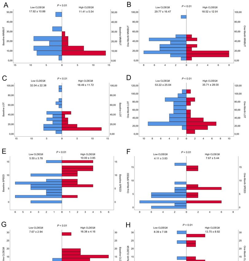
presented in Figure 1. Pyramid graph about baseline and one-month mean NIBUT, lid
opening time, SPEED, and CLDEQ-8 were presented in Figure 2.Diagnostics 2023, 13, 939 5 of 12
Table 1. Baseline outcomes divided by CLDEQ-8 group.
Variable Low CLDEQ-8 High CLDEQ-8 p Value
1.08 ± 0.73 1.08 ± 0.48
Conjunctival Redness Classification (Efron Scale) 0.96
(0.00 to 2.00) (0.00 to 2.00)
2.14 ± 1.33 1.92 ± 1.78
Lipid Layer Thickness Interferometry (Guillon Pattern) 0.60
(1.00 to 5.00) (0.00 to 5.00)
0.22 ± 0.05 0.19 ± 0.04
Tear Meniscus Height (Millimeters) 0.04 *
(0.11 to 0.32) (0.13 to 0.29)
5.01 ± 1.03 5.06 ± 1.08
First NIBUT (seconds) 0.86
(3.60 to 7.36) (3.92 to 7.80)
17.92 ± 10.88 11.41 ± 5.54
Mean NIBUT (seconds)Diagnostics 2023, 13, 939 6 of 12
Diagnostics 2023, 13, x FOR PEER REVIEW 6 of 12
Figure 1. Baseline and One Month Comparison of (A,B) Conjunctival Redness, (C,D) Lipid Layer
Figure 1. Baseline and One Month Comparison of (A,B) Conjunctival Redness, (C,D) Lipid Layer
Interferometry, (E,F) Tear Meniscus Height (TMH), and (G,H) First Non-Invasive Break Up Time
Interferometry, (E,F) Tear Meniscus Height (TMH), and (G,H) First Non‐Invasive Break Up Time
(FNIBUT).
(FNIBUT).The figure
The figureconsists
consistsofofeight
eightpyramid-shaped
pyramid‐shapedgraphs,
graphs,with
withfour
four left
left representing the base‐
representing the baseline
measurements and four right representing the measurements taken one month later.
line measurements and four right representing the measurements taken one month later.Diagnostics 2023,
Diagnostics 13,
2023, 13,939
x FOR PEER REVIEW 7 of 12
7 of 12
Figure 2. Baseline and One Month Comparison of (A,B) Mean Non-invasive Break-up Time, (C,D) Lid
Opening
Figure 2. Time (LOT),
Baseline and (E,F) Standard
One Month Patient Evaluation
Comparison of Eye
of (A,B) Mean Dryness (SPEED)
Non‐invasive Break‐upand (G,H)
Time, Contact
(C,D)
Lens
Lid Opening Time (LOT), (E,F) Standard Patient Evaluation of Eye Dryness (SPEED) and (G,H)with
Dry Eye Questionnaire (CLDEQ-8). The figure consists of eight pyramid-shaped graphs,
Contact Lens Dry Eye
four left representing theQuestionnaire (CLDEQ‐8).
baseline measurements The
and figure
four rightconsists
representing the pyramid‐shaped
of eight measurements taken
graphs, with four
one month later. left representing the baseline measurements and four right representing the meas‐
urements taken one month later.Diagnostics 2023, 13, 939 8 of 12
4. Discussion
4.1. Summary of Findings
Our findings could be summarized comparing the two groups, the following changes
were observed: conjunctival redness increased in high CLDEQ-8 group. Lipid layer de-
creased in both groups, especially in low CLDEQ-8. TMHs remain stable. FNIBUT de-
creased in both groups without statistically signification. MNIBUT and LOT achieved
relevant and significantly increased in both groups. Finally, subjective dry eye disease
sensation questionnaire specially decreased in high CLDEQ-8 group.
4.2. Comparison with Other Authors Outcomes
The daily version of Lehfilcon A has been previously studied. Marx et al. [23] con-
ducted to determine the level of satisfaction and comfort among first-time contact lens
users who were given Delefilcon A (DAILIES TOTAL1) daily disposable lenses. The study
was conducted in various European locations and spanned a period of two weeks. Partici-
pants were initially fitted with Delefilcon A lenses and then evaluated at the beginning of
the trial and after the first and second weeks. The results revealed that the study lenses
were found to have a higher mean score for subject-reported quality of vision and comfort
compared to their traditional glasses. Over 90% of the subjects stated that the lenses were
more comfortable than they had anticipated and 92% expressed an interest in purchasing
them. The investigators found that the fit of the study lenses was acceptable in at least 97%
of the subjects. The study ultimately concluded that practitioners could expect positive
results when transitioning first-time contact lens wearers from glasses to Delefilcon A daily
disposable contact lenses.
Szczesna-Iskander et al. [24] conducted to evaluate the pre-lens tear film surface
quality (TFSQ) of a new water gradient silicone hydrogel material that is used in daily
disposable lenses, in comparison to another daily disposable lens from the same manufac-
turer. Eleven subjects participated in the study and wore two different types of lenses for
two non-consecutive days. The TFSQ was measured using non-invasive interferometry
and subjective comfort was also assessed. The results showed that both lenses resulted
in a reduction of TFSQ compared to the bare eye condition. The new water gradient
silicone hydrogel material had a statistically significant smaller impact on TFSQ than the
high-water content material. The measurement methodology was found to have a high
level of linearity with respect to the lens material and there was a statistically significant
correlation between the TFSQ results of the two lenses. However, the correlation between
subjective comfort and the lenses was found to be insignificant. This research supports the
minimal effect on the tear meniscus height of Delefilcon A.
In a similar line of research Fujimoto et al. [25] aimed to determine if the water
gradient technology of Delefilcon A-based soft contact lenses improves tear film dynamics.
The study was observational and retrospective and included 50 asymptomatic users of
Delefilcon A or Narafilcon A SCLs. The study measured the thin aqueous layer break,
non-invasive tear break-up time, tear meniscus height, subjective dryness, and higher-
order aberrations. The measurements were taken at three visits: the first with the bare
eye, the second with the SCL-worn eye after 15 min, and the third 30 ± 5 days after the
second visit after the SCL was worn for at least 5 h. The study found that the water
gradient technology of Delefilcon A reduced thin aqueous layer break and increased non-
invasive tear break-up time. Additionally, it reduced tear meniscus height and total ocular
higher-order aberrations, and improved lens performance.
Lipid layer thickness reduction was determined by the presence of the biomimetic
content on the surface of the contact lens. Mao et al. [26] observed that surfaces with high-
aspect ratio nanostructure have the ability to kill bacteria. The physical interaction between
the nanostructure and the cell membrane causes the cells to break apart. Recent research
has been able to transfer this ability to artificial surfaces. However, these surfaces may have
different properties compared to those found in nature. The review looks at recent progress
in developing bactericidal surfaces and analyses the factors that influence their effectivenessDiagnostics 2023, 13, 939 9 of 12
and the mechanism behind the cell rupture. It uses a holistic approach, combining different
factors such as interaction mechanisms, material properties, and fabrication techniques, to
understand the effect of surface topography and its potential use in soft contact lenses.
Wesley et al. [27] investigated Lehfilcon A, but applied a subjective methodology in
the wettability measurements. Some 115 subjects completed the study and were divided
into two groups: one wearing the investigational Delefilcon A lens and the other wearing
the control Comfilcon A lens, both for 3 months. The study measured distance visual
acuity, lens fit and movement, centration, front and back surface deposits, and front surface
wettability. The results showed that both lenses provided excellent visual acuity, optimal
lens fitting characteristics, a clean surface, high wettability, and no ocular adverse events
after 3 months of lens wear. The Delefilcon A lens showed better performance in terms of
centration and lens fit/movement than the Comfilcon A lens.
4.3. Strengths and Limitations
The strengths of the current study include the use of a non-invasive methodology
approach to assess tear film dynamics in individuals with varying levels of contact lens dry
eye disease (CLDEQ-8 scores) using a Lehfilcon A silicone hydrogel water gradient contact
lens. This approach allowed for the objective measurement of tear film parameters without
the need for invasive methods. Additionally, the use of the CLDEQ-8 questionnaire pro-
vided a standardized and validated method for assessing dry eye symptoms in individuals
wearing contact lenses.
Certainly, using a non-invasive technology to measure tear film dynamics provides
several advantages over traditional questionnaires. Firstly, non-invasive methods, such as
the ones used in our study, do not require physical contact with the eye, making them more
comfortable for the patient and reducing the risk of infection or injury. This is particularly
important in individuals with dry eye disease, as their eyes may be more sensitive to
physical contact [28].
Secondly, non-invasive methods can provide more objective and quantitative data
compared to subjective questionnaires. By directly measuring tear film parameters such
as lipid layer thickness, tear meniscus height, and non-invasive break-up time, we can
obtain accurate and precise measurements that can be used to monitor changes in tear film
dynamics over time [21].
Finally, non-invasive methods are less dependent on patient interpretation and recall,
which can be a source of bias in questionnaires. This makes non-invasive methods more
reliable and reproducible, which is important for ensuring the accuracy and validity of
research findings [29].
By highlighting these advantages, we can better demonstrate the innovation of our
study and the importance of using non-invasive methods to assess tear film dynamics. Com-
paring our non-invasive methods with other questionnaires can help to further emphasize
the unique contribution of our study to the field of dry eye disease research.
The sample size was also appropriate and calculated using an appropriate sample size
calculator. The study also used multiple measurements to evaluate the tear film dynamics,
which provide a more comprehensive assessment of the tear film. Furthermore, the use
of a silicone hydrogel water gradient contact lens is a relevant and clinically significant
lens type, which is widely used today. Finally, the study compared the tear film dynamics
between low and high CLDEQ-8 score groups, which is an important aspect to understand
the impact of dry eye symptoms on tear film parameters.
One limitation of the current study is that it only included a single type of contact
lens, the Lehfilcon A silicone hydrogel water gradient contact lens. While this lens type is
widely used and clinically relevant, it may not be representative of all contact lens types
and the findings may not be generalizable to other lens types. Additionally, the study only
included a single follow-up time point of 30 days, which may not capture the full extent of
tear film dynamics over a longer period of time. Furthermore, the study was conducted
on a small sample size which may not be representative of the entire population. AnotherDiagnostics 2023, 13, 939 10 of 12
limitation is that the study did not include a control group of individuals not wearing
contact lenses, thus it is not possible to determine if the tear film dynamics observed are
specific to contact lens wear or if they would have been observed in non-lens wearers as
well. Additionally, the study only measured tear film dynamics at specific time points, it is
not possible to understand the tear film dynamics over time, and also, the study did not
include any subjective measurements of dry eye symptoms, it would have been beneficial
to have both objective and subjective measures to provide a more complete understanding
of the relationship between contact lens wear and dry eye symptoms.
Moreover, the study only used a single non-invasive method, Ocular Surface Ana-
lyzer (OSA) and Meibographer, to assess tear film dynamics. Other methods such as the
Interferometer or Schirmer test may have provided additional information about tear film
dynamics. Also, the study did not include any measurement of the lens surface, which
could have provided information about the lens surface and how it contributes to the tear
film dynamics. In addition, the study did not include any measure of the lens movement
during the blink, this would have provided important information about the lens–eyelid
interaction and how it affects the tear film dynamics. Finally, the study did not include any
measure of the tear film break-up time, which is an important measure of tear film stability
and dry eye. The current study provides important information about tear film dynamics
in individuals wearing a specific type of contact lens, but additional studies with larger
sample sizes and longer follow-up periods, including a control group and other lens types,
and using a combination of objective and subjective measures, and other non-invasive
methods, are needed to fully understand the relationship between contact lens wear and
tear film dynamics.
4.4. Future Lines of Research
Future research on this topic could explore the potential relationship between the
severity of dry eye symptoms as measured by the CLDEQ-8 questionnaire and the tear film
parameters measured by the non-invasive methods. This could provide insight into which
specific tear film parameters are most affected by dry eye symptoms and could potentially
be used as biomarkers for dry eye in contact lens wearers. Another line of research could be
to investigate the impact of various lens care solutions on the tear film dynamics. Different
lens care solutions have different compositions, and some may have a greater impact on
the tear film than others. Additionally, the impact of the lens replacement schedule (daily,
weekly, monthly) on the tear film dynamics could also be studied.
It would also be interesting to explore the relationship between the tear film dynamics
and the lens design. For example, different lens materials, designs, and geometries could
affect the tear film dynamics differently. This could be relevant in the development of new
contact lenses with improved comfort and performance. Additionally objective tear film
dynamics measurements could be added as future research. TF-OSI dynamic objective
scattering index of tear film provide a more complete understanding of the effect of contact
lens wear on tear film stability. Finally, future research could include a greater diversity of
subjects by including different age groups, ethnicities, and genders to better understand
the tear film dynamics in different populations. This would allow the generalization of the
findings to a larger population and improvement of the clinical significance of the results.
5. Conclusions
This study demonstrates the effectiveness of Lehfilcon A silicone hydrogel water
gradient contact lenses in improving tear film stability and reducing subjective dry eye
symptoms in individuals with low and high CLDEQ-8 scores. However, it also led to
an increase in conjunctival redness and a decrease in tear meniscus height. CLDEQ-8
questionnaire provides a standardized and quantitative measure of dry eye symptoms in
contact lens wearers.
Water gradient contact lenses present a promising option for patients who experience
dry eye symptoms related to contact lens wear. They have been shown to improve objectiveDiagnostics 2023, 13, 939 11 of 12
measures such as artificial tear film dynamics and subjective measures such as dry eye
questionnaire scores. However, it is important to have close supervision of the ocular
surface to monitor and control parameters such as ocular redness and tear layer volume.
Author Contributions: Conceptualization, R.C.-P., M.-J.B.-L. and J.-M.S.-G. methodology, R.C.-P.,
M.-J.B.-L. and J.-M.S.-G.; validation, R.C.-P., M.-J.B.-L. and J.-M.S.-G.; formal analysis, R.C.-P., M.-
J.B.-L. and J.-M.S.-G.; investigation, R.C.-P., M.-J.B.-L., and J.-M.S.-G.; resources, R.C.-P., M.-J.B.-L.
and J.-M.S.-G.; data curation, R.C.-P., M.-J.B.-L., and J.-M.S.-G.; writing—original draft preparation,
R.C.-P., M.-J.B.-L., and J.-M.S.-G.; writing—review and editing, R.C.-P., M.-J.B.-L. and J.-M.S.-G.;
visualization, R.C.-P., M.-J.B.-L. and J.-M.S.-G.; supervision, R.C.-P., M.-J.B.-L. and J.-M.S.-G. All
authors have read and agreed to the published version of the manuscript.
Funding: This research received no external funding.
Institutional Review Board Statement: The study was conducted in accordance with the Declaration
of Helsinki and approved by the Ethics Committee of the University of Seville (0384-N-22).
Informed Consent Statement: Informed consent was obtained from all subjects involved in the study.
Data Availability Statement: The data presented in this study are available on request from the
corresponding author. The data are not publicly available due to their containing information that
could compromise the privacy of research participants.
Acknowledgments: The authors appreciate the support offered by the members of the Department
of Physics of Condensed Matter, Faculty of Physics, University of Seville, with special thanks to
Javier Romero-Landa and Clara Conde-Amiano. In addition, the authors also appreciate the technical
support offered by the members and facilities of the Faculty of Pharmacy, University of Seville, with
special thanks to María Álvarez-de-Sotomayor.
Conflicts of Interest: The authors declare no conflict of interest.
References
1. Nelson, J.D.; Craig, J.P.; Akpek, E.K.; Azar, D.T.; Belmonte, C.; Bron, A.J.; Clayton, J.A.; Dogru, M.; Dua, H.S.; Foulks, G.N.; et al.
TFOS DEWS II Introduction. Ocul. Surf. 2017, 15, 269–275. [CrossRef]
2. Craig, J.P.; Nichols, K.K.; Akpek, E.K.; Caffery, B.; Dua, H.S.; Joo, C.-K.; Liu, Z.; Nelson, J.D.; Nichols, J.J.; Tsubota, K.; et al. TFOS
DEWS II Definition and Classification Report. Ocul. Surf. 2017, 15, 276–283. [CrossRef] [PubMed]
3. Pflugfelder, S.C.; Stern, M.E. Biological functions of tear film. Exp. Eye Res. 2020, 197, 108115. [CrossRef] [PubMed]
4. Golden, M.I.; Meyer, J.J.; Patel, B.C. Dry Eye Syndrome. In Statpearls; Statpearls Publishing: Treasure Island, FL, USA, 2022.
5. Bron, A.J.; de Paiva, C.S.; Chauhan, S.K.; Bonini, S.; Gabison, E.E.; Jain, S.; Knop, E.; Markoulli, M.; Ogawa, Y.; Perez, V.; et al.
TFOS DEWS II pathophysiology report. Ocul. Surf. 2017, 15, 438–510. [CrossRef] [PubMed]
6. Pflugfelder, S.C.; de Paiva, C.S. The Pathophysiology of Dry Eye Disease: What We Know and Future Directions for Research.
Ophthalmology 2017, 124, S4–S13. [CrossRef]
7. Willcox, M.D.P.; Argüeso, P.; Georgiev, G.A.; Holopainen, J.M.; Laurie, G.W.; Millar, T.J.; Papas, E.B.; Rolland, J.P.; Schmidt, T.A.;
Stahl, U.; et al. TFOS DEWS II Tear Film Report. Ocul. Surf. 2017, 15, 366–403. [CrossRef]
8. Stapleton, F.; Alves, M.; Bunya, V.Y.; Jalbert, I.; Lekhanont, K.; Malet, F.; Na, K.-S.; Schaumberg, D.; Uchino, M.; Vehof, J.; et al.
TFOS DEWS II Epidemiology Report. Ocul. Surf. 2017, 15, 334–365. [CrossRef]
9. Belmonte, C.; Nichols, J.J.; Cox, S.M.; Brock, J.A.; Begley, C.G.; Bereiter, D.A.; Dartt, D.A.; Galor, A.; Hamrah, P.; Ivanusic, J.J.; et al.
TFOS DEWS II pain and sensation report. Ocul. Surf. 2017, 15, 404–437. [CrossRef]
10. Richdale, K.; Cox, I.; Kollbaum, P.; Bullimore, M.A.; Bakaraju, R.C.; Gifford, P.; Plainis, S.; McKenney, C.; Newman, S.; Tomiyama,
E.S.; et al. CLEAR—Contact lens optics. Contact Lens Anterior Eye 2021, 44, 220–239. [CrossRef]
11. Stapleton, F.; Bakkar, M.; Carnt, N.; Chalmers, R.; Vijay, A.K.; Marasini, S.; Ng, A.; Tan, J.; Wagner, H.; Woods, C.; et al.
CLEAR—Contact lens complications. Contact Lens Anterior Eye 2021, 44, 330–367. [CrossRef]
12. Morgan, P.B.; Murphy, P.J.; Gifford, K.L.; Gifford, P.; Golebiowski, B.; Johnson, L.; Makrynioti, D.; Moezzi, A.M.; Moody, K.;
Navascues-Cornago, M.; et al. CLEAR—Effect of contact lens materials and designs on the anatomy and physiology of the eye.
Contact Lens Anterior Eye 2021, 44, 192–219. [CrossRef]
13. Chalmers, R.L.; Begley, C.G.; Moody, K.; Hickson-Curran, S.B. Contact Lens Dry Eye Questionnaire-8 (CLDEQ-8) and opinion of
contact lens performance. Optom. Vis. Sci. Off. Publ. Am. Acad. Optom. 2012, 89, 1435–1442. [CrossRef]
14. Chalmers, R.L.; Keay, L.; Hickson-Curran, S.B.; Gleason, W.J. Cutoff score and responsiveness of the 8-item Contact Lens Dry Eye
Questionnaire (CLDEQ-8) in a Large daily disposable contact lens registry. Contact Lens Anterior Eye 2016, 39, 342–352. [CrossRef]
15. Koh, S.; Chalmers, R.; Yamasaki, K.; Kawasaki, R.; Nishida, K. Factors influencing the 8-item contact lens dry eye questionnaire
score and comparison of translations in Japanese soft contact lens wearers. Contact Lens Anterior Eye 2022, 45, 101519. [CrossRef]Diagnostics 2023, 13, 939 12 of 12
16. Pucker, A.D.; Dougherty, B.E.; Jones-Jordan, L.A.; Kwan, J.T.; Kunnen, C.M.E.; Srinivasan, S. Psychometric Analysis of the SPEED
Questionnaire and CLDEQ-8. Investig. Ophthalmol. Vis. Sci. 2018, 59, 3307–3313. [CrossRef]
17. López-de la Rosa, A.; Arroyo-Del Arroyo, C.; Enríquez-de-Salamanca, A.; Pinto-Fraga, J.; López-Miguel, A.; González-García, M.J.
The ability of the Contact Lens Dry Eye Questionnaire (CLDEQ)-8 to detect ocular surface alterations in contact lens wearers.
Contact Lens Anterior Eye 2019, 42, 273–277. [CrossRef]
18. Wolffsohn, J.S.; Arita, R.; Chalmers, R.; Djalilian, A.; Dogru, M.; Dumbleton, K.; Gupta, P.K.; Karpecki, P.; Lazreg, S.; Pult, H.; et al.
TFOS DEWS II Diagnostic Methodology report. Ocul. Surf. 2017, 15, 539–574. [CrossRef]
19. Itokawa, T.; Suzuki, T.; Koh, S.; Hori, Y. Evaluating the Differences between Fluorescein Tear Break-up Time and Noninvasive
Measurement Techniques. Eye Contact Lens 2022, 49, 104–109. [CrossRef]
20. Yang, F.; Chang, Y.; Yang, L.; Jia, Z.; Liu, J.; Wang, Y. Evaluation of the repeatability of optical coherence tomography in patients
with age-related cataract associated with dry eye. Int. Ophthalmol. 2022, 43, 233–238. [CrossRef]
21. Sánchez-González, M.C.; Capote-Puente, R.; García-Romera, M.-C.; De-Hita-Cantalejo, C.; Bautista-Llamas, M.-J.; Silva-Viguera,
C.; Sánchez-González, J.-M. Dry eye disease and tear film assessment through a novel non-invasive ocular surface analyzer: The
OSA protocol. Front. Med. 2022, 9, 938484. [CrossRef]
22. Capote-Puente, R.; Bautista-Llamas, M.-J.; Manzoni, C.; Sánchez-González, J.-M. Pre-Lens Tear Meniscus Height, Lipid Layer
Pattern and Non-Invasive Break-Up Time Short-Term Changes with a Water Gradient Silicone Hydrogel Contact Lens. Life 2022,
12, 1710. [CrossRef] [PubMed]
23. Marx, S.; Lauenborg, B.; Kern, J.R. Performance evaluation of delefilcon a water gradient daily disposable contact lenses in
first-time contact lens wearers. Contact Lens Anterior Eye 2018, 41, 335–341. [CrossRef] [PubMed]
24. Szczesna-Iskander, D.H. Comparison of tear film surface quality measured in vivo on water gradient silicone hydrogel and
hydrogel contact lenses. Eye Contact Lens 2014, 40, 23–27. [CrossRef] [PubMed]
25. Fujimoto, H.; Ochi, S.; Yamashita, T.; Inoue, Y.; Kiryu, J. Role of the Water Gradient Structure in Inhibiting Thin Aqueous Layer
Break in Silicone Hydrogel-Soft Contact Lens. Transl. Vis. Sci. Technol. 2021, 10, 5. [CrossRef]
26. Mao, T.; Fang, F. Biomimetic Functional Surfaces towards Bactericidal Soft Contact Lenses. Micromachines 2020, 11, 835. [CrossRef]
27. Wesley, G.; Giedd, B.; Hines, B.; Bickle, K.; Pearson, C.; Lorentz, H. Safety and Efficacy of a New Water Gradient Biomimetic
Monthly Replacement Spherical Contact Lens Material (Lehfilcon A). Clin. Ophthalmol. 2022, 16, 2873–2884. [CrossRef]
28. Singh, S.; Srivastav, S.; Modiwala, Z.; Ali, M.H.; Basu, S. Repeatability, reproducibility and agreement between three different
diagnostic imaging platforms for tear film evaluation of normal and dry eye disease. Eye 2022, 1–6. [CrossRef]
29. Amparo, F.; Schaumberg, D.A.; Dana, R. Comparison of Two Questionnaires for Dry Eye Symptom Assessment: The Ocular
Surface Disease Index and the Symptom Assessment in Dry Eye. Ophthalmology 2015, 122, 1498–1503. [CrossRef]
Disclaimer/Publisher’s Note: The statements, opinions and data contained in all publications are solely those of the individual
author(s) and contributor(s) and not of MDPI and/or the editor(s). MDPI and/or the editor(s) disclaim responsibility for any injury to
people or property resulting from any ideas, methods, instructions or products referred to in the content.You can also read