The potential ameliorative impacts of cerium oxide nanoparticles against fipronil induced hepatic steatosis - Nature
←
→
Page content transcription
If your browser does not render page correctly, please read the page content below
www.nature.com/scientificreports

                OPEN              The potential ameliorative impacts
                                  of cerium oxide nanoparticles
                                  against fipronil‑induced hepatic
                                  steatosis
                                  Lamiaa Wasef1,12, Atef M. K. Nassar2, Yasser S. El‑Sayed3, Dalia Samak3, Ahmed Noreldin4,
                                  Norhan Elshony1, Hamida Saleh3, Yaser H. A. Elewa5,6,12, Shaimaa M. A. Hassan7,8,
                                  Abdullah A. Saati9, Helal F. Hetta10*, Gaber El‑Saber Batiha1*, Masakazu Umezawa11 &
                                  Hazem M. Shaheen1*

                                  Fipronil (FIP) is a phenylpyrazole insecticide that is commonly used in agricultural and veterinary fields
                                  for controlling a wide range of insects, but it is a strong environmentally toxic substance. Exposure
                                  to FIP has been reported to increase the hepatic fat accumulation through altered lipid metabolism,
                                  which ultimately can contribute to nonalcoholic fatty liver disease (NAFLD) development.
                                  The present study aimed to examine the function of cerium oxide nanoparticles (CeNPs) in protecting
                                  against hepatotoxicity and lipogenesis induced by FIP. Twenty-eight male albino rats were classified
                                  into four groups: FIP (5 mg/kg/day per os), CTR, CeNPs (35 mg/kg/day p.o.), and FIP + CeNPs (5
                                  (FIP) + 35 (CeNPs) mg/kg/day p.o.) for 28 consecutive days. Serum lipid profiles, hepatic antioxidant
                                  parameters and pathology, and mRNA expression of adipocytokines were assessed. The results
                                  revealed that FIP increased cholesterol, height-density lipoprotein, triacylglyceride, low-density
                                  lipoprotein (LDL-c), and very-low-density lipoprotein (VLDL-c) concentrations. It also increased nitric
                                  oxide (NO) and malondialdehyde (MDA) hepatic levels and reduced glutathione peroxidase (GPx) and
                                  superoxide dismutase (SOD) enzyme activities. Additionally, FIP up-regulated the fatty acid-binding
                                  protein (FABP), acetyl Co-A carboxylase (ACC1), and peroxisome proliferator-activated receptor-alpha
                                  (PPAR-α). Immunohistochemically, a strong proliferation of cell nuclear antigen (PCNA), ionized
                                  calcium-binding adapter molecule 1 (Iba-1), cyclooxygenase-2 (COX-2) reactions in the endothelial
                                  cells of the hepatic sinusoids, and increased expression of caspase3 were observed following FIP
                                  intoxication. FIP also caused histological changes in hepatic tissue. The CeNPs counteracted the
                                  hepatotoxic effect of FIP exposure. So, this study recorded an ameliorative effect of CeNPs against
                                  FIP-induced hepatotoxicity.

                                  1
                                   Department of Pharmacology and Therapeutics, Faculty of Veterinary Medicine, Damanhour University,
                                  Damanhour 22511, ElBeheira, Egypt. 2Pesticides Chemistry and Toxicology, Plant Protection Department, Faculty
                                  of Agriculture, Damanhour University, Damanhour, Egypt. 3Department of Veterinary Forensic Medicine and
                                  Toxicology, Faculty of Veterinary Medicine, Damanhour University, Damanhour, Egypt. 4Department of Histology
                                  and Cytology, Faculty of Veterinary Medicine, The Scientific Campus, Damanhour University, Damanhour,
                                  Egypt. 5Department of Histology and Cytology, Faculty of Veterinary Medicine, Zagazig University, Zagazig,
                                  Egypt. 6Laboratory of Anatomy, Department of Biomedical Sciences, Graduate School of Veterinary Medicine,
                                  Hokkaido University, Sapporo, Hokkaido 060‑0818, Japan. 7Histology and Cell Biology Department, Faculty of
                                  Medicine, Menoufia University, Shebeen El‑Kom, Egypt. 8Department of Histology, College of Medicine, Batterjee
                                  Medical College, Aseer, Saudi Arabia. 9Department of Community Medicine & Pilgrims Healthcare, Faculty of
                                  Medicine, Umm Al-Qura University, Mecca 24382, Saudi Arabia. 10Department of Medical Microbiology and
                                  Immunology, Faculty of Medicine, Assiut University, Assiut 71515, Egypt. 11Department of Materials Science
                                  and Technology, Faculty of Industrial Science and Technology Soga Laboratory, Tokyo University of Science, 9th
                                  Floor, Research Labs Building, 6‑3‑1 Niijuku, Katsushika, Tokyo 125‑8585, Japan. 12These authors contributed
                                  equally: Lamiaa Wasef and Yaser H. A. Elewa. *email: helal.hetta@uc.edu; gaberbatiha@gmail.com; dr_
                                  hazemshaheen3010@yahoo.com

Scientific Reports |   (2021) 11:1310                | https://doi.org/10.1038/s41598-020-79479-5                                             1

                                                                                                                                          Vol.:(0123456789)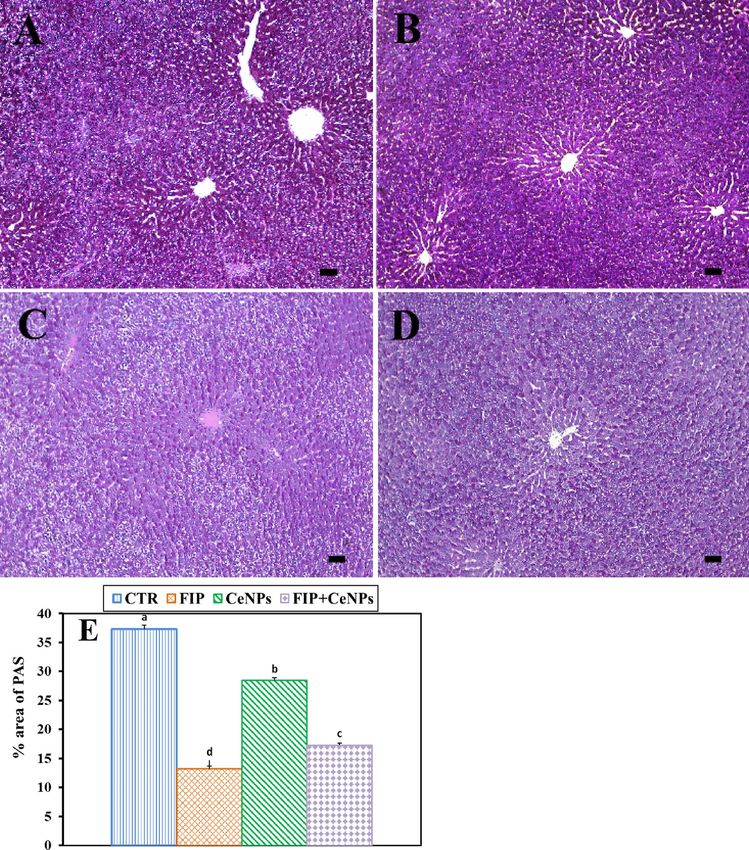
www.nature.com/scientificreports/
Around the world, pesticides are widely used to control plant and animal pests but they have direct and/or indi-
rect toxic effects on beneficial organisms1. Among pesticides, fipronil (FIP) [5-amino-3-cyano-1-(2,6-dichloro-
4-trifluoromethylphenyl) 4-fluoromethylsulfinyl pyrazole] is a second-generation phenylpyrazole insecticide2
extensively used in the veterinary clinical field against many pests, and e ctoparasites3,4. It blocked the chloride
channels regulated by GABA, resulting in an uncontrolled central nervous system (CNS) hyperexcitation and
death5. Fipronil toxic effect depends on the disturbance of the antioxidant and oxidant system balance through
the over-accumulation of reactive oxygen species (ROS) in the cells6, especially of liver tissue, which is the
main organ for insecticides-detoxification7. This balance is a good indicator of exposure to toxic chemicals in
hepatocytic mitochondria and endoplasmic reticulum8. Antioxidant/oxidant imbalance might lead to chemical
changes in proteins and fat molecules structure resulting in the activation of lipid peroxidation (LPO) process9
and is often believed to enhance the hepatic cells oxidative i njury10.
The accumulation of lipids in hepatic cells is more common in cases of exposure to toxic c hemicals11 and leads
brosis11. Increased synthesis of triglycerides results in increased fatty acids (FA) accumulation
to hepatitis and fi
in the liver. Consequently, mitochondrial oxidation of the free fatty acids (FFAs) damages the liver tissues, result-
ing in infiltration of neutrophil with subsequent ROS release and exacerbation of inflammatory r eactions12,13.
Alongside, cerium oxide nanoparticles (CeNPs) are among the most crucial metal-oxide nanoparticles, which
play a technologically important role in synthesizing different industrial materials including polishing materials
in the glass and optics industry, oxygen sensors, and ultraviolet fi lters14. CeNPs have gained much interest in
biological applications due to their antioxidant properties. Based on the C e3+/Ce4+ratio in CeNPs surface, this
antioxidant activity will be as follows: superoxide dismutase (SOD) mimetic a ctivity15, catalase (CAT) mimetic
activity, hydroxyl and nitric oxide (NO) scavenging property16. Therefore, CeNPs have been used in medicine15
and pharmacy17 due to their potent antioxidant activity in scavenging free radicals produced in the process of
oxidative damage. Reducing ROS production could reduce inflammation and consequent tissue injury18. No
earlier studies explained the correlation between CeNPs and FIP, thus the objective of the existing study was to
examine the ameliorative impacts of CeNPs on hepatic tissue against sub-acute toxicity caused by FIP admin-
istration in male albino rats.
Materials and methods
Chemicals. Cerium oxide nanopowder < 25 nm particle size, (CAS Number: 1306-38-3) was purchased
from Sigma-Aldrich Co., USA. The CeNPs were suspended in demineralized water at a 35 mg/kg bwt concen-
tration. Fipronil solution (5 mg/kg bwt) was prepared by dissolving the commercial product FIPROGENT 80%
(MAC-GmbH, Sigmarszell, Germany) in demineralized water.
Experimental animals. Twenty-eight healthy adult male albino rats (average weight 180 ± 10 g) were
obtained from the Animal Breeding Unit, Faculty of Agriculture, Alexandria University. The animals were
housed under a pathogen-free environment with controlled humidity, temperature (22 °C) and a 12 h light/dark
cycle. The animal experiments were performed according to the Laboratory Animals of the National Institutes of
Health (NIH) Care and Use Guidelines, and the study protocol was approved by the local authorities of Daman-
hour University, Egypt (DMU-2019-0023). Two weeks before the experiment was conducted, the animals were
allowed to acclimatize the testing facility condition. Afterward, the rats were caged equally into four experimen-
tal groups, each consisting of seven rats. Group 1—control group (CTR) rats were orally received saline. Group
2—animals have orally received FIP solution (5 mg/kg bwt, 1/20 of the LD50)19,20. Group 3—the rats have orally
received CeNPs solution (35 mg/kg body weight)21. Group 4—animals orally received FIP (5 mg/kg) and CeNPs
(35 mg/kg) solutions using gastric tube daily. During the experimental period, rats were daily observed for any
abnormal behavior and clinical signs.
Blood and tissue sampling. On the 28th day of the experiment, all rats were prohibited from feeding
overnight, weighed individually, and euthanized using an anesthesia system containing xylazine and ketamine
HCl. Blood was gathered from the aortic vein, kept in anticoagulant-free test tubes, and the serum was isolated
and kept at − 20 °C until lipid profile determination. The liver was excised and rinsed using physiological saline
(NaCl 0.9%), wiped using filter paper and split into two sections – the first section kept rapidly at − 80 °C for
biochemical and gene expression levels and the second section was used for histopathological and immunohis-
tochemical examination after fixing in four percent paraformaldehyde (PFA) diluted in phosphate buffer saline
(PBS) solution.
Serum biochemical test. For examination of the serum lipid profile of the treated rats; triacylglycerol,
cholesterol, HDL-c, LDL-c, and VLDL-c commercial kits were used, which were purchased from Bio-Diagnos-
tics Co., Cairo, Egypt. The experiments were conducted following the manufacturer’s instruction guidelines.
Oxidant/antioxidant parameters in liver tissue homogenates. Hepatic tissues were moistened
using phosphate-buffered saline (PBS, 0.1 M, pH 7.4). Using a disposable homogenizer (Biomasher; Nippi, Inc.,
Tokyo, Japan), homogenates of ten percent from the hepatic tissues were then prepared in cold potassium phos-
phate buffer (50 mM, pH 7.5). The resulted homogenates were centrifuged at 4 °C for 15 min at 10,000×g. The
supernatants were used for evaluating lipid peroxidation indicators: NO and malondialdehyde (MDA)22 and
antioxidant biomarkers: glutathione peroxidase (GPx) and SOD activity23,24 following the manufacturers’ guide-
lines (Bio-diagnostics CO., Cairo, Egypt).
Scientific Reports | (2021) 11:1310 | https://doi.org/10.1038/s41598-020-79479-5 2
Vol:.(1234567890)
www.nature.com/scientificreports/
Antibody Source Dilution Antigen retrieval Heating condition
(9662, Cell Signaling Technology,
Rabbit polyclonal anti-Caspase3 1:300 10 mM citrate buffer (pH 6.0) 105 °C, 20 min
Danvers, Ma, USA)
Rabbit polyclonal anti-Iba-1 (019-19741, Wako Osaka, Japan) 1:1200 10 mM citrate buffer (pH 6.0) 105 °C, 20 min
(sc-9857, Santa Cruz Biotechnology,
Goat polyclonal anti-PCNA 1:2000 Dako, 105 °C, 20 min 105 °C, 20 min
CA, USA)
Rabbit polyclonal anti- Bax (PU347-UP, San Ramon, Ca, USA) 1:30 None None
(RM-9121-S0, Thermo Fisher
Rabbit monoclonal anti-Cox-2 1:100 10 mM citrate buffer (pH 6.0) 105 °C, 20 min
Scientific, Fremont, CA, USA)
Table 1. List of antibodies, sources, working dilutions, and methods for antigen retrieval.
Primer name Accession number Sequences
F: GTTGGACAACGCCTTCACAC
Rattus norvegicus acetyl-CoA carboxylase alpha (Acaca), mRNA NM_022193.1
R: GCGCATGGAATGGCAGTAAG
F: AGGACCTCATCCAGAAAGGGA
Rattus norvegicus fatty acid binding protein 1 (Fabp1), Mrna NM_012556.2
R: TGACCTTTTCCCCAGTCATGG
Rattus norvegicus peroxisome proliferator activated receptor alpha F: ACCTTGTGCATGGCTGAGAA
NM_013196.1
(Ppara), mRNA R: CCTTGGCAAATTCCGTGAGC
F: AGTTGGACCCACCTTGTGAG
Rattus norvegicus Caspase3 NM_001284409.1
R: AGTCTGCAGCTCCTCCACAT
F: CACCAGCTCTGAACAGATCATGA
Rattus norvegicus Bcl-2 like protein 4 NM_007527.3
R: TCAGCCCATCTTCTTCCAGATGGT
Table 2. Primer sequences and accession number used for RT-qPCR.
Hepato‑histological examination. The fixed samples were treated using the traditional paraffin embed-
ding method, including drying in ascending grades of ethanol, disinfected in three changes of xylene and melted
paraffin finished by inserting it at 65 °C in paraffin wax. Hematoxylin and eosin (H&E) were used to stain four
µm thick sections as previously detected by Bancroft and Layton25 and Periodic acid Schiff (PAS) based on Lay-
ton and B ancroft26. Semi-quantitative scoring of cardiac lesions was calculated based on Gibson-Corley, et al.27.
Briefly, the lesions in 10 fields were collected and summed randomly from each slide for each rat. The lesions
were blindly recorded (Score scale: 0 = normal; 1 ≤ 25%; 2 = 26–50%; 3 = 51–75%; 4 = 76–100%).
Immunohistochemical examination. Working dilutions, sources, methods, and antibodies for antigen
recovery were listed in Table 1. The immunohistochemical technique in liver sections was investigated based
on the method identified by Noreldin, et al.28 and Noreldin, et al.29. Briefly, the paraffin sections were prepared
with a thickness of 4 µm, deparaffinized by xylene and re-moistened in graded alcohol and washed with distilled
water. Afterward, endogenous peroxidase was deactivated by 3% H2O2 in absolute methanol for 30 min at 4 °C
and washed again using PBS, blocking the nonspecific reaction at room temperature with 10% normal block-
ing serum for 60 min. Then, the sections were incubated overnight at 4 °C with the primary antibodies, washed
with PBS, incubated for 60 min with biotin-conjugated goat anti-rabbit IgG antiserum or rabbit anti-goat IgG
antiserum (Histofine kit, Nichirei Corporation) according to the species’ primary antibody hosted. Then the
sections were incubated for 30 min with streptavidin-peroxidase conjugate (Histofine kit, Nichirei Corporation)
after washing with PBS. The streptavidin–biotin complex was further incubated for 3 min with a solution of
3,3′-diaminobenzidine tetrahydrochloride (DAB)-H2O2, pH 7.0. Finally, the sections were rinsed with distilled
water and used Mayer’s hematoxylin counterstain. A digital camera (Leica EC3, Leica, Germany) connected
to a microscope (Leica DM500, Leica, Germany) was used to capture micrographs of the prepared sections.
ImageJ software (National Institutes of Health, Bethesda, MD, USA) was used for immunostaining intensities’
quantification30. The mean of the inverse density of 10 randomly selected fields from different parts of 8 rats in
each group was calculated byVis et al.31.
Quantitative reverse transcription‑polymerase chain reaction (RT‑qPCR). RNA-Spintm total
RNA extraction kits (Cat. #17211) (INTRON biotechnology Inc. was used to extract total RNA from the liver
tissues. Total RNA (1 mg) was used as a template for the first strand of complementary DNA (cDNA) using
Maxima First Strand cDNA synthesis Kits from iNtRON Biotechnology Inc (Cat. #EZ00SS). Quantitative RT-
PCR was conducted using Thermo Scientific Maxima SYBR Green/ROX qPCR PreMix kits from iNtRON Bio-
technology Inc (Cat. #RT500S). The primers were acetyl-CoA carboxylase alpha Rattus norvegicus, peroxisome
proliferator-activated receptor alpha (PPAR-α), Fabp1, Rattus norvegicus caspase3, and Rattus norvegicus Bcl-2
like protein (Table 2). The values of target genes have been standardized to household expression level, GAPDH.
Statistical analysis. Data were described as mean ± SEM. Results were analyzed statistically by one-way
(ANOVA) test using the Statistical Analysis System (SAS) software version 13.2 (SAS, 2016). Considerable
Scientific Reports | (2021) 11:1310 | https://doi.org/10.1038/s41598-020-79479-5 3
Vol.:(0123456789)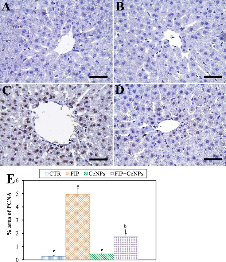
www.nature.com/scientificreports/
Group TAG (mg/dL) VLDLc (mg/dL) LDLc (mg/dL) Cholesterol (mg/dL) HDLc (mg/dL)
CRT 70.4125 ± 0.38534b 14.1825 ± 0.78979a 39.9725 ± 2.64559b 115.0000 ± 0.40825b 45.3500 ± 1.28225b
FIP 88.2000 ± 1.37174a 15.9900 ± 1.01985a 66.0900 ± 2.70945a 137.1250 ± 1.63777a 66.8750 ± 1.73656a
b a b c
CeNPs 66.0250 ± 0.83902 13.2175 ± 0.16815 31.3875 ± 2.64483 105.2125 ± 0.79441 41.7750 ± 1.18348b
b a b c
FIP + CeNPs 65.8125 ± 1.31694 14.4725 ± 1.06092 34.3950 ± 2.84348 104.3425 ± 1.55696 46.8500 ± 1.73133b
Table 3. Effects of Fipronil and CeNPs intake on Serum lipid profile indices in rats. The effect of cerium oxide
nanoparticles (CeNPs) on serum lipid profile concentrations in FIP intoxicated rats. All values are expressed
as the mean ± SE, n = 7. Means with different superscript letters (a, b, c) are statistically significant at (p ≤ 0.05).
FIP Fipronil, CeNPs cerium oxide nanoparticles, TAGtriacylglycerides, VLDLc very-low density lipoproteins,
LDLc low density lipoproteins, HDLc high density lipoproteins. Values with different superscript letters within
the same column are significantly different (P ≤ 0.05, One-way ANOVA with Tukey’s HSD post hoc test).
Group MDA (nmol/mg) NO (µmol/mg) GPx (U/mg) SOD(U/mg)
CRT 0.3825 ± 0.00479b 56.5825 ± 1.14776bc 42.4325 ± 2.19678a 7.7000 ± 0.48642a
a ab b
FIP 0.6475 ± 0.02016 86.5925 ± 0.50688 25.6025 ± 1.30649 4.1650 ± 0.26247b
c c a
CeNPs 0.2375 ± 0.00750 49.8525 ± 1.33769 45.9525 ± 1.16427 9.1800 ± 0.47450a
b bc a
FIP + CeNPs 0.4250 ± 0.01708 53.8875 ± 1.99681 41.9800 ± 1.12886 8.1450 ± 0.27834a
Table 4. Effects of Fipronil and CONPs intake on hepatic oxidative/antioxidative indices in rats. The effect of
cerium oxide nanoparticles (CeNPs) on liver tissue lipid peroxidation and activities of antioxidant enzymes
in FIP intoxicated rats All values are expressed as the mean ± SE, n = 7. Means with different superscript
letters (a, b, c) were significantly different at (P ≤ 0.05). FIP Fipronil, CeNPs cerium oxide nanoparticles, MDA
Malondialdehyde, SOD Superoxide dismutase, GPx Glutathione peroxidase, NO Nitrogen oxide. Values with
different superscript letters within the same row are significantly different (P ≤ 0.05, One-way ANOVA with
Tukey’s HSD post hoc test).
means have been compared with multiple reference checks of Tukey’s post-hoc. Results at P < 0.05 were consid-
ered statistically significant.
Results
Signs of toxicity. No obvious clinical signs or symptoms noticed all over the experimental period on rats
that were exposed to FIP or/and CeNPs.
Serum biochemical findings. The FPN-intoxicated group showed significant elevation (P < 0.05) in the
serum levels of cholesterol (19.0%), TAG (25.2%), LDL-c (66.0%) and HDL-c (47.6%) when compared with
the CTR one (Table 3). Meanwhile, rats treated with CeNPs and FIP + CeNPs exhibited remarkable reductions
(P ≤ 0.05) in serum cholesterol levels by 8.5 and 9.3%, respectively. Rats treated with CeNPs and FIP + CeNPs
had similar content of TAG, VLDL-c, LDL-c, and HDL-c to that of the CTR group. The VLDL-c levels were not
different in the serum of all treated groups (Table 3).
Liver lipid peroxidation and antioxidative indices. The results presented in Table 4 revealed that the
FIP-treated group showed substantial increase (P ≤ 0.05) in MDA (68%) concentrations and decrease in GPx
(39%) and SOD (46%) enzymes activities in the liver tissue in relative to the CTR one. The concentrations
of MDA in liver tissue (37%) in CeNPs-treated rats was reduced significantly (P ≤ 0.05), while GPx and SOD
enzyme activities were similar to normal CTR values (Fig. 2). Concentrations of serum NO were nearly compa-
rable in CTR, CeNPs, and FIP + CeNPs groups. The FIP toxic effects on hepatic GPx, MDA, SOD, and NO were
substantially reduced (P ≤ 0.05) by CeNPs administration indicating the effect of CeNPs in alleviating oxidative
damage caused by FIP.
Histopathological investigation of hepatic tissue. The histological findings revealed a polygonal
hepatocyte producing substantial anastomosis plaques in the liver tissue in the CTR animals with acidophilic
cytoplasm (Fig. 1A). Hepatic cells exhibited one or two large central round heavy hematoxylin-stained nuclei.
Moreover, one or more nucleoli were detected in some hepatic cells (Fig. 1A). Hepatocyte plates and the hepatic
capillaries barriers (sinusoids) laminated the Disse’s space, while hepatic capillary walls were bordered by Kupffer
cells (Fig. 1A) and distinguished primarily by elongated and heavily stained nuclei. The tissues of the CeNPs
group didn’t show any histopathological effects (Fig. 1B). In contrast, the FIP group revealed portal vein conges-
tion surrounded with lymphocytic infiltration, lymphocytic aggregation in between massive fatty degeneration,
congested central vein, necrotic foci, and nuclear condensation (Fig. 1C–E). It was noticed that CeNPs protected
hepatic cells against FIP adverse effects, where it restored the normal architecture of liver tissues (Fig. 1F). The
Scientific Reports | (2021) 11:1310 | https://doi.org/10.1038/s41598-020-79479-5 4
Vol:.(1234567890)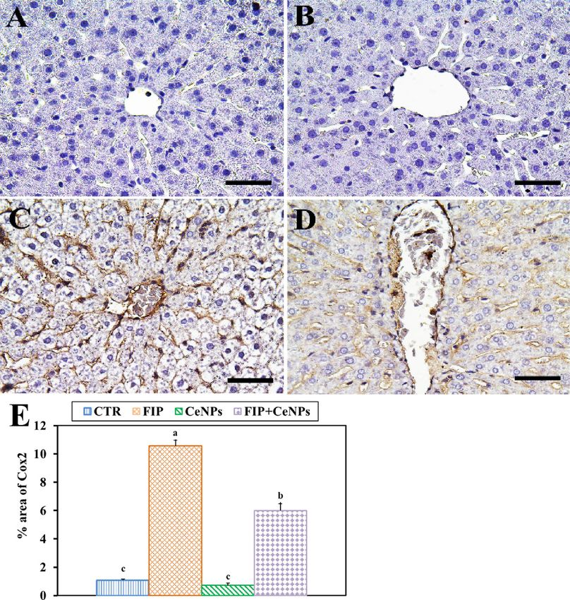
www.nature.com/scientificreports/

                                  Figure 1.  Histopathological examination of rat liver. (A) Negative control group. (B) CeNPs group. (C–E)
                                  FIP group showing in (C) congested central vein (arrow), massive fatty degeneration in the periportal areas
                                  (arrowheads) and lymphocytic aggregation (arrow) in between massive fatty degeneration (arrowheads).
                                  (D) Highly congested portal vein surrounded with lymphocytic infiltration (arrowhead). (E) Necrotic foci
                                  (thick arrow), congested liver sinusoids (arrowheads) and fatty degeneration (thin arrows). (F) FIP group that
                                  treated with CeNPs. Scale bar = 50 µm. (G) H&E semi quantitative scoring of hepatic fatty degeneration and
                                  lymphocytic aggregations. Data expressed as Mean ± SE, analyzed using one-way ANOVA at P ≤ 0.05, column
                                  with different letters (a, b & c) indicate significant difference among the values of different groups.

Scientific Reports |   (2021) 11:1310 |               https://doi.org/10.1038/s41598-020-79479-5                                                   5

                                                                                                                                            Vol.:(0123456789)
www.nature.com/scientificreports/
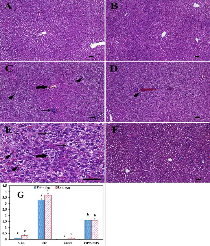
Figure 2. Histochemical staining of rat liver by periodic acid Schiff (PAS). (A) Negative control group.
(B) CeNPs group. (C) FIP group. (D) FIP group that treated with the CeNPs group. Scale bar = 50 µm. (E)
Quantification of PAS in the hepatic tissues in different groups Data expressed as Mean ± SE, analyzed using
one-way ANOVA at P ≤ 0.05, column with different letters (a, b, c, & d) indicate significant difference among the
values of different groups.
FIP treatment significantly increased the fatty bodies’ degeneration and lymph aggregation compared to CeNPs
and the CTR group, while CeNPs significantly countered these effects (Fig. 1G).
CeNPs groups had the greatest PAS distribution in all hepatocytes (Fig. 2A,B). Also, FIP caused a weak and
uneven distribution of PAS and decreased glycogen content (Fig. 2C) in relation to the CTR one. Additionally, the
FIP group that was treated with CeNPs had a moderate PAS reaction (Fig. 2D). FIP showed a significant decrease
Scientific Reports | (2021) 11:1310 | https://doi.org/10.1038/s41598-020-79479-5 6
Vol:.(1234567890)www.nature.com/scientificreports/
Figure 3. Immunohistochemical staining of rat liver by Caspase3. (A) Negative control group. (B) CeNPs
group. (C) FIP group. (D) FIP group that treated with CeNPs. Scale bar = 50 µm. (E) Quantification of Caspase3
in the hepatic tissues in different groups. Data expressed as Mean ± SE, analyzed using one-way ANOVA at
P ≤ 0.05, column with different letters (a, b & c) indicate significant difference among the values of different
groups.
in PAS distribution concerning CTR and the CeNPs-treated group. CeNPs showed a high even distribution of
PAS reaction in all hepatic lobules when combined with the FIP (Fig. 2E).
Immunohistochemistry. The FIP treatment showed a significant increase in caspase3 reaction concerning
CTR- and CeNPs-treated groups. On the other hand, the FIP-treated group was protected with cerium as shown
by a very weak caspase3 reaction (Fig. 3).
Scientific Reports | (2021) 11:1310 | https://doi.org/10.1038/s41598-020-79479-5 7
Vol.:(0123456789)www.nature.com/scientificreports/
Figure 4. Immunohistochemical staining of rat liver by proliferating cell nuclear antigen (PCNA). (A) Negative
control group. (B) CeNPs group. (C) FIP group. (D) FIP group that treated with CeNPs. Scale bar = 50 µm. (E)
Quantification of PCNA in the hepatic tissues in different groups. Data expressed as Mean ± SE, analyzed using
one-way ANOVA at P ≤ 0.05, column with different letters (a, b & c) indicate significant difference among the
values of different groups.
Also, FIP-treated group showed strong PCNA reaction in the nuclei of most hepatocytes compared to CTR
and CeNPs groups that showed negative PCNA reaction in the nuclei of hepatocytes, while FIP + CeNPs-treated
group showed nearly negative PCNA reaction in nuclei of the hepatocytes (Fig. 4).
Scientific Reports | (2021) 11:1310 | https://doi.org/10.1038/s41598-020-79479-5 8
Vol:.(1234567890)www.nature.com/scientificreports/
Immunostaining of rat liver tissues by massive ionized calcium-binding adapter molecule1 (Iba-1) in the case
of the FIP-intoxicated group showed a strong inflammatory process via strong cyclooxygenase (COX-2) reaction
and Iba-1 positive hepatic macrophage in the hepatic sinusoids in comparison to fipronil group that treated with
cerium nanoparticles. The latter group showed low IBA1 positive hepatic macrophage in the hepatic sinusoids.
CTR and CeNPs groups showed normal distribution of the Iba-1positive hepatic macrophage in the hepatic
sinusoids (Fig. 5) with a negative COX-2 reaction (Fig. 6). CeNPs attenuated FIP impacts on the endothelial
cells of the hepatic sinusoids and showed a moderate reaction (Fig. 6).
mRNA expression of FABP, ACC, PPAR‑α, Caspase3, and Bcl‑2. The results of mRNA expressions
of FABP, ACC, PPAR-α, caspase3 and Bcl-2 genes in liver tissues were presented in Table 5. The results revealed
up-regulation (P ≤ 0.05) in the comparative mRNA expressions of FABP, ACC, PPAR-α, and caspase3, while
exhibited down-regulation in the Bcl-2 gene in the liver tissue of rats that received FIP when compared with
the CTR animals. Meanwhile, CeNPs treatment down-regulated the mRNA expression of ACC and caspase3
and up-regulated the FABP, PPAR-α, and Bcl-2 compared to CTR. In the case of the FIP group that was treated
with CeNPs, significant up-regulation in FABP, PPAR-α and Bcl-2 were observed. On the other hand, ACC and
caspase3 mRNA expression were significantly down-regulated (Table 5).
Discussion
Hepatoxicity changes induced by FIP might be due to the imbalance of the antioxidant system, which might lead
to the formation of ROS that activates the apoptotic process and disturbances in the lipid profile of r ats32,33. Cur-
rent results showed that CeNPs was a chemoprotective agent against FIP-induced hepatotoxicity. The hepatic cells
were documented to have a significant and critical function in lipogeneses and glucose metabolism processes34.
The disturbance of lipid and glucose metabolism might be observed by the excess accumulation of FA in the
hepatic cells35. Consequently, increasing hepatic FA uptake36 leads to an increase in the hepatic triglyceride (TG)
levels, which induces NAFLD d evelopment37. The changes in lipid profile might be caused by the accumulation of
a high level of FA in circulation and hepatic lipogenesis, resulting in TG accumulation in the liver while dietary
lipids represent a large percentage of intrahepatic lipids38,39.
The current study explained a decreased weight gain in CeNPs-treated rats relative to CTR ones. Also, the ani-
mals treated with FIP showed a remarkable elevation in serum lipid profile (TAG, HDL-c, LDL-c, and cholesterol)
compared to CTR, CeNPs, and FIP + CeNPs groups. The significant modification in lipids might be due to the
oxidative stress produced by FIP, which ultimately leads to liver inflammation and injury. These results indicated
that FIP induced lipogenesis and lipid accumulation via increased expression of FABP and ACC1 in comparison
to the CeNPs-treated group, which showed downregulation of ACC1 along with the up-regulation of FABP like
the CTR groups. Activated AMPK (adenosine monophosphate-activated protein kinase) that phosphorylates the
main proteins associated with controlling lipid and carbohydrate metabolism, followed by ATP inhibition that
consumes anabolic pathways, such as cholesterol, isoprenoid, and FA synthesis, hepatic gluconeogenesis40 and
induces lipolysis41,42. Some studies revealed that FIP increases adipogenesis by downregulating AMPKα, which
disrupts lipid metabolism, which may demonstrate the high serum cholesterol l evel20,33. Fipronil insecticide has
a+2 by altering the plasma membrane p
been reported to raise the levels of intracellular C ermeabilization43, leading
to a disruption of adipogenesis and lipid metabolism process by the pathway mediated by Ca MKKβ and AMPK44.
FIP substantially elevated fatty acid synthase (FAS) and acetyl Co-A carboxylase (ACC) expression (the
two rate-limiting enzymes for lipogenesis)45 when compared with CTR group. Likewise, FABP (a protein that
transports FA in the cytoplasm for metabolic process or storage) e xpression46, was significantly enhanced by FIP
therapy relative to CTR45. Regarding CeNPs, interestingly, Rocca et al.47 identified CeNPs as a new anti-obesity
pharmaceutical formula after being tested in vitro and in vivo. They stated that CeNPs interfere with the adipo-
genic mechanism by decreasing the mRNA transcription of genes included in adipogenesis through inhibiting
the accumulation of triglycerides found in 3T3-L1 pre-adipocytes. Intraperitoneal injection of CeNPs at a dose
of 0.5 mg/kg did not show toxic symptoms in rats, however, a significant decrease in body weight and insulin,
leptin, glucose, and TG plasma levels are observed in comparison to CTR group47. Our results are consistent with
other studies that demonstrate the ability of CeNPs to activate AMPK which indicates inhibition of adipogenesis
with decreased PPARαC/EBPα expression as well as adipogenic markers including ACC and F AS48,49.
Lipogenesis inhibition due to ACC phosphorylation may be the major regulatory step in FA synthesis and
oxidation50. ACC stimulates the malonyl-CoA synthesis, which is a FA synthesis substrate and is inhibited by
AMPK-mediated ACC p hosphorylation51. Metabolism enhancement is considered as one of the anti-inflamma-
tory actions of CeNPs mechanisms since that results reported herein have demonstrated that CeNPs substan-
tially decreased the weight gain of rats, which might reduce the visceral obesity of rats (A. Rocca, S. Moscato, F.
Ronca, S. Nitti, V. Mattoli, M. Giorgi, G. Ciofani, Pilot in vivo investigation of cerium dioxide nanoparticles as a
novel anti-obesity pharmaceutical formulation, Nanomed. Nanotechnol., Biol. Med. 11 (7) (2015) 1725–1734).
The mass of visceral adipose tissue in CeNPs-treated rats was approximately twice less than CTR52. PPARα is
the most prevalent isotype in hepatic cells and participates in several lipid metabolism aspects53, such as FA
synthesis, degradation, storage, transport, lipoprotein metabolism and ketogenesis throughout fasting. PPARα
coordinates various de novo lipid synthesis pathways in the fed state to provide FA for hepatic TG storage dur-
ing starvation times54. PPARα’s mRNA expression in rats was the highest in tissues with high FA oxidation
levels, such as kidney, heart, liver, and brown adipose tissue55,56. High-fat diet administration is often related
to PPARα target genes hepatic expression participates in FA oxidation in wild-type mice, and it has been pro-
posed the adaptive or protective effect of PPARα57,58. Few reports have investigated the outcomes of PPARα for
treatments for N AFLD59. The improvement of β-FAO by PPARα a gonist60 might protect the mice from induced
liver injury61. Also, the recent study explained the high PPARα expression in FIP-treated rats that might be
Scientific Reports | (2021) 11:1310 | https://doi.org/10.1038/s41598-020-79479-5 9
Vol.:(0123456789)www.nature.com/scientificreports/
Figure 5. Immunohistochemical staining of rat liver by ionized calcium-binding adapter molecule 1 (Iba-1).
(A) Negative control group. (B) CeNPs group. (C) FIP group. (D) FIP group that treated with CeNPs. Scale
bar = 50 µm. (E) Quantification of I Iba-1 in the hepatic tissues in different groups. Data expressed as Mean ± SE,
analyzed using one-way ANOVA at P ≤ 0.05, column with different letters (a & b) indicate significant difference
among the values of different groups.
due to the increased level of FFAs and b-oxidation that usually caused by FFAs load in the mitochondria. This
increases the load on the endoplasmic reticulum, which leads to ROS production causing oxidative stress and
Scientific Reports | (2021) 11:1310 | https://doi.org/10.1038/s41598-020-79479-5 10
Vol:.(1234567890)www.nature.com/scientificreports/
Figure 6. Immunohistochemical staining of rat liver by COX-2. (A) negative control group. (B) CeNPs group.
(C) FIP group. (D) FIP group that treated with CeNPs. Scale bar = 50 µm. (E) Quantification of COX-2 in the
hepatic tissues in different groups. Data expressed as Mean ± SE, analyzed using one-way ANOVA at P ≤ 0.05,
column with different letters (a, b & c) indicate significant difference among the values of different groups.
Gene group FABP PPARα ACC Bcl2 like protein 4 Caspase3
FIP 2.230447721 ± 0.423 4.363039 ± 0.953 2.17347 ± 0.65 0.736624 ± 0.125 1.596966 ± 0.313
CeNPs 1.28877463 ± 0.329 1.395388 ± 0.351 0.076875 ± 0.021 1.670562 ± 0.346 0.240149 ± 0.051
FIP + CeNPs 1.428333957 ± 0.376 1.813781 ± 0.40459 0.846941 ± 0.203 1.081225 ± 0.165 0.82302 ± 0.143
Table 5. mRNA expression of fatty acid-binding protein. The effect of cerium oxide nanoparticles (CeNPs)
on liver mRNA expressions of FABP (fatty acid-binding protein), PPARα (peroxisome proliferator activated
receptor- alpha), ACC (Acetyl COA Carboxylase), Bcl2 Like protein 4 and Caspase3 in FIP intoxicated rats.
Data are expressed as Mean ± SE (n = 7). FIP Fipronil, CeNPs cerium oxide nanoparticles.
Scientific Reports | (2021) 11:1310 | https://doi.org/10.1038/s41598-020-79479-5 11
Vol.:(0123456789)www.nature.com/scientificreports/
inflammatory pathway a ctivation62. Besides its role in metabolism regulation, PPARα is also believed to have
anti-inflammatory activity63.
There has been cumulative evidence that pesticide toxicity inhibits redox homeostasis as well as oxidative
damage induction. Several studies suggested that redox homeostasis disturbance was triggered during FIP toxicity
was because of high ROS production33,64,65. Hepatotoxicity induced by FIP in this study might be due to increased
levels of MDA and NO (the indicator of LPO) because of the high reactive oxygen metabolites production
especially the hydroxyl r adicals33,66, LPO play role in the disruption of the integrity of cellular membranes and
implicated in liver i njuries67. ROS can target cell membranes and other cellular molecules, resulting in protein
oxidation, lipid peroxidation, caspase3 activation, and DNA damage33,68, leading to cell dysfunction69,70. The
protective mechanisms toward oxidative stress mainly through balances mediated by non-enzymatic and enzy-
matic antioxidants71. In current study, the decrease in SOD and GPx activities of rats subjected to FIP might be
due to the excess production of O 2 that is rapidly transformed by SOD and GPx to H 2O2 and water, respectively
as reported in the liver of pregnant rats and their offspring due to the inadequate ROS detoxification produced
by FIP in h epatocyte33,66.
It was recognized that liver cells showed the most active absorption and retention of CeNPs stored in the
liver for eight weeks at least62,72,73. Recently62, demonstrated that the antioxidant activity of CeNPs was through
increasing the antioxidant enzymes (SOD and GPx) with decreasing the lipid peroxidation markers (MDA and
NO). CeNPs have a high ability to eliminate free radicals as soon as they are produced during ROS imbalance
as they can inversely transform between C e3+ and C e4+ found on the surface74. The lipid peroxidation reduction
after CeNPs administration diminishes ROS harmful effects on the hepatic tissue. The mechanism of scavenging
of ROS/RNS of CeNPs depends on the physicochemical properties, the ability of CeNPs to absorb and release
oxygen75. CeNPs can act as a catalyst that mimics the antioxidant enzyme S OD76. So, there are two ways that
CeNPs might act as an antioxidant against FIP toxicity. The first is linked to its antioxidant activity because the
intensification of lipid peroxidation in the liver is among the causes of the inflammation, while the second is
controlled by AMPK-PPAR-α–signaling mechanism77. The inflammatory processes involved in the pathogenesis
of liver damage and obesity-related NAFLD78. Furthermore, fat destroys liver tissues and induces neutrophil
infiltration by releasing ROS and aggravating inflammatory p rocesses12,13. High accumulation of neutral lipid
(mostly TG in hepatic cell lipid droplets) begins the early NAFLD pathological s tages79. NAFLD’s pathogenesis
is not well known but is suggested as a “two-hit” mechanism80. The first “hit” results in lipid aggregation and its
mechanisms will likely include dysregulated lipid homeostasis such as de novo lipogenesis, β-oxidation, lipid
storage and trafficking, and VLDL-c s ecretion81. This hepatic steatosis characterizes the liver to a “second hit”
that results in inflammation, a primary pathophysiological symptom of steatohepatitis and advanced hepatic
disorders80,82. Oxidative stress has been suggested as the main mediator of this “second hit”80,83. So, increased
FFAs levels accompanied by PPAR-α stimulation, which in turn leads to ROSproduction and cell injury 81.
The efficiency of CeNPs on liver tissues was demonstrated by restoring the tissues architecture; necrosis,
inflammation, and a decline of dystrophy in rats exposed to the FIP corroborated the histopathological lesions
observed in the s tudy84. FIP group revealing congested central vein, massive fatty degeneration in the peripor-
tal areas, lymphocytic aggregation in between massive fatty degeneration, with lymphocytic infiltration. Also,
necrotic foci, congested liver sinusoids, and fatty degeneration might be due to that FIP increase lipid peroxi-
dation. The FIP group that was protected with cerium nanoparticles showed normal liver architecture similar
f72. Studies showed an important connection between PCNA, Iba-1, and COX-2 expression and
to the results o
the inflammatory reaction as well as mitotic d ivision85. Iba-1 participates in macrophage inflammatory path-
ways, including proliferation, migration, and signal t ransduction86. Proliferating cell nuclear antigen (PCNA)
is believed to have an important role in controlling DNA synthesis as well as cell proliferation87. Also, COX
isozymes (COX-1 and 2) are particularly important, as they are the main NSAIDs t argets88, that parameters
detected using the immunohistochemical.
In current study, Iba-1 cells were observed in the FIP-treated group with strong PCNA and COX-2 reaction
concentrated in the nuclei of the most hepatocytes that may be due to FIP induced liver injury while in case
of FIP group that protected by CeNPs show low Iba-1 positive hepatic macrophage and nearly negative PCNA
and COX-2 reaction in nuclei of the hepatocytes, So, liver regeneration is the expected physiological response.
Also, FIP induces apoptosis by strong caspase3 reaction in all nuclei of hepatocytes in opposite to FIP group
previously protected with cerium showing very weak caspase3 reaction in the nuclei of hepatocytes. That might
be due to the cytotoxic activity of fatty acid that influences cell survival. Long term accumulation of lipids may
lead to hepatocyte necrosis or a poptosis89. MDA and NO interacted directly with the DNA and triggered DNA
adducts and nuclear condensation that promoted apoptosis via cytochrome C and further caspase3 activations
as detected by immunostaining, along with the induced mitochondrial dysfunction. Several reports revealed
that FIP triggered cell death through apoptotic pathways20.
Conclusions
In this study, FIP induced hepatotoxicity through disturbance in the serum lipid profile (VLDL-c, HDL-c, TAG,
LDL-c) and cholesterol level might be due to increasing the mRNA fatty acid-binding protein expression and
ACC genes and FIP can cause serious tissue injury in the liver caused by oxidative stress through increasing
MDA and NO with decreasing SOD and GPx and apoptosis formation by increment caspase3 and decreasing
BCL-2. While CeNPs could be used to activate the protective mechanisms against oxidative damage caused by
FIP in the liver. Moreover, CeNPs effectively relieve the inflammatory processes in the blood of rats that may
reduce obesity defects in liver damage.
Scientific Reports | (2021) 11:1310 | https://doi.org/10.1038/s41598-020-79479-5 12
Vol:.(1234567890)www.nature.com/scientificreports/
Received: 16 May 2020; Accepted: 7 December 2020
References
1. Al-Badran, A. A., Fujiwara, M., Gatlin, D. M. & Mora, M. A. Lethal and sub-lethal effects of the insecticide fipronil on juvenile
brown shrimp Farfantepenaeus aztecus. Sci. Rep. 8, 10769. https://doi.org/10.1038/s41598-018-29104-3 (2018).
2. Simon-Delso, N. et al. Systemic insecticides (neonicotinoids and fipronil): trends, uses, mode of action and metabolites. Environ.
Sci. Pollut. Res. 22, 5–34 (2015).
3. Tavares, M. A. et al. Comparative effects of fipronil and its metabolites sulfone and desulfinyl on the isolated rat liver mitochondria.
Environ. Toxicol. Pharmacol. 40, 206–214. https://doi.org/10.1016/j.etap.2015.06.013 (2015).
4. Bonneau, J. et al. SoK: Research Perspectives and Challenges for Bitcoin and Cryptocurrencies (Springer, New York, 2015).
5. Das, P. C., Cao, Y., Cherrington, N., Hodgson, E. & Rose, R. L. Fipronil induces CYP isoforms and cytotoxicity in human hepato-
cytes. Chem. Biol. Interact. 164, 200–214 (2006).
6. Khan, S., Jan, M., Kumar, D. & Telang, A. Firpronil induced spermotoxicity is associated with oxidative stress, DNA damage and
apoptosis in male rats. Pestic. Biochem. Physiol. 124, 8–14 (2015).
7. Ahn, B.-E. & Baker, T. A. Oxidization without substrate unfolding triggers proteolysis of the peroxide-sensor, PerR. Proc. Natl.
Acad. Sci. 113, E23–E31 (2016).
8. Poljsak, B., Šuput, D. & Milisav, I. Achieving the balance between ROS and antioxidants: when to use the synthetic antioxidants.
Oxid. Med. Cell. Long. 2013 (2013).
9. Young, I. & Woodside, J. Antioxidants in health and disease. J. Clin. Pathol. 54, 176–186 (2001).
10. Pizzimenti, S. et al. Interaction of aldehydes derived from lipid peroxidation and membrane proteins. Front. Physiol. 4, 242 (2013).
11. Diehl, A. M. Nonalcoholic fatty liver disease: implications for alcoholic liver disease pathogenesis. Alcohol. Clin. Exp. Res. 25,
8s–14s (2001).
12. Mittal, M., Siddiqui, M. R., Tran, K., Reddy, S. P. & Malik, A. B. Reactive oxygen species in inflammation and tissue injury. Antioxid.
Redox Signal. 20, 1126–1167 (2014).
13. Newsholme, P. et al. Molecular mechanisms of ROS production and oxidative stress in diabetes. Biochem. J. 473, 4527–4550. https
://doi.org/10.1042/bcj20160503c (2016).
14. Dahle, J. & Arai, Y. Environmental geochemistry of cerium: applications and toxicology of cerium oxide nanoparticles. Int. J.
Environ. Res. Public Health 12, 1253–1278 (2015).
15. Xu, C. & Qu, X. Cerium oxide nanoparticle: a remarkably versatile rare earth nanomaterial for biological applications. NPG Asia
Mater. 6, e90 (2014).
16. Dowding, J. M., Seal, S. & Self, W. T. Cerium oxide nanoparticles accelerate the decay of peroxynitrite (ONOO−). Drug Deliv.
Transl. Res. 3, 375–379 (2013).
17. Caputo, F., De Nicola, M. & Ghibelli, L. Pharmacological potential of bioactive engineered nanomaterials. Biochem. Pharmacol.
92, 112–130. https://doi.org/10.1016/j.bcp.2014.08.015 (2014).
18. Olmedo, D. G., Tasat, D. R., Evelson, P., Guglielmotti, M. B. & Cabrini, R. L. Biological response of tissues with macrophagic activity
to titanium dioxide. J. Biomed. Mater. Res. A 84, 1087–1093 (2008).
19. Caballero, M. et al. Fipronil induces CYP isoforms in rats. Food Chem. Toxicol. 83, 215–221 (2015).
20. Abdel-Daim, M. M. & Abdeen, A. Protective effects of rosuvastatin and vitamin E against fipronil-mediated oxidative damage and
apoptosis in rat liver and kidney. Food Chem. Toxicol. 114, 69–77. https://doi.org/10.1016/j.fct.2018.01.055 (2018).
21. Khaksar, M. R. et al. Protective effects of cerium oxide and yttrium oxide nanoparticles on reduction of oxidative stress induced
by sub-acute exposure to diazinon in the rat pancreas. J. Trace Elem. Med Biol. 41, 79–90 (2017).
22. Renaudin, J. Determination of Nitrite and Nitrate Content in Water (Springer, New York, 2001).
23. Nishikimi, M., Rao, N. A. & Yagi, K. The occurrence of superoxide anion in the reaction of reduced phenazine methosulfate and
molecular oxygen. Biochem. Biophys. Res. Commun. 46, 849–854 (1972).
24. Paglia, D. E. & Valentine, W. N. Studies on the quantitative and qualitative characterization of erythrocyte glutathione peroxidase.
J. Lab. Clin. Med. 70, 158–169 (1967).
25. Bancroft, J. D. & Layton, C. in Bancroft s Theory and practice of histological techniques (ed Christopher Layton and John D. Bancroft
S. Kim suvarna) 173–186 (Churchill Livingstone:, 2013).
26. 27Layton, C. & Bancroft, J. D. in Bancroft s Theory and practice of histological techniques (ed C. Layton, J. D. B. S. Kim Suvarna)
215–238 (Churchill Livingstone, 2013).
27. Gibson-Corley, K. N., Olivier, A. K. & Meyerholz, D. K. Principles for valid histopathologic scoring in research. Vet. Pathol. 50,
1007–1015. https://doi.org/10.1177/0300985813485099 (2013).
28. Noreldin, A. E. et al. Spatial distribution of osteoblast activating peptide in the rat stomach. Acta Histochem. 118, 109–117. https
://doi.org/10.1016/j.acthis.2015.12.001 (2016).
29. Noreldin, A. E., Elewa, Y. H. A., Kon, Y., Warita, K. & Hosaka, Y. Z. Immunohistochemical localization of osteoblast activating
peptide in the mouse kidney. Acta Histochem. 120, 323–328 (2018).
30. Sysel, A. M., Valli, V. E., Nagle, R. B. & Bauer, J. A. Immunohistochemical quantification of the vitamin B12 transport protein
(TCII), cell surface receptor (TCII-R) and Ki-67 in human tumor xenografts. Anticancer Res. 33, 4203–4212 (2013).
31. Vis, A. N., Kranse, R., Nigg, A. L. & van der Kwast, T. H. Quantitative analysis of the decay of immunoreactivity in stored prostate
needle biopsy sections. Am. J. Clin. Pathol. 113, 369–373. https://doi.org/10.1309/CQWY-E3F6-9KDN-YV36 (2000).
32. Badgujar, P. C., Pawar, N. N., Chandratre, G. A., Telang, A. & Sharma, A. Fipronil induced oxidative stress in kidney and brain of
mice: Protective effect of vitamin E and vitamin C. Pestic. Biochem. Physiol. 118, 10–18 (2015).
33. Romero, A. et al. Fipronil sulfone induced higher cytotoxicity than fipronil in SH-SY5Y cells: Protection by antioxidants. Toxicol.
Lett. 252, 42–49 (2016).
34. Sun, Q. C., Clark, J. & Park, Y. Environmental pollutants and type 2 diabetes: A review of human studies. Toxicol. Environ. Chem.
99, 1–42. https://doi.org/10.1080/02772248.2017.1393818 (2017).
35. Alves-Bezerra, M. & Cohen, D. E. Triglyceride metabolism in the liver. Compr. Physiol. 8, 1–8. https://doi.org/10.1002/cphy.c1700
12 (2017).
36. Leung, T.-M. & Nieto, N. CYP2E1 and oxidant stress in alcoholic and non-alcoholic fatty liver disease. J. Hepatol. 58, 395–398
(2013).
37. Gaggini, M. et al. Non-alcoholic fatty liver disease (NAFLD) and its connection with insulin resistance, dyslipidemia, atherosclerosis
and coronary heart disease. Nutrients 5, 1544–1560 (2013).
38. Neuschwander-Tetri, B. A. Non-alcoholic fatty liver disease. BMC Med. 15, 45 (2017).
39. Donnelly, K. L. et al. Sources of fatty acids stored in liver and secreted via lipoproteins in patients with nonalcoholic fatty liver
disease. J. Clin. Investig. 115, 1343–1351 (2005).
40. Hawley, S. A. et al. Use of cells expressing γ subunit variants to identify diverse mechanisms of AMPK activation. Cell Metab. 11,
554–565 (2010).
41. Yin, W., Mu, J. & Birnbaum, M. J. Role of AMP-activated protein kinase in cyclic AMP-dependent lipolysis in 3T3-L1 adipocytes.
J. Biol. Chem. 278, 43074–43080 (2003).
Scientific Reports | (2021) 11:1310 | https://doi.org/10.1038/s41598-020-79479-5 13
Vol.:(0123456789)www.nature.com/scientificreports/
42. Koh, H.-J. et al. Adrenaline is a critical mediator of acute exercise-induced AMP-activated protein kinase activation in adipocytes.
Biochem. J. 403, 473–481 (2007).
43. Trump, B. F. & Berezesky, I. K. The role of cytosolic Ca2+ in cell injury, necrosis and apoptosis. Curr. Opin. Cell Biol. 4, 227–232
(1992).
44. Sun, Q. et al. Fipronil promotes adipogenesis via AMPKalpha-mediated pathway in 3T3-L1 adipocytes. Food Chem. Toxicol. 92,
217–223. https://doi.org/10.1016/j.fct.2016.04.011 (2016).
45. Sun, Q. et al. Fipronil promotes adipogenesis via AMPKα-mediated pathway in 3T3-L1 adipocytes. Food Chem. Toxicol. 92, 217–223
(2016).
46. Terra, X. et al. FABP 4 is associated with inflammatory markers and metabolic syndrome in morbidly obese women. Eur. J. Endo-
crinol. 164, 539–547 (2011).
47. Rocca, A. et al. Pilot in vivo investigation of cerium oxide nanoparticles as a novel anti-obesity pharmaceutical formulation.
Nanomed. Nanotechnol. Biol. Med. 11, 1725–1734 (2015).
48. Arya, A. et al. Cerium oxide nanoparticles promote neurogenesis and abrogate hypoxia-induced memory impairment through
AMPK–PKC–CBP signaling cascade. Int. J. Nanomed. 11, 1159 (2016).
49. Habinowski, S. A. & Witters, L. A. The effects of AICAR on adipocyte differentiation of 3T3-L1 cells. Biochem. Biophys. Res. Com-
mun. 286, 852–856 (2001).
50. Minokoshi, Y. et al. Leptin stimulates fatty-acid oxidation by activating AMP-activated protein kinase. Nature 415, 339–343. https
://doi.org/10.1038/415339a (2002).
51. Daval, M. et al. Anti-lipolytic action of AMP-activated protein kinase in rodent adipocytes. J. Biol. Chem. 280, 25250–25257 (2005).
52. Kobyliak, N. et al. Cerium dioxide nanoparticles possess anti-inflammatory properties in the conditions of the obesity-associated
NAFLD in rats. Biomed. Pharmacother. 90, 608–614. https://doi.org/10.1016/j.biopha.2017.03.099 (2017).
53. Kersten, S. Integrated physiology and systems biology of PPARα. Mol. Metab. 3, 354–371 (2014).
54. Hashimoto, T. et al. Defect in peroxisome proliferator-activated receptor α-inducible fatty acid oxidation determines the severity
of hepatic steatosis in response to fasting. J. Biol. Chem. 275, 28918–28928 (2000).
55. Bookout, A. L. et al. Anatomical profiling of nuclear receptor expression reveals a hierarchical transcriptional network. Cell 126,
789–799 (2006).
56. Escher, P. et al. Rat PPARs: quantitative analysis in adult rat tissues and regulation in fasting and refeeding. Endocrinology 142,
4195–4202 (2001).
57. Patsouris, D., Reddy, J. K., Müller, M. & Kersten, S. Peroxisome proliferator-activated receptor α mediates the effects of high-fat
diet on hepatic gene expression. Endocrinology 147, 1508–1516 (2006).
58. Kim, S. et al. Hepatic gene expression profiles in a long-term high-fat diet-induced obesity mouse model. Gene 340, 99–109 (2004).
59. Peyrou, M., Ramadori, P., Bourgoin, L. & Foti, M. PPARs in liver diseases and cancer: epigenetic regulation by MicroRNAs. PPAR
Res. 2012, 16. https://doi.org/10.1155/2012/757803 (2012).
60. Mandard, S., Muller, M. & Kersten, S. Peroxisome proliferator-activated receptor alpha target genes. Cell. Mol. Life Sci. 61, 393–416.
https://doi.org/10.1007/s00018-003-3216-3 (2004).
61. Zhao, Q., Yang, R., Wang, J., Hu, D.-D. & Li, F. PPARα activation protects against cholestatic liver injury. Sci. Rep. 7, 9967. https://
doi.org/10.1038/s41598-017-10524-6 (2017).
62. Oro, D. et al. Cerium oxide nanoparticles reduce steatosis, portal hypertension and display anti-inflammatory properties in rats
with liver fibrosis. J. Hepatol. 64, 691–698. https://doi.org/10.1016/j.jhep.2015.10.020 (2016).
63. Berghe, W. V. et al. Peroxisomal Disorders and Regulation of Genes 181–196 (Springer, New York, 2003).
64. Ortiz-Ortiz, M. A. et al. Nitric oxide-mediated toxicity in paraquat-exposed SH-SY5Y cells: a protective role of 7-nitroindazole.
Neurotox. Res. 16, 160–173 (2009).
65. Abdel-Daim, M. M. Synergistic protective role of ceftriaxone and ascorbic acid against subacute diazinon-induced nephrotoxicity
in rats. Cytotechnology 68, 279–289 (2016).
66. Guelfi, M., Maioli, M. A., Tavares, M. A. & Mingatto, F. E. Citotoxicity of fipronil on hepatocytes isolated from rat and effects of
its biotransformation. Braz. Arch. Biol. Technol. 58, 843–853 (2015).
67. Karami-Mohajeri, S., Ahmadipour, A., Rahimi, H.-R. & Abdollahi, M. Adverse effects of organophosphorus pesticides on the liver:
a brief summary of four decades of research. Arhiv Rada Toksikol. 68, 261–275 (2017).
68. Zhang, B. et al. Fipronil induces apoptosis through caspase-dependent mitochondrial pathways in Drosophila S2 cells. Pestic.
Biochem. Physiol. 119, 81–89. https://doi.org/10.1016/j.pestbp.2015.01.019 (2015).
69. Small, D. M., Coombes, J. S., Bennett, N., Johnson, D. W. & Gobe, G. C. Oxidative stress, anti-oxidant therapies and chronic kidney
disease. Nephrology 17, 311–321. https://doi.org/10.1111/j.1440-1797.2012.01572.x (2012).
70. Klaunig, J. E., Kamendulis, L. M. & Hocevar, B. A. Oxidative stress and oxidative damage in carcinogenesis. Toxicol. Pathol. 38,
96–109. https://doi.org/10.1177/0192623309356453 (2010).
71. Abdollahi, M., Ranjbar, A., Shadnia, S., Nikfar, S. & Rezaiee, A. Pesticides and oxidative stress: A review. Med. Sci. Monit. 10,
141–147 (2004).
72. Kumari, M., Kumari, S. I. & Grover, P. Genotoxicity analysis of cerium oxide micro and nanoparticles in Wistar rats after 28 days
of repeated oral administration. Mutagenesis 29, 467–479 (2014).
73. Saleh, H. et al. Chemo-protective potential of cerium oxide nanoparticles against fipronil-induced oxidative stress, apoptosis,
inflammation and reproductive dysfunction in male white albino rats. Molecules 25, 3479 (2020).
74. Naganuma, T. & Traversa, E. Stability of the Ce 3+ valence state in cerium oxide nanoparticle layers. Nanoscale 4, 4950–4953
(2012).
75. Deshpande, S., Patil, S., Kuchibhatla, S. V. & Seal, S. Size dependency variation in lattice parameter and valency states in nanocrys-
talline cerium oxide. Appl. Phys. Lett. 87, 133113 (2005).
76. Hirst, S. M. et al. Anti-inflammatory properties of cerium oxide nanoparticles. Small 5, 2848–2856 (2009).
77. Kobyliak, N. et al. Cerium Dioxide Nanoparticles Possess Anti-inflammatory Properties in the Conditions of the Obesity-Associated
NAFLD in Rats Vol. 90 (Springer, New York, 2017).
78. Tilg, H. The role of cytokines in non-alcoholic fatty liver disease. Dig. Dis. 28, 179–185 (2010).
79. Montagner, A. et al. Liver PPARα is crucial for whole-body fatty acid homeostasis and is protective against NAFLD. Gut 65,
1202–1214. https://doi.org/10.1136/gutjnl-2015-310798 (2016).
80. Salt, W. B. Nonalcoholic fatty liver disease (NAFLD): A comprehensive review. J. Insurance Med. 36, 27–41 (2004).
81. Saadeh, S. Nonalcoholic fatty liver disease and obesity. Nutr. Clin. Pract. 22, 1–10 (2007).
82. Day, C. P. & James, O. F. W. Steatohepatitis: A tale of two & #x201c;hits”?. Gastroenterology 114, 842–845. https://doi.org/10.1016/
S0016-5085(98)70599-2 (1998).
83. Allard, J. P. et al. Nutritional assessment and hepatic fatty acid composition in non-alcoholic fatty liver disease (NAFLD): a cross-
sectional study. J. Hepatol. 48, 300–307. https://doi.org/10.1016/j.jhep.2007.09.009 (2008).
84. Amiri, F. T., Hamzeh, M., Beklar, S. Y. & Hosseinimehr, S. J. Anti-apoptotic and antioxidant effect of cerium oxide nanoparticles
on cyclophosphamide-induced hepatotoxicity. Erciyes Med. J. 40, 1–2 (2018).
85. Wang, D., Shi, J. Q. & Liu, F. X. Immunohistochemical detection of proliferating cell nuclear antigen in hepatocellular carcinoma.
World J. Gastroenterol. 3, 101–103. https://doi.org/10.3748/wjg.v3.i2.101 (1997).
Scientific Reports | (2021) 11:1310 | https://doi.org/10.1038/s41598-020-79479-5 14
Vol:.(1234567890)You can also read