Topical Treatment of Colquhounia Root Relieves Skin Inflammation and Itch in Imiquimod-Induced Psoriasiform Dermatitis in Mice - Hindawi.com
←
→
Page content transcription
If your browser does not render page correctly, please read the page content below
Hindawi
Mediators of Inflammation
Volume 2022, Article ID 5782922, 15 pages
https://doi.org/10.1155/2022/5782922

Research Article
Topical Treatment of Colquhounia Root Relieves Skin
Inflammation and Itch in Imiquimod-Induced Psoriasiform
Dermatitis in Mice

 Fei Li ,1 Dan Han ,1 Bo Wang ,2 Wentao Zhang ,3 Yan Zhao ,1 Jing Xu ,3,4
 Liesu Meng ,3,4 Kuanhou Mou ,1 Shemin Lu ,3,4 Wenhua Zhu ,3,4 and Yan Zhou 1

 1
 Department of Dermatology, The First Affiliated Hospital of Xi’an Jiaotong University, Xi’an, Shaanxi, China
 2
 Center for Translational Medicine, The First Affiliated Hospital of Xi’an Jiaotong University, Xi’an, Shaanxi, China
 3
 Institute of Molecular and Translational Medicine, and Department of Biochemistry and Molecular Biology, School of Basic
 Medical Sciences, Xi’an Jiaotong University Health Science Center, Xi’an, Shaanxi, China
 4
 Key Laboratory of Environment and Genes Related to Diseases (Xi’an Jiaotong University), Ministry of Education, Xi’an,
 Shaanxi, China

 Correspondence should be addressed to Wenhua Zhu; zhuwenhua@xjtu.edu.cn and Yan Zhou; yanzhou7798@xjtu.edu.cn

 Fei Li and Dan Han contributed equally to this work.

 Received 18 June 2021; Revised 15 December 2021; Accepted 27 December 2021; Published 11 January 2022

 Academic Editor: Shushan Yan

 Copyright © 2022 Fei Li et al. This is an open access article distributed under the Creative Commons Attribution License, which
 permits unrestricted use, distribution, and reproduction in any medium, provided the original work is properly cited.

 Itch is one of the major clinical manifestations of psoriasis, which is closely related with neurogenic inflammation and difficult to
 control. Colquhounia Root (CR) is a Chinese herb exhibiting broad bioactivities on anti-inflammation. This study was designed to
 explore the antipsoriatic and anti-itch potential of CR and its underlying mechanisms. Mice in a model of imiquimod-induced
 psoriasiform dermatitis were treated topically with CR for 7 days, and the severity of skin lesions and itch was significantly
 ameliorated. CR reduced the inflammatory cell infiltration, as well as mast cells in skins. Particularly, the expression of
 inflammatory cytokines and chemokine including Il17a, Il22, and Ccl20 and itch-related molecules such as SP, CGRP, and NGF
 in lesions were decreased in diseased mice upon application with CR. The normal human epidermal keratinocytes were
 stimulated with the M5 cytokine cocktail, the mixture of IL-17A, IL-22, Oncostatin M, IL-1α, and TNF-α, and cell viability
 and mRNA expression levels of inflammatory factors and itch-related molecules were measured after being treated with CR.
 We found that CR inhibited both cell hyperproliferation and overexpression of inflammatory cytokines and itch-related
 molecules in vitro. Altogether, we conclude that CR relieves psoriatic lesions and itch via controlling immunological and
 neurogenic inflammation.

1. Introduction The pathophysiological mechanism of itch in psoriasis,
 which is mainly related to mental stress and neurogenic inflam-
Psoriasis is a chronic immune-mediated inflammatory disease mation, remains poorly understood. Studies have shown that
mainly manifested as scaly erythema and patches in the skin. itch-related molecules released by epidermal keratinocytes and
Patients suffering from psoriasis often complain of disgusting other cells in the dermis such as calcitonin gene-related peptide
appearance, chronic itch, and joint pain [1–3]. Itch is one of (CGRP), substance P (SP), nerve growth factor (NGF), and
the most troublesome symptoms of psoriasis, affecting the TRPV1 and inflammatory cytokines (IL-17 and IL-22), as well
quality of life and sleep of patients. It is probably unrelated as chemokine CCL20, are important in mechanisms of itch
to the severity of psoriasis due to its particular mechanisms [4]. [5–10]. Their expressions are significantly increased in the skin
2 Mediators of Inflammation

specimens of psoriasis patients, involved in the occurrence and group mice. CR were daily administered to the dorsal skins
development of psoriatic itch [2, 11]. for 9 consecutive days from two days before IMQ applica-
 In the last few decades, considerable progress has been tion. Mice in the normal group were treated with Vaseline.
made in the treatment of psoriasis including topical or oral A blinded observer measured the thickness of the dorsal
traditional medications, novel biologics, and phototherapies. skins by a vernier caliper daily and observed and photo-
With the in-depth study of psoriasis, psoriatic itch has grad- graphed the back skin with a dermoscope simultaneously.
ually been noticed. However, traditional antipruritic thera- Psoriasis Severity Index (PSI) scores were used to evaluate
pies (such as antihistamines) are limited in the treatment the severity of skin lesions every day [21], which were graded
of itch in psoriasis. Traditional topical drugs like vitamin from 0 to 4 for erythema, thickness, scaling, and total. All
D3 derivatives and coal tar for clinical treatment of psoriasis mice were euthanized, and their skin specimens were stored
have little effect on itch or even aggravate it [12]. Biological for determination and further experiments. The experiment
agents, such as TNF-α and IL-17 inhibitors, can quickly and scheme is shown in Figure 1(a). Liver and renal injury bio-
effectively relieve itch, but some patients could not afford markers were carried out by an autoanalyzer (LABOSPECT
them because of high prices or high-risk adverse reactions 008 ASi, Hitachi, Japan).
[13], such as infection, urticaria, and tumor in the long term
[14]. Therefore, successful treatment of psoriatic itch is still 2.4. Behavioral Tests. We did the behavioral tests following
challenging. It is necessary to further explore and develop the methods of Sakai et al. [22]. Firstly, we pretreated and
treatment strategies and drugs to relieve psoriatic itch. habituated all mice to a transparent acrylic box for 30 min
 Nowadays, the use of botanical therapeutics has been twice. Twenty hours after each topical application, we video-
taken attention by dermatologists, such as Tripterygium wil- taped the number of scratch bouts of mice for 30 minutes.
fordii and Colquhounia Root (CR) [15]. CR is a traditional Then, the scratch bouts of mice in videos were counted by
Chinese medicine, containing several bioactive ingredients a blinded observer. A scratch bout was defined as once or
such as triptolide and tripterine [16], is widely used in the more times rapidly front and backward pointing and touch-
treatment of autoimmune-related diseases such as systemic ing movement with hind claws in the skin lesions and end-
lupus erythematosus, rheumatoid arthritis, chronic nephri- ing by licking or biting the toes or placing the back claws
tis, vasculitis, and psoriasis, with anti-inflammatory and on the floor. Other movements away from the treated area
analgesic effects [17–19]. However, it has not been reported as ear scratching and grooming were excluded.
whether CR has the effect of inhibiting itch in psoriasis.
 2.5. Histopathology and Immunohistochemistry (IHC). Skin
Therefore, we aim to explore the effects and mechanisms
 specimens in paraformaldehyde-fixed (4%) were embedded
of topical treatment with CR.
 in paraffin for haematoxylin and eosin staining (H&E) and
 IHC. Mast cells were stained by Toluidine blue following
2. Materials and Methods the way of Puebla-Osorio et al. [23]. We assessed the histo-
 pathological alterations with computer-assisted quantitative
2.1. Mice. Forty-one female C57BL/6J mice, weighing 18-
 image analysis in lesions, including epidermal thickness,
20 g, were purchased from Xi’an Jiaotong University Labora-
 area of Munro’s microabscesses (MM), and the number of
tory Animal Center and were kept at a standardized breed-
 mast cells. Tissue sections were prepared for IHC with SP
ing environment. Standard diet and water were available to
 (Cat, No. sc-21715, 1 : 50, Santa Cruze), CGRP (Cat, No.
all mice anytime. All animal experiments were approved by
 sc-57053, 1 : 50, Santa Cruze), and NGF (Cat, No. sc-
the Institutional Animal Ethics Committee of Xi’an Jiaotong
 32300, 1 : 50, Santa Cruze) antibodies. The integrated optical
University.
 density (IOD) of SP, CGRP, and NGF in the image was mea-
 sured by Image Pro plus 6 software.
2.2. Preparation of CR. CR tablets (Z20027411) were pro-
vided by Pharmaceutical Factory of the Chongqing Acad- 2.6. mRNA Quantification. RNA, extracted from skin speci-
emy of Chinese Mataria Medica. We grounded 5 g CR mens with TRIzol reagent (Thermo Fisher Scientific) accord-
tablets into powder and then mixed it in 250 g Vaseline to ing to the instruction, was converted into cDNA with the
keep the effective concentration which was 0.002%, and then RevertAid First Strand cDNA Synthesis kit (Thermo Fisher
stored it at 4°C. Scientific). Quantitative real-time polymerase chain reaction
 (qPCR) was performed on a PCR machine (CFX CNNNECT
2.3. Imiquimod- (IMQ-) Induced Psoriasiform Dermatitis Real-time system; Bio Rad, Hercules, CA, USA). The gene-
(PsD) in Mice and Treatment. Mice were randomly divided specific primers are summarized in Table 1. The data were
into three groups, the normal control group, disease group analyzed with the 2−ΔΔCT method and normalized by
(IMQ), and CR treated group (IMQ+CR). We followed the GAPDH.
methods of William et al. to establish the IMQ-induced pso-
riasiform dermatitis mouse model [20]. Mice were anesthe- 2.7. Flow Cytometry. Skin samples from each group were cut
tized with isoflurane, and their hair on the back skin was into pieces using ophthalmic scissors and digested using colla-
shaved (3 cm × 3 cm). Then, 62.5 mg of IMQ creams (Min- genases (2.6 mg/ml, Sigma) and DNase (0.1 mg/ml, Sigma) for
gxin Pharmaceutical Co., Ltd., Sichuan, China) was applied 1.5 h. The cell suspension was filtered by a 40 μm nylon net.
daily onto the dorsal skins for 7 consecutive days to induce Then, cells from the skin were stained with anti-mouse-
psoriasiform dermatitis in the IMQ group and IMQ+CR BV785-CD45.2 (Cat, No. 109839, 1 : 200, BioLegend), anti-
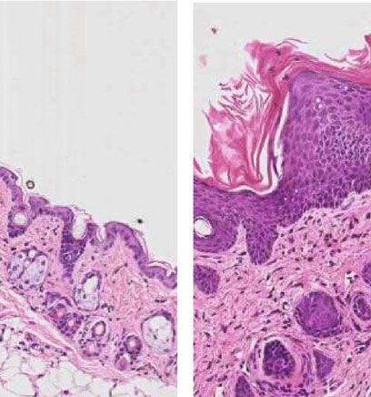
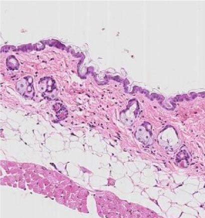
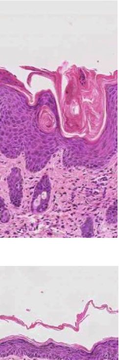
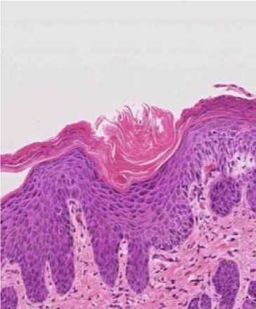
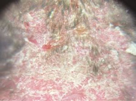
Mediators of Inflammation 3

mouse-AF700-TCRβ (Cat, No. 109224, 1 : 200, BioLegend), was reduced by the coadministration of CR (Figures 1(b)
anti-mouse-PE-Cy5-CD4 (Cat, No. 100514, 1 : 200, BioLe- and 1(c)). In these mice, reduced scales, skin thickness,
gend), anti-mouse-BV711-CD8 (Cat, No. 100759, 1 : 200, and cumulative scores were observed from day 5 to day 7
BioLegend), anti-mouse-APC-CD11b (Cat, No. 101212, compared with mice in the model group (Figure 1(d)). How-
1 : 200, BioLegend), anti-mouse-FITC-F4/80 (Cat, No. ever, the erythema of skin in the CR-treated group was not
123108, 1 : 200, BioLegend), anti-mouse-Pacific blue-Ly6G relieved (Figure 1(d)). Under the dermoscope, regularly dis-
(Cat, No. 127612, 1 : 200, BioLegend), and anti-mouse-PE- tributed dotted vessels and scales were observed in the IMQ
γδT (Cat, No. 553178, 1 : 200, Becton Dickinson). Finally, we group, while they were diminished in the CR-treated group.
detected and analyzed all samples with a flow cytometer. Gat- The sections of the IMQ-treated group showed the
ing strategy of cells in flow cytometry is shown in supplemen- representative histopathological changes of psoriasis with
tary Figure 1. hyperkeratosis, parakeratosis, and acanthosis, while these
 features were absent in normal mice (Figure 2(a)). However,
2.8. Cell Culture and Treatment. We performed all culture CR treatment partially inhibited these characteristic changes
experiments on the normal human epidermal keratinocytes in psoriatic lesions (Figure 2(a)). Statistical analysis also
(NHEKs) which were obtained from ATCC (Manassas, showed a significant decrease in the epidermal thickness in
VA). Cells were cultured with Dulbecco’s modified Eagle’s CR-treated mice (Figure 2(b)). In human psoriasis, neutro-
medium (DMEM, Hyclone) supplemented with 10% heat- phils aggregation in the cornified layer known as MM, indi-
inactivated fetal bovine serum (FBS), 100 U/ml of penicillin, cated that neutrophils may play an important role in disease
and 100 μg/ml of streptomycin and were placed in an incu- pathogenesis [27]. IMQ-treated skins also showed MM for-
bator at a 37°C humidified atmosphere with 5% CO2. mation (Figure 2(c)). MM in the model group were larger
NHEKs were treated with 10 ng/ml of M5 cocktail (IL-17A, and more numerous than those observed in CR-treated mice
IL-22, Oncostatin M, IL-1α, and TNF-α) [24], with or with- (Figure 2(c)). The average area of total MM of each mouse
out CR dissolved in DMSO for 24 h. was dramatically diminished in CR-treated mice vs. model
 mice (P < 0:01) (Figure 2(d)).
2.9. Cell Viability Assay. We tested the effect of CR on the
 Taken together, these results suggest that CR treatment
growth of NHEKs by CCK-8 assay following the methods
 could alleviate the severity of IMQ-induced PsD in mice.
of Ru et al. 2020 [25]. We seeded NHEKs onto 96-well plates
 Therefore, it is necessary to study how this treatment exerts
at a density of 2000 cells per well. Then, the medium was
 its therapeutic effect on psoriasis.
replaced with fresh medium containing 10 ng/ml M5 and
various concentrations (0, 0.25 μg/ml, 0.5 μg/ml, 1 μg/ml,
 3.2. CR Treatment Improves the Immune Microenvironment of
and 2 μg/ml) of CR and medium containing 0.1% DMSO
 the Psoriatic Skins of Mice. The immune cell infiltration in
were used as the vehicle control after 24 h. After 24 h incuba-
 mouse skins was then evaluated by flow cytometry. After
tion, 10 μl of CCK8 solution (Cat. No. AR1160, Boster) was
 administration of IMQ cream for 7 days, a significant increase
added to each well. Following 4 h incubation at 37°C, the
 of CD45+ cells ratio was observed in the IMQ group (P < 0:001
absorbance of each well at 450 nm was determined.
 ) compared with the normal group (Figure 3(a)). The ratio of
2.10. Statistical Analysis. Results are expressed as the mean neutrophils located in the skins was also increased after apply-
 ± SEM and were analyzed statistically using Graphpad ing IMQ (P < 0:01) (Figure 3(a)). CR treatment significantly
Prism software. A t-test was used to compare the differences decreased both CD45+ cells and neutrophils (P < 0:01). How-
between two groups. One-way ANOVA with a post hoc ever, the ratio of macrophages and αβT cells in the skin showed
comparison (Tukey’s HSD) test was used to compare the dif- no statistical differences between each group, whereas the pro-
ferences in more than two groups. Statistical significance was portion of CD4+ and CD8+αβT cells was increased in the IMQ
defined as P < 0:05. model group and decreased in the CR treatment group
 (Figure 3(a)).
3. Results Th17-related cells play an important role in inducing
 dermal inflammation and epidermal hyperplasia in psoriasis
3.1. Topical Treatment of CR Alleviates the Psoriasis-Like [28], so we analyzed the expression levels of inflammatory
Lesions Induced by IMQ. The IMQ-induced PsD model is cytokines and chemokines in the mouse skins. IMQ treat-
one of the best psoriatic mouse models with similar immu- ment significantly induced the expression of Il17a, Il22,
nological alteration to human [26]. In order to understand and Ccl20 compared with the normal group, which showed
the anti-inflammatory effects of CR, we applied CR to the proinflammatory effect of IMQ by promoting the release
IMQ-induced mice. Topical treatment of CR has shown no of inflammatory factors. Moreover, a significant decrease in
obvious damage to liver and renal function of mice or even the levels of Il17a, Il22, and Ccl20 was observed after CR
improved related index compared with the IMQ group (Sup- treatment (Figure 3(b)).
plementary figure 2). These results suggest that topical CR treatment is more
 The erythema, thickness, and scaling were observed after likely to have a direct anti-inflammatory effect on IMQ-
IMQ application (Figures 1(b) and 1(c)). The severity of induced PsD in mice.
these lesions was increased continuously with daily IMQ
application (Figure 1(d)). By comparison, the severity of 3.3. Topical CR Application Relieves Psoriatic Itch and
the skin lesions observed in the IMQ-induced PsD mice Reduces Itch-Related Mediators of Skins. As the IMQ mouse
4 Mediators of Inflammation

 Sacrificed

 C57BL/6J –2 1 7 (Days)

 IMQ
 CR
 (a)
 Normal IMQ IMQ+CR

 (b)

 (c)

 Figure 1: Continued.
Mediators of Inflammation 5

 Thickness Erythema
 4 3
 ⁎⁎⁎

 PSI Score (0–4) 3
 2

 PSI Score (0–4)
 ⁎⁎
 2 ⁎⁎
 ⁎
 1
 1

 0 0
 1 2 3 4 5 6 7 1 2 3 4 5 6 7
 Days
 Days Cumulative score
 4 Scaling
 ⁎⁎⁎
 1.0 ⁎⁎⁎
 ⁎⁎⁎
 8 ⁎⁎⁎

 PSI score (1-12)
 3 ⁎ ⁎⁎
 PSI Score (0–4)

 6
 2
 4

 1 2
 *
 0 0
 1 2 3 4 5 6 7 1 2 3 4 5 6 7

 Days Days

 Normal
 IMQ
 IMQ+CR
 (d)

Figure 1: CR treatment attenuated the severity of IMQ-induced PsD in mice. (a) The scheme of IMQ-induced murine model and CR
treatment. Clinical presentation of mice (b) and dermoscopic presentation (scale bar = 1 mm) (c) of the back skin of mice after 7 days of
IMQ treatment with or without the topical application CR. (d) The daily scores of erythema, skin thickness, scaling of back skin, and
PSI scores were compared among normal group (n = 11), IMQ group (n = 15), and IMQ + CR group (n = 15). Data are shown as the
Mean mean ± SEM and were analyzed by using t-test. ∗ P < 0:05, ∗∗ P < 0:01, and∗∗∗ P < 0:001, showing the difference between the IMQ
group and IMQ+CR group.

model is widely used in itch research of psoriasis, we observed by RT-qPCR in skins. Sp (P < 0:01) and Cgrp (P < 0:05) in
the CR’s effect on mice’s itch behavior [22]. The counts of the IMQ group were increased vs. those in the control group
spontaneous scratch bouts were gradually increased in IMQ- (Figure 5(a)). Of note, Sp (P < 0:05) and Cgrp (P < 0:05) in
treated mice compared with the control group (Figure 4(a)). the CR group were both reduced compared with those in
CR application significantly reduced the scratch bouts since the IMQ group (Figure 5(a)). But there were no differences
day 2 to day 7 (Figure 4(a)) and also diminished mast cells in mRNA levels of Ngf among the three groups (P > 0:05)
in dermis (Figures 4(b) and 4(c)). Mast cells are found to play (Figure 5(a)). The same results were also confirmed from
a role in the induction or aggravation of psoriatic itch in protein level by IHC. The Sp, Cgrp, and Ngf in the epidermis
previous studies [29]. The number of mast cells in the IMQ of psoriatic lesions were all increased markedly (P < 0:01)
group was significantly higher than that in the normal control with IMQ application compared with the normal group,
group. The reduction of mast cells by CR treatment was which were reduced (P < 0:01) after CR treatment
consistent with the results of itch behavioral test (Figures 4(b) (Figures 5(b) and 5(c)). Thus, it suggests that CR could neg-
and 4(c)). Both results suggest that CR can alleviate itch in atively regulate itch-related molecules in PsD mice’s skin to
psoriatic mice. control itch.
 The pathogenesis of itch in psoriasis is still not fully
understood. Nowadays, it is reported to be closely related 3.4. CR Downregulates M5-Induced Inflammatory Cytokines
with neurogenic inflammation [2]. To determine how CR and Itch-Related Molecules in NHEKs. Hyperproliferative
treatment regulated itch in the IMQ model, we measured keratinocytes not only mediate inflammation but also influ-
mRNA expression of itch-related genes Sp, Cgrp, and Ngf ence itch during psoriasis [30]. Consequently, the effects of
6 Mediators of Inflammation

CR on keratinocytes were then investigated. The cell viability Table 1: Primer sequences for quantitative reverse-transcription
of NHEKs treated with CR was measured using CCK-8 assay. polymerase chain reaction.
After NHEKs being treated with different concentrations of
CR, no significant toxicity was observed. In contrast, 0.5, 1, Gene symbol Sequence
and 2 μg/ml of CR could slightly promote cell viability F: 5 ′ -TTTTCAGCAAGGAATGTGGA-3 ′
(P < 0:05) (Figure 6(a)). In M5 (10 ng/ml) induced Il17a
 R: 5 ′ -TTCATTGTGGAGGGCAGAC-3 ′
inflammatory condition in vitro, the viability of NHEKs was
significantly enhanced after 24 h (P < 0:001) (Figure 6(a)). F: 5 ′ -ATGAGTTTTTCCCTTATGGGGAC-3 ′
 Il22
However, when we combined CR (0.5 or 2 μg/ml) with M5 R: 5 ′ -GCTGGAGTTGGACACCTCAA-3 ′
to treat keratinocytes, the induced cell viability was inhibited
 F: 5 ′ -GCCTCTCGTACATACAGACGC-3 ′
(Figure 6(a)). In addition, the inflammatory cytokines and Ccl20
itch-related molecules were determined in NHEKs with M5 R: 5 ′ -CCAGTTCTGCTTTGGATCAGG-3 ′
stimulation. We found that M5 (10 ng/ml) could significantly F: 5 ′ -AAGCGGGATGCTGATTCCTC-3 ′
upregulate mRNA levels of IL-6, CXCL8, IL-1β, and CCL20 Sp
(P < 0:05) (Figure 6(b)). The expression of NGF was also R: 5 ′ -TCTTTCGTAGTTCTGCATTGCG-3 ′
increased in the M5 group (P < 0:05), while SP and CGRP F: 5 ′ -GAGGGCTCTAGCTTGGACAG-3 ′
did not. Interestingly, CR treatment partially decreased Cgrp
 R: 5 ′ -AAGGTGTGAAACTTGTTGAGGT-3 ′
expression of inflammatory cytokines IL-6, CXCL8, IL-1β,
and CCL20 and itch-related molecules SP, CGRP, and NGF F: 5 ′ -TGATCGGCGTACAGGCAGA-3 ′
 Ngf
induced by M5 (P < 0:05) (Figure 6(b)). In general, these R: 5 ′ -GCTGAAGTTTAGTCCAGTGGG-3 ′
results suggest that CR treatment could regulate keratinocytes
 F: 5 ′ -AGGTCGGTGTGAACGGATTTG-3 ′
viability and reduce inflammation and the expression of some Gapdh
itch mediators in a psoriatic cell model. R: 5 ′ -TGTAGACCATGTAGTTGAGGTCA-3 ′
 F: 5 ′ -CCAAGAGGTGAGTGCTTCCC-3 ′
4. Discussion IL6
 R: 5 ′ -CTGTTGTTCAGACTCTCTCCCT-3 ′
Nowadays, most treatment protocols are targeted at the F: 5 ′ -CAAGGCTGGTCCATGCTCC-3 ′
inflammatory response of psoriasis, only a few of them spe- CXCL8
 R: 5 ′ -TGCTATCACTTCCTTTCTGTTGC-3 ′
cifically for improving psoriasis itch. The main compositions
of CR tablets were analyzed by HPLC as triptolide and epi- F: 5 ′ -GCAACTGTTCCTGAACTCAACT-3 ′
 IL1β
catechin in Zhou et al.’s study in 2018 [16]. Oral administra- R: 5 ′ -ATCTTTTGGGGTCCGTCAACT-3 ′
tion of CR has also been successfully used in the treatment of
 F: 5 ′ -CTGCTACTCCACCTCTGCG-3 ′
psoriasis [19]. However, the mechanisms of topical treat- CCL20
ment with CR in psoriasis are still unclear. Long-term oral R: 5 ′ -TTGCGCACACAGACAACTTT-3 ′
CR always accompanies with liver, kidney, and reproductive F: 5 ′ -TGATCTGAATTACTGGTCCGACT-3 ′
systemic damage, so that topical preparations of CR could be SP
a better choice preventing these side effects. R: 5 ′ -TCCGGCAGTTCCTCCTTGA-3 ′
 Itch is a subjective sensation, which could be quantified by F: 5 ′ -ATGCAGCACCATTCAGGTCTG-3 ′
observing the spontaneous behaviors of experimental animals. CGRP
 R: 5 ′ -CCAGCCGATGAGTCACACAG-3 ′
Rapid back-and-forth movements of the hind paw around
skin lesions in mice can imitate patients’ scratching behaviors. F: 5 ′ -GGCAGACCCGCAACATTACT-3 ′
 NGF
Therefore, we apply this model in our research. The results R: 5 ′ -CACCACCGACCTCGAAGTC-3 ′
showed that scales and thickness of skin were significantly
reduced in CR-treated mice, consistent with previous studies F: 5 ′ -CTGGGCTACACTGAGCACC-3 ′
 GAPDH
of Tripterygium wilfordii [31]. However, the erythema in the R: 5 ′ -AAGTGGTCGTTGAGGGCAATG-3 ′
CR group was not improved than that in the IMQ group.
We speculate that it may be related to irritant contact derma-
titis with external application by CR. Even so, the overall riasis, keratinocytes regulate differentiation and activation of
inflammation of the psoriatic lesions was greatly improved, Th17 and Th22 cells by producing IL-1β and IL-6 [28]. In
and the number of scratches was significantly reduced in the our study, the total ratio of αβT cells did not change signifi-
CR group. These observations suggest that CR can relieve cantly among three groups. However, the increased proportion
inflammation and pruritus in psoriasis. of CD4+/CD8+αβT cells in the IMQ group was decreased by
 Excessive inflammatory response in psoriasis has been topical application of CR. The ratio of CD4+/CD8+αβT cells
confirmed to be related with itch [2]. Several studies reported is positively related with Koebner phenomenon caused by
that CD4+ T cells were crucial for initiating and maintaining scratching in psoriasis [34, 35]. Scratch-induced skin injury is
the pathogenic process of psoriasis. The percentage of definitely correlated with itch. Consistent with previous
CD4+T cells was increased in the blood of psoriasis patients research, we observed an increase in neutrophil infiltration in
[32]. CD4+ T cells in skins consist of different helper T (Th) IMQ-induced PsD in mice, as well as scratch behaviors [22,
cells (Th1, Th2, Th9, Th17, Th22, and Treg cells) [33]. In pso- 27]. Previous studies have shown that scratch injury to
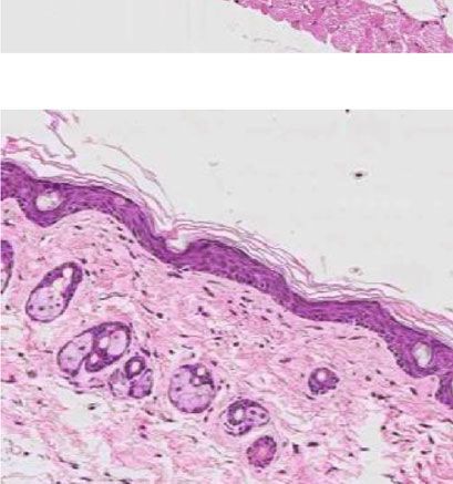
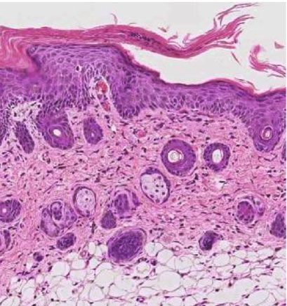
Mediators of Inflammation 7

 Epidermal thickness
 ⁎⁎⁎ ⁎⁎⁎
 200
 Normal IMQ

 Epidermal thickness (μm)
 150

 100

 50

 0
 IMQ+CR al

 R
 rm Q

 +C
 No IM

 Q
 IM
 (a) (b)

 MM

 ⁎⁎
 IMQ
 8×10⁴
 Relative MM area (μm2)

 6×10⁴

 4×10⁴

 2×10⁴

 0
 IMQ+CR R
 Q
 IM +C
 Q
 IM
 (c) (d)

Figure 2: CR treatment reduced the epidermal thickness and diminished neutrophil accumulation in IMQ-induced PsD in mice. (a)
Representative microscopic images of H&E-stained skin sections from each group (scale bar = 145 μm). (b) Epidermal thickness was
evaluated using Leica Microsystems software under a microscope. n = 11 in the normal group; n = 15 in IMQ group and IMQ+CR
group. (c) Representative image of MM (scale bar = 145 μm). (d) Quantification of MM area. n = 15 in both the IMQ and IMQ+CR
group. Epidermal thicknesses were compared by using one-way ANOVA and post hoc Tukey’s test, and MM areas were compared using
a t-test. Data are shown as the Mean ± SEM. ∗∗ P < 0:01 and∗∗∗ P < 0:001.
8 Mediators of Inflammation

 CD45+ cells Neutrophils Macrophages
 ⁎⁎⁎ ⁎⁎ ⁎⁎⁎
 60 ⁎⁎⁎
 8 4

 6 3
 % of live cells

 40

 % of live cells
 % of live cells
 4 2

 20
 2 1

 0 0 0
 al al al
 rm IM
 Q CR rm Q CR rm Q CR
 No Q+ No IM Q+ No IM Q+
 IM IM IM
 α β T cells CD4+/CD8+αβ T cells
 0.8 50 ⁎ ⁎⁎

 0.6 40
 CD4+/CD8+
 % of live cells

 30
 0.4
 20
 0.2
 10

 0.0 0
 al al
 rm Q CR rm C R
 No IM Q+ o IM
 Q Q+
 IM N IM
 (a)
 Il17a Il22 Ccl20

 3 ⁎⁎ ⁎⁎ 1.5 15 ⁎ ⁎
 ⁎⁎⁎ ⁎⁎
 Relative mRNA expression

 Relative mRNA expression
 Relative mRNA expression

 2 1.0 10

 1 0.5 5

 0 0.0 0
 R R R
 +C +C +C
 al

 al

 al
 m

 m

 m

 Q Q Q Q Q Q
 or

 or

 or

 IM IM IM IM IM IM
 N

 N

 N

 (b)

Figure 3: CR treatment decreased immune cell infiltration and cytokine production in murine skins of IMQ-induced PsD. (a) Flow
cytometry analysis of immune cells (CD45+ immune cells, neutrophils, macrophages, αβ T cells, and CD4+/CD8+αβ T cells) from skin
tissues among groups. n = 5 in the normal group, n = 8 in the IMQ group, and n = 10 in the IMQ+CR group. (b) mRNA expression of
Il17a, Il22, and Ccl20 in mouse skin was measured via RT-qPCR, and the relative mRNA expression was normalized to Gapdh
expression. n = 6 in the normal group, n = 8 in the IMQ group, and n = 8 in the IMQ+CR group. Statistical analysis was performed using
one-way ANOVA followed by post hoc Tukey’s test. Data are shown as the Mean ± SEM. ∗ P < 0:05, ∗∗ P < 0:01, and∗∗∗ P < 0:001.
Mediators of Inflammation 9

 Normal

 40

 30 ⁎⁎⁎ ⁎⁎⁎
 ⁎⁎

 ⁎⁎
 Scratch bouts/30min

 ⁎ IMQ
 20

 ⁎

 10

 0
 1 2 3 4 5 6 7
 IMQ+CR
 Days

 Normal
 IMQ
 IMQ+CR
 (a) (b)
 Number of mast cells
 ⁎⁎⁎ ⁎⁎⁎
 250

 200
 Number of mast cells

 150

 100

 50

 0
 al
 R
 +C

 m
 Q

 or
 IM

 Q

 N
 IM

 (c)

Figure 4: Pruritus was improved after CR treatment in IMQ-induced PsD in mice. (a) Spontaneous scratching was measured and compared
among groups. n = 11 in the normal group and n = 15 in both the IMQ group and IMQ+CR group. (b) Mast cells were stained using
Toluidine blue (scale bar = 73 μm). (c) Numbers of mast cells in each group were counted. n = 5 in the normal group and n = 6 in both
the IMQ group and IMQ+CR group. Statistical analysis was performed using one-way ANOVA followed by post hoc Tukey’s test. Data
are shown as the Mean ± SEM. ∗ P < 0:05, ∗∗ P < 0:01, and∗∗∗ P < 0:001.
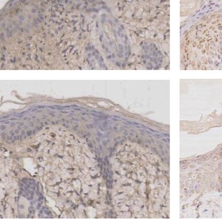
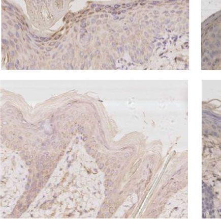
10 Mediators of Inflammation

 SP Cgrp Ngf

 4 ⁎⁎ ⁎ 5 ⁎ ⁎ 4

 Relative mRNA expression
 Relative mRNA expression

 Relative mRNA expression
 4
 3 3

 3
 2 2
 2

 1 1
 1

 0 0 0
 R
 CR +C +C
 R

 al
 al

 al
 +

 m
 Q
 m

 m
 Q Q

 Q
 Q

 Q
 or
 or

 IM

 or
 IM

 IM
 IM
 IM

 IM
 N
 N

 N
 (a)
 Sp Cgrp Ngf

 Normal

 IMQ

 IMQ+CR

 (b)

 ⁎⁎⁎ ⁎⁎⁎ ⁎⁎ ⁎⁎
 15 20 15 ⁎⁎⁎ ⁎⁎⁎

 15
 Mean of IOD

 Mean of IOD

 10 10
 Mean of IOD

 10

 5 5
 5

 0 0 0
 CR
 CR

 CR

 al
 al

 al

 Q+
 Q+

 Q+

 m
 m

 m

 Q Q Q
 or
 or

 or

 IM IM IM
 IM
 IM

 IM

 N
 N

 N

 (c)

Figure 5: CR treatment reduced the expression of itch-related molecules in IMQ-induced PsD in mice. (a) mRNA expression of Sp, Cgrp,
and Ngf in skins was detected by RT-qPCR. n = 6 mice in the control group and n = 8 mice in both the IMQ group and IMQ+CR group.
Results of mRNA were normalized to Gapdh expression. (b) Representative immunohistochemical images of SP, CGRP, and NGF
expression from the epidermis of back skins among groups (scale bar = 73 μm). (c) Mean of integrated optical density (IOD) of Sp, Cgrp,
and Ngf in skins from each group. n = 5 mice in three groups. Statistical analysis was performed using one-way ANOVA followed by
post hoc Tukey’s test. Mean ± SEM values are indicated, ∗ P < 0:05, ∗∗ P < 0:01, and∗∗∗ P < 0:001.
Mediators of Inflammation 11

 ⁎⁎⁎ ⁎
 2.5
 ⁎⁎ ⁎⁎⁎
 ⁎
 2.0
 ⁎

 OD Value (450nm)
 1.5

 1.0

 0.5

 0.0
 5 5 1
 0.2

 2
 0.5
 0

 25

 5

 1

 2
 M 5+

 5+
 0.
 0.

 5+

 5+
 M

 M
 M
 M
 (a)
 IL6 CXCL8
 ⁎ ⁎ ⁎⁎ ⁎
 200 600
 Relative mRNA expression

 Relative mRNA expression

 150
 400
 100
 50 200

 5 5
 4 4
 3 3
 2 2
 1 1
 0 0
 CR
 5

 CR
 l
 CR

 CR
 l

 5
 ro

 ro
 M

 M
 nt

 nt
 5+

 5+
 Co

 Co
 M

 M

 IL1β CCL20
 ⁎⁎ ⁎
 20 ⁎⁎⁎ ⁎ 400
 Relative mRNA expression

 Relative mRNA expression

 300
 15
 200

 10 100
 5
 4
 5 3
 2
 1
 0 0
 5

 5

 CR
 CR
 CR

 CR
 l

 l
 M

 M
 ro

 tro

 5+
 5+
 nt

 n
 Co

 Co

 M
 M

 SP CGRP NGF
 4 ⁎ 3 ⁎ 8 ⁎ ⁎
 Relative mRNA expression

 Relative mRNA expression
 Relative mRNA expression

 3 6
 2
 2 4
 1
 1 2

 0 0 0
 l

 5

 l

 5

 l

 5
 CR

 CR

 CR

 CR

 CR

 CR
 ro

 ro

 ro
 M

 M

 M
 nt

 nt

 nt
 5+

 5+

 5+
 Co

 Co

 Co
 M

 M

 M

 (b)

Figure 6: CR treatment regulated cell viability and function of M5-induced keratinocytes in vitro. M5 (10 ng/ml) was used to stimulate NHEKs
with or without CR treatment ((a): 0, 0.25 μg/ml, 0.5 μg/ml, 1 μg/ml, and 2 μg/ml; (b): 0.5 μg/ml). (a) Cell viability was measured by using CCK-8
(n = 4). (b) The mRNA expression of IL-6, CXCL8, IL-1β, CCL20, SP, CGRP, and NGF was detected by RT-qPCR (n = 3). The relative mRNA
expression was normalized to GAPDH expression. Statistical analysis was performed using one-way ANOVA followed by post hoc Tukey’s test.
Mean ± SEM values are indicated. ∗ P < 0:05, ∗∗ P < 0:01, and∗∗∗ P < 0:001.
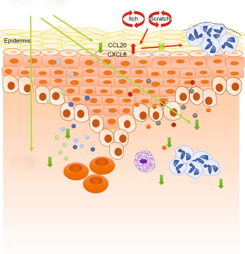
12 Mediators of Inflammation

 Colquhounia root
 Munro microabscesses

 Dermis Neurogenic
 CGRP
 inflammation
 IL-17a

 IL-22
 SP
 IL-17 IL-6 NGF

 Immunogenic
 Inflammation

 Mast Cells Neutrophils
 T cells

Figure 7: Characterization of the mechanism of CR for psoriasis pruritus improvement. CR treatment relieves the symptoms of psoriasis
pruritus by inhibiting increased mast cells and neutrophils, as well as the upregulation of the inflammatory factors IL-17a, IL-22, IL-6,
CXCL8, IL-1β, and CCL20 and itch-related molecules SP, CGRP, and NGF in psoriasis.

epidermal keratinocytes promoted the release of CCL20 and observed that CR could promote cell viability slightly in
CXCL8, which was related to the recruitment of both neutro- NHEKs, while in NHEKs treated with M5, CR inhibited cell
phils and IL-17a-producing immune cells [36]. CCL20 was viability. We speculate that CR might regulate the viability of
expressed abundantly in human psoriatic epidermis [9, 37]. keratinocytes under different circumstances, which should be
Dupilumab inhibits the IL-4/IL-13 signaling pathway to reduce explored in further study.
itching and itching in atopic dermatitis, which may reduce the Notably, the number of mast cells was significantly
release of CCL20 from keratinocytes and also reduce Th17 cell reduced in lesions of CR application. Several researches
infiltration [38]. As well as in psoriasis, the enrichment of IL- observed that mast cells are hyperactivated in psoriatic
17A-producing immune cells leads to produce high levels of lesions of progressive stage. The use of certain medications
IL-17A, which further stimulates keratinocytes producing to treat psoriasis, such as glucocorticosteroids, may lead a
CCL20, CXCL8, and IL-36γ to recruit more IL-17A- decline of the mast cell count [11]. Our observation indicates
producing immune cells and neutrophils [39]. In our findings, that CR may function as an inhibitor of itch in psoriasis by
the neutrophil infiltration and also increasing expression of decreasing the number of mast cells in skin lesions.
CCL20 in IMQ treated mice were both inhibited by CR. As well In addition, the itch sensation is transmitted to the brain
in vitro, dexamethasone partially inhibited scratch-induced via itch neural pathways from skin lesions [40, 41]. Sensory
CXCL8 and CCL20 secretion in the keratinocyte model [36]. nerve fibers increased in the psoriatic epidermis and dermal
In order to mimic a psoriatic environment, we chose an M5- papilla, which were stimulated (endogenous or exogenous)
induced keratinocyte model to investigate the effects of CR. by various neuropeptides and neurotrophic molecule, such
As expected, CR reduced the expression levels of inflammatory as CGRP, SP, and NGF. Also, in the epidermis, neuropep-
cytokines IL-6, IL-8, CXCL8, and CCL20. Interestingly, we tides released from the nerve fibers stimulate keratinocytes
Mediators of Inflammation 13

to release proinflammatory cytokines such as IL-6 and IL-8. Supplementary Materials
On the other hand, CGRP and SP induce the release of vaso-
active amine by mast cells, promoting the infiltration of neu- Supplementary 1 Supplementary Figure 1: gating strategy of
trophils and T cells closely related with itch [42–45]. The skin cells in flow cytometry. Supplementary 2 Supplementary
main source of NGF in the skin is keratinocytes [46]. The Figure 2: effect of psoriatic inflammation and CR on liver
expression of NGF in psoriatic patients with pruritus is and renal function of mice. (Supplementary Materials)
higher than that without pruritus [47]. These molecules
work directly or indirectly on the nerve and lead to or aggra- References
vate the degree of itch in psoriasis [48, 49]. In our research,
 [1] J. C. Szepietowski and A. Reich, “Pruritus in psoriasis: an
topical CR preparation can significantly relieve itch accom-
 update,” European Journal of Pain, vol. 20, no. 1, pp. 41–46,
panied with reduced levels of CGRP, SP, and NGF in lesions
 2016.
from psoriatic mice. Additionally, in a psoriatic cell model,
 [2] E. Komiya, M. Tominaga, Y. Kamata, Y. Suga, and
itch-related molecules SP, CGRP, and NGF were found
 K. Takamori, “Molecular and cellular mechanisms of itch in
decreased in NHEK cotreated with CR and M5. However, psoriasis,” International Journal of Molecular Sciences,
the expression of CGRP and SP in vitro was not upregulated vol. 21, no. 21, p. 8406, 2020.
by M5. We speculate that M5-induced keratinocytes may
 [3] G. Yosipovitch, A. Goon, J. Wee, Y. H. Chan, and C. L. Goh,
not be an optimal itching cell model in psoriasis. “The prevalence and clinical characteristics of pruritus among
 patients with extensive psoriasis,” The British Journal of Der-
5. Conclusion matology, vol. 143, no. 5, pp. 969–973, 2000.
 [4] B. Elewski, A. F. Alexis, M. Lebwohl et al., “Itch: an under-
In summary, we firstly prove that CR can not only inhibit the recognized problem in psoriasis,” Journal of the European
increased mast cells and neutrophils in lesions but also down- Academy of Dermatology and Venereology, vol. 33, no. 8,
regulate the inflammatory factors and neuropeptides and neu- pp. 1465–1476, 2019.
rotrophic molecule to reduce the inflammation and itch in [5] B. Amatya, H. el-Nour, M. Holst, E. Theodorsson, and
IMQ-induced murine model of psoriasis, which is also vali- K. Nordlind, “Expression of tachykinins and their receptors
dated in a psoriatic cell model (Figure 7). In conclusion, CR in plaque psoriasis with pruritus,” The British Journal of Der-
is considered to be an ideal topical therapeutic drug for anti- matology, vol. 164, no. 5, pp. 1023–1029, 2011.
itch treatment not just anti-inflammation in psoriasis. [6] R. Saraceno, C. E. Kleyn, G. Terenghi, and C. E. M. Griffiths,
 “The role of neuropeptides in psoriasis,” The British Journal
Data Availability of Dermatology, vol. 155, no. 5, pp. 876–882, 2006.
 [7] J. Yamaguchi, M. Aihara, Y. Kobayashi, T. Kambara, and
The datasets generated during and/or analyzed during the Z. Ikezawa, “Quantitative analysis of nerve growth factor
current study are available from the corresponding author (NGF) in the atopic dermatitis and psoriasis horny layer and
on reasonable request. effect of treatment on NGF in atopic dermatitis,” Journal of
 Dermatological Science, vol. 53, no. 1, pp. 48–54, 2009.
Conflicts of Interest [8] F. Cevikbas, X. Wang, T. Akiyama et al., “A sensory neuron-
 expressed IL-31 receptor mediates T helper cell-dependent
The authors declare no commercial or financial conflict of itch: involvement of TRPV1 and TRPA1,” The Journal of
interest. Allergy and Clinical Immunology, vol. 133, no. 2, pp. 448–
 460.e7, 2014.
 [9] J. Pène, S. Chevalier, L. Preisser et al., “Chronically inflamed
Authors’ Contributions human tissues are infiltrated by highly differentiated Th17
 lymphocytes,” Journal of Immunology, vol. 180, no. 11,
Fei Li and Dan Han designed the study, performed the
 pp. 7423–7430, 2008.
majority of the experiments, analyzed the data, and wrote
 [10] B. Strober, B. Sigurgeirsson, G. Popp et al., “Secukinumab
the manuscript; Bo Wang, Wentao Zhang, Yan Zhao, and
 improves patient-reported psoriasis symptoms of itching,
Jing Xu performed experiments and analyzed data; Liesu pain, and scaling: results of two phase 3, randomized,
Meng and Shemin Lu provided expert technical assistance; placebo-controlled clinical trials,” International Journal of
Kuanhou Mou provided clinical advice and critical discus- Dermatology, vol. 55, no. 4, pp. 401–407, 2016.
sion of work; Wenhua Zhu and Yan Zhou designed the [11] K. Jaworecka, J. Muda-Urban, M. Rzepko, and A. Reich,
study, supervised the project, and wrote the manuscript. “Molecular aspects of pruritus pathogenesis in psoriasis,”
 International Journal of Molecular Sciences, vol. 22, no. 2,
Acknowledgments p. 858, 2021.
 [12] A. W. Armstrong and C. Read, “Pathophysiology, clinical pre-
This work is supported by the National Natural Science sentation, and treatment of psoriasis: a review,” JAMA,
Foundation of China (81703130 and 82171724), Key vol. 323, no. 19, pp. 1945–1960, 2020.
Research and Development Program of Shaanxi Province [13] G. Yosipovitch, J. Soung, J. Weiss et al., “Secukinumab pro-
of China (2020KW-047), and the Clinical Research Award vides rapid relief from itching and pain in patients with
of the First Affiliated Hospital of Xi’an Jiaotong University, moderate-to-severe psoriasis: patient symptom diary data
China (XJTU1AF-CRF-2019-027 and 2021ZXY-12). from two phase 3, randomized, placebo-controlled clinical
14 Mediators of Inflammation

 trials,” Acta Dermato-Venereologica, vol. 99, no. 9, pp. 820- Producing γδ T Cells,” The Journal of Investigative Dermatol-
 821, 2019. ogy, vol. 134, no. 7, pp. 1912–1921, 2014.
[14] M. Kamata and Y. Tada, “Safety of biologics in psoriasis,” The [29] K. Gupta and I. T. Harvima, “Mast cell-neural interactions
 Journal of Dermatology, vol. 45, no. 3, pp. 279–286, 2018. contribute to pain and itch,” Immunological Reviews,
[15] B. Farahnik, D. Sharma, J. Alban, and R. K. Sivamani, “Topical vol. 282, no. 1, pp. 168–187, 2018.
 botanical agents for the treatment of psoriasis: a systematic [30] H. S. Kim and G. Yosipovitch, “The skin microbiota and itch:
 review,” American Journal of Clinical Dermatology, vol. 18, is there a link?,” Journal of Clinical Medicine, vol. 9, no. 4,
 no. 4, pp. 451–468, 2017. p. 1190, 2020.
[16] W. Zhou, G. Shi, J. Bai, S. Ma, Q. Liu, and X. Ma, “Colquhou- [31] J. Zhao, T. di, Y. Wang et al., “Multi-glycoside of Tripterygium
 nia Root Tablet Protects Rat Pulmonary Microvascular Endo- wilfordii Hook. f. ameliorates imiquimod-induced skin lesions
 thelial Cells against TNF–Induced Injury by Upregulating the through a STAT3-dependent mechanism involving the inhibi-
 Expression of Tight Junction Proteins Claudin-5 and ZO-1,” tion of Th17-mediated inflammatory responses,” Interna-
 Evidence-based Complementary and Alternative Medicine, tional Journal of Molecular Medicine, vol. 38, no. 3, pp. 747–
 vol. 2018, Article ID 1024634, 11 pages, 2018. 757, 2016.
[17] M. Jiang, H. Zhang, and Y. Ding, “Research progress on phar- [32] R. M. El-Gharabawy, A. S. Ahmed, and A. H. Al-Najjar,
 macological activities and clinical applications of Tripterygium “Mechanism of action and effect of immune-modulating
 glycosides,” Chinese Archives of Traditional Chinese Medicine, agents in the treatment of psoriasis,” Biomedicine & Pharma-
 vol. 39, no. 3, pp. 59–63, 2021. cotherapy, vol. 85, pp. 141–147, 2017.
[18] X. L. Wu, J. B. Li, and S. L. Mo, “Clinical observation on colqu- [33] T. Nomura, K. Kabashima, and Y. Miyachi, “The panoply of
 hounia root tablet in treating lipid metabolism disturbance αβT cells in the skin,” Journal of Dermatological Science,
 secondary to nephrotic syndrome,” Zhongguo Zhong Xi Yi Jie vol. 76, no. 1, pp. 3–9, 2014.
 He Za Zhi, vol. 22, no. 1, pp. 30–32, 2002. [34] E. Theodorakopoulou, Z. Z. N. Yiu, C. Bundy et al., “Early- and
[19] X. Han, H. Mou, and L. M. Wang, “Clinical effect analysis of late-onset psoriasis: a cross-sectional clinical and immunocy-
 colquhounia root tablet in the treatment of 46 cases of psoria- tochemical investigation,” The British Journal of Dermatology,
 sis vulgaris,” China Journal of Leprosy and Skin Diseases, vol. 6, vol. 175, no. 5, pp. 1038–1044, 2016.
 pp. 588-589, 2004. [35] B. S. Baker, A. V. Powles, S. Lambert, H. Valdimarsson, and
[20] W. R. Swindell, K. A. Michaels, A. J. Sutter et al., “Imiquimod L. Fry, “A prospective study of the Koebner reaction and T
 has strain-dependent effects in mice and does not uniquely lymphocytes in uninvolved psoriatic skin,” Acta Dermato-
 model human psoriasis,” Genome Medicine, vol. 9, no. 1, Venereologica, vol. 68, no. 5, pp. 430–434, 1988.
 p. 24, 2017. [36] K. Furue, T. Ito, Y. Tanaka et al., “Cyto/chemokine profile of
[21] S. R. Feldman, D. M. Bushnell, P. A. Klekotka et al., “Differ- in vitro scratched keratinocyte model: implications of signifi-
 ences in psoriasis signs and symptom severity between patients cant upregulation of CCL20, CXCL8 and IL36G in Koebner
 with clear and almost clear skin in clinical practice,” The Jour- phenomenon,” Journal of Dermatological Science, vol. 94,
 nal of Dermatological Treatment, vol. 27, no. 3, pp. 224–227, no. 1, pp. 244–251, 2019.
 2016. [37] T. G. Kim, H. Jee, J. Fuentes-Duculan et al., “Dermal clusters of
[22] K. Sakai, K. M. Sanders, M. R. Youssef et al., “Mouse model of mature dendritic cells and T cells are associated with the
 imiquimod-induced psoriatic itch,” Pain, vol. 157, no. 11, CCL20/CCR6 chemokine system in chronic psoriasis,” The
 pp. 2536–2543, 2016. Journal of Investigative Dermatology, vol. 134, no. 5,
[23] N. Puebla-Osorio, S. N. E. Sarchio, S. E. Ullrich, and S. N. pp. 1462–1465, 2014.
 Byrne, “Detection of infiltrating mast cells using a modified [38] E. L. Simpson, T. Bieber, E. Guttman-Yassky et al., “Two phase
 toluidine blue staining,” Methods in Molecular Biology, 3 trials of dupilumab versus placebo in atopic dermatitis,” The
 vol. 1627, pp. 213–222, 2017. New England Journal of Medicine, vol. 375, no. 24, pp. 2335–
[24] K. Guilloteau, I. Paris, N. Pedretti et al., “Skin inflammation 2348, 2016.
 induced by the synergistic action of IL-17A, IL-22, Oncostatin [39] E. G. Harper, C. Guo, H. Rizzo et al., “Th17 Cytokines
 M, IL-1{alpha}, and TNF-{alpha} recapitulates some features Stimulate CCL20 Expression in Keratinocytes In Vitro and
 of psoriasis,” Journal of Immunology, vol. 184, no. 9, In Vivo: Implications for Psoriasis Pathogenesis,” The Jour-
 pp. 5263–5270, 2010. nal of Investigative Dermatology, vol. 129, no. 9, pp. 2175–
[25] Y. Ru, H. Li, R. Zhang et al., “Role of keratinocytes and 2183, 2009.
 immune cells in the anti-inflammatory effects of Tripterygium [40] S. Bourane, B. Duan, S. C. Koch et al., “Gate control of
 wilfordii Hook. f. in a murine model of psoriasis,” Phytomedi- mechanical itch by a subpopulation of spinal cord interneu-
 cine, vol. 77, article 153299, 2020. rons,” Science, vol. 350, no. 6260, pp. 550–554, 2015.
[26] K. Nakajima and S. Sano, “Mouse models of psoriasis and their [41] M. Lay and X. Dong, “Neural mechanisms of itch,” Annual
 relevance,” The Journal of Dermatology, vol. 45, no. 3, pp. 252– Review of Neuroscience, vol. 43, no. 1, pp. 187–205, 2020.
 263, 2018. [42] A. Dallos, M. Kiss, H. Polyánka, A. Dobozy, L. Kemény, and
[27] L. van der Fits, S. Mourits, J. S. A. Voerman et al., “Imiquimod- S. Husz, “Effects of the neuropeptides substance P, calcitonin
 induced psoriasis-like skin inflammation in mice is mediated gene-related peptide, vasoactive intestinal polypeptide and
 via the IL-23/IL-17 axis,” Journal of Immunology, vol. 182, galanin on the production of nerve growth factor and inflam-
 no. 9, pp. 5836–5845, 2009. matory cytokines in cultured human keratinocytes,” Neuro-
[28] R. Yoshiki, K. Kabashima, T. Honda et al., “IL-23 from Langer- peptides, vol. 40, no. 4, pp. 251–263, 2006.
 hans Cells Is Required for the Development of Imiquimod- [43] G. J. Burbach, K. H. Kim, A. S. Zivony et al., “The neurosen-
 Induced Psoriasis-Like Dermatitis by Induction of IL-17A- sory tachykinins substance P and neurokinin A directly induce
Mediators of Inflammation 15 keratinocyte nerve growth factor,” The Journal of Investigative Dermatology, vol. 117, no. 5, pp. 1075–1082, 2001. [44] I. S. Song, N. W. Bunnett, J. E. Olerud et al., “Substance P induction of murine keratinocyte PAM 212 interleukin 1 pro- duction is mediated by the neurokinin 2 receptor (NK-2R),” Experimental Dermatology, vol. 9, no. 1, pp. 42–52, 2000. [45] Y. M. Park and C. W. Kim, “The effects of substance P and vasoactive intestinal peptide on interleukin-6 synthesis in cul- tured human keratinocytes,” Journal of Dermatological Sci- ence, vol. 22, no. 1, pp. 17–23, 1999. [46] C. Pincelli, “Nerve growth factor and keratinocytes: a role in psoriasis,” European Journal of Dermatology, vol. 10, no. 2, pp. 85–90, 2000. [47] M. Nakamura, M. Toyoda, and M. Morohashi, “Pruritogenic mediators in psoriasis vulgaris: comparative evaluation of itch-associated cutaneous factors,” The British Journal of Der- matology, vol. 149, no. 4, pp. 718–730, 2003. [48] K. Sakai, K. M. Sanders, M. R. Youssef et al., “Role of neurturin in spontaneous itch and increased nonpeptidergic intraepider- mal fiber density in a mouse model of psoriasis,” Pain, vol. 158, no. 11, pp. 2196–2202, 2017. [49] X. Kodji, K. L. Arkless, Z. Kee et al., “Sensory nerves mediate spontaneous behaviors in addition to inflammation in a murine model of psoriasis,” The FASEB Journal, vol. 33, no. 2, pp. 1578–1594, 2019.
You can also read