Untangling the Small Intestine in 3D cine-MRI using Deep Stochastic Tracking
←
→
Page content transcription
If your browser does not render page correctly, please read the page content below
Proceedings of Machine Learning Research – Under Review:1–11, 2021        Full Paper – MIDL 2021 submission

            Untangling the Small Intestine in 3D cine-MRI
                   using Deep Stochastic Tracking

Louis D. van Harten1                                                 l.d.vanharten@amsterdamumc.nl
Catharina S. de Jonge2
Jaap Stoker2
Ivana Išgum1,3
1
  Department of Biomedical Engineering and Physics, Amsterdam UMC, Location AMC, University
of Amsterdam, Amsterdam, The Netherlands.
2
  Department of Radiology and Nuclear Medicine, Amsterdam Gastroenterology, Endocrinology and
Metabolism, Amsterdam UMC, University of Amsterdam, Amsterdam, The Netherlands.
3
  Department of Radiology and Nuclear Medicine, Amsterdam UMC, Location AMC, University of
Amsterdam, Amsterdam, The Netherlands.

Editors: Under Review for MIDL 2021

                                                 Abstract
     Motility of the small intestine is a valuable metric in the evaluation of gastrointestinal
     disorders. Cine-MRI of the abdomen is a non-invasive imaging technique allowing evalu-
     ation of this motility. While 2D cine-MR imaging is increasingly used for this purpose in
     both clinical practice and in research settings, the potential of 3D cine-MR imaging has
     been largely underexplored. In the absence of image analysis tools enabling investigation
     of the intestines as 3D structures, the assessment of motility in 3D cine-images is generally
     limited to the evaluation of movement in separate 2D slices. Hence, to obtain an untangled
     representation of the small intestine in 3D cine-MRI, we propose a method to extract a
     centerline of the intestine, thereby allowing easier (visual) assessment by human observers,
     as well as providing a possible starting point for automatic analysis methods quantifying
     peristaltic bowel movement along intestinal segments. The proposed method automatically
     tracks individual sections of the small intestine in 3D space, using a stochastic tracker built
     on top of a CNN-based orientation classifier. We show that the proposed method outper-
     forms a non-stochastic iterative tracking approach.

     Keywords: Deep learning, cine-MRI, centerline extraction, small intestine, motility.

1. Introduction
Intestinal motility consists of contractions of the bowel wall, providing peristaltic movement
and intestinal content mixing. Changes in small bowel motility are associated with a wide
variety of functional gastrointestinal diseases and disorders such as Crohn’s disease and
chronic intestinal pseudo-obstruction (CIPO) (Menys et al., 2018; Paine et al., 2013; van
Rijn et al., 2020). While motility in the proximal small intestine can be measured inva-
sively using antroduodenal manometry, the high patient burden of this procedure has driven
the investigation of cine-MRI as a non-invasive alternative for the complete small bowel.
The resulting images can be visually assessed by a radiologist (Guglielmo et al., 2015; Heye
et al., 2012), or quantitatively evaluated using techniques like displacement mapping (Odille

© 2021 L.D. van Harten, C.S. de Jonge, J. Stoker & I. Išgum.
Deep Stochastic Tracking in Abdominal 3D cine-MRI

Figure 1: A bowel section with two unambiguous directions (left) and an ambiguous case
          where the classifier may predict directions for different intestinal sections (right).

et al., 2012), or diameter measurements (Wakamiya et al., 2011). While diameter measure-
ments traditionally involve labour-intensive annotations, recent works have shown promise
in automating this task using convolutional neural networks (CNNs) (Wu et al., 2020).
    Currently, literature on quantifying intestinal motility in cine-MRI is generally centered
around 2D cine-MRI (De Jonge et al., 2018). Although some works acquire 3D cine-MRI,
the analysis is generally limited to sequences of 2D slices (Menys et al., 2018; Odille et al.,
2012; Wakamiya et al., 2011; Wu et al., 2020). As movement of the small intestines occurs
continuously in all directions, motility movement and through-plane motion are entangled
in such methods: in 2D analysis it is impossible to differentiate intestinal contractions
from bowel sections moving (partially) in and out of plane, as both lead to a change in
perceived luminal diameter between time points. Furthermore, 2D analysis is ill suited
for differentiating peristaltic from intestinal content mixing motion and for identification
of dysfunctional propagation of contractions, as doing so requires correlating contractions
along intestinal segments.
    The low-dimensional analysis can be largely attributed to the lack of (semi-)automatic
image analysis tools enabling investigation of the intestines as 3D structures. A stretched-
out, untangled representation of the intestines would allow easier assessment by human
observers and it could enable detailed automatic analysis of the functional peristaltic motion.
Such a representation can be generated by piecewise resampling of the intestines along
their centerline, creating a multi-planar reformation (MPR), but manually annotating such
centerlines is unfeasible in routine clinical patient work-up. Previous work on automatic
extraction of centerlines from the small intestine in MR has relied on the availability of
an accurate segmentation mask of the small intestine (Spuhler et al., 2006). However,
recent work on deep learning-based segmentation has shown that state-of-the-art methods
achieve much lower performance in the small intestine compared to other organs, producing
clinically acceptable segmentations in less than 40% of the tested subjects (Liu et al., 2020).
The authors indicate that the large diversity in shapes, sizes and locations in the abdomen
among different patients make the small intestine especially difficult to segment using deep
learning. This means the extraction of centerlines through segmentation is currently not a
promising approach for application in the clinic. To the best of our knowledge, more recent
work on the extraction of centerlines in the small intestine is not available.

                                               2
Deep Stochastic Tracking in Abdominal 3D cine-MRI

Figure 2: Center-cropped slices from three subjects, illustrating variety in characteristics.

    Alternative to using segmentation maps as an intermediate, centerlines can also be ex-
tracted directly from the MR images. Existing methods for centerline extraction in medical
images have been developed for application in vessels in various anatomical regions, as well
as the airways in chest images (Friman et al., 2010; Bauer et al., 2009). These methods have
traditionally relied on modelling the target anatomy as tubular structures (Frangi et al.,
1998). More recent work has used machine learning approaches to predict centerlines either
directly (Sironi et al., 2015) or through use of intermediate flow fields (Gülsün et al., 2016).
Recent work in cardiac CTA has shown the efficacy of using deep neural orientation classi-
fiers for extracting centerlines in the coronary artery tree (Wolterink et al., 2019). Inspired
by this work, we develop a tracking method for the small intestine.
    In abdominal 3D cine-MRI, substantial sections of the small intestine are outside of the
field of view (FOV), meaning the centerline has to be extracted as unconnected segments.
The small FOV is a consequence of the short time available to scan each time point. Limited
image context is available and there is a possibility of the tracker crossing the intestinal wall
into an adjacent bowel segment (Figure 1), incorrectly fusing unconnected segments. To
overcome these challenges, we introduce a novel stochastic tracking strategy, which improves
the robustness of the tracking compared to non-stochastic neural centerline extraction.

2. Data
The method was developed and evaluated with 3D cine-MR scans from fourteen healthy
volunteers, retrospectively selected from a previous study (de Jonge et al., 2019). Se-
quences were imaged at 1.0 image per second during a 20 second breath-hold, acquired at
2.5x2.5x2.5 mm and reconstructed to 1.4x1.4x2.5 mm. Image FOV is 400x400x35 mm. The
exact anterior-posterior planning position differs, but all scans contain the terminal ileum.
    Prior to scanning, subjects were orally administered 1000 mL mannitol solution to im-
prove MR contrast in the intestine. The images were acquired using a balanced fast field
echo sequence, aimed at maximising the contrast of the interface between the intestinal wall
and the surrounding fatty tissue by means of anti-phase annihilation. Due to the sensitiv-
ity to body composition of this effect, the images present a large variability in contrast.
Furthermore, contrast at the interface of adjacent intestinal sections without fatty tissue in
between them varies strongly among subjects (Figure 2).

                                                3
Deep Stochastic Tracking in Abdominal 3D cine-MRI

    The reference centerlines were annotated as segments by manually placing markers along
the direction of the small intestine until the intestine would leave the FOV, or until the
annotator could no longer distinguish the direction of the segment due to image artifacts
or low contrast. Annotations were made in one of the 3D time points for each subject. An
average of 1150 mm of reference centerlines were annotated per subject, divided over 186
segments of on average 88 mm. Segments shorter than 25 mm were excluded, resulting in
a total of 181 annotated segments in the reference set.

3. Methods
3.1. Deep neural centerline tracking
Building on a method for coronary artery tracking in CTA (Wolterink et al., 2019), we
develop a method to track centerlines through the small intestine. The method uses a
deep neural network to predict direction proposals. This orientation classifier consists of a
fully convolutional neural network that operates on 32x32x32 voxel patches from a single 3D
cine-MR time point. The output is a set of 500 logits that correspond to equidistant vectors
on the unit sphere. During training, the network is provided with patches sampled around
a centerline and is trained to predict a probability distribution for its directions. Once the
classifier is trained, a centerline can be tracked by starting at a seed point and iteratively
stepping into the direction with maximum probability pmax . By masking the probabilities
for backward directions, this allows stepping through an estimate of the centerline until a
stopping criterion is met. If a moving average of the maximum value of the probability
distribution drops below a threshold, the tracker is terminated. For each starting point, the
method produces two centerlines (one in each direction), which are fused by reversing the
direction of one of them and concatenating the two segments.

3.2. Stochastic tracking strategy
To improve the robustness and reliability of the tracking system, we propose the concept of a
stochastic agent. Stochastic agents behave similarly to a non-stochastic iterative tracker, but
these agents do not always step into the direction with the maximum probability. Instead,
they stochastically sample a direction on the unit sphere from the generated probability
distribution (i.e. a direction with estimated probability p will have a p chance of being
chosen by the agent). By initialising multiple such agents, the probability space for possible
centerline paths can be explored.
    For the stochastic tracker, we initialise n stochastic agents at random points in a sphere
with radius r around the seed point. All agents start tracking simultaneously. To combine
these agents into a single centerline, the median location of all living agents is computed
after each step. If an agent strays more than distance d away from the median location,
the aberrant agent is terminated; if more than t agents are terminated, tracking stops. By
enforcing the distance-to-median constraint, the system effectively tracks by majority vote:
decisions to move into a direction are only taken if the majority of agents agree. Agents are
not re-initialized throughout; the progress of the agents is independent from the predicted
centerline points apart from the aberrant termination condition.

                                              4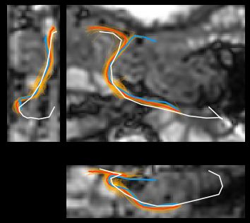
Deep Stochastic Tracking in Abdominal 3D cine-MRI

             Recall          Precision          Overlap              Recall          Precision          Overlap
1.00

0.75

0.50

0.25

       NST            ST   NST           ST   NST         ST   NST            ST   NST           ST   NST         ST

Figure 3: Comparison of results achieved by non-stochastic tracking (NST, blue) and
          stochastic tracking (ST, red) in terms of recall, precision and overlap. Left:
          Each point represents an intestinal segment. Right: Each point represents a sub-
          ject. Lines connect results for the same segments/subjects in both experiments,
          their colour indicates which strategy performed better on each metric. When
          both methods achieve similar performance (metric difference < 0.01), points are
          connected by a dashed grey line.

3.3. Evaluation
The centerline segments produced by the non-stochastic and the stochastic method are
evaluated based on their overlap with the reference, following the definition in (Schaap et al.,
2009): The harmonic mean between recall and precision, similar to the Dice coefficient. As
radius annotations are not available, we use a static distance threshold of 10 mm to define
true positive, true negative, false positive and false negative points (TP/TN/FP/FN). Due
to the incomplete reference standard, precision may be underestimated in some intestinal
segments. Beside evaluating the results per segment, we analyse the results per subject by
pooling all TP/TN/FP/FN.

4. Experiments and results
4.1. Experimental settings
The reference centerlines were quantized at a resolution of 0.5 mm to uniformly generate
training points along the segments. During training, patches were sampled at an isometric
resolution of 1.5 mm around the training points using linear interpolation, resulting in
a receptive field of 48 mm. Full 3D rotation augmentations were used. Due to the low
number of subjects in this study, our experiments were performed with leave-one-subject-
out cross-validation. The networks were trained using a cosine annealed learning rate policy
with warm restarts (Loshchilov and Hutter, 2016). Suitable values for maximum learning
rate, number of epochs and cut-off values for the stopping criteria were selected based on
preliminary experiments on a single image. This image was excluded from the evaluation.
Tracking step size was set to 0.5 mm and confidence-based stopping criterion thresholds were
set to pmax > 0.025 with a moving average window of 3 steps for both tracking methods.

                                                           5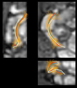
Deep Stochastic Tracking in Abdominal 3D cine-MRI

Figure 4: Visualisation of tracking results for an intestinal segment in two different subjects.
          Triplar view centered on the point where the trackers were initialized and zoomed
          to fit the results to the view. Projections are plotted for the manual reference
          (white), the non-stochastic tracking result (blue), the stochastic tracking result
          (red) and the individual stochastic agents (orange).

For the stochastic tracker, we used n = 64 agents, initialized in a sphere of r = 5 mm
around the seed point, equivalent to the thinnest non-contracted regions of the intestinal
lumen. The maximum median-distance for individual agents was set to d = 10 mm and
the stopping criterion threshold for the maximum number of terminated agents was set to
t = 34 n. During evaluation, a single seed point on the reference centerline was randomly
selected in each intestinal segment, serving as a starting point for the automatic methods.

4.2. Results
The quantitative results for both methods are listed in Table 1 and shown in Figure 3.

  Table 1: Quantitative results for the non-stochastic and stochastic tracking methods.

     Strategy                    Segments                           Subjects
                        recall   precision     overlap     recall   precision    overlap
     Non-stochastic     0.69     0.77          0.67        0.57     0.66         0.61
     Stochastic         0.72*    0.88*         0.75*       0.61*    0.83*        0.69*
*statistically significant improvement (Wilcoxon signed rank test, p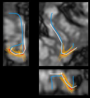
Deep Stochastic Tracking in Abdominal 3D cine-MRI

Figure 5: Untangled representations of the intestinal segments shown in Figure 4, generated
          from the manual centerline (top), the non-stochastic tracking result (middle) and
          the stochastic tracking result (bottom), aligned along the horizontal axis. Deviant
          segment lengths are caused by low precision and low recall, respectively.

intestinal wall into an adjacent bowel loop. In both cases, at least one stochastic agent
agreed with the non-stochastic method, but was terminated as aberrant. The images on the
right show a segment where both the non-stochastic and the stochastic method have near-
perfect precision, but the non-stochastic method fails due to collision with the intestinal wall
in both directions. This is caused by a region in the segment where orientation classification
performance is poor. While the stochastic agents have the same problem, a large cluster of
them survives the problematic area, causing the complete section to be tracked correctly.
Additional qualitative results can be found in the appendix.
    Finally, Figure 5 shows the untangled representations for both of these intestinal seg-
ments, generated from both the manual and the automatic centerlines. Rotation angle
around the centerline and horizontal axis position were matched for easier visual compar-
ison. Differences in length are caused by late and early termination of the non-stochastic
method on these two segments. In the top-left view, the intestine exhibits a slight wobble
around the centerline, revealing an imperfection in the manual annotation. In the correctly
tracked section, both methods produce a more straight result. The representations pro-
duced by the stochastic method contain more high-frequency resampling noise in the outer
regions of the MPR, indicating a lower smoothness of the centerline.

5. Discussion
We have presented a novel stochastic method for tracking centerlines through the small
intestine in 3D cine-MRI. To the best of our knowledge, this is the first method for tracking
centerlines in the small intestine that does not depend on the availability of an accurate
segmentation mask. The method is inspired by a recent method that accurately extracted
centerlines of the coronary arteries in cardiac CTA using a deep learning-based iterative

                                               7
Deep Stochastic Tracking in Abdominal 3D cine-MRI

tracker. We have presented measurable improvement by adding stochasticity: centerlines
produced by our method are less prone to crossing the intestinal wall and leaking out into
surrounding tissues. Our method is fully parallelizable, meaning no performance penalty is
incurred by the stochastic strategy if sufficient computation cores are available.
    Quantitatively, the stochastic method outperformed the non-stochastic method in terms
of precision and overlap scores. While the improvement in recall was also statistically
significant on both patient and segment levels, the difference was much smaller than for the
other two metrics. The reason is that the stochastic method was more likely than the non-
stochastic method to terminate early in regions where network performance is compromised,
for example due to the presence of artifacts. It may be beneficial to relax the thresholds on
the stopping criteria, trading off improvements in precision for additional gains in recall.
Results of the non-stochastic method shown in Figure 5 illustrate that for visual assessment,
low precision is not as problematic as low recall. Qualitatively, the MPRs generated from
the results of the stochastic method look noticeably different from MPRs generated from
the non-stochastic results. The reason for this is lower smoothness of the centerline: when a
stochastic agent is terminated, the median tracking location jumps away from the direction
of the dying agent. Should this pose a problem for downstream tasks, the noise could be
removed by applying a smoothing filter to the extracted centerline.
    Performance of our method may be affected by the noisy reference annotations. They
were created by a single annotator and because of the difficulty of the task, this likely re-
sulted in a number of inaccuracies. Furthermore, due to the annotation protocol dictating
human uncertainty as a stopping criterion, the annotations are biased to avoid difficult
decisions. For this reason, precision may have been underestimated in evaluation for some
intestinal segments, caused by incomplete reference centerlines. Future work will employ
multi-observer consensus to alleviate these issues. Furthermore, unlike in work proposed
by (Wolterink et al., 2019), our reference annotations did not define radii around the cen-
terline, preventing the methods from using variable step-sizes and orientation correcting
off-centerline translation augmentations. Prior work has shown such augmentations can re-
sult in substantial performance improvements. Future work could focus on acquiring radius
annotations in the small intestine, or develop a functionally similar augmentation strategy
that circumvents the need for radius annotations.
    While the method was developed for a 4D modality, it only operates on one time point
in the sequence. Hence, it does not exploit the available 4D information. Future work
could investigate methods to augment the orientation classifier with 4D input patches, or
to employ tracking agents in multiple time points to further improve robustness.

6. Conclusion
In this work, we have demonstrated the feasibility of automatic centerline tracking through
the small intestine in 3D cine-MR images using deep neural trackers. We have presented
a novel stochastic tracking strategy, which improves tracking robustness by exploiting a
multi-agent consensus. The presented method outperforms non-stochastic iterative tracking
across all of the used evaluation metrics. Automatic untangling of the small intestine paves
the way to automatic motility analysis in 4D.

                                             8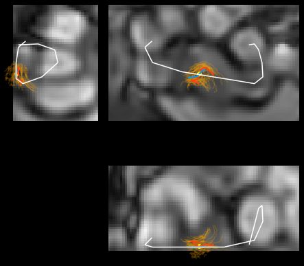
Deep Stochastic Tracking in Abdominal 3D cine-MRI

Acknowledgments
The authors would like to thank Jelmer Wolterink for his valuable input at the early stages
of this project.

References
Christian Bauer, Thomas Pock, Horst Bischof, and Reinhard Beichel. Airway tree re-
 construction based on tube detection. In Proc. of Second International Workshop on
 Pulmonary Image Analysis, pages 203–213, 2009.

Catharina S De Jonge, André JPM Smout, Aart J Nederveen, and Jaap Stoker. Evaluation
  of gastrointestinal motility with MRI: advances, challenges and opportunities. Neurogas-
  troenterology & Motility, 30(1):e13257, 2018.

Catharina S de Jonge, Alex Menys, Kyra L van Rijn, Arjan J Bredenoord, Aart J Nederveen,
  and Jaap Stoker. Detecting the effects of a standardized meal challenge on small bowel
  motility with MRI in prepared and unprepared bowel. Neurogastroenterology & Motility,
  31(2):e13506, 2019.

Alejandro F Frangi, Wiro J Niessen, Koen L Vincken, and Max A Viergever. Multiscale
  vessel enhancement filtering. In International Conference on Medical Image Computing
  and Computer-Assisted Intervention, pages 130–137. Springer, 1998.

Ola Friman, Milo Hindennach, Caroline Kühnel, and Heinz-Otto Peitgen. Multiple hy-
  pothesis template tracking of small 3d vessel structures. Medical Image Analysis, 14(2):
  160–171, 2010.

Flavius F Guglielmo, Donald G Mitchell, Patrick L O’Kane, Sandeep P Deshmukh, Christo-
  pher G Roth, Ilene Burach, Aaron Burns, Susan Dulka, and Laurence Parker. Identifying
  decreased peristalsis of abnormal small bowel segments in Crohn’s disease using cine MR
  enterography: the frozen bowel sign. Abdominal Imaging, 40(5):1150–1156, 2015.

Mehmet A Gülsün, Gareth Funka-Lea, Puneet Sharma, Saikiran Rapaka, and Yefeng Zheng.
 Coronary centerline extraction via optimal flow paths and CNN path pruning. In Inter-
 national Conference on Medical Image Computing and Computer-Assisted Intervention,
 pages 317–325. Springer, 2016.

Tobias Heye, Daniel Stein, Dalibor Antolovic, Margret Dueck, Hans-Ulrich Kauczor, and
  Waldemar Hosch. Evaluation of bowel peristalsis by dynamic cine MRI: detection of rele-
  vant functional disturbances—initial experience. Journal of Magnetic Resonance Imaging,
  35(4):859–867, 2012.

Zhikai Liu, Xia Liu, Bin Xiao, Shaobin Wang, Zheng Miao, Yuliang Sun, and Fuquan Zhang.
  Segmentation of organs-at-risk in cervical cancer CT images with a convolutional neural
  network. Physica Medica, 69:184–191, 2020.

Ilya Loshchilov and Frank Hutter. SGDR: Stochastic gradient descent with warm restarts.
   arXiv preprint arXiv:1608.03983, 2016.

                                            9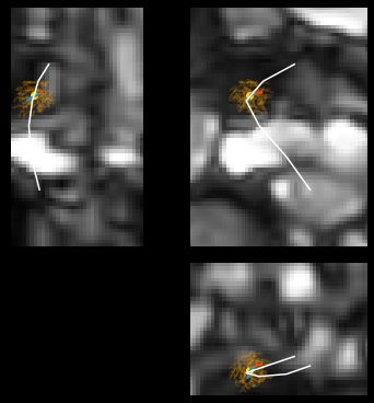
Deep Stochastic Tracking in Abdominal 3D cine-MRI

Alex Menys, Carl Puylaert, Charlotte E Tutein Nolthenius, Andrew A Plumb, Jesica
  Makanyanga, Jeroen A Tielbeek, Doug Pendse, Lodewijk A Brosens, Manuel Rodriguez-
  Justo, David Atkinson, et al. Quantified terminal ileal motility during MR enterography
  as a biomarker of Crohn disease activity: prospective multi-institution study. Radiology,
  289(2):428–435, 2018.

Freddy Odille, Alex Menys, Asia Ahmed, Shonit Punwani, Stuart A Taylor, and David
  Atkinson. Quantitative assessment of small bowel motility by nonrigid registration of
  dynamic MR images. Magnetic Resonance in Medicine, 68(3):783–793, 2012.

P Paine, J McLaughlin, and S Lal. the assessment and management of chronic severe
  gastrointestinal dysmotility in adults. Alimentary Pharmacology & Therapeutics, 38(10):
  1209–1229, 2013.

Michiel Schaap, Coert T Metz, Theo van Walsum, Alina G van der Giessen, Annick C
 Weustink, Nico R Mollet, Christian Bauer, Hrvoje Bogunović, Carlos Castro, Xiang
 Deng, et al. Standardized evaluation methodology and reference database for evaluating
 coronary artery centerline extraction algorithms. Medical Image Analysis, 13(5):701–714,
 2009.

Amos Sironi, Engin Türetken, Vincent Lepetit, and Pascal Fua. Multiscale centerline detec-
 tion. IEEE Transactions on Pattern Analysis and Machine Intelligence, 38(7):1327–1341,
 2015.

Christoph Spuhler, Matthias Harders, and Gábor Székely. Fast and robust extraction of
 centerlines in 3d tubular structures using a scattered-snakelet approach. In Medical Imag-
 ing 2006: Image Processing, volume 6144, page 614442. International Society for Optics
 and Photonics, 2006.

Kyra L van Rijn, Albert J Bredenoord, André JPM Smout, Gerd Bouma, Jeroen AW Tiel-
 beek, Karin Horsthuis, Jaap Stoker, and Catharina S de Jonge. Fasted and fed small
 bowel motility patterns at cine-MRI in chronic intestinal pseudo-obstruction. Neurogas-
 troenterology & Motility, page e14062, 2020.

Makoto Wakamiya, Akira Furukawa, Shuzo Kanasaki, and Kiyoshi Murata. Assessment of
 small bowel motility function with cine-MRI using balanced steady-state free precession
 sequence. Journal of Magnetic Resonance Imaging, 33(5):1235–1240, 2011.

Jelmer M Wolterink, Robbert W van Hamersvelt, Max A Viergever, Tim Leiner, and Ivana
  Išgum. Coronary artery centerline extraction in cardiac CT angiography using a CNN-
  based orientation classifier. Medical Image Analysis, 51:46–60, 2019.

Xing Wu, Mingyu Zhong, Yike Guo, and Hamido Fujita. The assessment of small bowel
  motility with attentive deformable neural network. Information Sciences, 508:22–32,
  2020.

                                            10
Deep Stochastic Tracking in Abdominal 3D cine-MRI
Appendix A. Additional visual examples
(a) (b) (c)
(d ) (e) (f )
(g) (h)
Figure 6: Additional qualitative examples, showing various situations. Projections are
plotted for the manual reference (white), non-stochastic tracking results (blue),
stochastic tracking results (red) and individual stochastic agents (orange). (a-
c) Both methods perform similarly. (d) Result from non-stochastic tracker crosses
the intestinal wall. (e-f) Cases with the largest decrease in recall: Stochastic
tracker terminates early as more than t agents hit the confidence-based and aber-
rant stopping criteria. (g-h) Seed points inside an air bubble and near the FOV
border, compromising network performance.
11You can also read