2021 Capstone Project Presenta ons Monday, April 21st - University of Colorado School of Medicine
←
→
Page content transcription
If your browser does not render page correctly, please read the page content below
2021
Capstone Project PresentaƟons
      Monday, April 21st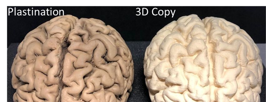
Page |1

                    2021 Modern Human Anatomy Program
                         Capstone Poster Presentations
                                   Agenda

                                   April 21, 2021

9:00 AM                     Welcome and overview, Dr. Ernesto Salcedo

9:05 AM – 9:55 AM           Capstone Presentations for Session I
                            Moderator:   Dr, Ernesto Salcedo

      https://ucdenver.zoom.us/j/92657385066?pwd=WXdCV0EzU3Zrd1lNRldHNGszUEdSQT09
                                      Meeting ID: 926 5738 5066
                                          Passcode: 842536

10:00 AM – 11:05 AM         Capstone Presentations for Session II
                            Moderator:   Dr. John Caldwell

      https://ucdenver.zoom.us/j/99521658290?pwd=Qlp5bmxUa3RqZzl5a1B0V0ZVUFpHUT09
                                     Meeting ID: 995 2165 8290
                                         Passcode: 107044

11:10 AM – 12:15 PM         Capstone Presentations for Session III
                            Moderator:   Dr. John Thompson

      https://ucdenver.zoom.us/j/91906750694?pwd=c3hqQXpwejhLTDgwZzlYam5FQk5kQT09
                                     Meeting ID: 919 0675 0694
                                         Passcode: 180362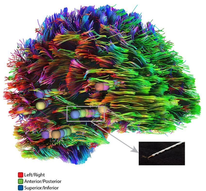
Page |2
Session I Presenters
9:05 AM – 9:55 AM
https://ucdenver.zoom.us/j/92657385066?pwd=WXdCV0EzU3Zrd1lNRldHNGszUEdSQT09
Meeting ID: 926 5738 5066
Passcode: 842536
Capstone Presenter Abstract Presentation Title
Presenter # Page #
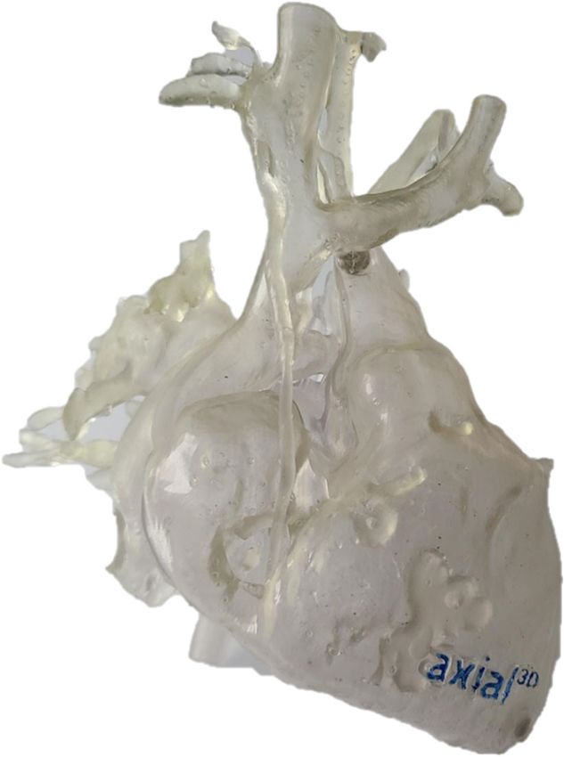
Evaluation of 3D Printed Models on Cardiac
Alexis MacDonald 1 6 Catheterization Staff Education of Single Ventricle
Palliation poster
That costs How Much: 3D printing a female pelvic
Marisa Flores 2 7
ultrasound phantom
Traumatic Brain Injury Exacerbates Alzheimer’s Disease
Conner Secora 3 8
in the Retina
We’re on the right tract: Diffusion tensor imaging
Caleb Fiebig 4 9 assessment of epileptogenic zone using depth electrode
locations to investigate white matter networks. poster
Targeting Effects of rZI vs. STN in Deep Brain
Kimberly Thies 5 10
Stimulation for Parkinson’s Disease. poster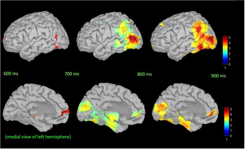
Page |3

                                 Session II Presenters
                                10:00 AM – 11:05 AM

        https://ucdenver.zoom.us/j/99521658290?pwd=Qlp5bmxUa3RqZzl5a1B0V0ZVUFpHUT09
                                       Meeting ID: 995 2165 8290
                                           Passcode: 107044

    Capstone       Presenter Abstract                    Presentation Title
    Presenter          #      Page #

                                        Animating and 3D Printing the Cranial Nerves
Jessica Bryant        1         11
                                        poster

                                        Evaluation of 3D Digital Models in Head and Neck
Lauren Wahl           2         12
                                        Anatomy poster

                                        What the head and neck? A study of integrated gross
Hailey Angus          3         13      anatomy with embryology and its effect on learning
                                        and retention. poster

                                        Change of Heart: Virtual Embryonic 3D Heart Model
Marissa Hight         4         14      with Interchangeable Conotruncal Septa Promotes
                                        Clinically Oriented Learning. poster

                                        Embryorigami: The Educational Value of 4D Virtual
Andrew Hull           5         15      Embryo Models on Student Learning and Confidence.
                                        poster

                                        Anatomy Educators’ Ethical Views on the Re-creation
Peter Therriat        6         16
                                        of Human Tissue via 3D Printing Technology. poster
Page |4

                                       Session III Presenters
                                       11:10 AM – 12:15 PM

         https://ucdenver.zoom.us/j/91906750694?pwd=c3hqQXpwejhLTDgwZzlYam5FQk5kQT09
                                        Meeting ID: 919 0675 0694
                                            Passcode: 180362

      Capstone         Presenter Abstract                         Presentation Title
      Presenter            #      Page #

                                               Can you stand it? A comparison of vertical ground
 Eliza Biondi              1          17
                                               reaction force in patients with osteoarthritis. poster

                                               Long-term Cardiometabolic Risk Among Acute
 Cassandra
                           2          18       Lymphoblastic Leukemia Childhood Cancer Survivors
 Gonzalez
                                               Treated by Bone Marrow Transplant

                                               Assessment of Cone Beam CT Accuracy for Creating
 Zachary Zylstra           3          19       Digital Scale Models: an Essential Step for Actualizing
                                               3D Printed Dental Implants poster

                                               Heart Grew Three Sizes: Improving the Health Literacy
 Deion Peña                4          20       of Patients Utilizing 3D Heart Model of Hypoplastic
                                               Left Heart Syndrome

                                               Integrating Endoscopy: Visual Scoring and Lung Fluid
 Canaan Kerr               5          21       Cellularity Associated with Eosinophilic Esophagitis.
                                               poster

 Rawan Jarrar              6          22       Neural correlates behind selective attention and visual
                                               conflict monitoring using a Stroop task

Posters can also be found here in this ONEDrive folder
Page |5

Thank you to faculty and industry experts serving on capstone committees, as these
  projects would not be possible without your commitment to the success of our
                                    students.

     MSMHA              Capstone Committee                  Capstone Mentor                  Committee Member
     Student                  Chair
Angus, Hailey         Caley Orr, PhD                  Lisa Lee, PhD                       Jennifer Stratford, PhD

Biondi, Eliza         Chelsea Lohman Bonfiglio, PhD   Jennifer Stevens‐Lapsley, PT, PhD   Jacob Capin, DPT, PhD

Bryant, Jessica       John Thompson, PhD              Ernesto Salcedo, PhD                Jake Shearer, MS

Fiebig, Caleb         Ernesto Salcedo, PhD            John Thompson, PhD                  Jennifer Stratford, PhD

Flores, Marisa        Caley Orr, PhD                  Juliana Wilson, DO                  Chelsea Lohman Bonfiglio, PhD

Gonzalez, Cassandra   John Caldwell, PhD              Jill Kaar, PhD                      Hesham Eissa, MD

Hight, Marissa        Ernesto Salcedo, PhD            Lisa Lee, PhD                       Nicholas Jacobson, MDesD

Hull, Andrew          Ernesto Salcedo, PhD            Lisa Lee, PhD                       Jennifer Stratford, PhD

Jarrar, Rawan         John Thompson, PhD              Isabelle Buard, PhD                 Peter Teale, MSEE

Kerr, Canaan          Thomas Finger, PhD              Emily DeBoer, MD                    Joel Friedlander, MD

MacDonald, Alexis     Chelsea Lohman Bonfiglio, PhD   Jenny Zablah, MD                    Michael Shorofsky, MD

Peña, Deion           Caley Orr, PhD                  Michael DiMaria, MD                 Jenny Zablah, MD

Secora, Conner        Chelsea Lohman Bonfiglio, PhD   Natalia Vergara, PhD                Ernesto Salcedo, PhD

Therriat, Peter       Chelsea Lohman Bonfiglio, PhD   Daniel Goldberg, JD, PhD            John Caldwell, PhD

Thies, Kimberly       Caley Orr, PhD                  John Thompson, PhD                  Drew Kern, MD

Wahl, Lauren          Danielle Royer, PhD             Caley Orr, PhD                      Jake Shearer, MS

Zylstra, Zachary      Chelsea Lohman Bonfiglio, PhD   Thomas Greany, DDS                  Kerri Font, DDS
Page |6

                               Session I:             9:05 AM – 9:55 AM

Presenter #1                  Alexis MacDonald

Evaluation of 3D Printed Models on Cardiac Catheterization Staff Education of Single
Ventricle Palliation

Capstone Committee: Chelsea Lohman Bonfiglio, PhD (chair), Jenny Zablah, MD (mentor), Michael
Shorofsky, MD

ABSTRACT:
Three-dimensional printed models (3DPM) have applications in medicine that have seen rapid growth and
diversification in the past decade. Education has been a large area of application, with patient and medical
student learning extensively evaluated. Less investigated has been the usefulness of 3DPM in clinical staff
education. The objective of this study is to assess the value and efficacy of using 3DPM of the heart for clinical
education of cardiac catheterization laboratory staff. The complex and changing anatomy of patients with a
single ventricle deformity presents the condition as a good case for the use of 3DPM. In particular, the 3 stages
of single ventricle palliation were selected to be used as a model in testing the efficacy of 3DPM printing for
staff member education. In this study, three-dimensional rotational angiography (3DRA) was used to create 3D
printed models of each stage of single ventricle palliation. The aim of this study is to identify how 3DPM are
perceived and understood by trained staff members as well as investigate how they may be useful in the future
for both staff and cross-discipline training. We hypothesized that cardiac catheterization laboratory and
pre/post-operative unit staff members will perceive 3D printed models to be more useful in understanding the
anatomy of all stages of single ventricle repair than 2D angiograms, a more traditional form of depicting
variations of congenital heart disease. Staff members associated with either the cardiac catheterization
laboratory or cardiology pre/postoperative unit were given a pre-educational survey, participated in both two-
dimensional and three-dimensional educational modalities, and asked to reassess their knowledge in a post-
education survey. The deidentified participant data was analyzed using a Kruskal Wallis test for non-parametric
data followed by a thematic analysis performed on survey comments received. We found the reported ratings of
knowledge at each stage of single ventricle repair after the 3DPM module to be significantly higher from the
pre-educational values at all stages (all p0.05) suggesting that 3DPM could be a strong educational modality for staff
members. Lastly, the thematic analysis of participants comments allowed us to identify strengths and
weaknesses of 3DPM in congenital cardiology for staff education which may have future significance and
implications on staff member training. This study implicates that 3DPM could play a larger future role in staff
education and may have other benefits in training applicable to interventional congenital cardiology.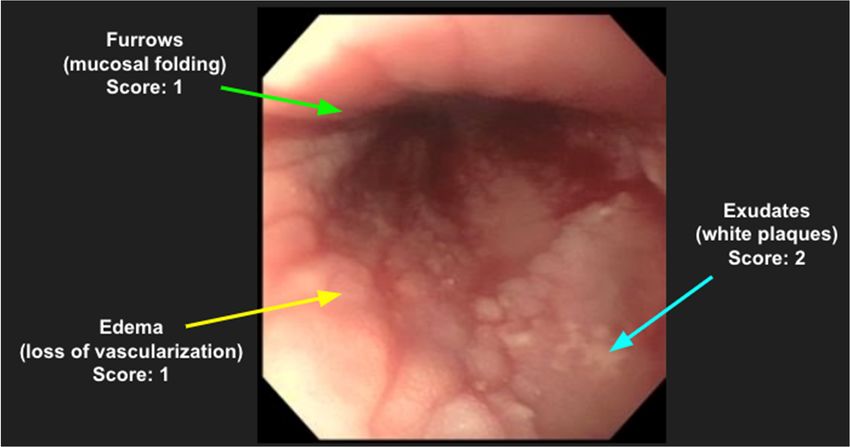
Page |7

Presenter #2                  Marisa Flores
That costs How Much: 3D printing a female pelvic ultrasound phantom

Capstone Committee: Cale Orr, PhD (chair), Juliana Wilson, DO (mentor), Chelsea Lohman Bonfiglio, PhD

ABSTRACT:
Objective: Point of care ultrasound (POCUS) exams have demonstrated to lead to more timely and accurate
diagnosis. Although most physicians begin ultrasound training in medical school, most emergency medicine
residents complete their first female pelvic and transvaginal ultrasounds on emergency room patients as
residents. Ultrasound phantoms have been proven to be a reliable educational resource. Unfortunately, female
pelvic ultrasound phantoms on the market can be cost-prohibitive in ultrasound training. This study aims to test
the hypothesis that a 3-dimensional (3D) printed self-assembled female pelvic phantom embedded in ballistic
gel will prove to be a cost-effective and adequate resource for student training of female pelvic ultrasound.

Methods: In this study, we digitally modeled, and 3D printed a uterus, ovaries, and female pelvic bones. First
models were created using a perishable material to test the reliability of the 3D printed models under ultrasound
scans. The final phantom is to be composed of ballistics gel for longevity.

Results: The different materials used during the printing process did not show a significant density difference
between structures under ultrasound. As currently constructed, the phantom is perishable, but imaging results
suggest that it is an adequate design warranting the development of our more permanent version using ballistics
gel.

Conclusions: The overall cost and quality of the new phantom model suggests it is a suitable resource for
student training while also being more cost-effective than other phantoms currently on the market.
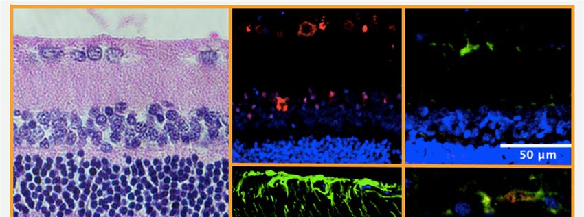
Page |8

Presenter #3                 Conner Secora
Traumatic Brain Injury Exacerbates Alzheimer’s Disease in the Retina

Capstone Committee: Chelsea Lohman Bonfiglio, PhD (chair), Natalia Vergara, PhD (mentor), Ernesto
Salcedo, PhD

ABSTRACT:
Alzheimer’s disease (AD) is a neurodegenerative condition that affects 5.7 million people in the U.S. alone,
representing the leading cause of dementia. Additionally, traumatic brain injury (TBI), which can arise from
sport concussions, military combat and other causes, is associated with a 2.3-fold higher risk of developing AD
and AD-related dementias. Moreover, AD can also lead to visual impairment, and recent studies have found that
AD histopathology manifests in the retina, which is an extension of the central nervous system. However, the
pathophysiological link between TBI and AD remains poorly understood. In this study we set out to evaluate
the effects of TBI, AD, and their combination, on retinal histopathology.
Several animal models have been developed to investigate the mechanisms of AD but have been limited by
imperfect recapitulation of human pathology. Here we take advantage of a transgenic rat model (Tg-F344) that
more faithfully mimics human AD pathology, and subject it to a TBI paradigm. Wild-type and transgenic rats
were randomly selected to undergo a two-time unilateral controlled cortical impact (2xCCI). At 16 months of
age, the rats were sacrificed and the eyes subsequently removed, fixed, and processed. Retinas were sectioned
and analyzed using immunofluorescent and standard histological staining techniques. Our results confirm the
presence of AD markers in transgenic retinas, and an increased severity of AD pathology due to TBI. This has
meaningful implications in understanding the pathophysiology of AD in relation to TBI within the retina, which
could lead to better treatments and to the development of retinal biomarkers for improved AD diagnosis.
Page |9

Presenter #4                  Caleb Fiebig
We’re on the right tract: Diffusion tensor imaging assessment of epileptogenic zone using
depth electrode locations to investigate white matter networks.

Capstone Committee: Ernesto Salcedo, PhD (chair), John Thompson, PhD (mentor), Jennifer Stratford, PhD

ABSTRACT:
Background: In patients with drug-resistant epilepsy, it can be difficult to pinpoint the precise regions of the
brain that create and propagate abnormal brainwave activity. Stereoencephalography (SEEG) is a minimally
invasive approach with reliable accuracy in the localization of the epileptogenic zone in patients with drug-
resistant epilepsy (Serletis et al., 2014). Diffusion tensor imaging (DTI) has been used to estimate and model
white matter tracts of the brain, allowing for 3D visualization of brain region connections and networks. To
date, the combination of patient-specific brain imaging and 3D extracted depth electrode contacts and white
matter tracts has not been used to build patient-specific seizure network models. The goal of this project is to
utilize the combination of these methods to define the epileptogenic zone in order to investigate and potentially
locate shared networks across patients with drug-resistant epilepsy that predict susceptible networks.

Methods: Human participants (N=10) in an IRB-approved protocol underwent SEEG along with pre- and post-
operative imaging. Implanted probes were segmented from the post-operative images and 3D white matter
tracts were generated from the pre-operative DTI. A novel structural analysis approach was applied to
extrapolated association fiber bundles representing the major known human white matter tracts. This structural
shape analysis was also applied to the fiber bundles derived from SEEG electrode contacts. An independent t-
test was run on 23 canonical fiber bundles with associated shape parameters in comparison to hippocampal tract
data to determine the probability that fiber bundles were shared between the structures.

Results: 8 of the 23 canonical fiber bundles were indicated in sharing a significant number of fibers with the
hippocampus-associated tracts.

Conclusion: This study indicates that the combination of DTI tractography along with SEEG data can bridge
the gap between the anatomical and physiological components of the brain, allowing us to better understand and
study neuropathologies such as drug-resistant epilepsy.
P a g e | 10

Presenter #5                      Kimberly Thies
Targeting Effects of rZI vs. STN in Deep Brain Stimulation for Parkinson’s Disease

Capstone Committee: Caley Orr, PhD (chair), John Thompson, PhD (mentor), Drew Kern, MD

ABSTRACT:
Parkinson’s Disease (PD) is one of the most common neurodegenerative disorders with a range of associated
motor and nonmotor symptoms. There is currently no cure for this disease, and treatment of PD mainly focuses
on symptom management. Deep brain stimulation (DBS) surgery is a highly efficacious therapy for several
movement disorders, including PD, as it modulates pathologic brain activity in deep brain structures. The main
targets for DBS in PD are the subthalamic nucleus (STN) and globus pallidus interna (GPi) because of their
regulating function in basal ganglia output. However, STN-targeted DBS has been associated with the onset of
adverse effects including dyskinesias (involuntary/continuous movements) and neuropsychiatric manifestations.
Another deep brain structure located dorsal to the STN is the zona incerta (ZI). Limited research has been
published on the use of ZI as a target for DBS in PD. The rostral zona incerta (rZI) is of particular interest in
this study due to its location of many converging white matter pathways important for motor related activity.
The aims of the current study include (1) to localize electrodes and build a trajectory model within each
patient’s bilateral STN using neuroradiology data and 3D imaging software, (2) to evaluate each patient in a
double-blind clinic visit while on three different optimized DBS stimulation settings: STN alone, rZI alone, and
STN + rZI co-stimulation using rating scales defined by the Movement Disorder Society (MDS-UPDRS and
UDysRS), and (3) to incorporate these optimized stimulation parameters into a software creating a volume of
tissue activated (VTA) model for each patient. Within STN, DBS stimulation parameters induce an electric field
with a unique geometric shape modeled as a VTA for each patient. By quantifying this VTA, clinicians can
estimate the percent of STN activated and elucidate how
this stimulation is spreading to other areas of the brain such
as rZI. Methods of this study tested the hypothesis that a
contact in the region of rZI further improves the benefit
patients obtain with DBS compared with STN stimulation
alone. Findings suggest no significant differences in
outcome data between the three stimulation settings, likely
because sensitivity of the rating scales used may not be
amenable to the acute transitions that took place in this
study. However, it was found that--of the three conditions--
patients largely preferred (50%) STN + rZI stimulation
which is highly clinically meaningful. These results show
therapeutic equivalence across the three stimulation
paradigms and demonstrate no adverse effects occurring
with rZI stimulation. These results, if replicated in larger
studies, have important implications for current DBS
therapy of PD and point to a promising target area for
future neurosurgical interventions.

Figure 1: Volume of tissue activated (VTA) models for two study patients. The teal structure represents STN & the red structure
represents VTA
P a g e | 11

                             Session II:            10:00 AM – 11:05 AM

Presenter #1                  Jessica Bryant
Animating and 3D Printing the Cranial Nerves

Capstone Committee: John Thompson, PhD (chair), Ernesto Salcedo, PhD (mentor), Jake Shearer, MS

ABSTRACT:
Cranial nerves are widely taught in health science education yet lack a complete learning resource. Students
require a complete visualization of the anatomy of the cranial nerves, which is not widely available in
education. Learners typically rely on dissection that destroys the passage of the nerves or textbook images
showing only a 2D rendering. This project sought to develop a new virtual and 3D printed educational resource
depicting the passage of the cranial nerves through the skull to bridge that educational gap. This project
segmented the cranial nerves, and 3D modeled them from origin to endpoint using the Visible Human Male
(VHM) dataset kindly provided by Touch of Life Technologies (Toltech). The VHM models provide better
realism than others available, as they are compiled from real cadaveric sections. The 3D models were animated
in Maya to create a short summary video highlighting each cranial nerve. The animations were uploaded to a
YouTube playlist and provided to first and second-year Modern Human Anatomy (MHA) students. The entire
model was also 3D printed using clear resins on a Form2 printer. The skull printed successfully after 41 hours,
while the brainstem and attached nerves failed after 19 hours. The physical models will thus be provided to
students later. Students filled out a survey rating the effectiveness of the animations as a learning resource and
comparing it to other methods available. Feedback from students proved difficult to garner during COVID-19.
The results were analyzed to determine if it is more effective for graduate level students learning anatomy and if
it represented a complete learning resource for the cranial nerves. Different methods of printing, including
collaborating with Inworks, are being explored as different approaches to the project. Until a 3D print can be
provided in conjunction with the cranial nerve animations, the hypothesis regarding them being a complete
learning resource could not fully be evaluated. However, early survey data provides promising results showing
that the project has the potential to be a complete learning resource in the future.
P a g e | 12
Presenter #2 Lauren Wahl
Evaluation of 3D Digital Models in Head and Neck Anatomy
Capstone Committee: Danielle Royer, PhD (chair), Caley Orr, PhD (mentor), Jake Shearer, MS
ABSTRACT:
Head and neck anatomy has been regarded as one of the most difficult subjects of human anatomy to learn and
mentally visualize. Appropriate models and three-dimensional (3D) representations of this complex anatomy
are either unaffordable and/ or do not detail the advanced anatomy of the head and neck regions that
professional students in a human anatomy graduate program are required to know (Park et al 2018). The lack of
proper detailed and affordable 3D resources for advanced head and neck anatomy leaves a gap in the kinds of
resources that professional students in anatomical sciences have voiced that they prefer to learn and study from
for an anatomical region that is more difficult to visualize and view in a 3D representation outside of traditional
lecture or laboratory materials (cadaveric dissection, lecture slide decks, etc.). The goal of this project was to
create a 3D representation of the parasympathetic cranial nerve fiber distribution within the head and neck as an
example of complex anatomy professional students are required to know and evaluate whether using this 3D
representation had any effect on student assessment performance and confidence. In order to generate a useful
3D resource of complex head and neck anatomy that detailed anatomy sufficient for the studying purposes of a
professional student in anatomical sciences, a 3D digital resource of the parasympathetic cranial fiber
distribution was crafted from a microcomputed tomography dataset, segmented in 3D Slicer, further modeled
and developed in Maya, and uploaded to an Sketchfab, an open-access online platform, for easy student access.
In an IRB-approved protocol, students were then randomly selected to use either the 3D digital model or
traditional lecture resources in an optional review session to complete a worksheet on cranial nerve
parasympathetics and were also given pre-review and post-review content assessments and confidence
questionnaires to gauge their mastery and comfort level with the material. With a total n = 23, no significant
differences were found in pre- versus post-review assessment scores (p = 0.590) nor in pre- versus post-review
confidence ratings (p = 0.478, 0.416, 0.274). These results were expected as previous literature on similar
studies have produced similar results; however, feedback from students indicated that the 3D digital resource
was “easy to visualize”, “helpful and easy to connect to 2D resources”, but they also commented that it could be
improved by “overlaying sympathetics as well” and “changing opacity gradients and nerve colors”. Future
directions of this study include incorporating a 3D printed model for more hands-on student learners and testing
the efficacy of these 3D resources on other professional students at the same institution. Ultimately, this study
shows that affordable 3D models that represent complex anatomy that is required for mastery by professional
anatomy students can be helpful ancillary educational tools.
Figure 1. Glossopharyngeal parasympathetic fiber distribution to the parotid gland.P a g e | 13 Presenter #3 Hailey Angus What the head and neck? A study of integrated gross anatomy with embryology and its effect on learning and retention. Capstone Committee: Caley Orr, PhD (chair), Lisa Lee, PhD (mentor), Jennifer Stratford, PhD ABSTRACT: Horizontal integration in curricula is the combining of basic science courses together to give students a better understanding of how the different sciences work together. Integration is thought to improve student learning and retention. Many health professional schools are switching to integration in the first two years to combat reduced contact hours in the basic sciences. Along with integration, the introduction of online resources for students is growing, especially within this last year thanks to the Covid-19 pandemic. There is evidence to suggest that integrated curriculums benefit student learning, but it is not well studied if integration of embryology alone in gross anatomy content improves student understanding of gross anatomy. It is also not known how this applies in the use of online resources. In addition, few studies have focused on these effects in dental student populations. This study was designed to determine if integration of embryology in an online gross anatomy tutorial led to better learning, retention, and confidence in cranial nerve anatomy for dental students than a stand-alone gross anatomy tutorial. To that end two digital tutorials on cranial nerve anatomy were created with the only difference between the two being integrated embryology content in the experimental video. First- and second- year dental students at CU Anschutz were recruited to participate in this single blind randomized study. Participants took a pretest, watched the assigned tutorial, and took a posttest. After data analysis, no differences in scores, score improvement, perceived learning, or confidence was found between control and experimental groups. This demonstrates that embryology integrated content may not improve gross anatomy understanding. This offers a new insight into how integration affects student learning. Although there were not differences between the control and experimental groups, there were significant improvements in student performance, confidence, and perceived learning in all groups and cohorts. Adding to research in favor of using digital resources. Student feedback in open-ended questions reiterates benefits of digital resources, integration, and some of the difficulties that coinside with their use. Finally, this study offers a model of study design that is repeatable, brief, and widely distributable for educational research.
P a g e | 14

Presenter #4                  Marissa Hight
Change of Heart: Virtual Embryonic 3D Heart Model with Interchangeable Conotruncal
Septa Promotes Clinically Oriented Learning

Capstone Committee: Ernesto Salcedo, PhD (chair), Lisa Lee, PhD (mentor), Nicholas Jacobson, MDesD

ABSTRACT:
One of the most challenging and clinically relevant concepts in embryology is the heart outflow tract septation
involving 180-degree spatial rotation and precise alignment of the growing septa that can often go awry,
resulting in various congenital cardiac septal defects. Teaching and learning embryology pose challenges due to
the rapid and complex developmental changes that occur across time and space. . Previous investigations have
shown that virtual and 3D printed embryo models increase learning outcomes and interest in embryology.
Unfortunately, there are no visual aids that effectively demonstrate heart outflow tract septation in 3D. Towards
this end, we created a 3D embryonic virtual heart development model (EVH) with interchangeable conotruncal
septa. The educational efficacy of this virtual tool was tested in a COMIRB exempt (# 20-2124), randomized
single-blind study with the first-year medical students completing a flipped-classroom embryology active
learning event occurring in a virtual platform due to COVID-19 restriction. As a part of the course requirement,
all students first watched a pre-recorded lecture video and completed a test on their own before attending the
synchronous virtual active learning event. During the active learning event, students were randomized into
control or experimental breakout session groups. Experimental breakout session groups (16 groups, n=80) were
provided access to the EVH with accompanying prompts designed to reinforce 3D spatial orientation and
congenital septal defects demonstrated by the EVH. The control groups (14 groups, n=70) were tasked to
complete a similar learning activity but on a different developmental process. All students completed a second
test and a survey after the active learning event. Data analysis revealed that pre-test and post-test scores were
not significantly between groups (Two-Way ANOVA F(1,151) = 0.16, p=0.69); however, survey data revealed
that students positively perceived the educational value of the EVH, especially for demonstrating embryonic
heart development (4.6 + 0.84 on a 5-point agreement scale). Students also felt the EVH helped in
understanding spatial and developmental changes (4.8 + 0.80). Consistently, thematic analysis of the survey
comments indicated that the spatial and visual aspects of the EVH demonstrating the congenital defects were
perceived as essential. The significantly higher learning outcomes immediately after the resource interaction
with EVH and the perceived favorability of the EVH warrant further development and research. A future aim of
this project is to enhance the EVH to represent the sequelae of abnormal conotruncal septum formation. Group
and virtual classroom effects are other confounds that should be reduced in future studies.
P a g e | 15

Presenter #5                  Andrew Hull
Embryorigami: The Educational Value of 4D Virtual Embryo Models on Student Learning
and Confidence

Capstone Committee: Ernesto Salcedo, PhD (chair), Lisa Lee, PhD (mentor), Jennifer Stratford, PhD

ABSTRACT:
In human development, normal formation of the body cavity involves intricate interplay of tissues folding from
a flat disc form into a complex tube-within-tube 3D structure over a short period of time. Understanding the
normal body cavity formation is imperative to understanding anatomic organizations, variations, and congenital
conditions. Unfortunately, resources that effectively demonstrate changing body cavity anatomy during the
folding process are sparse and their educational effectiveness is not well understood. Therefore, an interactive
digital resource, the “Virtual Folding Embryo” (VFE), demonstrating the 4-dimensional transformation of the
folding embryo (Figure 1) was created and its educational value was assessed in a COMIRB exempt (#20-2125)
randomized single blind study. As a part of the flipped curriculum, 155 first-year medical students enrolled in
an integrated anatomy course first viewed a pre-recorded lecture on embryology of the thorax then completed a
quiz to test their foundational knowledge before attending a synchronous online active learning session
conducted on the Zoom platform. Students were randomized into small breakout groups of 4-5 students and half
of the groups were given access to the VFE (Figure 2), while the other half were given access to a resource
demonstrating a different embryonic process. After 15 minutes, all students completed a quiz and an optional
survey.

A two way repeated measures ANOVA showed there were no statistical differences between the control and
VFE groups for pre or post quiz containing all questions, nor were there differences in post quiz scores between
control and VFE groups, F(1,151) = 0.28, p=0.59. Additionally, no difference in pre quiz score between control
and VFE groups when measuring body cavity quiz items only F(1,151)= 1.06 p=0.3 (Figure 4).

Descriptive statistics on the qualitative survey analysis indicate that students positively perceived the VFE for
visually elucidating the transformation of the body cavity (n=74 mean Likert score = 4 on 5 point agreement
scale; sd=0.81) and its spatial relationship to organ systems (mean=3.83; sd=0.93). Despite the favorable
perception of VFE and improved quiz performance, students rated their level of confidence in the material low
(mean=2.18, sd=0.37). Thematic analysis of survey comments reinforced positive perception of the VFE and
indicated desire for more time for VFE interaction.

These results do not conclusively demonstrate direct correlation between the VFE and learning outcomes.
However, qualitative results warrant further development and investigation of the VFE under different
educational circumstances to determine efficacy. Virtual 4D resource such as VFE, designed for active learning
may hold potential to enrich student engagement in the post COVID-19 era, and serve as a model for creating
similar resources in the future.
P a g e | 16

Presenter #6                  Peter Therriat
Anatomy Educators’ Ethical Views on the Re-creation of Human Tissue via 3D Printing
Technology

Capstone Committee: Chelsea Lohman Bonfiglio, PhD (chair), Daniel Goldberg, JD, PhD (mentor), John
Caldwell, PhD

ABSTRACT:
Cadavers are the gold standard in teaching anatomical education. Contact hours in labs have decreased, which
has created a need for different methods of teaching anatomical education. 3D printing has emerged in tandem
with plastination to offer a tactile approach to learning anatomy, but with their emergence, ethical issues have
arisen. 3D printing has not had the same ethical scrutiny that plastination has seen up until the present. Current
ethical guidelines may not properly cover 3D printed re-creations; therefore, the aims of this study are to
determine what ethical guidelines anatomy educators find most critical in the re-creation of human tissue via 3D
printing for the purpose of training future health professionals. Overall findings suggest that anatomy educators
find that the informed consent process could be updated on body donation forms, respect should be on a similar
level as plastinations and other cadaver data sets, and accessibility/ownership of data from body donors may be
decided on a case-by-case basis.
P a g e | 17

                         Session III:                  11:10 AM – 12:15 PM

Presenter #1                 Eliza Biondi

Can you stand it? A comparison of vertical ground reaction force in patients with
osteoarthritis.

Capstone Committee: Chelsea Lohman Bonfiglio, PhD (chair), Jennifer Stevens-Lapsley, PT, PhD (mentor),
Jason Capin, DPT, PhD

ABSTRACT:
Weight bearing asymmetry (WBA) between limbs is present in patients with advanced osteoarthritis (OA) and
can be quantified using 90-second quiet standing trials. Causes of WBA include habitual movement patterns,
pain, and reduced muscle strength. Studies that have previously examined quiet standing trials have approached
quantifying asymmetries by averaging the entire time sequence, or a specific section in the trial. While these
approaches work, the objective of this project was to examine variation in weight distribution throughout every
section of a trial. 77 patients with OA were included in the study (mean age=65). Vicon Nexus was used to
collect force plate data, Visual3D was used to process vertical ground reaction force (vGRF) data and export to
MATLAB. These trials were then split into three even sections (first 30 seconds, middle 30 seconds, last 30
seconds), normalized by patients’ body weight, and used to calculate an asymmetry ratio. The ANOVA showed
no differences between groups (p=0.99). These results demonstrate that there is no variation in WBA from the
beginning to end of a 90-second quiet standing trial. However, participants do exhibit WBA immediately and
continuously during the trial suggesting this is a habitual stance not related to pain or muscle fatigue.
P a g e | 18 Presenter #2 Cassandra Gonzalez Long-term Cardiometabolic Risk Among Acute Lymphoblastic Leukemia Childhood Cancer Survivors Treated by Bone Marrow Transplant Capstone Committee: John Caldwell, PhD (chair), Jill Kaar, PhD (mentor), Hesham Eissa, MD ABSTRACT: Acute lymphoblastic leukemia (ALL), is the most prevalent of all childhood cancers with the majority of patients experiencing long term survival rates. Childhood cancer survivors (CCS) are at greater risk for developing cardiometabolic disease later in life compared to non-CCS. More specifically, disorders of the endocrine system such as obesity, type 2 diabetes, and metabolic syndrome. However, little is known regarding the health of ALL survivors who were treated by bone marrow transplant (BMT). The goal of this study will fill this research gap by examining cardiometabolic health outcomes in a sample of ALL survivors at Children’s Hospital Colorado (CHCO). The outcomes will identify early cardiometabolic risk factors and document the need for improved treatment and clinical care for ALL survivors. We hypothesized that children who undergo more invasive treatments, such as BMT, will have a higher incidence of cardiometabolic risk factors compared to the general population. A COMIRB approved retrospective chart review of 68 patients diagnosed with ALL and treated with BMT between January 2008 to October 2020 was conducted via electronic medical records in the EPIC database. Demographics, comorbidities, treatment protocols, and clinical outcomes including height, weight, blood pressure, fasting glucose, HbA1c, HDL, triglycerides, and liver enzymes were collected at baseline, pre-BMT, and subsequent visits up to 24 months (3-, 6-, 9-, 12-, and 24-months) post-transplant. Preliminary data show survivors were predominantly non-Hispanic (59%), white (60%), and male (68%) between the ages of 6-22 at the time of transplant. The mean age at the time of transplant was 11 years old. At baseline, 37% of the population was overweight/obese and over 24 months post-transplant, this value was maintained. This study highlighted the need for ALL survivors at CHCO to receive a full metabolic workup beginning 2 years post-transplant. Survivors diagnosed with cardiometabolic risk factors will be placed in an intervention for treatment. An animated educational resource will accompany the intervention to enhance patient and parent understanding of cardiometabolic diseases.
P a g e | 19

                              Presenter #3                  Zachary Zylstra

Assessment of Cone Beam CT Accuracy for Creating Digital Scale Models: an Essential Step
for Actualizing 3D Printed Dental Implants

Capstone Committee: Chelsea Lohman Bonfiglio, PhD (chair), Thomas Greany, DDS (mentor), Kerri Font,
DDS

ABSTRACT:
Although there are many forms of preventative care for a patient, dental implants can play an incredible role in
restoring someone’s mouth and protecting both their self-image and livelihood from orofacial trauma, disease,
or even following orthodontic treatment. However, dental implant therapy still lacks solutions for shortening the
timeline of the therapy as a whole: extraction, bone grafting, implant placement, and healing abutment
placement. In addition, controlling the cost of these various procedures, maintaining peri-implant tissue
architecture, gingival embrasure cleansability, and delivering the aesthetic of a properly contoured coronal
emergence profile remains top priority for dental professionals. Recently, Cone Beam Computed Tomography
(CBCT) images have been used to diagnose bone volume and density in establishing a treatment plan that may
involve dental implants. This study has shown that those same CBCT images can be used beyond diagnostics in
dentistry to build a three-dimensional (3D) model of a patient’s existing dentition and root structure. Using open
GL software (CloudCompare), both CBCT and Medical Computed Tomography (MEDCT) segmentation
models (3D Slicer) were overlayed onto digital impressions (TRIOS) of identical human cadaveric tooth
samples in order to develop a standard of measurement through Boolean subtraction techniques. This
comparative study gives a perspective on how well CBCT and MEDCT diagnostic techniques align in regard to
their precision and accuracy. In addition, this model analysis is the first step in creating a 3D printed dental
implant model that duplicates the patient’s existing root structure morphology well enough to replicate ideal
esthetics and function while shortening the restorative timeline and eliminating potentially unnecessary
procedures.
P a g e | 20 Presenter #4 Deion Peña Heart Grew Three Sizes: Improving the Health Literacy of Patients Utilizing 3D Heart Model of Hypoplastic Left Heart Syndrome Capstone Committee: Caley Orr, PhD (chair), Michael DiMaria, MD (mentor), Jenny Zablah, MD ABSTRACT: Hypoplastic left heart syndrome (HLHS) is a congenital heart disease that is the underdevelopment of the left side of the heart and can lead to worsening complications if not treated appropriately. Symptoms that could occur from HLHS include cyanosis (bluish skin coloring), weak pulse, and shortness of breath. The treatment of HLHS is done through three surgical procedures that are the Norwood procedure, the Glenn procedure, and the final surgery that is the Fontan procedure. Due to the relatively high mortality rates and the complexity of Hypoplastic Left Heart Syndrome (HLHS), quality educational resources are needed to assist patients and their families to manage their treatment effectively. The current standard for patient education was through either diagrams or 3D models that were not effectively showcasing the anatomy affected. We hypothesized that by creating a 3D heart model that represents the anatomy of HLHS, this could be used as an educational resource to improve the health literacy of patients and families who are being treated at the Single Ventricle Heart Clinic at Children’s’ Hospital Colorado. The effect of the 3D heart model was then evaluated with a survey that consisted of basic anatomical concepts of a normal heart and a heart that had HLHS. It was then evaluated if the 3D model was helpful for understanding HLHS and if there was an overall improvement in the understanding HLHS. There was an improvement of the averages of the survey scores after the educational intervention, but there was no statistical significance within the study. This was able to show that a 3D model has the capability to improve the understanding of a congenital heart disease while guiding patients through an active educational discussion.
P a g e | 21

Presenter #5                 Canaan Kerr
Integrating Endoscopy: Visual Scoring and Lung Fluid Cellularity Associated with
Eosinophilic Esophagitis

Capstone Committee: Thomas Finger, PhD (chair), Emily DeBoer, MD (mentor), Joel Friedlander, MD

ABSTRACT:
Eosinophilic esophagitis (EoE) is a disease characterized by an invasion of eosinophils within the lining mucosa
of the esophagus and is diagnosed via histological analysis of esophageal tissue. While there is a basic
understanding of how EoE presents clinically, how EoE co-occurs and presents in children with multi-system
aerodigestive disorders is less clear. In multidisciplinary aerodigestive clinics, diagnostic procedures such as
flexible bronchoscopy may be able to enhance the diagnostic capability of upper intestinal endoscopy with
biopsy. We hypothesized that eosinophilic esophagitis would cause clinically relevant gross presentations in the
upper digestive and lower respiratory gross anatomy as well as increase lung fluid cellularity. To explore this
question, this study analyzed bronchoscopies and esophagogastroduodenoscopies (EGDs) from ten pediatric
aerodigestive patients with eosinophilic esophagitis (+EoE) and ten patients without eosinophilic esophagitis (-
EoE) to evaluate presentations of EoE in the lower airway. Two validated visual scoring systems to assess the
bronchoscopies and EGDs were used. The bronchoscopy scoring method analyzed secretion amount and
mucosal appearance throughout nine regions of the bronchial tree. Bronchoalveolar lavage (BAL) fluid was also
collected during bronchoscopies and analyzed for cellularity. The EoE Endoscopic Reference Score (EREFS)
assessed the esophagus based on mucosal appearance. Based on bronchoscopy scores, we found that secretion
amount and ridging of the mucosa varies between +EoE and -EoE patients. Based on EGD scores, we found
that severity of exudates and furrows also varies between the two groups. Based on BAL cell counts, there was
no significant difference in cellularity between +EoE and -EoE patients. These promising preliminary findings
permit us to advocate for expansion of this analysis to increase sample size and integrate other variables
including sex and the presence of comorbidities.
P a g e | 22

Presenter #6                         Rawan Jarrar
Neural correlates behind selective attention and visual conflict monitoring using a Stroop
task

Capstone Committee: John Thompson, PhD (chair), Isabelle Buard, PhD (mentor), Peter Teale, MSEE

ABSTRACT:
Selective attention and conflict monitoring are phenomena that humans use every day, yet the neurological
underpinnings behind these processes are not yet fully understood. This study was an attempt to identify the
spatial and temporal activity of the neural networks behind selective attention and conflict monitoring using a
Stroop task. Evoked magnetic fields (EMFs) from a group of 28 healthy adults between the ages of 19 and 36
were measured while performing a computerized cued switch Stroop task using magnetoencephalography
(MEG). We hypothesized that there would be stronger activation in the left dorsolateral prefrontal cortex
(DLPFC) in the incongruent-neutral color contrast and stronger activation in the left anterior cingulate cortex
(ACC) in the incongruent-congruent color contrast. Results revealed sustained, simultaneous activity in the left
DLPFC and the left temporoparietal junction (TPJ) between 700-800ms post stimulus onset in the incongruent
versus congruent contrast for the color condition, with no activation of the left ACC. The incongruent-neutral
contrast revealed activation in the left ACC during the 600-700ms averaged time window, followed by
activation in posterior brain regions, which supports a top-down biasing model of selective attention and
conflict resolution. MEG has demonstrated to provide valuable insight into the spatial and temporal dynamics of
these processes and should be implemented into standard practice for studying neural phenomena.

    Cortical map of left hemisphere showing patterns of activation based on local maximum t-values with p
You can also read