A biomechanical study on post-scoliotic deformity correction - IOPscience
←
→
Page content transcription
If your browser does not render page correctly, please read the page content below
IOP Conference Series: Materials Science and Engineering

PAPER • OPEN ACCESS

A biomechanical study on post-scoliotic deformity correction
To cite this article: S Balamurugan et al 2020 IOP Conf. Ser.: Mater. Sci. Eng. 912 022020

View the article online for updates and enhancements.

                               This content was downloaded from IP address 46.4.80.155 on 13/07/2021 at 08:25
3rd International Conference on Advances in Mechanical Engineering (ICAME 2020)            IOP Publishing
IOP Conf. Series: Materials Science and Engineering 912 (2020) 022020 doi:10.1088/1757-899X/912/2/022020

      A biomechanical study on post-scoliotic deformity correction

                     S Balamurugan1, Kunal Pandey2, Shraddha R Iyer2, Appaji Krishnan1 and
                     Shantanu Patil1
                     1
                       Department of Mechanical Engineering, SRM Institute of Science and Technology,
                     Chennai, India.
                     1
                       Department of Translational Medicine and Research, SRM Institute of Science and
                     Technology, Chennai, India.
                     1
                       Consultant Spine Surgeon, SIMS Hospital Vada Palani, Chennai, India
                     2
                       Students, Department of Mechanical Engineering, SRM Institute of Science and
                     Technology, Chennai, India.

                     E-mail: balamurs3@srmist.edu.in

                     Abstract. Scoliosis is the deformity of the growing human spinal column such that the
                     vertebral alignment is distorted in a corkscrew fashion. A person with severely deformed spine
                     may find it difficult to breathe as the ribcage may press against the internal organs
                     compromising the functions of the lungs and the heart. Due to the altered load transfer, these
                     patients also suffer from back pain and early arthritis. The scoliotic deformity is surgically
                     corrected by using implants which are screwed into the vertebra. In severe cases, complete
                     correction may not be achievable. As a result, the loads experienced by the implants may not
                     be optimal, leading to their early failure. The objective of this work is to study the effect on the
                     deformity correction in a scoliotic spine. A three-dimensional model of the surgically corrected
                     spine was segmented from the computed tomography scan and converted into a surface model.
                     This model was imported in to ANSYS for meshing and subjected to compression load to
                     simulate weight bearing. The stress concentration and displacement across the entire spine,
                     individual vertebrae and discs was analysed. The effect on the implants was separately
                     analysed as well.

     1. Introduction
     Scoliosis is a deformity of the growing human spine [1]. It deforms the torso and also affects the
     shape of the body [2]. Sometimes the spine anomaly is present at the birth and sometimes it can be
     observed only when the child reaches adolescence. There are mainly three types of scoliosis
     suggested by American Association of Neurological Surgeons (AANS) namely neuromuscular,
     idiopathic and congenital [3]. In idiopathic scoliosis [4] the cause of deformation is unknown
     whereas in congenital scoliosis the cause of the spine deformation present in the foetus during
     spine development present during birth. There are different reasons for congenital anomaly [3].

     1.1 Incomplete formation of vertebrae
     Due to incomplete formation of a vertebra during gestation, a sharp wedge is formed at the spine.
     Such kind of vertebra is known as a hemivertebra. The angle at the spine can get even worse while
     reaching the adolescence. There are cases in which more than one hemivertebra has been formed
     in the spine which tries to neutralise each other to stabilize the spine hence giving it a sharper
     angle increasing the severity of the deformation.

              Content from this work may be used under the terms of the Creative Commons Attribution 3.0 licence. Any further distribution
              of this work must maintain attribution to the author(s) and the title of the work, journal citation and DOI.
Published under licence by IOP Publishing Ltd                          1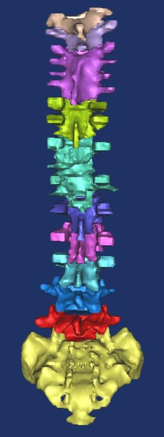
3rd International Conference on Advances in Mechanical Engineering (ICAME 2020)            IOP Publishing
IOP Conf. Series: Materials Science and Engineering 912 (2020) 022020 doi:10.1088/1757-899X/912/2/022020

    1.2 Incomplete separation of vertebrae
    During the intra-uterine gestation of the foetus, the spine is developed as a single column of tissue
    which in later stages get segmented into bony vertebrae but sometimes the separation is
    incomplete which leads to formation of partial fusion and joins two or more vertebrae [5]. Due to
    fusion the growth of spine is averted on one side and a curvature is developed.

    1.3 Formation of a hemivertebrae and partial fusion
   In such cases the deformation arises due to both aforementioned deformities. Such cases need early
   diagnosis and corrective measure to arrest the increased curvature of the spine [5].
   The model used in the analysis was of a patient with congenital scoliosis due to development of a
   hemivertebrae. The patient underwent surgical correction of the deformity, excision of the
   hemivertebrae and spinal fusion with intervertebral cage [6, 7]. The correction was maintained
   with the help of twelve pedicular screws and two titanium rods stabilised with cross bars at one
   level.

    2. Materials and Methods

    2.1. Model Generation
    A linear three-dimensional thoracolumbar model of the spine which consists of vertebrae from T1
    to T12, L1-L5, implant rod and screws [5] was developed using a Computed Tomography (CT)
    scan which is a combination of the series of X-rays images taken from various angles of the
    human body. It generates the cross-sectional images of the bones, blood vessels and tissues using
    computer processing. CT scan of the patient post-surgery was procured from the SIMS Hospital,
    Chennai. The scan was imported to MIMICS 14.0 software. MIMICS is an image processing
    software used for 3D designing and modelling. It uses the image data obtained from CT scan to
    calculate the three-dimensional model through segmentation as shown in figure 1 & 2.

   Figure 1. Scan                    Figure 2. Segmented Model segmented in different views.
  model.

                                                    2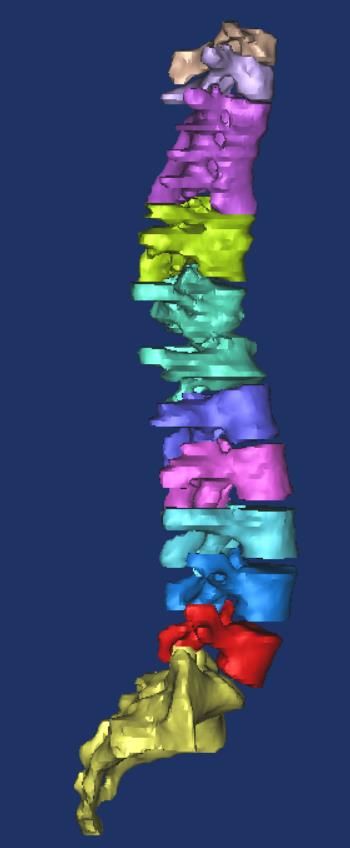
3rd International Conference on Advances in Mechanical Engineering (ICAME 2020)            IOP Publishing
IOP Conf. Series: Materials Science and Engineering 912 (2020) 022020 doi:10.1088/1757-899X/912/2/022020

       The imported post correction model is segmented using this software to create a 3D surface
    model. 0.5 mm gap was created between each slice. The model is converted into a .stl file and
    imported to Geomagics Design. The imported model is then subdivided, smoothened, re-wrapped
    and the surface model was generated as shown in figure. 3. The model is exported to ANSYS 18.0
    for meshing and to assign the material properties, was shown in table 1 [6-8]. Beam element was
    created in the surgical region to represent the implant. The normal body load to simulate standing
    condition was applied on the spine in different locations (figure 4).

                              Figure 3. Surface generation in Geomagics.

        Table 1. Material properties of the FEM of thoracolumbar spine.

                               Young’s           Poisson’s          Cross-             Element
         Components            Modulus           Ratio              Sectional
                               (MPa)                                Area (mm2)
         Cortical bone         1200              0.26               -
         Cancellous            100               0.2                -                  Solid 185
         bone Posterior        3500              0.25               -
         elements
         Disc
         Nucleus               1                 0.499              -
         Ground                4.2               0.45               -                  Link 10
         Substance             0.3               0.3                0.76
         Fibre
         Ligament
         ALL                   20                -                  63.7
         PLL                   20                -                  20                 Link 10
         TL                    58.7              -                  3.6
         LF                    19.5              -                  40
         ISL                   11.6              -                  40
         SSL                   15                -                  30
         CL                    32.9              -                  60

                                                     3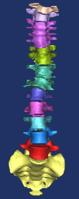
3rd International Conference on Advances in Mechanical Engineering (ICAME 2020) IOP Publishing
IOP Conf. Series: Materials Science and Engineering 912 (2020) 022020 doi:10.1088/1757-899X/912/2/022020
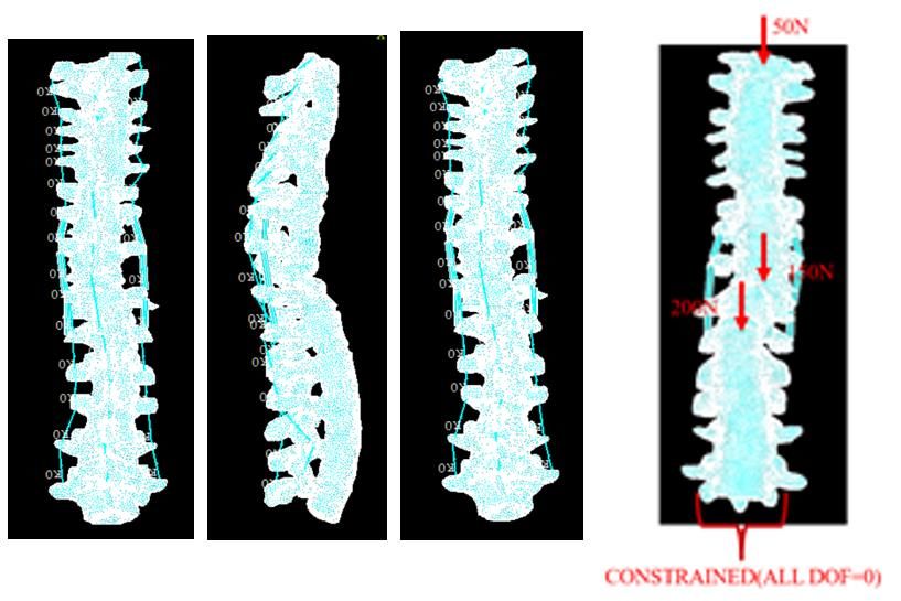
Figure 4. Meshed model of corrected spine with boundary condition.
a. Boundary Conditions
The L5 vertebra was constrained from all the degrees of freedom. Assuming the weight of the
patient to be 800N (80kg), a compressive force of 50N along the Z- axis was applied on T1
vertebra. This load replicates weight of the human head. A load of 150N was applied on the T9
vertebra replicating the weight of the upper proximity of the human body. Another load of 200N
was applied on T12 vertebra on the thoraco-lumbar junction (Figure. 4)
3. Results and Discussion
Finite element meshed model of post-surgery scoliosis affected spine was developed and analysed
for Maximum stress and vertical movement in vertebrae and disc. Static load was applied to the
model to understand the severity of the surgical region in terms of stress concentration and
deformation were investigated. After the surgical region, lumbar segments showed maximum
stress concentration compare to thoracic region. The maximum stress and deformations were
shown in figure 5 and 6.
Stress distribution in the vertebrae of post-operative condition for normal standing loading,
followed the stress pattern similar to intact spine. This shows that the non-uniform deformity in
the scoliosis affected spine were corrected in the spine curvature and mimicked the intact spine.
While, comparing the displacement in the vertebrae, adjacent vertebrae of the surgical region T7
and T9 showed the maximum displacement compare to other vertebrae. Similarly, comparing the
disc stress distribution and displacement the lumbar segment showed the maximum value. Further,
the stress and displacement on L3 to L5 vertebrae and disc were high because this segment is
below the surgical region. The maximum stress and displacement are shown in figure 7 and 8.
4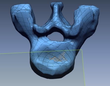
3rd International Conference on Advances in Mechanical Engineering (ICAME 2020)            IOP Publishing
IOP Conf. Series: Materials Science and Engineering 912 (2020) 022020 doi:10.1088/1757-899X/912/2/022020

     Figure 5. Stress distribution in vertebrae.        Figure 6. Axial displacement in vertebrae.

      Figure 7. Stress distribution in discs.              Figure 8. Axial displacement in discs.

        However, the stress distribution was more in the lumbar segment. L1 to L5 in vertebrae and L3
    to L5 in disc showed high values, this showed that below the surgical region experience maximum
    stress. But, last three vertebrae and disc were showed the stress raiser. This proves that the stress
    concentration is maximum at the end of the lumber region after post-surgery condition.

   4. Conclusion
    Three-dimensional model of post-surgery condition was modelled and analysed. After simulation,
    pre-surgery FE model helped to understand the stress and displacement patterns. The study
    revealed that high stress concentration on the vertebra and the disc comprised in the lumbar
    region. The extracted model was subject specific and studying the corrected spine curvature after
    surgery was critical and more complex. This study may pave pathways for future studies to be
    conducted in this field.

    Acknowledgements
    This work was supported by Translational Medicine and Research (TMR) of SRM Medical
    College Hospital & Research Centre and Biomechanics lab of SRM institute of science and
    technology.

     5. References
     [1] Nusret Kose R M C J 2004 Congenital scoliosis Med. Sci. Monit. 10 104-10
     [2] Chen X, Cai H, Zhang G, Zheng F, Wu C and Lin 2020 The construction of the scoliosis 3D
               finite element model and the biomechanical analysis of PVCR orthopaedy Saudi. J.
               Biol. Sci. 27 695-700

                                                    5
3rd International Conference on Advances in Mechanical Engineering (ICAME 2020) IOP Publishing
IOP Conf. Series: Materials Science and Engineering 912 (2020) 022020 doi:10.1088/1757-899X/912/2/022020
[3] El Hawary R and Chukwunyerenwa C 2014 Update on evaluation and treatment of scoliosis
Pediatric clinics of North America 61223-41
[4] Weiss H-R 2007 Idiopathic scoliosis: How much of a genetic disorder? Report of five pairs of
monozygotic twins Developmental Neurorehabilitation 10 67-73
[5] Martin Weyreuther CEH, Michael Westphal, Jan Zierski and Ulrich 2007 Weber MRI Atlas:
Orthopedics and Neurosurgery Springer
[6] Chiang MF, Zhong Z C, Chen C S, Cheng C K, and Shih S L 2006 Biomechanical
Comparison of Instrumented Posterior Lumbar Interbody Fusion With One or Two
Cages by Finite Element Analysis Spine 31 682-689
[7] Polikeit A, Ferguson SJ, Nolte LP and Orr TE 2003 Factors influencing stresses in the lumbar
spine after the insertion of intervertebral cages: finite element analysis European spine
journal 12 413-20
[8] Pitzen T, Matthis D and Steudel WI 2002 The effect of posterior instrumentation following
PLIF with BAK cages is most pronounced in weak bone Acta Neurochirurgica 144
121-8
6You can also read