A multispeaker dataset of raw and reconstructed speech production real-time MRI video and 3D volumetric
←
→
Page content transcription
If your browser does not render page correctly, please read the page content below
A multispeaker dataset of raw and reconstructed speech
                                              production real-time MRI video and 3D volumetric
                                                                   images?

                                             Yongwan Lim1?? , Asterios Toutios1?? , Yannick Bliesener1 , Ye Tian1 , Sajan Goud
                                             Lingala1 , Colin Vaz1 , Tanner Sorensen2 , Miran Oh2 , Sarah Harper2 , Weiyi Chen1 ,
arXiv:2102.07896v1 [eess.SP] 16 Feb 2021

                                             Yoonjeong Lee2 , Johannes Töger1 , Mairym Lloréns Montesserin2 , Caitlin Smith2 ,
                                             Bianca Godinez3 , Louis Goldstein2 , Dani Byrd2 , Krishna S. Nayak1 , and Shrikanth
                                                                              Narayanan1,2? ? ?
                                                           1
                                                             Ming Hsieh Department of Electrical and Computer Engineering,
                                                Viterbi School of Engineering, University of Southern California, Los Angeles, CA, USA
                                               2
                                                 Department of Linguistics, Dornsife College of Letters, Arts and Sciences, University of
                                                                      Southern California, Los Angeles, CA, USA
                                             3
                                                Department of Linguistics, California State University Long Beach, Long Beach, CA, USA

                                                     Abstract. Real-time magnetic resonance imaging (RT-MRI) of human speech
                                                     production is enabling significant advances in speech science, linguistics, bio-
                                                     inspired speech technology development, and clinical applications. Easy access
                                                     to RT-MRI is however limited, and comprehensive datasets with broad access
                                                     are needed to catalyze research across numerous domains. The imaging of the
                                                     rapidly moving articulators and dynamic airway shaping during speech demands
                                                     high spatio-temporal resolution and robust reconstruction methods. Further, while
                                                     reconstructed images have been published, to-date there is no open dataset pro-
                                                     viding raw multi-coil RT-MRI data from an optimized speech production experi-
                                                     mental setup. Such datasets could enable new and improved methods for dynamic
                                                     image reconstruction, artifact correction, feature extraction, and direct extraction
                                                     of linguistically-relevant biomarkers. The present dataset offers a unique corpus
                                                     of 2D sagittal-view RT-MRI videos along with synchronized audio for 75 sub-
                                                     jects performing linguistically motivated speech tasks, alongside the correspond-
                                                     ing first-ever public domain raw RT-MRI data. The dataset also includes 3D volu-
                                                     metric vocal tract MRI during sustained speech sounds and high-resolution static
                                                     anatomical T2-weighted upper airway MRI for each subject.

                                            Background & Summary

                                            Human upper airway functions such as swallowing, breathing, and speech production
                                            are the result of a well-coordinated choreography of various mobile soft tissue and
                                            muscular structures such as the tongue, lips, and velum, as well as bony structures such
                                            as the plate, mandible, and hyoid [1,2]. The complexity and sophistication of human
                                             ?
                                                 Article submitted to Nature Scientific Data.
                                            ??
                                                 YL and AT should be considered joint first-authors
                                           ???
                                                 Corresponding author(s): Shrikanth S. Narayanan (shri@sipi.usc.edu)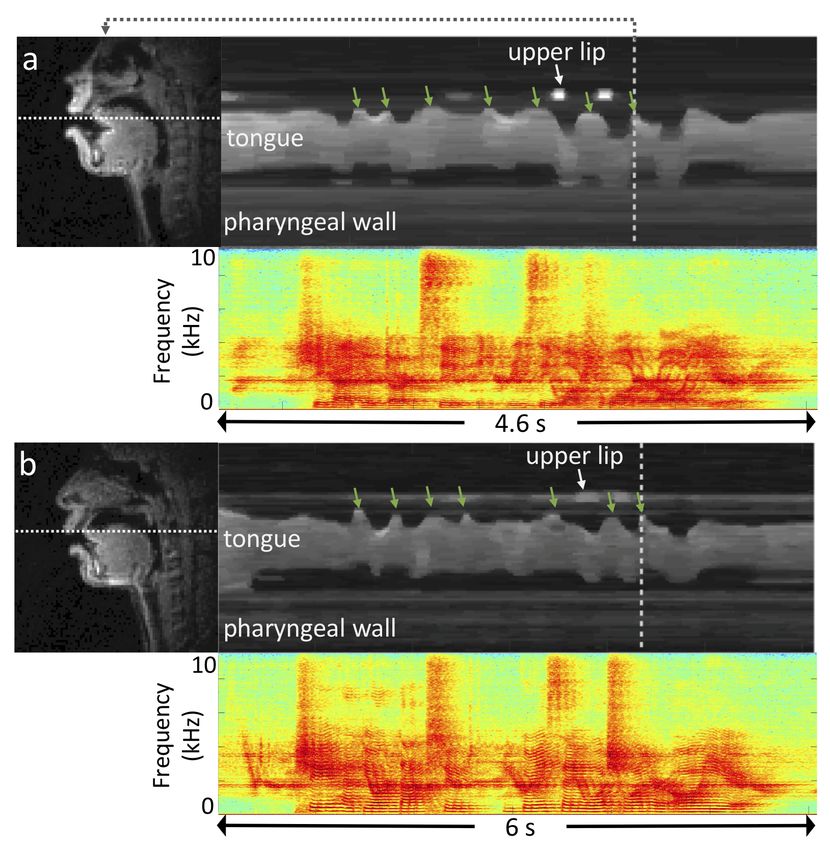
2

speech production poses a multitude of open research questions with implications for
linguistics and speech science, as well as clinical and technological applications, creat-
ing a demand for improved methods for observing and measuring the vocal instrument
in action [3,4].
     Real-Time Magnetic Resonance Imaging (RT-MRI) with concurrent audio record-
ing has emerged as an imaging modality that can provide new insights into speech
production with all its inherent systematicities and variation between languages, con-
texts, and individuals [1,3,5,6]. This technique has the unique advantage of monitoring
the complete vocal tract safely and non-invasively at relatively high spatial and tem-
poral resolution. Applications of RT-MRI span multiple realms of research including
study of: (i) phonetic and phonological phenomena, (ii) spoken language acquisition
and breakdown, including the assessment and remediation of speech disorders [1,3];
(iii) dynamics of vocal tract shaping during communicative speech or vocal perfor-
mance; (iv) articulatory modeling and motor control; (v) speech synthesis and recogni-
tion technologies, and (vi) speaker modeling and biometrics.
     Several examples of recently published MRI data repositories [7,8,9,10,11] have
demonstrated the value of publicly available datasets to address a multitude of open
challenges not only in scholarly research but also in translation into clinical and sci-
entific applications. Open raw datasets have been used to validate and refine advanced
algorithms and to train, validate, and benchmark promising recent applications of ideas
inspired by artificial intelligence/machine learning methods [12]. They are also valued
by commercial vendors to showcase performance and generalizability of reconstruction
methods. Despite a recognized impact in, for example, the domains of musculoskeletal
and brain MRI, there are currently no raw MRI datasets involving the moving vocal
tract.
     The vocal tract contains multiple rapidly moving articulators, which can change
position significantly on a millisecond timescale – an imaging challenge necessitat-
ing high temporal resolution adequate for observing these dynamic speech process [6].
While under-sampling of MRI measurements on time-efficient trajectories enables the
desired resolution, such measurements are hampered by prolonged computation time
for advanced image reconstruction, low signal-to-noise-ratio, and artifacts due to under-
sampling and/or rapid differences of magnetic susceptibility [13,14,15] at the articula-
tor boundaries, which are of utmost interest in characterizing speech production. These
limitations often render present day RT-MRI’s operating point beneath application de-
mands and can introduce bias and increased variance during data analysis. Thus, there is
much room for improvements in the imaging technology pipeline. Despite the promise
of deep learning/machine learning approaches to provide superior performance both in
reconstruction time and image quality, their application to dynamic imaging of fast ape-
riodic motion with high spatiotemporal resolution and low latency is still in its infancy.
We speculate that this is in large measure due to the lack of large-scale public MRI
datasets – the cornerstone of machine learning.
     This paper presents a unique dataset that offers videos of the entire vocal tract in
action, with synchronized audio, imaged along the sagittal plane from 75 subjects while
they performed a variety of speech tasks. The dataset also includes 3D volumetric vocal
tract MRI during sustained speech sounds and high resolution static anatomical T2-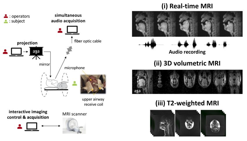
3

weighted upper airway MRI for each subject. Unlike other open datasets for dynamic
speech RT-MRI [16,17,18,19,20], the present dataset includes raw, multi-receiver-coil
MRI data with non-Cartesian, spiral sampling trajectory.
     The dataset can be used to aid the development of algorithms that monitor fast ape-
riodic dynamics of speech articulators at high spatiotemporal resolution while offering
simultaneous suppression of noise and artifacts due to sub-Nyquist sampling or suscep-
tibility. The inclusion of an unprecedented number of 75 speakers can also help improve
our scientific understanding of how vocal tract morphology and speech articulation in-
teract and shed light on the stable and variable aspects of speech signal properties across
speakers, providing for improved models of speech production in linguistics and speech
science research.
     In sum, it is our hope and anticipation that the public and free provision of this rich
dataset can further foster and stimulate research and innovation in the science of human
speech production and its imaging.

Methods
Participants

Seventy-five healthy subjects (40 females and 35 males; 49 native and 26 non-native
American English speakers; age 18-59 years) were included in this study. Each partic-
ipant filled out a questionnaire on basic demographic information including birthplace,
cities raised and lived, and first and second languages. Demographics are summarized
in Table 1. All subjects had normal speech, hearing, and reading abilities, and reported
no known physical or neurological abnormalities. All participants were cognizant of
the nature of the study, provided written informed consent, and were scanned under
a protocol approved by the Institutional Review Board of the University of Southern
California (USC). The data were collected at the Los Angeles County – USC Medical
Center between January 24, 2016 and February 24, 2019.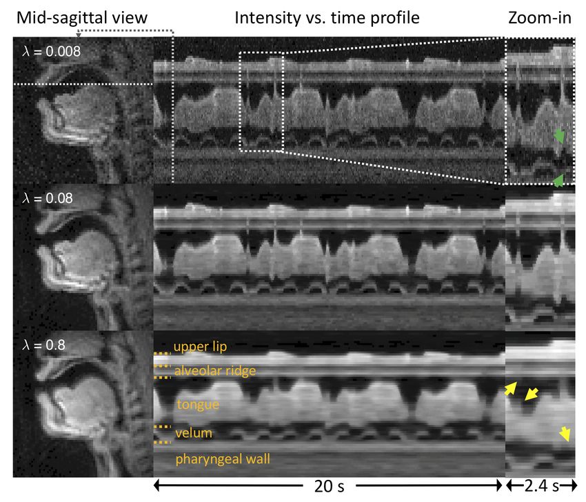
4

       Table 1: Demographic information of subjects; Age at the time of scan
       (years), F: Female, M: Male, L1: first language, L2: second language (if
       any)

    Subject
            Sex Age        L1              L2             City raised
      ID
    sub001 F    24      Mandarin        English         Hohhot, China
    sub002 F    26       English            -            Phoenix, AZ
    sub003 F    19       English       Mandarin         Alhambra, CA
    sub004 M    21       English            -            Portland, OR
    sub005 F    27       English        Spanish          Ashland, KY
    sub006 M    27       English            -              Exton, PA
    sub007 F    30       English        Spanish          Alameda, CA
    sub008 M    32       Telugu          Hindi        Hyderabad, India
    sub009 M    27       English            -              Unknown
    sub010 F    21       English            -            Cranston, RI
    sub011 F    19       English        German          Ridgefield, CT
    sub012 F    27       German         English        Siegen, Germany
    sub013 M    23       English            -           Monrovia, CA
    sub014 F    26       English        Spanish          Riverside, CA
    sub015 M    26       English            -             Edison, NJ
    sub016 F    20       English         French           Gurnee, IL
    sub017 F    31       English            -             Queens, NY
    sub018 F    21       English         Italian     West Newbury, MA
    sub019 M    26       English            -        Virginia Beach, VA
    sub020 M Unknown     English        Spanish        Los Angeles, CA
    sub021 F    29       Korean         English       Karachi, Pakistan
    sub022 M    49        Tamil         English        New Delhi, India
    sub023 M    18       English        Spanish       San Clemente, CA
    sub024 F    22     Vietnamese       English           Austin, TX
    sub025 M    21       English       Mandarin          San Jose, CA
    sub026 F    21       English    Bahasa Indonesia Jakarta, Indonesia
    sub027 F    21       English    Native Hawaiian      Kaneohe, HI
    sub028 M    25       English         Hindi          Mumbai, India
    sub029 M    42        Greek         English         Serres, Greece
    sub030 M    28       Gujarati       English          Rajkot, India
    sub031 F    20       English            -        Thousand Oaks, CA
    sub032 F    21       Korean         English         Seoul, S. Korea
    sub033 M    18       English         French         San Diego, CA
    sub034 M    28   Telugu/Kannada     English        Bangalore, India
    sub035 M    21       English        Spanish       Castro Valley, CA
    sub036 F    25       English        German            Norfolk, NE
    sub037 M    32       Korean         English         Seoul, S. Korea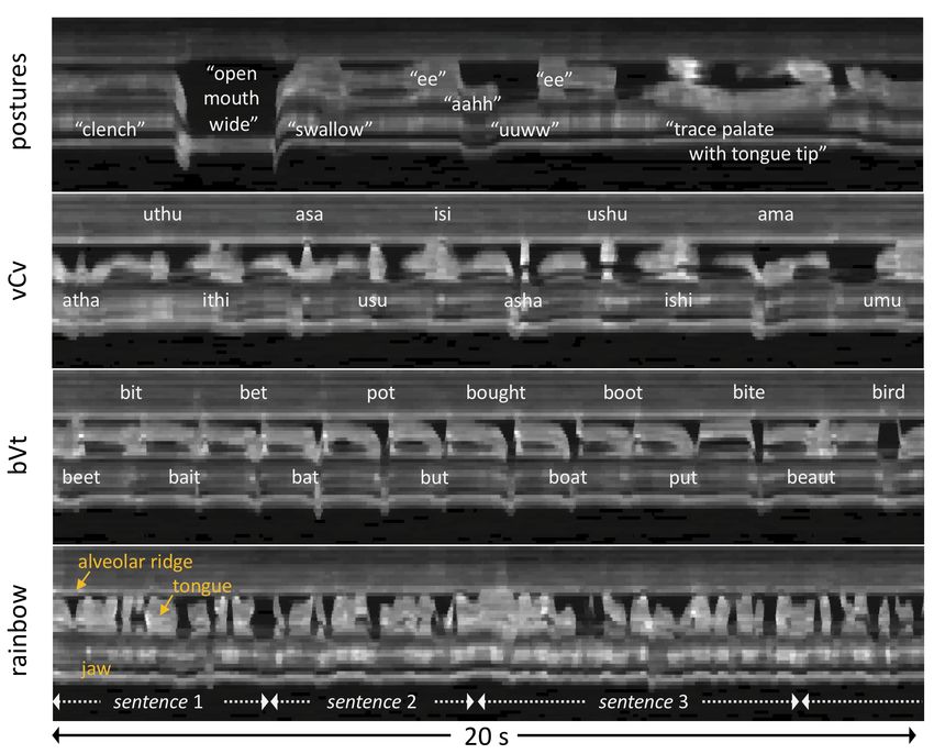
5

                       Table 1 . Continued
Subject
        Sex Age        L1                L2          City raised
  ID
sub038 M    30      Russian           English     Novosibirsk, Russia
sub039 M    27      Chinese           English      Shanghai, China
sub040 M    26       Korean           English       Seoul, S. Korea
sub041 F    22      English           French        Nanjing, China
sub042 F    22      English         Cantonese     Monterey Park, CA
sub043 F    28      English           Russian         Houston, TX
sub044 F    24      English           Spanish         Atlanta, GA
sub045 M    23      English               -        Los Angeles, CA
sub046 M    49      English           Spanish      Los Angeles, CA
sub047 F    18      English               -        Sacramento, CA
sub048 F    27      English           Spanish    Manhattan Beach, CA
sub049 F    29      English               -        Sacramento, CA
sub050 M    30      English           Korean         Barrington, RI
sub051 M    33       Korean           English       Seoul, S. Korea
sub052 M    26      English           German           Omaha, NE
sub053 M    29      English               -         Mill Valley, CA
sub054 F    24      English           Spanish          Oswego, IL
sub055 F    21      English           French        Cincinnati, OH
sub056 M    18     Portuguese         English      Diamond Bar, CA
sub057 M    19      English           Spanish        Abington, PA
sub058 F    28   Spanish/English       Italian   San Juan, Pueto Rico
sub059 F    22      English           Spanish        Chantilly, VA
sub060 F    22       Korean           English       San Diego, CA
sub061 F    26       Tamil             Hindi           Ooty, India
sub062 M    25      English              Twi          Tema, Ghana
sub063 F    26      English          Mandarin      Charlestown, IN
sub064 F Unknown     Telugu           English      Vijayawada, India
sub065 M    19   Spanish/English          -         New York, NY
sub066 F    18   Spanish/English          -        Los Angeles, CA
sub067 M    59      English           French       Washington, DC
sub068 M    28      Gujarati           Hindi        Junagadh, India
sub069 F    25      Mandarin          English        Suzhou, China
sub070 F    29      English               -           Brawley, CA
sub071 F    25      English           Spanish          Fresno, CA
sub072 M    36      English               -           Medina, OH
sub073 M    30       Korean           English       Iksan, S. Korea
sub074 F    30       Telugu            Hindi       Hyderabad, India
sub075 F    29      Spanish           English     Baldwin Park, CA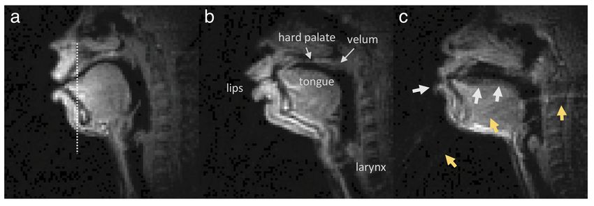
6
Experimental Overview
Three types of MRI data were recorded for each subject: (i) dynamic, real-time MRI of
the vocal tract’s mid-sagittal slice at 83 frames per second during production of a com-
prehensive set of scripted and spontaneous speech material, averaging 17 minutes per
subject, along with synchronized audio; (ii) static, 3D volumetric images of the vocal
tract, captured during sustained production of sounds from the full set of American En-
glish vowels and continuant consonants, 7 seconds each; (iii) T2-weighted volumetric
images at rest position, capturing fine detail anatomical characteristics of the vocal tract
(See Figure 1).
All data were collected using a commercial 1.5 Tesla MRI scanner (Signa Excite,
GE Healthcare, Waukesha, WI) with gradients capable of 40 mT/m amplitude and 150
mT/m/ms slew rate. A custom 8-channel upper airway receiver coil array [21], with four
elements on each side of the subject’s cheeks, was used for signal reception. Compared
to commercially available coils that are designed for neurovascular or carotid artery
imaging, this custom coil has been shown to provide 2-fold to 6-fold higher signal-to-
noise-ratio (SNR) efficiency in upper airway vocal tract regions of interests including
tongue, lips, velum, epiglottis, and glottis. The subjects, while imaged, were presented
with scripts of the experimental stimuli via a projector-mirror setup [22]. Acoustic audio
data were recorded inside the scanner using commercial fiber-optic microphones (Op-
toacoustics Inc., Yehuda, Israel) concurrently with the RT-MRI data acquisition using a
custom recording setup [23].
Stimuli and Linguistic Justification
The stimuli were designed to efficiently capture salient, static and dynamic, and articu-
latory and morphological aspects of speech production of American English in a single
90-minute scan session.
Table 2 lists the speech stimuli used for the RT-MRI data collection of the first 45-
minute sub scan session. Each individual task was designed to be performed within 30
seconds at a normal speaking rate, however the actual recordings varied in duration de-
pending on the length of the task and the natural speaking rate of the individual. The
stimulus set contained material to elicit both scripted speech and spontaneous speech.
The scripted speech tasks were consonant production in symmetric vowel-consonant-
vowel context, vowels /V/ produced between the consonants /b/ and /t/, i.e., in /bVt/
contexts, four phonologically rich sentences [24], and three reading passages commonly
used in speech evaluation and linguistic studies [25,26,27]. Scripted instructions to pro-
duce several gestures were also included. These gestures were clenching, wide opening
of mouth, yawning, swallowing, slow production of the sequence /i/-/a/-/u/-/i/, tracing
of the palate with the tongue tip, and singing “la” at their highest and lowest pitches.
The stimuli in the scripted speech were repeated twice. The spontaneous speech tasks
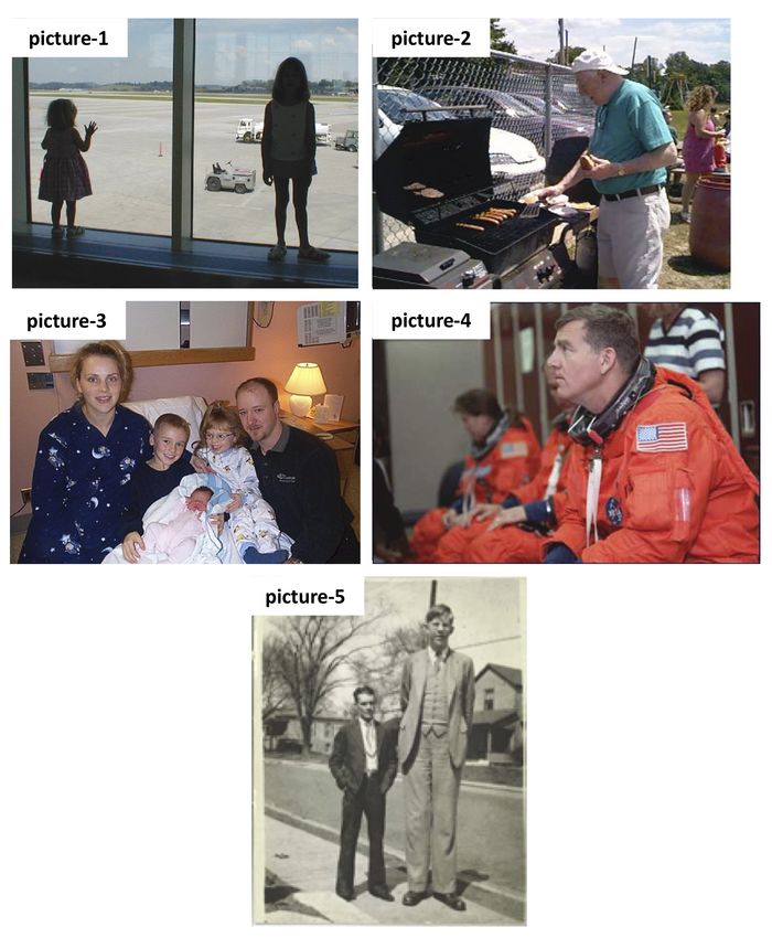
were to describe the content and context of five photographs (Supplemental Figure 1)
and to answer five open-ended questions (e.g., “What is your favorite music?”). The full
scripts and questions are provided in Table 3.7
Table 2. Speech experiment stimuli for 2D RT-MRI.
Stimulus Stimulus Duration
Category Description
Index Name (sec)
01, 02, 03 vcv[1-3] Consonants in symmetric /VCV/ 30 (x3)
04 bvt Vowels in /bVt/ 30
05 shibboleth Four phonologically rich sentences [34] 30
Scripted 06 rainbow Rainbow passage [35] 30
speech 07, 08 grandfather[1-2] Grandfather passage [36] 30 (x2)
09, 10 northwind[1-2] Northwind and the sun passage [37] 30 (x2)
11 postures Postures 30
Repetition of the above scripted speech tasks 30 (x11)
Spontaneous 12 – 16 picture[1-5] Description of pictures 30 (x5)
speech 17 – 21 topic[1-5] Discussion about topics 30 (x5)
Table 4 lists the speech stimuli used for the 3D volumetric data collection of a
30-minute part of the scan session. Each individual task was designed for a subject to
sustain vowels, consonant sounds, or to maintain postures for 7 seconds. The stimulus
set contained consonant production in symmetric vowel-consonant-vowel context, vow-
els /V/ produced between the consonants /b/ (or /p/) and /t/, and production of several
postures. All of the stimuli were repeated twice.
RT-MRI Acquisition
RT-MRI acquisition was performed using a 13-interleaf spiral-out spoiled gradient-echo
pulse sequence [28]. This is an efficient scheme for sampling MR measurements, each
with the different initial angle being interleaved by the bit-reversed order in time [29].
The 13 spiral interleaves, when collected together, fulfil the Nyquist sampling rate.
Imaging parameters were: repetition time = 6.004 ms, echo time = 0.8 ms, field-of-
view (FOV) = 200 × 200 mm2 , slice thickness = 6 mm, spatial resolution = 2.4 × 2.4
mm2 (84 × 84 pixels), receiver bandwidth = ± 125 kHz, flip angle = 15◦ . Imaging was
performed in the mid-sagittal plane, which was prescribed using a real-time interactive
imaging platform [30] (RT-Hawk, Heart Vista, Los Altos, CA). Real-time visualization
was implemented within the custom platform by using a sliding window gridding re-
construction [31] to ensure subject’s compliance with stimuli and to detect substantial
head movement.
Acquisition was divided into 20-40 second task intervals presented in Table 2, each
followed by a pause of the same duration so as to allow enough a brief break for the
subject prior to the next task and to avoid gradient heating.8
Table 3. Full scripts and questions of speech experiment stimuli for 2D RT-MRI.
Stimulus
Script or question/topic
Index
vcv1 apa upu ipi ata utu iti aka uku iki aba ubu ibi ada udu idi aga ugu igi
vcv2 atha uthu ithi asa usu isi asha ushu ishi ama umu imi ana unu ini ala ulu ili
vcv3 afa ufu ifi ava uvu ivi ara uru iri aha uhu ihi awa uwu iwi aya uyu iyi
bvt beet bit bait bet bat pot but bought boat boot put bite beaut bird boyd abbot
She had your dark suit in greasy wash water all year.
Don’t ask me to carry an oily rag like that.
shibboleth The girl was thirsty and drank some juice followed by a coke.
Your good pants look great however your ripped pants look like a cheap version
of a k-mart special is that an oil stain on them.
When the sunlight strikes raindrops on the air they act as a prism and form
a rainbow. The rainbow is a division of white light into many beautiful colors.
These take the shape of a long round arc with its path high above and its two
rainbow ends apparently beyond the horizon. There is according to legend a boiling pot
of gold at one end. People look but no one ever finds it. When a man looks for
something beyond his reach his friends say he is looking for the pot of gold
at the end of the rainbow.
You wish to know all about my grandfather. Well, he is nearly ninety three
years old yet he still thinks as swiftly as ever. He dresses himself in an
grandfather1 old black frock coat usually several buttons missing. A long beard clings
to his chin, giving those who observe him a pronounced feeling of the utmost
respect. When he speaks, his voice is just a bit cracked and quivers a bit.
Twice each day he plays skillfully and with zest upon a small organ.
Except in the winter when the snow or ice prevents, he slowly takes a short
grandfather2 walk in the open air each day. We have often urged him to walk more and
smoke less but he always answers, “banana oil” grandfather likes to be modern
in his language.
The north wind and sun were disputing which was the stronger, when a traveler
came along wrapped in a warm cloak. They agreed that the one who first
northwind1
succeeded in making the traveler take off his cloak should be considered
stronger than the other.
Then the north wind blew as hard as he could, but the more he blew
the more closely did the traveler fold his cloak around him and at last
northwind2 the north wind gave up the attempt. Then the sun shone out warmly and
immediately the traveler took off his cloak and so the north wind was
obliged to confess that the sun was stronger of the two.
clench, open wide & yawn, swallow, eee. . . aahh. . . uuww. . . eee,
postures trace palate with tongue tip, Sing “la” at your highest note,
Sing “la” at your lowest note
topic1 My favorite music
topic2 How do I like LA?
topic3 My favorite movie
topic4 Best place I’ve been to
topic5 My favorite restaurant9
Table 4. Speech experiment stimuli for 3D volumetric static MRI.
Stimulus Stimulus Duration
Category Description
Index Name (sec)
Sustain sound at vowel
01 beet beet
02 bit bit
03 bait bait
04 bet bet
05 bat bat
Vowels 06 pot pot
in /bVt/ 07 but but 7 (x13)
08 bought bought
09 boat boat
10 boot boot
11 put put
12 bird bird
13 abbot abbot
Sustain sound at consonant
14 afa afa as in food
15 ava ava as in voice
16 atha thing atha as in thing
17 atha this atha as in this
18 asa asa as in soap
Consonants 19 aza aza as in zipper
in symmetric 20 asha asha as in shoe 7 (x14)
/VCV/ 21 agea agea as in beige
22 aha aha as in happy
23 ama ama as in rim
24 ana ana as in pin
25 anga anga as in ring
26 ala ala as in late
27 ara ara as in rope
Breathe normally with your mouth
28 breathe
closed, resting.
29 clench Clench your teeth and hold.
Stick your tongue out
30 tongue
as far as you can, and hold.
7 (x6)
Postures Pull back your tongue as far into
31 yawn the mouth as you can,
and hold (like yawning).
Raise the tip of your tongue to
32 tip
the middle of your palate, and hold.
33 hold Hold your breath.
Repetition of the above tasks 7 (x33)10
RT-MRI Reconstruction
The dataset provides one specific image reconstruction that has been widely used in
speech production research [21,28]. This image reconstruction involves optimizing the
following cost function:
minm kAm − dk22 + λk 5t mk1 (1)
where A represents the encoding matrix that models for the non-uniform fast Fourier
transform and the coil sensitivity encoding, m is the dynamic image time series to be
reconstructed, d is the acquired multiple-coil raw data, λ is the regularization parameter,
5t is the temporal finite difference operator, and k · k22 and k · k1 are l2 and l1 norms,
respectively. The coil sensitivity maps were estimated using the Walsh method [32]
from temporally combined coil images. The regularization term encourages voxels to
be piecewise constant along time. This regularization has been successfully applied to
speech RT-MRI [21,33,34] and a variety of other dynamic MRI applications where the
primary features of interest are moving tissue boundaries [35,36,37].
Our reference implementation solves Equation (1) using nonlinear conjugate gra-
dient algorithm with Fletcher-Reeves updates and backtracking line search [38]. The
algorithm was terminated either at 150 maximum iterations or the step size fell below
< 1e-5 during line search. Reference images were reconstructed using 2 spiral arms per
time frame, resulting in a temporal resolution of 83.28 frames per second. This tempo-
ral resolution is enough to capture important articulator motions1, but reconstruction at
different temporal resolutions is also possible with the provided dataset by adjusting the
number of spiral arms per time frame. The algorithm was implemented in both MAT-
LAB (The MathWorks, Inc., Natick, MA) and Python (Python Software Foundation,
https://www.python.org/). The provided image reconstruction was performed
in MATLAB 2019b on a Xeon E5-2640 v4 2.4GHz CPU (Intel, Santa Clara, CA) and
a Tesla P100 GPU (Nvidia, Santa Clara, CA). Reconstruction time was 160.69 ± 1.56
ms per frame. Parameter selection of λ is described in the technical validation section
below.
Audio Data
One main microphone was positioned ∼ 0.5 inch away from the subject’s mouth. The
microphone signal was sampled at 100 kHz each. The data were recorded on a laptop
computer using a National Instruments NI-DAQ 6036E PCMCIA card. The audio sam-
ple clock was hardware-synchronized to the MRI scanner’s 10 MHz master clock. The
audio recording was started and stopped using the trigger pulse signal from the scanner.
The real-time audio data acquisition routine was written in MATLAB. Audio was first
low-pass filtered and decimated to a sampling frequency of 20 kHz. The recorded audio
was then enhanced using a normalized least-mean-square noise cancellation method
[23] and was aligned with the reconstructed MRI video sequence to aid linguistic anal-
ysis.11
3D Volumetric MRI of Sustained Sounds
An accelerated 3D gradient-echo sequence with Cartesian sparse sampling was imple-
mented to provide static high-resolution images of the full vocal tract during sustained
sounds or postures. Imaging parameters were: repetition time = 3.8 ms, spatial resolu-
tion = 1.25 × 1.25 × 1.25 mm3, FOV = 200 × 200 × 100 mm3 (respectively in the
anterior-posterior, superior-inferior, and left-right directions), image matrix size = 160
× 160 × 80, and flip angle = 5◦ . The central portion (40 × 20) of the ky -kz space was
fully sampled to estimate the coil sensitivities from the data itself. The outer portion
of the ky -kz space was sampled using a sparse Poisson-Disc sampling pattern, which
together resulted in 7-fold net acceleration, and a total scan time of 7 seconds.
Data were acquired while the subjects sustained for 7 seconds a sound from the full
set of American English vowels and continuant consonants (Table 4). The stimuli were
presented to the subject via a projector-mirror setup, and upon hearing a “GO” signal
given by the scanner operator, a subject started to sustain the sound or posture; this
was followed by the operator triggering the acquisition manually. Subjects sustained
the postures as long as they could hear the scanner operating. A recovery time of 5-10
seconds was given to the subject between the stimuli.
Image reconstruction was performed off-line by a sparse-SENSE constrained re-
construction similar to the optimization problem shown in Equation (1). In contrast to
Equation (1) , isotropic spatial total variation constraints were used [28,39]. The recon-
struction was achieved using the open-source Berkeley Advanced Reconstruction Tool-
box (BART) [40]; the image reconstruction was performed in MATLAB using GPU
acceleration.
T2-Weighted MRI at Rest
Fast spin echo-based sequence was performed to provide high-resolution T2-weighted
images with fine detail anatomical characteristics of the vocal tract at rest position.
Imaging was run to obtain full sweeps of the vocal tract in the axial, coronal, and sagittal
orientations, for each of which the number of slices ranges from 29 to 70, depending on
the size of the vocal tract. Imaging parameters were: repetition time = 4600 ms, echo
time = 120 - 122 ms, slice thickness = 3 mm, in-plane FOV = 300 × 300 mm2 , in-plane
spatial resolution = 0.5859 × 0.5859 mm2 , number of averages = 1, echo train length =
25, and scan time = approximately 3.5 minutes per orientation.
Data Records
This dataset is publicly available in figshare [41]. The total size of this dataset is ap-
proximately 966 GB. It contains (i) raw 2D sagittal-view RT-MRI data, reconstructed
images and videos, and synchronized denoised audio, (ii) 3D volumetric MRI images,
and (iii) T2-weighted MRI images. The data for subject XYZ is contained in folder with
identifier subXYZ (e.g., sub001/) and organized into three main folders: 2D RT-MRI
data are located in the subfolder 2drt/ (e.g., sub001/2drt/), 3D volumetric images in
3d/, and T2-weighted images in t2w/. The contents and data structures of the dataset
are detailed as follows.12
Table 5. Naming of folders and files. Subject identifiers correspond to Table 1, column Subject ID. Stimulus
indices correspond to Tables 2 and 4, column Stimulus Index. Stimulus names correspond to Tables 2 and 4,
column Stimulus Name.
Category Data folder Filename convention Description
subXYZ/2drt/ 2drt Raw RT-MRI
raw/ r raw.h5 k-space data
(e.g., sub001 2drt 01 vcv1 r1 raw.h5) in MRD format
subXYZ/2drt/ 2drt Reconstructed
recon/ r recon.h5 RT-MRI image data
in HDF5 format
RT-MRI
subXYZ/2drt/ 2drt Co-recorded
audio/ r audio.wav audio data
in WAV format
subXYZ/2drt/ 2drt Videos of speech task
video/ r video.mp4 with aligned audio
in MPEG-4 format
subXYZ/3d/ 3d Reconstructed
recon/ r recon.mat volumetric image data
3D (e.g., sub001 3d 13 abbot r1 recon.mat) in MAT format
volumetric subXYZ/3d/ 3d Mid-sagittal
snapshot/ r snapshot.png slice image
in PNG format
T2- subXYZ/t2w/ .dcm T2-weighted image
weighted dicom/ (e.g., 00010001.dcm) in DICOM format
RT-MRI
Raw RT-MRI data are provided in the vendor-agnostic MRD format (previously known
as ISMRMRD) [42,43], which stores k-space MRI measurements, k-space location ta-
bles, and sampling density compensation weights. Parameters for the acquisition se-
quence are contained in the file header information. In addition, this dataset includes
reconstructed image data for each subject and task in HDF5 format, audio files in WAV
format, and videos in MPEG-4 format.
For each subject, RT-MRI raw data is contained in the subfolder raw/ (e.g., sub001/2drt/raw/),
reconstructed image data in recon/, co-recorded audio (after noise cancellation) in au-
dio/, and reconstructed speech videos with aligned audio in video/. Table 5 summarizes
the data structure and naming conventions for this dataset.13
3D Volumetric MRI of Sustained Sounds
3D volumetric MRI data contains reconstructed image data and imaging parameters
in MAT format (MATLAB, The MathWorks, Inc., Natick, MA) in the subfolder recon/
(e.g., sub001/3d/recon/) and a mid-sagittal slice image in PNG format in the subfolder
snapshot/.
T2-Weighted MRI at Rest
T2-weighted image data from axial, coronal, and sagittal orientations are stored in Digi-
tal Imaging and Communications in Medicine (DICOM) format in the subfolder dicom/
(e.g., sub001/t2w/dicom/).
The imaging DICOM files were de-identified using the Clinical Trial Processor
(CTP) developed by the Radiological Society of North America (RSNA) [44]. Specif-
ically, data anonymization was completed using a command line tool developed in the
Java programming language [45]. All images followed the standardized DICOM format
and some of the attributes were removed or modified to preserve privacy of the subjects,
specifically: PatientName was modified to follow the subXYZ pattern, and the original
study dates were shifted by the same offset for all subjects.
Metadata
We provide presentation slides that contain the experimental stimuli including scripts
and pictures used for the visualization to the subjects, in PPT format in Stimuli.ppt. De-
mographic information for each subject is contained in XLSX format in Subjects.xlsx.
This meta file contains sex, race, age (at the time of scan), height (cm), weight (kg),
origin, birthplace, cities raised and lived, L1 (first language), L2 (second language, if
any), L3 (third language, if any), and the first language and birthplace of each subject’s
parents.
Additionally, meta information for each subject and RT-MRI task is contained in
JSON format in metafile public .json. For each subject, we include the
following demographic information: 1) L1 (first language), 2) L2 (second language, if
any), 3) sex (M/F), 4) age (years at the time of scan). Further, visual and audio quality
assessment scores are provided using a 5-level Likert scale for each subject stratified
into categories: off-resonance blurring (1, very severe; 2, severe; 3, moderate; 4, mild; 5,
none), video SNR (1, poor; 2, fair; 3, good; 4, very good; 5, excellent), aliasing artifacts
(1, very severe; 2, severe; 3, moderate; 4, mild; 5, none), and audio SNR (1, poor;
2, fair; 3, good; 4, very good; 5, excellent). Specifically, an MRI expert with 6 years
of experience in reconstructing and reading speech RT-MRI examined all the RT-MRI
videos reconstructed for each subject and assessed and scored their visual and audio
quality. For each task of the individual subjects, the information about task’s index,
name, existence of file, and notes taken during data inspection by inspectors are also
contained in the meta file.14
Technical Validation
Data Inspection
Four inspectors manually performed qualitative data inspection for the datasets. After
reconstruction, all images were converted from HDF5 format to MPEG-4 format, at
which time co-recorded audio (WAV format) was integrated. All qualitative data in-
spection was performed manually based on MPEG-4 format videos. Included in the
dataset were files that met all of the following criteria:
1. The video (MPEG-4) exists (no data handling failure).
2. The audio recording (WAV) exists (no data handling failure).
3. The video and audio are synchronized (based on inspection by a human).
After the inspection, 75 subjects were included in this dataset. It should be noted
that although three subjects (sub18, sub74, and sub75) present severe radio-frequency-
interference artifacts that were later determined to be from a leak in the MRI radio-
frequency cage, we included those three subjects in this dataset in the hopes of po-
tentially facilitating development of a digital radio-frequency interference correction
method. Files that did not exist (criteria 1) were annotated for each task and subject in
the meta file
(i.e., metafile public .json).
Figure 2 shows representative examples of the data quality from three subjects that
are included in this dataset (sub35, sub41, and sub58). Note that all 75 subjects ex-
hibited acceptable visualization of all soft tissue articulators. However, two types of
artifacts were commonly observed:
1. Blurring artifact due to off-resonance (green arrows, Figure 2c): This artifact ap-
pears as blurring or signal loss predominantly adjacent to air-tissue boundaries that
surround soft tissue articulators. It is induced in spiral imaging by rapid changes
of local magnetic susceptibility between the air and tissue. This artifact correc-
tion is still an active research area, including methods for a simple zeroth order
frequency demodulation to advanced model-based [14,46,47] and data-driven ap-
proaches [15,48]. At present, we perform zeroth order correction during data ac-
quisition as part of our routine protocol.
2. Ringing artifact due to aliasing (yellow arrows, Figure 2c): This artifact appears
as an arc-like pattern centered at the bottom-right corner outside the FOV. It is
caused by a combination of gradient non-linearity and non-ideal readout low-pass
filter when a strong signal source appears outside the unaliasing FOV. This artifact
does not overlap with the articulator surfaces that are most important in the study of
speech production. However, avoiding and/or correcting this artifact would improve
overall image quality and potentially enable the use of a smaller FOV.
Figure 3 shows representative examples of the diverse speech stimuli that are in-
cluded in this dataset. The intensity vs. time profiles visualize the first 20 seconds of
four representative stimuli from sub35, shown in Figure 2a. These examples show the
variety of patterns and speed of movement of the soft tissue articulators observed within15
a speaker as a function of the speech stimuli performed. Figure 4 contains a histogram
of the speaking rate in read sentences [49]. The overall mean speaking rate was 149.2 ±
31.2 words per minute. The statistics are calculated from the read “shibboleth,” “rain-
bow,” “grandfather[1-2],” and “northwind[1-2]” stimuli for all subjects. These selected
stimuli are composed of read full sentences.
Figure 5 illustrates variability within the same speech stimuli across different speak-
ers. The image time profiles correspond to the period of producing the first sentence
“She had your dark suit in greasy wash water all year” in the stimulus “shibboleth”
from sub35 and sub41. Although both speakers share the critical articulatory events
(see green arrows), the timing and pattern vary depending on the subject.
Regularization Parameter Selection for Image Reconstruction for RT-MRI
We performed a parameter sweep and qualitative evaluation on a subset of the data to
select a regularization parameter for the provided reconstructions. Ten stimuli from ten
different subjects were randomly selected. The regularization parameter λ was swept
in the range between 0.008C and 1C in a logarithmic scale. Here, C represents the
maximum intensity of the zero-filled reconstruction of the acquired data. Figure 6 shows
a representative example of the impact of λ on the reconstructed image quality. A small
λ (= 0.008C) exhibits high noise level in the reconstruction (top, Figure 6). A higher λ
(= 0.8C) reduces the noise level but exhibits unrealistic temporal smoothing as shown in
the intensity vs. time profiles (yellow arrows, Figure 6). The optimal parameter 0.08C
was selected by consensus among four experts in the area of MRI image reconstruction
and/or speech production RT-MRI. Once the λ was optimized for the subset of the data,
reconstruction was performed for all datasets. We have empirically observed that the
choice of λ appears robust across all of the datasets.
Usage Notes
Several papers have been published by our group in which methods are directly applied
to a subset of this dataset for the reconstruction and artifact correction of the RT-MRI
data. These include auto-calibrated off-resonance correction [14], deblurring using con-
volutional neural networks [15], aliasing artifact mitigation [50] (the artifact marked by
the yellow arrows in Figure 2c), and super-resolution reconstruction [under review].
Additionally, several tools have been developed at our site for the analysis and mod-
elling of reconstructed real-time MRI data. These include a graphical user interface
for efficient visual inspection [17] and implementations of grid-based tracking of air-
tissue boundaries [51,52,53], region segmentation and factor analysis [54,55,56], neural
network-based edge detection [57,58]; region of interest (ROI) analysis [59,60,61] and
centroid tracking [62]. Some of these tools are also made available; see code repository
at https://github.com/usc-mrel/usc_speech_mri.git, as well as Ra-
manarayanan et al [3] and Toutios et al [63] for detailed reviews.16
Code Availability
This dataset is accompanied by a code repository (https://github.com/usc-mrel/
usc_speech_mri.git) that contains examples of software and parameter configu-
rations necessary to load and reconstruct the raw RT-MRI in MRD format. Specifically,
the repository contains demonstrations to illustrate and replicate results of Figures 2-
6. Code samples are available in MATLAB and Python programming languages. All
software is provided free to use and modify under the MIT license agreement.
Acknowledgements
This work was supported by NSF Grant 1514544, NSF Grant 1908865, and NIH Grant
R01DC007124.
Author contributions
Y.Lim: wrote the manuscript and collected and curated data
A.T.: led the data collection and curation
Y.B.: contributed to data curation and the manuscript writing
Y.T.: contributed to data curation and the manuscript writing
S.G.L.: developed data acquisition protocols and collected data
C.V.: developed data acquisition protocols and collected data
T.S.: collected and curated data
M.O.: collected and curated data
S.H.: collected and curated data
Y.Lee: collected data
W.C.: collected data
J.T.: collected data
M.L.M.: collected data
C.S.: collected data
B.G.: curated data
L.G.: designed experimental stimuli
D.B.: designed experimental stimuli
K.N.: managed the project; developed data acquisition protocols; contributed to data
curation
S.N.: conceived the project; managed the project; contributed to data curation
All authors contributed to the paper preparation, reviewed drafts of the paper, and
approved of the final version.
Competing interests
The authors declare no competing interests.17 Figures and figures legends Figure 1. Data acquisition workflow and data records. (Left) Data were acquired at 1.5 Tesla using the custom upper-airway coil located in close proximity to the subject’s upper airway structures. The subject visualized the stimuli through a mirror-projector setup and audio was recorded through an MR-compatible microphone simultaneous with the RT-MRI. The scanner operator used a custom interactive imaging interface with the scanner hardware to control and acquire the data for the RT- MRI session. (Right) The recorded MRI data were: (i) dynamic, 2D real-time MRI of the vocal tract’s mid-sagittal slice at 83 frames per second during production of a comprehensive set of scripted and spontaneous speech material, along with synchronized audio recording; (ii) static, 3D volumetric images of the vocal tract, captured during sustained production of sounds or postures, 7 seconds each; (iii) T2-weighted volumetric images at rest position, capturing fine detail anatomical characteristics of the vocal tract.
18

Figure 2. Typical data quality of 2D real-time speech imaging, shown in mid-sagittal image frames
from three example subjects: (a) sub35 (male, 21yrs, native American English speaker), (b) sub51
(male, 33yrs, non-native speaker), (c) sub58 (female, 32yrs, non-native speaker). The mid-sagittal
image frames depict the event of articulating the fricative consonant [θ] in the word “uthu” (stimulus
“vcv2”), where the tongue tip contacts the upper teeth. (a) and (b) are considered to have very high
quality, based on high SNR and no noticeable artifact. (c) is considered to have moderate quality,
based on good SNR and mild image artifacts; the white arrows point to blurring artifacts due to
off-resonance while the yellow arrows point to ringing artifacts due to aliasing.
19

Figure 3. Image vs. time profiles during the first 20 seconds of four different stimuli for sub35.
Profiles show the time evolution of the cut depicted by the dotted line in the image frame shown
in Figure 2a (sub35). The rows visualize different examples of stimuli: “postures,” “vcv2,” “bvt,”
and “rainbow” passage. The set of stimuli covers a wide range of articulator postures and tongue
velocities.
20

                            150

                            100
                Frequency

                             50

                              0
                                  50   100      150        200        250
                                       Words Per Minute

Figure 4. Histogram of words per minute during scripted speech stimuli including “shibboleth,”
“rainbow,” “grandfather[1-2],” and “northwind[1-2].”
21 Figure 5. Variability in the articulation of the same sentence between two speakers: (a) sub35, (b) sub41. The time profile and audio spectrum correspond to the first sentence “She had your dark suit in greasy wash water all year” in the stimulus of “shibboleth” from each subject. The green arrows point to several noticeable time points at which the tongue tip contacts the upper teeth/alveolar ridge.
22

Figure 6. Illustration of the impact of reconstruction parameter λ on image quality. Data are from
sub15 (male, 26yrs, native American English speaker). (Left) Mid-sagittal image frames during
speaking. (Middle) The intensity vs. time profiles for stimulus “vcv1.” (Right) Zoomed-in time pro-
files. Different rows correspond to different λ values. For a smaller λ (= 0.008C), the reconstruction
shows a higher noise level and obscured articulatory event (green arrows), whereas for a higher λ (=
0.8C), the noise level decreases but the temporal smoothing artifact is evident in regions indicated by
yellow arrows. λ = 0.08C yields an acceptable noise level while showing adequate temporal fidelity
and therefore was selected for the optimal value for the reconstruction.
23

Supplemental Figure 1. Photographs corresponding to speech experiment stimuli of picture1 to
picture5. Photo source:
https://writefix.com/?page_id=411 (picture1),
https://writefix.com/?page_id=438 (picture2),
https://writefix.com/?page_id=443 (picture3),
https://writefix.com/?page_id=400 (picture4),
https://farmvilleherald.com/2020/03/the-worlds-biggest-man-visits-farmville/
(picture5)
24

References

 1. Lingala, S. G., Sutton, B. P., Miquel, M. E. & Nayak, K. S. Recommendations for real-time
    speech MRI. J. Magn. Reson. Imaging 43, 28–44 (2016).
 2. Scott, A. D., Wylezinska, M., Birch, M. J. & Miquel, M. E. Speech MRI: Morphology and
    function. Phys. Medica 30, 604–618 (2014).
 3. Ramanarayanan, V. et al. Analysis of speech production real-time MRI. Comput. Speech.
    Lang. 52, 1–22 (2018).
 4. Hagedorn, C. et al. Engineering Innovation in Speech Science: Data and Technologies. Per-
    spect. ASHA Spec. Interes. Groups 4, 411–420 (2019).
 5. Bresch, E., Kim, Y. C., Nayak, K., Byrd, D. & Narayanan, S. Seeing speech: Capturing vocal
    tract shaping using real-time magnetic resonance imaging. IEEE Signal Process. Mag. 25,
    123–129 (2008).
 6. Nayak, K. S., Lim, Y., Campbell-Washburn, A. E. & Steeden, J. Real-Time Magnetic Reso-
    nance Imaging. J. Magn. Reson. Imaging, (2020). https://doi.org/10.1002/jmri.27411
 7. Marcus, D. S., Wang, T. H., Parker, J., Csernansky & J. G. Open access series of imag-
    ing studies (OASIS): Cross-sectional MRI data in young, middle aged, nondemented, and
    demented older adults. J. Cogn. Neurosci. 19, 1498–1507 (2007).
 8. Souza, R. et al. An open, multi-vendor, multi-field-strength brain MR dataset and analysis of
    publicly available skull stripping methods agreement. Neuroimage 170, 482–494 (2018).
 9. Knoll, F. et al. fastMRI: A Publicly Available Raw k-Space and DICOM Dataset of Knee
    Images for Accelerated MR Image Reconstruction Using Machine Learning. Radiol. Artif.
    Intell. 2, e190007 (2020).
10. Chen, C. et al. OCMR (v1.0)–Open-access multi-coil k-space dataset for cardiovascular mag-
    netic resonance imaging. arXiv:2008.03410 (2020).
11. http://mridata.org/
12. Knoll, F. et al. Advancing machine learning for MR image reconstruction with an open
    competition: Overview of the 2019 fastMRI challenge. Magn. Reson. Med. 84, 3054–3070
    (2020).
13. Sutton, B. P., Conway, C. A., Bae, Y., Seethamraju, R. & Kuehn, D. P. Faster dynamic imag-
    ing of speech with field inhomogeneity corrected spiral fast low angle shot (FLASH) at 3 T.
    J. Magn. Reson. Imaging 32, 1228–1237 (2010).
14. Lim, Y., Lingala, S. G., Narayanan, S. S. & Nayak, K. S. Dynamic off-resonance correction
    for spiral real-time MRI of speech. Magn. Reson. Med. 81, 234–246 (2019).
15. Lim, Y., Bliesener, Y., Narayanan, S. S. & Nayak, K. S. Deblurring for spiral real-time MRI
    using convolutional neural network. Magn. Reson. Med. 84, 3438–3452 (2020).
16. Töger, J. et al. Test–retest repeatability of human speech biomarkers from static and real-time
    dynamic magnetic resonance imaging. J. Acoust. Soc. Am. 141, 3323–3336 (2017).
17. Narayanan, S. et al. Real-time magnetic resonance imaging and electromagnetic articulog-
    raphy database for speech production research (TC). J. Acoust. Soc. Am. 136, 1307–1311
    (2014).
18. Kim, J. et al. USC-EMO-MRI corpus: An emotional speech production database recorded
    by real-time magnetic resonance imaging. In Proc. the 10th Int. Semin. Speech Prod., 5–8
    (2014).
19. Sorensen, T. et al. Database of volumetric and real-time vocal tract MRI for speech science.
    In Proc. Annu. Conf. Int. Speech Commun. Assoc. (INTERSPEECH), 645–649 (2017).
20. Toutios, A. & Narayanan, S. S. Advances in real-time magnetic resonance imaging of the
    vocal tract for speech science and technology research. APSIPA Trans. Signal Inf. Process.
    5, e6 (2016).
25
21. Lingala, S. G. et al. A fast and flexible MRI system for the study of dynamic vocal tract
shaping. Magn. Reson. Med. 77, 112–125 (2017).
22. Lingala, S. G. et al. State-of-the-art MRI protocol for comprehensive assessment of vocal
tract structure and function. In Proc. Annu. Conf. Int. Speech Commun. Assoc. (INTER-
SPEECH), 475–479 (2016).
23. Bresch, E., Nielsen, J., Nayak, K. & Narayanan, S. Synchronized and noise-robust audio
recordings during realtime magnetic resonance imaging scans. J. Acoust. Soc. Am. 120,
1791–1794 (2006).
24. Garofolo, J. S., Lamel, L. F., Fisher, W. M., Fiscus, J. G. & Pallett, D. S. DARPA
TIMIT acoustic-phonetic continous speech corpus CD-ROM. NIST speech disc 1-1.1. NASA
STI/Recon Tech. Rep. N, 27403 (1993).
25. Fairbanks, F. The Rainbow Passage. Voice and Articulation Drillbook 2nd edn. (New York:
Harper & Row., 1960) 124–139.
26. Darley, F. L., Aronson, A. E. & Brown, J. R. Motor Speech Disorders. (Saunders, 1975).
27. Smith, C. L. Handbook of the International Phonetic Association: A guide to the use of the
International Phonetic Alphabet (Cambridge University Press, 1999).
28. Lingala, S. G. et al. State-of-the-art MRI protocol for comprehensive assessment of vocal
tract structure and function. In Proc. Annu. Conf. Int. Speech Commun. Assoc. (INTER-
SPEECH), 475–479 (2016).
29. Kerr, A. B. et al. Real-time interactive MRI on a conventional scanner. Magn. Reson. Med.
38, 355–367 (1997).
30. Santos, J. M., Wright, G. A. & Pauly, J. M. Flexible real-time magnetic resonance imaging
framework. In Proc. Annu. Int. Conf. IEEE Eng. Med. Biol. Soc. (EMBS), 1048–1051 (2004).
31. Narayanan, S. S., Nayak, K. S., Lee, S., Sethy, A. & Byrd, D. An approach to real-time
magnetic resonance imaging for speech production. J. Acoust. Soc. Am. 115, 1771–1776
(2004).
32. Walsh, D. O., Gmitro, A. F. & Marcellin, M. W. Adaptive reconstruction of phased array MR
imagery. Magn. Reson. Med. 43, 682–690 (2000).
33. Burdumy, M. et al. One-second MRI of a three-dimensional vocal tract to measure dynamic
articulator modifications. J. Magn. Reson. Imaging 46, 94–101 (2017).
34. Lim, Y. et al. 3D dynamic MRI of the vocal tract during natural speech. Magn. Reson. Med.
81, 1511–1520 (2019).
35. Bassett, E. C. et al. Evaluation of highly accelerated real-time cardiac cine MRI in tachycar-
dia. NMR Biomed. 27, 175–182 (2014).
36. Haji-Valizadeh, H. et al. Validation of highly accelerated real-time cardiac cine MRI with
radial k-space sampling and compressed sensing in patients at 1.5T and 3T. Magn. Reson.
Med. 79, 2745–2751 (2018).
37. Steeden, J. A. et al. Real-time assessment of right and left ventricular volumes and function
in children using high spatiotemporal resolution spiral bSSFP with compressed sensing. J.
Cardiovasc. Magn. Reson. 20, 79 (2018).
38. Lustig, M., Donoho, D. & Pauly, J. M. Sparse MRI: the application of compressed sensing
for rapid MR imaging. Magn. Reson. Med. 58, 1182–1195 (2007).
39. Kim, Y., Narayanan, S. & Nayak, K. Accelerated three-dimensional upper airway MRI using
compressed sensing. Magn. Reson. Med. 61, 1434–1440 (2009).
40. Uecker, M. et al. Berkeley Advanced Reconstruction Toolbox. In Proceedings of the Int. Soc.
Magn. Reson. Med. (ISMRM) 23, 2486 (2015).
41. Lim, Y. et al. A multispeaker dataset of raw and reconstructed speech
production real-time MRI video and 3D volumetric images. figshare
https://doi.org/10.6084/m9.figshare.13725546.v1 (2021).
42. Inati, S. J. et al. ISMRM Raw data format: A proposed standard for MRI raw datasets. Magn.
Reson. Med. 77, 411–421 (2017).26
43. https://ismrmrd.github.io/.
44. Radiological Society of North America I. CTP-The RSNA Clinical Trial Processor. Radio-
logical Society of North America, Inc.
45. http://mircwiki.rsna.org/index.php?title=The_
DicomAnonymizerTool.
46. Fessler, J. A. et al. Toeplitz-Based Iterative Image Reconstruction for MRI With Correction
for Magnetic Field Inhomogeneity. IEEE Trans. Signal. Process. 53, 3393–3402 (2005).
47. Sutton, B. P., Conway, C. A., Bae, Y., Seethamraju, R. & Kuehn, D. P. Faster dynamic imag-
ing of speech with field inhomogeneity corrected spiral fast low angle shot (FLASH) at 3 T.
J. Magn. Reson. Imaging 32, 1228–1237 (2010).
48. Zeng, D. Y. et al. Deep residual network for off-resonance artifact correction with application
to pediatric body MRA with 3D cones. Magn. Reson. Med. 82, 1398–1411 (2019).
49. Jacewicz, E., Fox, R. A., O’Neill, C. & Salmons, J. Articulation rate across dialect, age, and
gender. Lang. Var. Change 21, 233–256 (2009).
50. Ye, T. et al. Aliasing artifact reduction in spiral real-time MRI. Magn. Reson. Med. in press
(2021). https://doi.org/10.1002/mrm.28746
51. Proctor, M. I., Bone, D., Katsamanis, N. & Narayanan, S. Rapid Semi-automatic Segmenta-
tion of Real-time Magnetic Resonance Images for Parametric Vocal Tract Analysis. In Proc.
Annu. Conf. Int. Speech Commun. Assoc. (INTERSPEECH), 1576–1579 (2010).
52. Kim, J., Kumar, N., Lee, S. & Narayanan, S. Enhanced airway-tissue boundary segmenta-
tion for real-time magnetic resonance imaging data. In Proc. 10th Int. Semin. Speech Prod.
(ISSP), 5–8 (2014).
53. Kim, J., Toutios, A., Lee, S. & Narayanan, S. S. Vocal tract shaping of emotional speech.
Comput. Speech Lang. 64, 101100 (2020).
54. Bresch, E. & Narayanan, S. Region segmentation in the frequency domain applied to up-
per airway real-time magnetic resonance images. IEEE Trans. Med. Imaging 28, 323–338
(2009).
55. Toutios, A. & Narayanan, S. S. Factor analysis of vocal-tract outlines derived from real-time
magnetic resonance imaging data. In Proc. 18th Int. Congress of Phonetic Sciences (ICPhS)
(2015)
56. Sorensen, T., Toutios, A., Goldstein, L. & Narayanan, S. Task-dependence of articulator
synergies. J. Acoust. Soc. Am. 145, 1504 (2019).
57. Somandepalli, K., Toutios, A. & Narayanan, S. S. Semantic edge detection for tracking vocal
tract air-tissue boundaries in real-time magnetic resonance image. In Proc. Annu. Conf. Int.
Speech Commun. Assoc. (INTERSPEECH), 631–635 (2017).
58. Hebbar, S. A., Sharma, R., Somandepalli, K., Toutios, A. & Narayanan, S. Vocal Tract
Articulatory Contour Detection in Real-Time Magnetic Resonance Images Using Spatio-
Temporal Context. In IEEE International Conference on Acoustics, Speech and Signal Pro-
cessing, (ICASSP), 7354–7358 (2020).
59. Lammert, A. C., Proctor, M. I. & Narayanan, S. S. Data-Driven Analysis of Realtime Vo-
cal Tract MRI using Correlated Image Regions. In Proc. Annu. Conf. Int. Speech Commun.
Assoc. (INTERSPEECH), 1572–1575 (2010).
60. Lammert, A., Ramanarayanan, V., Proctor, M. & Narayanan, S. Vocal tract cross-distance
estimation from real-time MRI using region-of-interest analysis. In Proc. Annu. Conf. Int.
Speech Commun. Assoc. (INTERSPEECH), 959–962 (2013).
61. Proctor, M. et al. Direct estimation of articulatory kinematics from real-time magnetic reso-
nance image sequences. In Proc. Annu. Conf. Int. Speech Commun. Assoc. (INTERSPEECH),
281–284 (2011).
62. Oh, M. & Lee, Y. ACT: An Automatic Centroid Tracking tool for analyzing vocal tract
actions in real-time magnetic resonance imaging speech production data. J. Acoust. Soc. Am.
144, EL290–EL296 (2018).27
63. Toutios, A., Byrd, D., Goldstein, L. & Narayanan, S. Advances in vocal tract imaging and
analysis. The Routledge Handbook of Phonetics (Routledge, 2019).You can also read