Actinoporin-like Proteins Are Widely Distributed in the Phylum Porifera
←
→
Page content transcription
If your browser does not render page correctly, please read the page content below
marine drugs
Article
Actinoporin-like Proteins Are Widely Distributed in the
Phylum Porifera
Kenneth Sandoval                 and Grace P. McCormack *

                                          Molecular Evolution and Systematics Laboratory, Zoology, Ryan Institute & School of Natural Sciences, National
                                          University of Ireland Galway, 23 University Rd., H91 R8EC Galway, Ireland; k.sandoval1@nuigalway.ie
                                          * Correspondence: grace.mccormack@nuigalway.ie

                                          Abstract: Actinoporins are proteinaceous toxins known for their ability to bind to and create pores
                                          in cellular membranes. This quality has generated interest in their potential use as new tools, such
                                          as therapeutic immunotoxins. Isolated historically from sea anemones, genes encoding for similar
                                          actinoporin-like proteins have since been found in a small number of other animal phyla. Sequencing
                                          and de novo assembly of Irish Haliclona transcriptomes indicated that sponges also possess similar
                                          genes. An exhaustive analysis of publicly available sequencing data from other sponges showed
                                          that this is a potentially widespread feature of the Porifera. While many sponge proteins possess a
                                          sequence similarity of 27.70–59.06% to actinoporins, they show consistency in predicted structure.
                                          One gene copy from H. indistincta has significant sequence similarity to sea anemone actinoporins and
                                          possesses conserved residues associated with the fundamental roles of sphingomyelin recognition,
                                          membrane attachment, oligomerization, and pore formation, indicating that it may be an actinoporin.
                                          Phylogenetic analyses indicate frequent gene duplication, no distinct clade for sponge-derived
                                          proteins, and a stronger signal towards actinoporins than similar proteins from other phyla. Overall,
                                this study provides evidence that a diverse array of Porifera represents a novel source of actinoporin-

                                          like proteins which may have biotechnological and pharmaceutical applications.
Citation: Sandoval, K.; McCormack,
G.P. Actinoporin-like Proteins Are
Widely Distributed in the Phylum
                                          Keywords: Porifera; marine sponge; Haliclona; transcriptomics; actinoporins; pore-forming toxins
Porifera. Mar. Drugs 2022, 20, 74.
https://doi.org/10.3390/
md20010074
                                          1. Introduction
Academic Editors: Micha Ilan,
Shmuel Carmeli, Michelle Kelly and
                                                Actinoporins (APs) are proteinaceous α-pore-forming toxins originally isolated from
Mark T. Hamann                            and named after sea anemones [1]. This group of toxins typically exhibit several common
                                          characteristics, such as a common absence of cysteine residues, a high isoelectric point
Received: 15 December 2021                (>8.8), and a small size (~20 kDa) [2]. Furthermore, they comprise a compact β-sandwich
Accepted: 10 January 2022
                                          flanked on each side by an α-helix, as indicated by the crystal structures of the well-studied
Published: 15 January 2022
                                          equinatoxin II (EqT-II), stichyolysin II (Stn-II), and fragaceatoxin C (Fra-C) [3–5]. The
Publisher’s Note: MDPI stays neutral      molecular mechanism of cytolytic pore formation by APs has been extensively researched
with regard to jurisdictional claims in   and appears to involve several steps, which are briefly summarized. First, lipid recognition
published maps and institutional affil-   and membrane binding are accomplished via the interfacial binding site (IBS), which fea-
iations.                                  tures a cluster of prominent aromatic residues that bind to phosphocholine (POC) [4,6]. In
                                          particular, APs have an affinity for the POC group of sphingomyelin (SM) and are capable
                                          of discriminating between this target and other membrane lipids, such as phosphatidyl-
                                          choline [6,7]. After binding to a target membrane, APs then undergo a conformational
Copyright: © 2022 by the authors.
                                          change in which the N-terminal region, containing one of the α-helices, is translocated
Licensee MDPI, Basel, Switzerland.
                                          to lie flat upon the membrane surface [8,9]. This N-terminal region is then inserted into
This article is an open access article
                                          the target membrane and undergoes further conformational change to increase the overall
distributed under the terms and
                                          length of the amphipathic α-helix relative to its unbound state [10,11]. The pore is finally
conditions of the Creative Commons
                                          formed when oligomerization occurs via the recruitment of additional AP monomers,
Attribution (CC BY) license (https://
creativecommons.org/licenses/by/
                                          which undergo the same process in the same region of the membrane to bring about the
4.0/).
                                          death of targeted cells by osmotic shock [12,13]. For a more in-depth explanation on the

Mar. Drugs 2022, 20, 74. https://doi.org/10.3390/md20010074                                              https://www.mdpi.com/journal/marinedrugs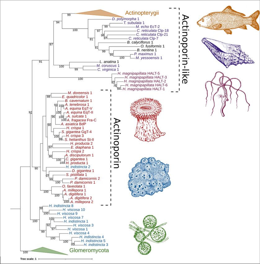
Mar. Drugs 2022, 20, 74                                                                                             2 of 20

                          molecular mechanisms of pore formation by APs, the reader is referred to reviews which
                          focus on this topic [13–15]. The qualities of APs which allow for their membrane-binding
                          and pore-forming activity have attracted attention regarding potential biotechnological
                          and therapeutic applications, such as the design of immunotoxins, nanopores, adjuvants,
                          and SM-specific probes [16–19].
                                 Historically, sea anemones have been the primary source of APs, although similar
                          cytolytic proteins can be found in other anthozoans [20,21]. Indeed, an exhaustive bioin-
                          formatic analysis indicated that actinoporin-like proteins (ALPs) are distributed across
                          multiple phyla with high structural similarity despite low sequence similarity [22]. In
                          particular, APs and ALPs have been detected in chordates (primarily teleost fish), cnidar-
                          ians, molluscs, mosses, and ferns. Furthermore, a structural similarity of APs and ALPs
                          to fungal-fruit body lectins has also been determined. A phylogenetic analysis of these
                          identified proteins revealed four distinct groups comprising ALPs primarily found in verte-
                          brates, hydrozoan ALPs, APs from cnidarians and plants, and fungal fruit-body lectins, all
                          of which were proposed to comprise the actinoporin-like proteins and fungal fruit-body
                          lectins superfamily (AF). The presence of ALP genes in non-vertebrate bilaterians has been
                          further illuminated in studies focused on polychaetes of the genus Glycera, the crustacean
                          Xibalbanus tulumensis, the brachiopod Lingula anatina, and many molluscs of the classes
                          Gastropoda and Bivalvia [23–25]. Several ALPs have been functionally characterized, in-
                          dicating that they can possess similar qualities to APs regarding membrane binding and
                          cytolytic activity.
                                 The first published example of an ALP was echotoxin II, isolated from the salivary
                          glands of the predatory mollusc Monoplex echo, which also has an amphipathic N-terminal
                          α-helix, a patch of aromatic residues, and hemolytic activity, but a specificity to gangliosides
                          rather than SM [26]. In further contrast to APs, an ALP from the zebrafish, Danio rerio,
                          possessed no cytolytic activity and its membrane-binding activity was not specific to
                          SM [22]. Yet bryoporin, from the moss Physcomitrella patens, showed consistency with
                          its close phylogenetic grouping to APs in that it also exhibited specificity for SM as well
                          as hemolytic activity, although its biological role appears to be related to dehydration
                          stress [27]. Similarly, clamlysin B from the bivalve Corbicula japonica also exhibits SM-
                          binding and cytolytic activity [28]. Finally, the ALP HALT-1 from the cnidarian Hydra
                          magnipapillata, which is phylogenetically distinct from anthozoan APs, exhibits lower
                          hemolytic activity, the creation of larger pores, and a lower affinity to SM in comparison
                          to EqT-II [29]. Altogether, the observation that ALPs from non-anthozoans can possess
                          similar biochemical properties to APs supports the notion that they may also be potential
                          targets for the aforementioned biotechnological and therapeutic applications which have
                          been investigated for EqT-II, Stn-II, and Fra-C.
                                 Sea sponges of the phylum Porifera are benthic, filter-feeding animals which can be
                          found in marine and freshwater environments throughout the world. Given the niche they
                          fill, challenges faced by these organisms include contact with pathogenic microorganisms,
                          spatial competition with other benthic life, and predation [30–32]. In order to deal with
                          these challenges, many sponges utilize complex chemical armaments which display an
                          array of bioactivities towards targets, such as pathogens, fouling organisms and cancer-
                          ous cells [33–35]. While a majority of these bioactivities have been attributed to small
                          molecules, some larger proteinaceous toxins have also been identified, such as suberitine
                          from Suberities domuncula, halilectin-3 from Haliclona caerulea, and chondrosin from Chon-
                          drosia reniformis [36–38]. In addition, sea sponges have also been shown to be a source
                          of cytolytic pore-forming proteins, one of which is an antibacterial, perforin-like protein
                          from S. domuncula, which was found to be upregulated upon exposure to lipopolysac-
                          charide [39,40]. No ALPs have been isolated and characterized from this phylum, but a
                          recent phylogenetic study has indicated that genes encoding for these proteins are present
                          in the genome of the species Oscarella pearsei (then O. carmela, when its genome was se-
                          quenced) [21,41,42]. Little was reported on this ALP other than it being phylogenetically
                          distant from both anthozoan APs, hydrozoan ALPs and mollusc ALPs. Similarly, while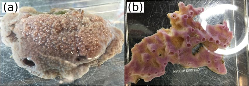
Mar. Drugs 2022, 20, 74                                                                                                                  3 of 20

                                       analyzing our transcriptome assemblies of native Irish Haliclona species, we noticed the
                                       presence of numerous genes encoding for proteins with the Pfam domain PF06369, rep-
                                       resenting sea anemone cytotoxic proteins. The presence of ALPs in sponges of both the
                                       classes Demospongiae and Homoscleromorpha prompted the questions of whether these
                                       proteins are widely distributed throughout the phylum and how similar they are to known
                                       APs. As discussed previously, such a quality would expand the possible biotechnological
                                       and therapeutic applications of sponges. To date, these organisms have been the subject of
                                       numerous transcriptomic and genomic studies, resulting in a wealth of public data with
                                       which to carry out such an inquiry [43]. To address these aforementioned questions and
                                       expand the knowledge of APs and ALPs, we herein present an exploration of the diversity,
                                       distribution, and predictive function of these proteins in the phylum Porifera.

                                       2. Results
                                       2.1. Transcriptome Sequencing and ALP Identification
                                             After processing with fastp, approximately 472.68, 69.80, 257.97, 207.46, and 94.64 Mbp
                                       of data were acquired for H. cinerea, H. indistincta, H. oculata, H. simulans, and H. viscosa,
                                       respectively. Five separate transcriptomes were then assembled using the Trinity RNA-Seq
                                       assembler (Table 1). More data were available for H. cinerea, H. oculata, and H. simulans,
                                       which appeared to be reflected in the generally larger assembly size and higher number of
                                       true genes when compared to H. indistincta and H. viscosa (the latter two species belong to a
                                       separate species group that is phylogenetically distinct from the first three). Furthermore,
                                       this division was also apparent regarding GC content, in which the first three exhibited
                                       a value around 39%, while the latter exhibited a value of 44.5%. The total amount of
                                       translated open reading frames was largely reflected by the size of the assemblies, with
                                       H. cinerea yielding the most protein sequences, while H. viscosa yielded the least (Table 2).
                                       All five Haliclona transcriptomes exhibited very high completeness when assessed with the
                                       BUSCO eukaryote dataset (Table 3).

                                       Table 1. Trinity assembly statistics for the five Haliclona transcriptomes.

                                                                    Number of Trinity ‘Genes’
    Species          Total (Mbp)         Number of Contigs                                            Contig N50 (Kbp)           GC (%)
                                                                      Excluding Isoforms
   H. cinerea             156.12               123,111                         64,261                         2.81               39.59
  H. indistincta          106.87               101,413                         48,788                         2.03               44.10
   H. oculata             142.18               122,855                         70,008                         2.46               38.56
  H. simulans             104.12               106,366                         55,501                         1.89               39.87
   H. viscosa              94.09               105,831                         59,949                         1.73               44.50

                                       Table 2. TransDecoder open reading frame statistics for the five Haliclona transcriptomes.

    Species               Total (aa)        Number of Complete ORFs           Number of 50 Partial ORFs         Number of 30 Partial ORFs
   H. cinerea             34,243,765                     58,606                          14,476                           6129
  H. indistincta          26,553,534                     29,232                          15,794                           7290
   H. oculata             30,530,592                     49,528                          16,191                           5687
  H. simulans             24,643,377                     31,081                          16,112                           7561
   H. viscosa             22,299,218                     24,435                          12,776                           7166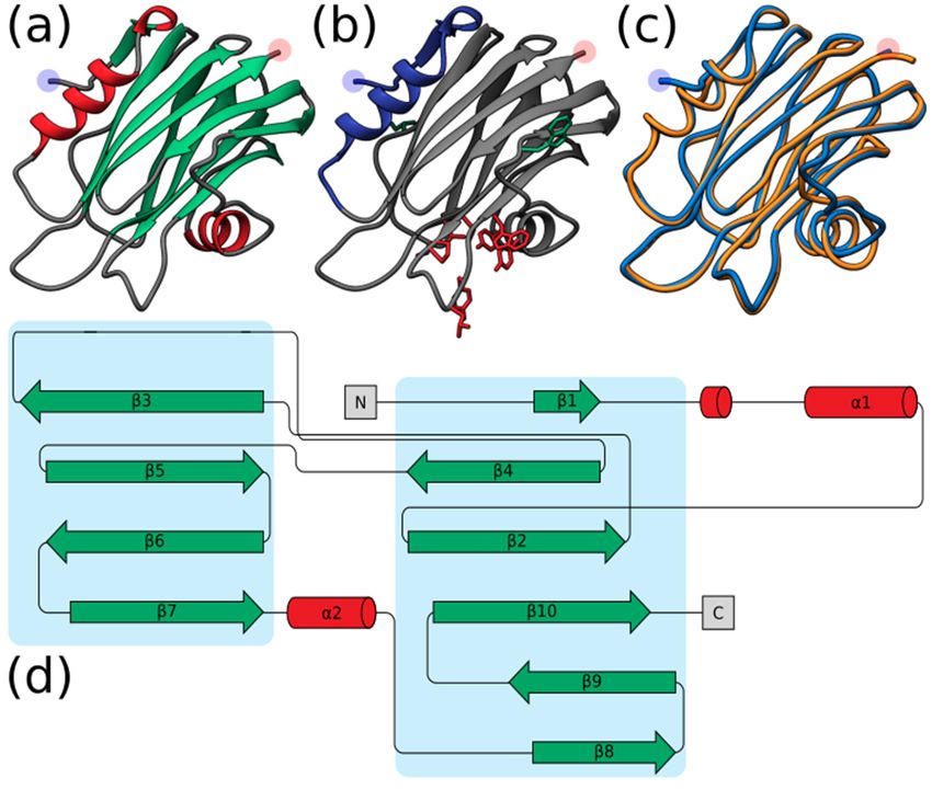
Mar. Drugs 2022, 20, 74                                                                                                       4 of 20

                               Table 3. BUSCO eukaryotic score for the five Haliclona transcriptomes.

        Species           Complete (%)          Single (%)          Duplicate (%)         Fragmented (%)          Missing (%)
      H. cinerea              97.6                 23.9                   73.7                   0.8                   1.6
     H. indistincta           99.6                 45.1                   54.5                   0.0                   0.4
      H. oculata              98.5                 31.4                   67.1                   0.4                   1.1
     H. simulans              97.6                 38.0                   59.6                   2.0                   0.4
      H. viscosa              95.3                 55.3                   40.0                   4.3                   0.4

                                    A total of 66 unique open reading frames encoding for proteins with the pfam domain
                               PF06369 were identified in the analyzed sponge NGS resources. These ORFs were derived
                               from 20 of the total 63 species that were screened (Table S1) Of these, 38 ORFs were
                               determined to be complete by TransDecoder. As none of these sponge-derived proteins
                               have been experimentally characterized, they are hence referred to as ALPs. Specifically,
                               these complete ALPs were found in C. orientalis (Co), C. varians (Cv), D. avara (Da), G. barretti
                               (Gb), H. indistincta (Hi), H. viscosa (Hv), O. pearsei (Op), a Scolapina sp. (S), S. officinalis (So),
                               and T. wilhelma (Tw). ALPs were absent from all freshwater sponge data. Their presence
                               in an order did not necessarily translate to this being an absolute feature of said order.
                               This is particularly exemplified by the order Haplosclerida, in which numerous paralogs
                               were detected in the sister species H. indistincta and H. viscosa, a few in H. amboinensis
                               and H. cinerea, and none in H. oculata, H. simulans, H. tubifera, or A. queenslandica. Only
                               C. varians and D. avara possessed the same degree of parology as H. indistincta and H.
                               viscosa. Of the sponge ALPs, only two from T. wilhelma were identified as having a signal
                               peptide by SignalP [44]. All the complete sponge ALPs aligned most closely with cnidarian
                               actinoporins when these were used in a blastp query against the NCBI non-redundant
                               database (Table S2). In particular, one ALP from H. indistincta named Hi2 exhibited the
                               highest sequence similarity with an actinoporin from Haloclava producta at 59.06% identity. It
                               was also observed that several ALPs from incomplete ORFs most closely aligned with pore-
                               forming proteins from other phyla, such as coluporins and tereporins from the Mollusca.
                               No ALPs were detected in the screened genomes or transcriptomes of choanoflagellates,
                               ctenophores, or placozoans. The theoretical isoelectric point of complete sponge ALPs
                               ranged from 4.66 to 9.46, whereas the average molecular weight ranged from 14,153.27 to
                               33,419.71 Da (Table S3).

                               2.2. Structural Prediction
                                     In contrast to the low-to-modest sequence similarity many sponge ALPs showed in
                               relation to actiniarian APs (27.70–59.06%) (Table S2), homology modeling with Phyre2
                               indicated that the aligned predicted structure of all complete sponge ALPs was highly
                               similar to that of Stn-II and EqT-II, with confidence values ranging from 97.3% to 100%.
                               Quantification of the similarity between the predicted sponge ALP models and the crystal
                               structure of EqT-II via TM-align showed that all produced models had a TM-align value
                               above 0.5, indicating that the structural similarity was not random (Figure 1), whereas the
                               RMSD values ranged from 0.54 to 1.74 (Å) (Table S4).
                                     It must be noted, however, that several sea sponge ALPs exhibited structural incon-
                               sistencies with the expected AP skeleton, such as the lack of an N-terminal α-helix. Hi2
                               exhibited one of the highest TM-scores at 0.96406. The quality of the predicted model for
                               Hi2 was further supported with a ProQ3D S-score of 0.697 (0.5–1.0 representing a good
                               model; Arne Elofsson, personal communication) and a ModFOLD8 p-value of 3.772 × 10⁻⁶
                               (less than a 1/1000 chance that the model is incorrect). As can be seen by the structure
                               generated by Phyre2, Hi2 shares many characteristics typical of cnidarian actinoporins,
                               such as comprising a β-sandwich flanked by two α-helices (Figure 2a,d). Furthermore, the
                               localization of the interfacial binding site can also be observed (Figure 2b). In general, the
                               predicted structure of Hi2 appears to overlay well with the crystal structure of EqT-II chain
                               A (1IAZ) (Figure 2c).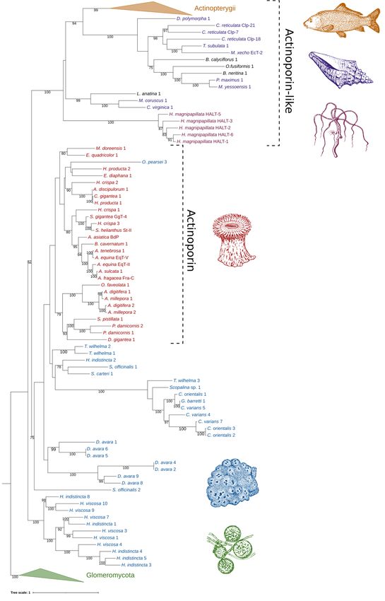
dicated that the aligned predicted structure of all complete sponge ALPs was highly sim-
ilar to that of Stn-II and EqT-II, with confidence values ranging from 97.3% to 100%. Quan-
tification of the similarity between the predicted sponge ALP models and the crystal struc-
ture of EqT-II via TM-align showed that all produced models had a TM-align value above
Mar. Drugs 2022, 20, 74 0.5, indicating that the structural similarity was not random (Figure 1), whereas 5the RMSD
of 20
values ranged from 0.54 to 1.74 (Å) (Table S4).
Figure 1. Comparison of complete sponge ALPs with EqT-II. Blue represents the TM-score of all
Figure 1. Comparison of complete sponge ALPs with EqT-II. Blue represents the TM-score of all
predicted sponge ALP structures via Phyre2 with the crystal structure of EqT-II (1IAZ). Orange
predicted sponge ALP structures via Phyre2 with the crystal structure of EqT-II (1IAZ). Orange
represents the sequence identity of the aligned region of the two structures. The orange bar rep-
resenting percent identity is overlaid upon the blue bar representing TM-score. Abbreviations are
as follows: Co, C. orientalis; Cv, C. varians; Da, Dysidea avara; Hi, H. indistincta; Hv, H. viscosa; Op,
O. pearsei; S, Scopalina sp.; So, S. officinalis; Tw, Tethya wilhelma. The protein Sc1 from S. carteri is
excluded due to having unknown residues. Sequence identity is expressed on a scale of 0–1 rather
than as a percentage.
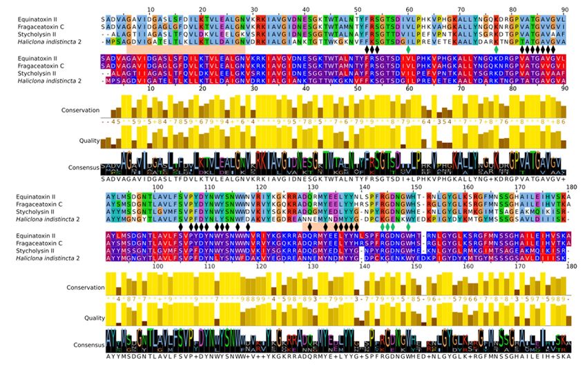
2.3. Multiple Sequence Alignment and Residue Analysis
Due to its high sequence similarity with cnidarian actinoporins, Hi2 was chosen
for further in-depth analyses to determine whether it exhibited the same membrane-
binding and pore-forming activities. A multiple sequence alignment of Hi2 with the final
mature peptides of the well-studied equinatoxin II (EqT-II; P61914), fragaceatoxin C (Fra-
C; B9W5G6), and stichyolysin II (Stn-II; P07845) indicated a percent identity of 50.56%,
50.00%, and 51.41%, respectively (Figure 3). Despite these modest values, the alignment
illustrated a high degree of conservation regarding residues and motifs critical for the
functional activities of actinoporins [15]. For example, a majority of the residues associated
with the interfacial binding site in Hi2 are consistent with those of EqT-II, Fra-C, and
Stn-II [6]. In addition, Hi2 possesses the conserved residue Tyr112, which is critical for
SM recognition; however, a substitution of Leu for Trp at residue 111 is also observed.
Furthermore, the presence of Ser53, Val86, Ser104, Pro106, Trp115, Tyre132, Tyr136, and
Tyr137 are consistent with the POC binding site found in cnidarian APs [4,6,15], and the
conserved P-[WYF]-D binding motif found in this region of APs is also present in Hi2 at
residues 106–108 [22]. Oligomerization of actinoporin monomers upon the cell membrane is
another crucial step towards pore formation and is known to be influenced by an Arg–Gly–
Asp motif. Hi2 shows inconsistency with this motif, as it instead possesses Lys142, Gly143,
and Glu144. However, Hi2 possesses the residue Lys76, which is consistent with similar
residues associated with oligomerization in other APs [45]. The presence of Ile59 and
Trp147 are also partially consistent with residues of Fra-C, associated with oligomerization
and protein–protein interaction between protomers; the observed substitution of Ile for
Val at this site can be seen in Stn-II [13,46]. Unlike most cnidarian APs, Hi2 exhibits the
presence of cysteine at residue 141, but such a characteristic is not unheard of in these
proteins [21].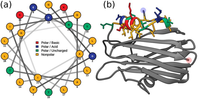
generated by Phyre2, Hi2 shares many characteristics typical of cnidarian actinoporins,
       such as comprising a β-sandwich flanked by two α-helices (Figure 2a,d). Furthermore, the
       localization of the interfacial binding site can also be observed (Figure 2b). In general, the
       predicted
Mar. Drugs 2022, 20, 74 structure of Hi2 appears to overlay well with the crystal structure of 6EqT-II
                                                                                                 of 20

       chain A (1IAZ) (Figure 2c).

                               Figure 2. (a) Predicted structure of Hi2 by Phyre2. Red represents α-helices. Green represents
     Figure 2. (a) Predicted   structure
                          β-sheets.            of Hi2
                                     (b) Significant     by Phyre2.
                                                     functional  residuesRed   represents
                                                                          of Hi2.               α-helices.
                                                                                  Blue represents             Green
                                                                                                    the N-terminal      represents
                                                                                                                    α-helix associated β-
     sheets. (b) Significant functional
                          with               residues
                                pore formation.           of Hi2. the
                                                   Red represents    Blue   represents
                                                                        residues            the N-terminal
                                                                                   of the interfacial            α-helix
                                                                                                      binding site.         associated
                                                                                                                    Green represents
     with pore formation. theRed    represents
                              residues   associatedthe
                                                     withresidues      of the(c)interfacial
                                                           oligomerization.                     bindingof site.
                                                                                  Structural alignment      Hi2 inGreen     represents
                                                                                                                    blue upon   EqT-II
                          chain  A (1IAZ)   in orange.  For all predicted structures   blue and  red highlights
     the residues associated with oligomerization. (c) Structural alignment of Hi2 in blue upon EqT-II          represent  the N- and
                          C-terminus, respectively. (d) Protein topology plot of Hi2 with the same color scheme as (a). The size
     chain A (1IAZ) in orange.     For all predicted structures blue and red highlights represent the N- and
                          of the topology plot was manually reduced to be more compact.
     C-terminus, respectively. (d) Protein topology plot of Hi2 with the same color scheme as (a). The
     size of the topology plotMembrane
                                 was manually          reduced
                                               penetration         to beformation
                                                             and pore      more compact.
                                                                                      by oligomerized APs is achieved by their
                        respective amphipathic N-terminal α-helices. In EqT-II, the N-terminal region undergoes a
                        conformational change, producing an α-helix comprising residues 6–28 which is capable of
     2.3. Multiple Sequence  Alignment and Residue Analysis
                        spanning a target membrane [10]. These residues correspond to a conserved N-terminal
                        glycine
          Due to its high        and C-terminal
                           sequence              asparagine
                                         similarity      withofcnidarian
                                                                the α-helix, which  are also present
                                                                             actinoporins,      Hi2 inwas
                                                                                                       Fra-C and Hi2.for
                                                                                                           chosen
                        Within this region, Hi2 also showed consistency with several previously determined highly
     further in-depth analysis     to determine whether it exhibited the same membrane-binding
                        conserved hydrophobic residues (Val7, Ile8, Leu13, Leu18, Leu22, and Ile25), as well as
                        Arg30, which is associated with the insertion of the α-helix into the target membrane [20].
                        Using these sequence boundaries allowed for the construction of an Edmundson peptide
                        helical wheel of the predicted Hi2 N-terminal α-helix after a hypothetical conformational
                        change (Figure 4a). Consistent with the amphipathic nature of the N-terminal α-helix of
                        EqT-II, Fra-C, and Stn-II, a side comprising a majority of polar amino acids opposite another
                        comprising a majority of nonpolar amino acids can be seen in that of Hi2. Furthermore,
                        the hydrophobic moment of Hi2, a measure of helix amphipathicity, was calculated to be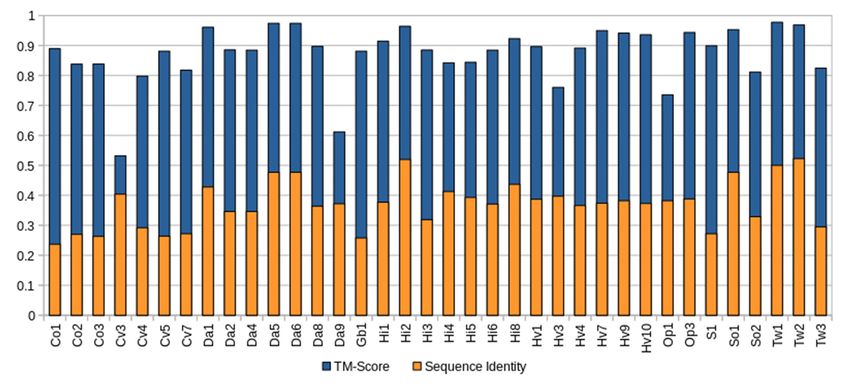
Tyr136, and Tyr137 are consistent with the phosphocholine binding site found in cnidar-
ian APs [4,6,15], and the conserved P-[WYF]-D binding motif found in this region of APs
is also present in Hi2 at residues 106–108 [22]. Oligomerization of actinoporin monomers
upon the cell membrane is another crucial step towards pore formation and is known to
Mar. Drugs 2022, 20, 74 be influenced by an Arg–Gly–Asp motif. Hi2 shows inconsistency with this motif, 7asofit20
instead possesses Lys142, Gly143, and Glu144. However, Hi2 possesses the residue Lys76,
which is consistent with similar residues associated with oligomerization in other APs
[45].
0.384The
µH,presence
which isofcomparable
Ile59 and Trp147
to that are also partially
of EqT-II at 0.337 consistent with residues
µH. The N-terminal of Fra-C,
α-helix of Hi2
associated with oligomerization and protein–protein interaction between
exhibits hydrophobicity of 0.607 and a net charge of 0. The two faces of the N-terminalprotomers; the
observed substitution of Ile for Val at this site can be seen in Stn-II [13,46].
α-helix prior to a hypothetical conformational change also display a hydrophobic and Unlike most
cnidarian
hydrophilic APs, Hi2which
side, exhibits
are,the presence oforiented
respectively, cysteinetowards
at residue
and141,
away butfrom
suchthe
a character-
rest of the
istic is not
protein unheard
(Figure 4b).of in these proteins [21].
Multiple
Figure3.3.Multiple
Figure sequence
sequence alignment
alignment of Hi2
of Hi2 with
with EqT-II,
EqT-II, Fra-C,
Fra-C, and and Stn-II.
Stn-II. Thealignment
The top top alignment
rep-
resents Clustalx color coding. The bottom alignment represents hydrophobicity color color
represents Clustalx color coding. The bottom alignment represents hydrophobicity coding.
coding. Be-
Between
tween these
these twotwo alignments,
alignments, black
black markers
markers represent
represent residues
residues importantfor
important formembrane
membranebinding,
binding,
green
greenmarkers
markersrepresent
representresidues
residuesimportant
importantforforoligomerization,
oligomerization,andandorange
orangerectangles
rectanglesrepresent
represent
the
the α-helices of EqT-II (the final length of the N-terminal α-helix after a conformationalchange
α-helices of EqT-II (the final length of the N-terminal α-helix after a conformational changeand
and
insertion
insertioninto
intothe
thetarget
targetmembrane
membraneisispresented)
presented)[10,12,13,15,45,46].
[10,12,13,15,45,46].
2.4. Phylogenetic Analysis
The relatedness between AP/ALP sequences from the two Haliclona species in which
they were present, AP sequences from cnidarians, and ALP sequences from other taxa
was visualized with an initial maximum likelihood tree (Figure 5). Here we find that the
sequence Hi2 from H. indistincta nestles well within the AP clade from cnidarians, but no
sequence of this type was found in its close sister species, H. viscosa. Additional (ALP)
sequences from H. indistincta and its sister species H. viscosa form a distinct and highly
supported clade well outside that of the cnidarians, indicating radiation from an additional
AP/ALP copy in the ancestor of that species group. This clade is distinct from the other
animal ALPs, which also form a monophyletic grouping supported by 91 BP.Furthermore, the hydrophobic moment of Hi2, a measure of helix amphipathicity, was
calculated to be 0.384 µH, which is comparable to that of EqT-II at 0.337 µH. The N-termi-
nal α-helix of Hi2 exhibits hydrophobicity of 0.607 and a net charge of 0. The two faces of
the N-terminal α-helix prior to a hypothetical conformational change also display a hy-
Mar. Drugs 2022, 20, 74 and hydrophilic side, which are, respectively, oriented towards and away from8 of 20
drophobic
the rest of the protein (Figure 4b).
Figure 4. (a) Edmundson
Figurepeptide helical wheel
4. (a) Edmundson peptideprojection ofprojection
helical wheel residuesof5–28 from5–28
residues Hi2. (b)Hi2.
from Residues
(b) Residues
5–28 of the Phyre2 predicted structure of Hi2 colored in the same manner
5–28 of the Phyre2 predicted structure of Hi2 colored in the same manner as (a). Blue and red high- as (a). Blue and red
highlights represent the N- and
lights represent the N- and C-terminus, respectively. C-terminus, respectively.
When all available AP/ALP sequences from Porifera are added to the analyses, the
2.4. Phylogenetic Analysis
high number of paralogs present in the dataset obscure the phylogenetic signal and reduce
confidence
The relatedness betweeninAP/ALP
generatingsequences
an accuratefrom
phylogeny.
the two However, the reconstructed
Haliclona maximum
species in which
likelihood tree provides insight into the sequence similarity and potential relatedness of
they were present,theAPseasequences from cnidarians, and ALP sequences from other taxa
sponge ALPs, as well as similar proteins from other phyla. Four distinct groups
was visualized with anstrong
with initialbootstrap
maximum likelihood
support tree (Figure
were produced: 5). Here
(1) ALPs we find
from fungi that
of the the
class Glom-
sequence Hi2 fromeromycota
H. indistincta
(usednestles well within
as outgroup), the AP clade
(2) the majority from cnidarians,
of sequences from the genusbutHaliclona,
no
(3) anthozoan
sequence of this type was found APsinwith
its other
closesponge
sisterALPs, and H.
species, (4) ALPs from
viscosa. other invertebrates
Additional (ALP) and
chordates (Figure 6). The strong grouping of the Haliclona ALPs, independent of all other
sequences from H. indistincta and its sister species H. viscosa form a distinct and highly
sequences and sitting at the base of the tree, was a consistently observed phenomenon
supported clade well
whileoutside thatalignments
testing other of the cnidarians, indicating
for the reconstruction of aradiation from
final tree (data notan addi-These
shown).
tional AP/ALP copy arein the ancestor
a sister ofother
group to all that APs
species group.
and ALPs This
in the clade
dataset. is remaining
The distinct from theALPs
APs and
other animal ALPs,form
which also form grouping
a monophyletic a monophyletic
within whichgrouping
there are supported
two clades; one,bysupported
91 BP. by 100 BP,
consisting of the freshwater Hydra and the bilaterians, the other containing the marine
cnidarians and sponges (75 BP). The presence of a large number of other sponge sequences
has the effect of pulling the Hi2 sequence outside the cnidarian clade, which itself is no
longer supported by bootstrapping. Relationships between the other sponge sequences are
unclear, as indicated by the low bootstrap support of internal nodes. This is particularly
exemplified by the likely spurious placement of Op3 within the clade of anthozoan APs.
Frequently, it was observed that additions or subtractions of sequences in the alignment
would result in this protein—along with Hi2, Sc1, So1, Tw1, and Tw2—being shifted in and
out of the cnidarian AP group. The main exception to this is a second strongly supported
clade consisting of ALPs from the genera Cliona, Geodia, Scopalina, and Tethya. While this
clade may move relative to other sponge sequences, the clade remained intact and distinct
from the anthozoan APs. Two groups of D. avara sequences were present, both highly
supported, but not always remaining together on trees, depending on the comparative
sequences included. They were also always distinct from anthozoan APs. Despite having
a high sequence similarity to both anthozoan APs and sponge ALPs, those derived from
plants, such as bryoporin, were only found to introduce additional noise into the data
without significantly changing tree topology and were thus excluded, being hypothesized
to be the result of a horizontal gene transfer event (data not shown) [27].Mar. Drugs 2022, 20, 74 9 of 20
Mar. Drugs 2022, 20, 74 8 of 20
Figure 5. Phylogenetic tree of ALPs from the genus Haliclona and similar proteins. Numbers on
Figure
nodes Phylogenetic
5. represent maximumtree likelihood
of ALPs from the genus
bootstrap Haliclona
values above 75%.and
The similar proteins.
color scheme Numbers on
is as follows:
nodes
greenrepresent
representsmaximum
ALPs fromlikelihood
fungi of thebootstrap values above
class Glomeromycota; 75%.
blue The color
represents ALPsscheme is as follows:
from Haliclona
sp.;represents
green red represents
ALPsAPsfrom
fromfungi
cnidarians
of theofclass
the class Anthozoa; reddish-purple
Glomeromycota; represents
blue represents ALPs ALPs
from from
Haliclona
sp.;cnidarians of the class
red represents APsHydrozoa; purple represents
from cnidarians of the classALPs from the phylum
Anthozoa; Mollusca; orange
reddish-purple rep- ALPs
represents
resents ALPs from fish of the class Actinopterygii; black represents ALPs from miscellaneous phyla.
from cnidarians of the class Hydrozoa; purple represents ALPs from the phylum Mollusca; orange
Abbreviations are as follows: EqT, equinatoxin; Fra, fragaceatoxin; St, sticholysin; BdP, bandaporin;
represents ALPs from
GgT, gigantoxin; fishhydra
HALT, of the class Actinopterygii;
actinoporin-like toxin; EcT,black represents
echotoxin; ALPs from
Clp, coluporin. miscellaneous
The glomero-
mycete
phyla. vector image is
Abbreviations a reproduced
are as follows:copyEqT,of equinatoxin;
the high-resolution image 4341-2_P1St,
Fra, fragaceatoxin; of Rhizophagus
sticholysin; BdP,
irregularis GgT,
bandaporin; (Błaszk., Wubet, Renker,
gigantoxin; and Buscot)
HALT, hydra C. Walker toxin;
actinoporin-like and A. Schüßler
EcT, derived
echotoxin; Clp,from
coluporin.
AAFC/CCAMF (https://agriculture.canada.ca/en/agricultural-science-and-innovation/agriculture-
The glomeromycete vector image is a reproduced copy of the high-resolution image 4341-2_P1 of
and-agri-food-research-centres-and-collections/glomeromycota-vitro-collection-ginco/catalogue-
Rhizophagus irregularis (Błaszk., Wubet, Renker, and Buscot) C. Walker and(accessed
arbuscular-mycorrhizal-fungi-strains-available-glomeromycetes-vitro-collection) A. Schüßler on 25derived
from AAFC/CCAMF
December 2021). The sea(https://agriculture.canada.ca/en/agricultural-science-and-innovation/
sponge, sea anemone, seashell, and carp vector images were derived from
those of Pearson Scott Foresman under a CC0 1.0 license (https://commons.wiki-
agriculture-and-agri-food-research-centres-and-collections/glomeromycota-vitro-collection-
media.org/wiki/File:Sponge_(PSF).png; https://commons.wikimedia.org/wiki/File:Anem-
ginco/catalogue-arbuscular-mycorrhizal-fungi-strains-available-glomeromycetes-vitro-collection)
one_2_(PSF).png; https://commons.wikimedia.org/wiki/File:Seashell_3_(PSF).png; https://com-
(accessed on 25 November 2021). The sea
mons.wikimedia.org/wiki/File:Carp_(PSF).jpg) sponge,
(accessed sea
on 26 anemone,
November seashell,
2021). The Hydra and carp vec-
vulgaris
tor vector
images image was derived from the Freshwater and Marine Image Bank at the University of Wash-license
were derived from those of Pearson Scott Foresman under a CC0 1.0
ington under a CC0 1.0 license (https://commons.wikimedia.org/wiki/File:FMIB_50097_Hydra_vul-
(https://commons.wikimedia.org/wiki/File:Sponge_(PSF).png; https://commons.wikimedia.org/
garis.jpeg) (accessed on 26 November 2021).
wiki/File:Anemone_2_(PSF).png; https://commons.wikimedia.org/wiki/File:Seashell_3_(PSF).png;
https://commons.wikimedia.org/wiki/File:Carp_(PSF).jpg) (accessed on 26 November 2021).
The Hydra vulgaris vector image was derived from the Freshwater and Marine Image Bank at the
University of Washington under a CC0 1.0 license (https://commons.wikimedia.org/wiki/File:
FMIB_50097_Hydra_vulgaris.jpeg) (accessed on 26 November 2021).Mar. Drugs 2022, 20, 74 10 of 20
Drugs 2022, 20, 74 10 of 20
Figure 6. Phylogenetic
Figure 6.tree of ALPs from
Phylogenetic treesea
ofsponges and sea
ALPs from similar proteins.
sponges and Numbers on nodesNumbers
similar proteins. rep- on nodes
resent maximum likelihood
represent bootstrap
maximum values above
likelihood 75%.values
bootstrap Blue represents
above 75%.sea sponge
Blue AP/ALPs.
represents All
sea sponge AP/ALPs.
other color schemes as well as image acknowledgements are identical to those of Figure 5.
All other color schemes as well as image acknowledgements are identical to those of Figure 5.Mar. Drugs 2022, 20, 74 11 of 20
3. Discussion
Cytolytic pore-forming toxins are widely distributed throughout prokaryotic and
eukaryotic life and often function as immunological defenses in the latter [47,48]. The
proposed AF superfamily includes α-pore-forming toxins derived from diverse eukaryotic
lineages with similar predicted protein structure despite low sequence similarity [22]. Mem-
bers of this family include anthozoan APs, plant APs, hydrozoan ALPs, ALPs from other
animals, and fungal fruit-body lectins. Herein, new additions from the phylum Porifera
are proposed to belong to the AF superfamily, as members of the classes Demospongiae,
Homoscleromorpha, and Calcarea have been shown to possess genes encoding for ALPs.
Many functionally characterized AF proteins appear to serve a role in envenomation as they
have been localized in the nematocysts of cnidarians and the salivary glands of predatory
molluscs [25,26,29,49]. However, AF proteins appear to also have functions other than
envenomation, as indicated by their presence in the mesenteric filaments of cnidarians,
as well as organisms which do not perform this process, such as bivalves [28,49]. With
this in consideration, it is not entirely unusual to observe the presence of these proteins in
non-venomous animals, such as sponges, in which they may serve a different ecological
function via a similar molecular mechanism.
When considering the taxonomic distribution of ALPs in sponges, no clear pattern can
be discerned. The majority of the identified ALPs were derived from the Demospongiae
and more specifically from the orders Axinellida, Bubarida, Clionaida, Dendroceratida,
Dictyoceratida, Haplosclerida, Poecilosclerida, Suberitida, Tethyida, and Tetractinellida,
but not Chondrillida, Spongillida, or Verongiida. However, further analysis at the genus
level indicated that these proteins appear to have been lost in species closely related to those
which possess ALPs. This is particularly exemplified by the genera Haliclona and Geodia, in
which the transcriptomes of numerous species have been reported [43,50]. Furthermore,
ALPs appear to be distributed throughout the phylum Porifera, as several were identified
in the classes Homoscleromorpha and Calcarea, but not in Hexactinellida. However,
two caveats should be considered regarding these observations. The first is that the high
prevalence of these proteins in the Demospongiae is most likely due to sampling bias, as
transcriptomes of this class have been disproportionately sequenced compared to the other
three. Second, as most of the data analyzed in this study are derived from transcriptomes,
a lack of gene expression or sequencing depth cannot be ruled out as an explanation for
these genes not having been identified in a species. That said, the absence of ALPs in the
Amphimedon queenslandica genome does appear to indicate that the loss of ALP genes has
occurred in the order Haplosclerida, which may be an explanation for their absence in other
species [51].
The predicted structure of most identified ALPs from sponges exhibited a high degree
of aligned structural similarity to anthozoan APs, despite a low to moderate sequence
similarity. Such an observation is common in studies of AF proteins from other organ-
isms [22,25]. That said, the observed high aligned structural similarity does not necessarily
equate to these sea sponge ALPs having the same membrane-binding and pore-forming
capabilities. This is particularly exemplified by the observation that the N-terminal region
of sea sponge ALPs appears to vary greatly. For example, this region is fully present
in Hi2, truncated in Hi3, and completely absent in Hi4. Such a quality is not unique to
sea sponge ALPs, as can be seen by analyzing those of hydrozoans [29]. Similarly, an
incomplete N-terminal α-helix was also observed on the ALP Dr1 from D. rerio and was
hypothesized to influence the lack of pore-forming capabilities and specificity towards SM
exhibited by the protein [22]. With this in mind, many of the identified ALPs from sea
sponges may serve functions other than those associated with well-characterized APs, such
as EqT-II. In contrast, the higher similarity of Hi2 to anthozoan APs at both a sequence
and structural level could indicate that it is capable of SM recognition and pore formation,
which prompted a further analysis on this concept.
A multiple sequence alignment of Hi2 with the model APs EqT-II, Fra-C, and Stn-II
indicated that this predicted protein shares numerous conserved residues associated withMar. Drugs 2022, 20, 74 12 of 20
the fundamental processes of lipid recognition and membrane binding, insertion of the N-
terminal α-helix into the target membrane, and oligomerization allowing for pore formation.
The presence of a patch of aromatic residues (Tyr114, Trp117, Tyr134, Tyr138, and Tyr139) in
Hi2 is consistent with the IBS observed in the model APs EqT-II, Fra-C, and Stn-II. The major
observed deviation is that a substitution of leucine for tryptophan occurs at residue 113
of Hi2. The importance of the equivalent residue in EqT-II, Trp112, in membrane binding
and SM recognition has been exemplified by studies in which this residue was mutated to
phenylalanine or subject to 19 F NMR studies [52,53]. This substitution also appears to be
prevalent in nature, as it exists in numerous anemone APs, as well as the ALP Dr1 from
D. rerio [20,22]. Furthermore, a mutant of EqT-II containing this substitution exhibited
similar SM specificity to the wild-type protein [6]. For these reasons, this substitution
in Hi2 is not expected to significantly inhibit any possible membrane-binding and SM-
recognition capabilities of the protein. While at a lower level of conservation the N-terminal
region of Hi2 also appears to form an α-helix of similar length to EqT-II, Fra-C, and Stn-
II. The notion that this α-helix is capable of inserting itself into a target membrane is
supported by the amphipathic nature of the predicted helical wheel and its hydrophobic
moment comparable to previously analyzed AP N-terminal α-helices [20]. Finally, residues
associated with oligomerization were somewhat consistent between Hi2 and the model
actinoporins. While Hi2 showed conservation at residues Lys76, Ile59, and Trp147, whose
equivalents in anthozoan APs are associated with oligomerization, its RGD motif was
another site of substitution [12,13,45,46]. Hi2 instead shows a substitution of Lys for Arg
and Glu for Asp. That said, Lys and Glu are of a similar charge and hydrophobicity to Arg
and Asp, respectively, and may allow for the retention of the oligomerization function [12].
Furthermore, this Lys substitution has been observed in natural actinoporins [20]. Hi2
and many other sponge ALPs also exhibited an acidic pI (Supplementary Table), which is
uncharacteristic of the typically basic anthozoan APs [15,54]. This observation is not entirely
unusual, however, as there is a precedent for acidic APs derived from anthozoans [20,54,55].
The identification of ALPs from glomeromycete fungi with high sequence similar-
ity to known APs supports the previous notion of a pre-metazoan origin of these pro-
teins [22]. The presence of these genes in fungi, sponges, and cnidarians, and their absence
in choanoflagellates, ctenophores, and placozoans, could possibly be explained by a series
of gene losses occurring throughout their history. However, additional assemblies from
these organisms should be assessed prior to making this conclusion. Furthermore, the
observation that many species of sea sponge were the source of numerous ALP isoforms
may be an indication that duplication of this gene is a common event in this phylum,
similar to the situation in cnidarians and molluscs [25,54]. This observed diversification
paired with the fact that many sponge ALPs were derived from transcriptomes indicates
that these are not simply genomic relics but do play some sort of functional role in sponges.
In the phylogenetic analysis, the sea sponge ALPs were not found to cluster together in a
clear monophyletic group, which appears to further exemplify the notion of a high degree
of divergence of these proteins in the porifera. However, based on the strong signal that
numerous sponge ALPs share with anthozoan APs, to the point of being grouped together,
and considering the higher sequence similarity many sponge ALPs have with cnidarian
APs, several of these proteins from sponges may instead be classifiable as APs.
4. Materials and Methods
4.1. Sample Collection
Haliclona indistincta (MIIG1388; Appendix A Figure A1a) was collected at Corranroo
on 17 May 2019 and H. viscosa (MIIG1389 and MIIG1390; Figure A1b) was collected at
Bridges of Ross on 1 August 2019. The following sample processing protocol was applied to
both species. Visible epibionts were removed. The sponges were rinsed in sterile artificial
seawater. The sponges were then dissected into ~1 cm3 pieces and flash-frozen with liquid
nitrogen. Samples were stored at −70 ◦ C until further use. Voucher specimens were stored
in ethanol.Mar. Drugs 2022, 20, 74 13 of 20
4.2. RNA Extraction
A ~1 cm3 piece of flash-frozen sponge tissue was submerged in 500 µL of Trizol in
a 2 mL microcentrifuge tube. The tissue was semi-homogenized by hand with a plastic
pestle. An additional 500 µL of Trizol was then added to the sample. The sample was
mixed by gently inverting five times and allowed to incubate at room temperature for
5 min. The sample was then inverted and vortexed with a VWR Analogue mini vortex
mixer at maximum speed for 2 min. A volume of 100 µL BCP was added to the sample.
The sample was mixed by hand for 20 s and then vortexed for 10 s with a VWR Analogue
mini vortex mixer at maximum speed. The sample was incubated at room temperature
for 5–10 min. The sample was then centrifuged at 16,000 g for 15 min at 6 ◦ C. The clear,
aqueous layer at the top was transferred to a fresh microcentrifuge tube. RNA was purified
by first adding 500 µL of 100% isopropanol to the aqueous phase sample. The sample was
then inverted and vortexed with a VWR Analogue mini vortex mixer at maximum speed
for 2 min. The sample was left to incubate for 10 min at room temperature. The sample was
then centrifuged at 16,000 g for 15 min at 4 ◦ C. The supernatant was discarded and 1 mL of
75% EtOH was added to the RNA pellet. The pellet was disrupted by vortexing. The RNA
sample was then centrifuged for 5 min at 4 ◦ C at 7500 g. The EtOH was carefully removed
without disturbing the pellet. The washing with 75% EtOH was repeated once. The RNA
sample was allowed to air dry until the edges of the pellet were visible. Finally, the pellet
was resuspended in 100 µL molecular-grade water. The RNA sample was kept frozen at
−70 ◦ C until further use. A subsample of each RNA extraction was used for quality and
quantity assessment on a 2100 Bioanalyzer RNA Eukaryotic Chip.
4.3. Transcriptome Sequencing
Samples were sent to Macrogen, Inc. for the preparation of Illumina TruSeq Stranded
mRNA libraries from poly-A selection, with insert sizes of 150 bp. The libraries were
sequenced on a Novaseq, with a targeted 40 million reads per sample.
4.4. Transcriptome Assembly
Previously sequenced raw cDNA Illumina reads of H. cinerea (culture held at Carna
Marine Research Station), H. oculata (MIIG1250 and MIIG1251), H. indistincta (MIIG1093,
MIIG1094, MIIG1095), and H. simulans (MIIG1248 and MIIG1249) were acquired from Prof.
Grace P. McCormack and Dr. Jose Maria Aguilar-Camacho for use in this study (personal
communication) [43].
Raw cDNA reads of H. cinerea, H. indistincta, H. oculata, H. simulans, and H. viscosa
were processed with fastp version 0.2 using default settings to remove adapters and low-
quality regions [56]. The processed reads were then assembled with Trinity version 2.8.5
using default settings [57]. Reads were pooled so that one transcriptome per species was
assembled. Isoforms and low-expressed transcripts were retained in the final assembly. The
longest translated open reading frames per transcript were extracted using TransDecoder
version 5.5.0 [58]. Homology searches using these open reading frames as a query against
the SwissProt database (accessed on 14 January 2020) with BLASTp version 2.9.0 [59,60], as
well as the Pfam database (accessed on 14 January 2020) with HMMER version 3.2.1 [61,62],
were performed. Significant homologous alignments were used to guide TransDecoder
in identifying additional open reading frames. The completeness of the transcriptomes
was then assessed using BUSCO version 5.1.2; specifically, the assemblies were queried
against the latest version of the eukaryota_odb10 dataset (downloaded 13 April 2021) [63].
Transcriptome assembly, open reading frame extraction, BUSCO analysis, and the gen-
eration of the maximum likelihood trees were performed with an account at the Leibniz
Supercomputing Centre.
4.5. Identification of Novel Actinoporin-like Proteins from Sea Sponges
While analyzing the output of the homology search against the Pfam database, it
was noticed that numerous translated protein sequences possessed the Pfam domainMar. Drugs 2022, 20, 74 14 of 20
PF06369 representing sea anemone cytotoxic proteins, such as actinoporins. Due to the
biotechnological potential of actinoporins, this prompted the screening of all publicly
available sea sponge genomes [41,51,64–68] and transcriptomes [42,50,65,68–86] to see
if other members of the phylum also encoded ALPs in their genome. If transcriptome
assemblies were not provided in the original publication, the data were assembled in
the same manner as the Irish Haliclona. The Haliclona ALPs were used as queries in a
tblasn search against the other sponge assemblies with an e-value cutoff of 1e-4. The
longest open reading frames were then extracted from the hits using TransDecoder ver-
sion 5.5.0 [58]. These protein sequences were screened against the Pfam database (ac-
cessed on 14 January 2020) with HMMER version 3.2.1 [61,62] and only those indicated as
possessing domain PF06369 were retained for further analysis. These identified sponge
ALPs were also screened against the NCBI Conserved Domain Database to confirm that
PF06369 was the primary conserved domain [87]. To explore the possible evolutionary
origin of ALPs in animals, the genomes of the choanoflagellates Monosiga brevicollis and
Salpingoeca rosetta, the ctenophores Mnemiopsis leidyi and Pleurobrachia bachei, and the placo-
zoans Trichoplax adhaerens and Hoilungia hongkongensis were also screened using EqT-II as a
query [88–93]. Furthermore, nineteen choanoflagellate transcriptomes were also screened
in a similar manner [94].
4.6. Sequence Analysis and Structural Prediction
All identified sea sponge ALPs were used as a query against the NCBI non-redundant
protein database to identify the closest homologous sequence [95]. SignalP 5.0 was used to
identify the presence of signal peptides in sea sponge ALPs contained within a complete
ORF [44]. The isoelectric point and molecular weight of complete, mature sponge ALPs
were determined using the compute pI/Mw tool of ExPASy [96]. Protein structure predic-
tion of all sponge ALPs was performed using Phyre2 Suite version 5.1 [97]. The quality of
the predicted protein structure for Hi2 was assessed with ProQ3D and ModFOLD8 [98,99].
Structural alignment of sponge ALPs upon the crystal structure of chain A from EqT-II
(1iaz) was performed with TM-Align [100]. Protein structures were visualized using UCSF
Chimera version 1.15 [101]. A protein topology plot was created using Pro-origami [102]. A
multiple sequence alignment of Hi2, EqT-II (P61914), Fra-C (B9W5G6), and Stn-II (P07845)
was performed with MAFFT v7.490, with the L-INS-i alignment method using default
settings [103]. The multiple sequence alignment was then visualized using Jalview version
2.11.1.4 [104]. Analysis of the N-terminal α-helix of Hi2 and generation of an Edmundson
wheel projection were accomplished using HeliQuest and NetWheels [105,106]. RNA 3D
structure prediction was performed with 3dRNA v2.0 [107].
4.7. Phylogenetic Analysis of Actinoporin-like Proteins from Sea Sponges
Sea sponge ALPs derived from complete ORFs were chosen for multiple sequence
alignment and phylogenetic analysis. All alignments were performed using MAFFT 7.490,
with the L-INS-i alignment method using default settings [103]. APs were represented by
well-characterized actiniarian proteins, such as the aforementioned EqT-II, Fra-C, and Stn-II,
as well as those from stony and soft corals. ALPs were represented by the series of HALT
proteins from H. magnipapillata. It was observed that in general the sea sponge ALPs most
consistently aligned with sequences from sea anemones, fungi of the class Glomeromycota,
and teleost fish, in that order. To get more sequences for the phylogenetic tree, all complete
sponge ALPs were queried against the NCBI nr database against these three taxonomic
groups, as well as against molluscs and invertebrates which did not fall under these
aforementioned phyla. The top hit from each category for each sponge sequence was then
retrieved. This resulted in seven groups of sequences: glomeromycete fungi, sponges,
anthozoan cnidarians, hydrozoan cnidarians, molluscs, miscellaneous invertebrates, and
teleost fish (Table S5). All signal peptides were removed prior to alignments using SignalP
version 5.0 [44]. Each of these groups were separately aligned to the mature sequences of
EqT-II, Fra-C, and Stn-II. The individual alignments were then trimmed correspondingMar. Drugs 2022, 20, 74 15 of 20
to the boundaries of EqT-II, Fra-C, and Stn-II using using Jalview version 2.11.1.4 [104].
Several sponge ALPs, while complete, where excluded due to being excessively truncated
compared to EqT-II or introducing significant gaps. All trimmed sequences were then
pooled together and once more aligned. Maximum likelihood trees were constructed in IQ-
TREE version 2.1.4 with 1000 bootstrap pseudoreplicates with the intention of visualizing
the degree of similarity the sponge ALPs had to APs and ALPs from other phyla [108].
The resulting phylogenetic trees were modified using the Interactive Tree of Life (iTOL)
v6 [109].
4.8. Generation of Figures
Several figures were further modified using the GNU Image Manipulation Program
2.10.28 [110] and Inkscape 0.92 [111].
5. Conclusions
Sea sponges, like some other invertebrates, are a source of ALPs. These proteins
exhibit a high degree of predicted structural similarity as well as a phylogenetic signal
to APs from cnidarians. One AP, Hi2, also possesses a majority of conserved residues
associated with essential functions, including the recognition of SM, binding to membranes,
oligomerization, and the formation of pores. While their ecological role in sponges remains
to be determined, the aforementioned qualities encourage the exploration of these proteins
for the biotechnological applications which have been proposed for anthozoan APs.
Supplementary Materials: The following are available online at https://www.mdpi.com/article/10.3
390/md20010074/s1, Table S1. Presence; Table S2. Blastp nr; Table S3. pI-Mw; Table S4. TM-align; Table
S5. Trees.
Author Contributions: K.S. conceived the experiments, aided in the collection of samples, carried
out all other methodological tasks, prepared figures, and wrote the paper. G.P.M. supervised the
research and assisted in interpreting the data and in writing the paper. All authors have read and
agreed to the published version of the manuscript.
Funding: This project has received funding from the European Union’s Horizon 2020 research
and innovation programme under the Marie Skłodowska-Curie grant agreement No. 764840. The
Department of Further and Higher Education, Research, Innovation and Science, through the Higher
Education Authority (HEA), provided funding to higher education institutions to cover costed
extensions for research activities that are at risk because of interruptions caused by the COVID-
19 pandemic.
Institutional Review Board Statement: Not applicable.
Informed Consent Statement: Not applicable.
Data Availability Statement: The raw RNA-seq reads are available at the NCBI BioProject PR-
JNA795170. Transcriptome assemblies, sea sponge ALP sequences, predicted protein structures,
multiple sequence alignments, and maximum likelihood trees are available at Mendeley (https:
//data.mendeley.com/datasets/w9t6zsjjb7/1, accessed on 14 December 2021).
Acknowledgments: The authors thank the technicians of the NUIG Zoology Department for their
help in collecting H. indistincta. Olivier P. Thomas and Daniel Rodrigues are thanked for the collection
of H. viscosa. Jose Maria Aguillar-Camacho provided raw data of H. cinerea. Members of the Marie
Skłodowska-Curie Actions innovative training network IGNITE—Comparative Genomics of Non-
Model Invertebrates are thanked for their advice on methodology. The Irish marine biorepository is
thanked for providing a high-quality image of Haliclona viscosa for the graphical abstract.
Conflicts of Interest: The authors declare no conflict of interest.Mar. Drugs 2022, 20, 74 16 of 20
Mar. Drugs 2022, 20, 74 16 of 20
Conflicts of Interest: The authors declare no conflict of interest.
Appendix A
Appendix A
FigureA1.
Figure A1.(a)
(a)Haliclona
Haliclonaindistincta
indistincta(MIIG1388).
(MIIG1388). (b)
(b) Haliclona
Haliclonaviscosa
viscosa(MIIG1389).
(MIIG1389).
References
References
1.1. Kem,
Kem,W.R.
W.R.Sea SeaAnemone
AnemoneToxins:Toxins:Structure
Structureand Action.Biol.
andAction. Biol.Nematocysts
Nematocysts1988, 1988,375–405.
375–405.
2.2. Anderluh,
Anderluh, G.; Maček,
Maček, P. P. Cytolytic
CytolyticPeptide
Peptideand andProtein
ProteinToxins
Toxins from
from SeaSea Anemones
Anemones (Anthozoa:
(Anthozoa: Actiniaria).
Actiniaria). Toxicon
Toxicon 2002,2002, 40,
40, 111–
111–124. [CrossRef]
124. https://doi.org/10.1016/S0041-0101(01)00191-X.
3.3. Athanasiadis,
Athanasiadis,A.; A.;Anderluh,
Anderluh,G.; Maček,
G.; Maček, P.; P.;
Turk, D. Crystal
Turk, D. CrystalStructure
Structureof the
ofSoluble FormForm
the Soluble of Equinatoxin II, a Pore-Forming
of Equinatoxin Toxin
II, a Pore-Forming
from
Toxinthe Seathe
from Anemone ActiniaActinia
Sea Anemone Structure
Equina.Equina. 2001, 9,2001,
Structure 341–346. [CrossRef]
9, 341–346. https://doi.org/10.1016/S0969-2126(01)00592-5.
4.4. Mancheño,
Mancheño,J.M.; J.M.;Martín-Benito,
Martı́n-Benito,J.;J.;Martínez-Ripoll,
Martı́nez-Ripoll, M.;M.;
Gavilanes,
Gavilanes, J.G.; Hermoso,
J.G.; Hermoso, J.A.J.A.
Crystal andand
Crystal Electron Microscopy
Electron MicroscopyStructures
Struc-
of Sticholysin
tures II Actinoporin
of Sticholysin RevealReveal
II Actinoporin Insights into the
Insights intoMechanism
the Mechanism of Membrane
of Membrane Pore Formation.
Pore Formation. Structure 11, 1319–1328.
2003, 2003,
Structure 11, 1319–
[CrossRef]
1328. https://doi.org/10.1016/j.str.2003.09.019.
5.5. Mechaly,
Mechaly,A.E.;A.E.;Bellomio,
Bellomio,A.; Gil-Cartón,
A.; Gil-Cartón, D.;D.;
Morante,
Morante, K.;K.;
Valle, M.;M.;
Valle, González-Mañas,
González-Mañas, J.M.;J.M.;
Guérin, D.M.A.
Guérin, Structural
D.M.A. Insights
Structural into
Insights
the
intoOligomerization
the Oligomerization and Architecture
and Architectureof Eukaryotic Membrane
of Eukaryotic Pore-Forming
Membrane Toxins. Structure
Pore-Forming Toxins. 2011, 19, 181–191.
Structure 2011, 19, [CrossRef]
181–191.
[PubMed]
https://doi.org/10.1016/j.str.2010.11.013.
6.6. Bakrač,
Bakrač, B.;
B.; Gutiérrez-Aguirre,
Gutiérrez-Aguirre, I.; I.; Podlesek,
Podlesek, Z.; Z.; Sonnen,
Sonnen, A.F.-P.;
A.F.-P.; Gilbert,
Gilbert, R.J.C.;
R.J.C.; Maček,
Maček, P.; P.;Lakey,
Lakey,J.H.;
J.H.;Anderluh,
Anderluh,G. G.Molecular
Molecular
Determinants
DeterminantsofofSphingomyelin
Sphingomyelin Specificity
Specificity of a ofEukaryotic Pore-Forming
a Eukaryotic Pore-Forming J. Biol. Chem.
Toxin. Toxin*. 2008,
J. Biol. 283, 2008,
Chem. 18665–18677. [CrossRef]
283, 18665–18677.
7. Bakrač, B.; Kladnik, A.; Maček, P.; McHaffie, G.; Werner, A.; Lakey, J.H.; Anderluh, G. A Toxin-Based Probe Reveals Cytoplasmic
https://doi.org/10.1074/jbc.M708747200.
7. Exposure
Bakrač, B.;ofKladnik, A.; Maček, P.; J.
Golgi Sphingomyelin. Biol. Chem.
McHaffie, G.;2010, 285,A.;
Werner, 22186–22195.
Lakey, J.H.;[CrossRef]
Anderluh, G. A Toxin-Based Probe Reveals Cytoplas-
8. Malovrh,
mic ExposureP.; Viero, G.; Serra,
of Golgi M.D.; Podlesek,
Sphingomyelin*. Z.; Lakey,
J. Biol. J.H.; Maček,
Chem. 2010, P.; Menestrina,
285, 22186–22195. G.; Anderluh, G. A Novel Mechanism of Pore
https://doi.org/10.1074/jbc.M110.105122.
8. Formation:
Malovrh, P.; Membrane
Viero, G.;Penetration
Serra, M.D.;byPodlesek,
the N-Terminal Amphipathic
Z.; Lakey, J.H.; Maček, Region of Equinatoxin.
P.; Menestrina, J. Biol. Chem.
G.; Anderluh, G. A2003, 278,Mechanism
Novel 22678–22685. of
[CrossRef] [PubMed]
Pore Formation: Membrane Penetration by the N-Terminal Amphipathic Region of Equinatoxin. J. Biol. Chem. 2003, 278, 22678–
9. Rojko,
22685. N.; Kristan, K.Č.; Viero, G.; Žerovnik, E.; Maček, P.; Serra, M.D.; Anderluh, G. Membrane Damage by an α-Helical
https://doi.org/10.1074/jbc.M300622200.
9. Pore-Forming
Rojko, N.; Kristan,Protein,
K.Č.;Equinatoxin II, Proceeds
Viero, G.; Žerovnik, throughP.;a Serra,
E.; Maček, Succession
M.D.; of OrderedG.
Anderluh, Step. J. Biol. Chem.
Membrane Damage 2013, 288,α-Helical
by an 23704–23715.
Pore-
[CrossRef]
Forming Protein, Equinatoxin II, Proceeds through a Succession of Ordered Step*. J. Biol. Chem. 2013, 288, 23704–23715.
10. Drechsler, A.; Potrich, C.; Sabo, J.K.; Frisanco, M.; Guella, G.; Dalla Serra, M.; Anderluh, G.; Separovic, F.; Norton, R.S. Structure
https://doi.org/10.1074/jbc.M113.481572.
10. and Activity
Drechsler, A.;ofPotrich,
the N-Terminal
C.; Sabo,Region of the Eukaryotic
J.K.; Frisanco, M.; Guella,Cytolysin Equinatoxin
G.; Dalla Serra, II. Biochemistry
M.; Anderluh, 2006, 45,
G.; Separovic, F.;1818–1828.
Norton, R.S. [CrossRef]
Structure
[PubMed]
and Activity of the N-Terminal Region of the Eukaryotic Cytolysin Equinatoxin II. Biochemistry 2006, 45, 1818–1828.
11. Lam, Y.H.; Hung, A.; Norton, R.S.; Separovic, F.; Watts, A. Solid-State NMR and Simulation Studies of Equinatoxin II N-Terminus
https://doi.org/10.1021/bi052166o.
11. Interaction
Lam, Y.H.; withHung, Lipid
A.; Norton, Proteins
Bilayers.R.S.; Struct.F.;
Separovic, Funct.
Watts,Bioinform. 2010, 78,
A. Solid-State NMR 858–872. [CrossRef]Studies of Equinatoxin II N-Termi-
and Simulation
12. García-Linares,
nus InteractionS.; Richmond,
with R.; García-Mayoral,
Lipid Bilayers. Proteins Struct. M.F.; Bustamante,
Funct. Bioinform.N.; Bruix,
2010, 78,M.; Gavilanes,
858–872. J.G.; Martínez-del-Pozo, Á. The Sea
https://doi.org/10.1002/prot.22612.
12. Anemone Actinoporin (Arg-Gly-Asp) Conserved Motif Is Involved in Maintaining
García-Linares, S.; Richmond, R.; García-Mayoral, M.F.; Bustamante, N.; Bruix, M.; Gavilanes, J.G.; the Competent Oligomerization State of Á.
Martínez-del-Pozo, These
The
Pore-Forming Toxins. FEBS J. 2014, 281, 1465–1478. [CrossRef] [PubMed]
Sea Anemone Actinoporin (Arg-Gly-Asp) Conserved Motif Is Involved in Maintaining the Competent Oligomerization State of
13. Tanaka, K.; Caaveiro, Toxins.
These Pore-Forming J.M.M.; FEBS
Morante, K.; González-Mañas,
J. 2014, J.M.; Tsumoto, K. Structural Basis for Self-Assembly of a Cytolytic
281, 1465–1478. https://doi.org/10.1111/febs.12717.
13. Pore Lined
Tanaka, K.;by Protein and
Caaveiro, Lipid.
J.M.M.; Nat. Commun.
Morante, 2015, 6, 6337. J.M.;
K.; González-Mañas, [CrossRef]
Tsumoto, K. Structural Basis for Self-Assembly of a Cytolytic
14. Črnigoj Kristan,
Pore Lined K.; Viero,
by Protein andG.; DallaNat.
Lipid. Serra, M.; Maček,
Commun. 2015,P.;6,Anderluh, G. Molecular Mechanism of Pore Formation by Actinoporins.
6337. https://doi.org/10.1038/ncomms7337.
14. Toxicon
Črnigoj2009, 54, 1125–1134.
Kristan, K.; Viero, G.;[CrossRef]
Dalla Serra,[PubMed]M.; Maček, P.; Anderluh, G. Molecular Mechanism of Pore Formation by Actino-
15. Rojko,
porins.N.; Dalla2009,
Toxicon Serra,54,M.; Anderluh, G. Pore Formation by Actinoporins, Cytolysins from Sea Anemones. Biochim.
Maček, P.;https://doi.org/10.1016/j.toxicon.2009.02.026.
1125–1134.
15. Biophys.
Rojko, N.;Acta (BBA)-Biomembr.
Dalla Serra, M.; Maček, 1858,
2016,P.; 446–456.G.[CrossRef]
Anderluh, [PubMed]
Pore Formation by Actinoporins, Cytolysins from Sea Anemones. Biochim.
16. Mutter, N.L.; Soskine, M.; Huang, G.; Albuquerque, I.S.; Bernardes, G.J.L.; Maglia, G. Modular Pore-Forming Immunotoxins with
Et Biophys. Acta (BBA)-Biomembr. 2016, 1858, 446–456. https://doi.org/10.1016/j.bbamem.2015.09.007.
16. Caged
Mutter,Cytotoxicity
N.L.; Soskine, Tailored by Directed
M.; Huang, Evolution. ACS
G.; Albuquerque, I.S.;Chem. Biol. 2018,
Bernardes, 13,Maglia,
G.J.L.; 3153–3160. [CrossRef]
G. Modular Pore-Forming Immunotoxins
17. Wloka, C.; Mutter,
with Caged N.L.; Soskine,
Cytotoxicity TailoredM.; byMaglia,
Directed G.Evolution.
Alpha-Helical ACSFragaceatoxin
Chem. Biol. 2018, C Nanopore
13, 3153–3160.Engineered for Double-Stranded and
https://doi.org/10.1021/acschem-
Single-Stranded
bio.8b00720. Nucleic Acid Analysis. Angew. Chem. Int. Ed. 2016, 55, 12494–12498. [CrossRef]
17. Wloka, C.; Mutter, N.L.; Soskine, M.; Maglia, G. Alpha-Helical Fragaceatoxin C Nanopore Engineered for Double-Stranded and
Single-Stranded Nucleic Acid Analysis. Angew. Chem. Int. Ed. 2016, 55, 12494–12498. https://doi.org/10.1002/anie.201606742.You can also read