Anatomical Features of Some Bones of the Forelimbs of Lions (Panthera leo)
←
→
Page content transcription
If your browser does not render page correctly, please read the page content below

Int. J. Morphol.,
39(2):378-385, 2021.

                       Anatomical Features of Some Bones of the
                           Forelimbs of Lions (Panthera leo)

                                 Características Anatómicas de Algunos Huesos de los
                                     Miembros Torácicos de Leones (Panthera leo)

                           Md. Shahriar Hasan Sohel1; Kh. Nurul Islam2 & Mohammad Lutfur Rahman2

SOEL, M. S. H.; ISLAM, K. N. & RAHMAN, M. L. Anatomical features of some bones of the forelimbs of lions (Panthera leo). Int.
J. Morphol., 39(2):378-385, 2021.

         SUMMARY: We studied the bones of forelimb of four adult lions (Panthera leo) of both sexes to record the gross anatomical
and morphometrical features of the scapula, humerus, radius and ulna. We observed some unique anatomical features that will be helpful
for radiographic interpretation and forensic investigations. The lateral surface of scapula was unequally divided into supraspinous (fossa
supraspinata) and infraspinous fossa (fossa infraspinata) by a well developed spine (spina scapulae). The acromion process was subdivided
into suprahamate process (processus suprahamatus)and hamate process (processus hamatus); the later one was over hanged the glenoid
cavity (cavitas glenoidalis), but the supraglenoid tubercle (tuberculum supraglenoidalis) was absent. The shaft (diaphysis) of humerus
was compressed craniocaudally in proximal part, rounded to oval in middle part and compressed mediolaterally in distal part. A long,
narrow supracondyloid foramen was found at distal limb just above the medial epicondyle (epicondylus medialis) which didn’t connect
the radial fossa (fossa radialis) with the olecranon fossa (fossa olecrani). The radius and ulna were twin bones where radius was
articulated craniolateral to the ulna proximally and craniomedial to the ulna distally. However, the ulna was the longest bone in the
forelimb of lion. The olecranon tuberosity of this bone had three prominences - two were cranially, whereas the caudal one was the
largest and rounded. Distally projected styloid processes (processus styloideus) were found in the distal limb of both radius and ulna.

        KEY WORDS: Lion; Anatomy; Scapula; Humerus; Radius; Ulna.

INTRODUCTION

       The lion (Panthera leo), popularly known as the “King              equivalent to an architectural column; the downward scapula
of jungle” as possessing both beauty and strength. It is a                is aligned with the humerus and ulna (Nzalak et al., 2010).
member of the Felidae (cat) family and one of four big cats
in the genus Panthera (Nowak & Walker, 1999). Both in                              Many scientists have been studied on the skeletal
structure and in kinematic patterns, the bones of the lion                system of large animals, for example horse, cattle, small
mainly the bones of forelimb reveals many peculiarities                   ruminants such as sheep, goat (Sisson et al., 1975), carnivores
(Howell, 1944; Hildebrand & Hurley, 1985). Like other                     such as dog (Miller et al., 1964), wild carnivores such as
felines it is strongly muscular and contain very powerful                 tiger (Tomar et al., 2018), leopard (Podhade, 2007), Asiatic
muscles in their chests and forelimbs as well as their manus              cheetah (Nazem et al., 2017) and Indian wild cat (Palanisamy
can be supinated. These unique characteristics allow them to              et al., 2018). Only few literaturesare available on some bones
capture large prey such as buffalo, zebra, etc. Furthermore,              of the Asiatic lion (Pandey et al., 2004; Nzalak et al.), but
these allow them to reach speeds of over 80 kilometers per                the morphometrical study of the skeletal system of the lion
hour while chasing prey (Kirberger et al., 2005; Lucky &                  has not been studied in details. Beside this, in the field of
Harshan, 2014). The bones of the forelimb are twisted in such             radiology and forensic studies, the osteomorphometrical
a manner as to give a vast range of motion to the forelimb.               features of the scapula, humerus, radius and ulna are very
The shoulder, elbow and radiocarpal joints of the lion are                important. Therefore, considering the above facts the present
placed one above the other to hold up its heavy muscles                   study was conducted.

1
Laboratory of Veterinary Anatomy, Joint Graduate School of Veterinary Sciences, Gifu University, 1-1 Yanagido, Gifu 501-1193, Japan.
2
Department of Anatomy and Histology, Chattogram Veterinary & Animal Sciences University, Zakir Hossain Road, Khulshi, Chattogram-4225, Bangladesh.

378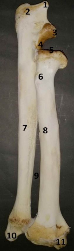
SOEL, M. S. H.; ISLAM, K. N. & RAHMAN, M. L. Anatomical features of some bones of the forelimbs of lions (Panthera leo). Int. J. Morphol., 39(2):378-385, 2021.

MATERIAL AND METHOD                                                                                 Though, Nzalak et al. called them as acromion and
                                                                                            metacromion process, respectively. The hamate process
                                                                                            was triangular with thick blunted ends that over hanged
        The scapula, humerus, radius and ulna of four adult                                 the glenoid cavity (cavitas glenoidalis). At the tip of the
lions of both sexes were examined at the Anatomy Museum                                     end it was slightly backward directed (Fig. 1). This finding
of Chattogram Veterinary and Animal Sciences University,                                    was consistent with the findings in lion (Nzalak et al.),
Bangladesh. Recently these four lions died in the Bangladesh                                although it was not over hanged to the glenoid cavity in
National Zoo, Dhaka and buried in different isolated places                                 cattle, sheep and goat (Sisson et al.). The suprahamate
of the zoo burial ground with aseptic measures. After six                                   process was resembling thick triangular plate and
months the bones were collected and subsequently processed                                  backward directed (Fig. 1) as previously observed in lion
by removing the mud and boiled with water and hydrogen                                      (Nzalak et al.).
peroxide (H2O2) for one hour to remove the remaining mus-
cular structure from the bones. After removing the muscu-                                           The supraglenoid tubercle (tuberculum
lar structures through knife all the bones were properly                                    supraglenoidalis) was absent in this study, which was
washed with fresh water and finally all the bones were dried                                agreed with lion (Pandey et al.; Nzalak et al.) but disagreed
under sunlight for a week. For the gross morphometric study,                                with the horse (Sisson et al.) and cattle (Budras & Habel,
the length, width, height and circumference were measured                                   2011). The surface of supraspinous fossa was centrally
by using a calibrated scale and were recorded in centimeter                                 undulating, concave dorsally then became convex and
(cm). The weight was also measured by using a digital ba-                                   finally concave towards the spine and the infraspinous fossa
lance and recorded in gram (g). The data were statistically                                 was almost similar but it was less undulating (Fig. 1). This
analyzed.                                                                                   result was strongly agreed with previous result of lion
                                                                                            (Nzalak et al.) but partially agreed with tiger (Tomar et
                                                                                            al.) and with Indian wild cat (Palanisamy et al.), where
RESULTS AND DISCUSSION                                                                      the authors pointed out that the infraspinous fossa was
                                                                                            flattened.

Scapula. The scapula of lion was downward and forward                                               The dorsal margin of scapula was extended from
directed triangular shape flat bone placed in the cranio-late-                              the level of the proximal limb of 1st rib to the middle of
ral aspect of thorax with the dorsal end being relatively wide                              the 6th rib. The outline was rough for the attachment of
and the ventral end being narrow. However, the quadrangular                                 scapular cartilage, but this cartilage was lost during the
shaped scapula was found in tiger (Tomar et al.), Indian                                    collection of specimens. The cranial margin of scapula was
wild cat (Palanisamy et al.) and civet cat (Sarma et al., 2017).                            slightly convex which extended from scapular notch (in-
It was slightly sloped that helped to adapt the form of the                                 cisura scapulae) to cranial angle(anguluscranialis) (Fig. 1).
forelimb laterally. The morphometrical data for different                                   The outline of this margin was circular and smooth,
parameters of scapula of the lions are illustrated in Table I.                              however it was thin and strongly circular in Indian wild
                                                                                            cat (Palanisamy et al.). The caudal (axillary) margin was
       The scapula possessed two surfaces, three margins                                    straight with thick and smooth outline and extended from
and three angles. The lateral surface was divided by a well                                 the caudal angle (angulus caudalis) to the glenoid cavity
developed spine (spina scapulae) into two unequal fossae,                                   (Fig. 1), which was similar to the study of Indian lion
i.e. supraspinous fossa (fossa supraspinata) and                                            (Nzalak et al.), tiger (Tomar et al.), leopard (Podhade) and
infraspinous fossa (fossa infraspinata) (Fig. 1) as studied                                 Indian wild cat (Palanisamy et al.).
previously in lion (Pandey et al.). However, the equal
fossae were found in dog (Miller et al.; Sisson et al.) and                                        The cranial angle (angulus cranialis) was not well
Indian wild cat (Palanisamy et al.). The height of the spine                                distinct but fused with the adjacent margins, whereas the
gradually decreased towards the proximal limb, which was                                    caudal angle (angulus caudalis) was thick, rough and
similar to the findings of Nzalak et al. and Pandey et al.                                  tuberous. Moreover, the ventral angle (angulus ventralis)
The edge of spine was inclined towards the infraspinous                                     of scapula was articulated with humerus by glenoid cavity
fossa except in its distal 1/4th part, whereas the proximal                                 (cavitas glenoidalis) of scapula and head of humerus. The
1/3rd was slightly rough and thickened as reported in tiger                                 glenoid cavity (cavitas glenoidalis) was elongated oval
(Tomar et al.). The distal continuation of the spine namely                                 shaped (Fig. 2), which was variable in some other species
acromion process was composed of hamate process                                             such as it was elongated in elephant (Ahasan et al., 2016),
(processus hamatus) and suprahamate process (processus                                      oval to quadrangular in tiger (Tomar et al.) and oval shape
suprahamatus) (Fig. 1).                                                                     in Indian wild cat (Palanisamy et al.).
                                                                                                                                                                            379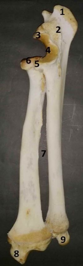
SOEL, M. S. H.; ISLAM, K. N. & RAHMAN, M. L. Anatomical features of some bones of the forelimbs of lions (Panthera leo). Int. J. Morphol., 39(2):378-385, 2021.
Table I. Morphometrical data for different parameters of scapula, humerus, radius and ulna (N=4).
Bone Parameters Mean ± SE
Right Left
Weight (g) 216.5 ± 22.70 217.5 ± 23.97
Maximum length (Dorsal margin to glenoid cavity) (cm) 25.98 ± 0.94 25.85 ± 1.02
Maximum width (Cranial margin to caudal angle) (cm) 20.55 ± 0.79 20.48 ± 0.88
Length of cranial margin (cm) 22.20 ± 0.89 22.18 ± 0.94
Length of caudal margin (cm) 21.43 ± 0.97 21.38 ± 1.05
Length of dorsal margin (cm) 15.48 ± 0.95 15.63 ± 0.95
Length of scapular spine (cm) 19.70 ± 0.79 19.53 ± 0.72
Scapula Height of scapular spine from supraspinous fossa (cm) 3.55 ± 0.16 3.7 ± 0.16
Height of scapular spine from infraspinous fossa (cm) 4.25 ± 0.23 4.33 ± 0.18
Maximum width of supraspinous fossa (cm) 8.45 ± 0.48 8.65 ± 0.41
Maximum width of infraspinous fossa (cm) 10.13 ± 0.45 10.23 ± 0.45
Length of glenoid cavity (cm) 5.33 ± 0.33 5.45 ± 0.37
Width of glenoid cavity (cm) 3.63 ± 0.24 3.75 ± 0.26
Distance between glenoid cavity and acromion process (cm) 3.83 ± 0.24 3.75 ± 0.26
Weight (g) 407 ± 56.48 400.7 ± 56.64
Total length (cm) 31.23 ± 1.48 31.10 ± 1.47
Shaft
Length (cm) 25.55 ± 1.22 25.45 ± 1.22
Circumference of upper part (cm) 14.10 ± 0.66 13.92 ± 0.65
Circumference of middle part (cm) 11.58 ± 0.85 11.45 ± 0.82
Circumference of lower part (cm) 11.25 ± 0.47 11.20 ± 0.44
Humerus Circumference of head (cm) 17.28 ± 1.21 17.22 ± 1.15
Proximal limb
Circumference (cm) 18.4 ± 1.32 18.27 ± 1.32
Width (cm) 9.20 ± 0.67 9.07 ± 0.67
Distal limb
Circumference (cm) 15.05 ± 1.09 14.97 ± 1.06
Width (cm) 8.08 ± 0.39 8.0 ± 0.35
Depth of olecranon fossa (cm) 2.20 ± 0.21 2.1 ± 0.20
Total length (cm) 28.62 ± 1.23 28.57 ± 1.22
Proximal limb
Circumference (cm) 13.97 ± 1.08 13.85 ± 1.06
Width (cm) 3.97 ± 0.28 3.92 ± 0.30
Radius Distal limb
Circumference (cm) 14.1 ± 1.32 13.97 ± 1.33
Width (cm) 4.2 ± 0.38 4.1 ± 0.38
Circumference at mid shaft (cm) 7.57 ± 0.52 7.47 ± 0.48
Total length (cm) 34.97 ± 1.59 35.0 ± 1.66
Circumference
Proximal limb (cm) 10.92 ± 0.86 10.8 ± 0.84
Ulna
Distal limb (cm) 7.85 ± 0.78 7.72 ± 0.79
Length of olecranon (cm) 9.17 ± 0.53 9.12 ± 0.52
Circumference at distal limb of olecranon (cm) 9.82 ± 0.56 9.77 ± 0.55
On the medial surface, the subscapular fossa (fossa comparatively shallow subscapular fossa with two ridges
subscapularis) was deep (Fig. 3) and contain two were found in tiger (Tomar et al.), whereas four ridges
prominent ridges namely the anterior ridge and posterior were observed in civet cat (Sarma et al.). This discrepancy
ridge. The anterior one was curved and started from the due to the species differences. In this study, an almost
lower one third of the cranial margin, became prominent rounded tiny coracoid process (processus coracoideus) also
towards the distal limb. On the other hand, the posterior observed that was directed medially backward and
ridge was straight, started immediately below the caudal downward (Fig. 3). However, this result was partially
angle ran parallel to the caudal margin and became analogous to the study in tiger (Tomar et al.) and civet cat
prominent towards the distal limb and ended at above the (Sarma et al.), where they mentioned hook like prominent
rim of the glenoid cavity (cavitas glenoidalis). However, coracoid process.
380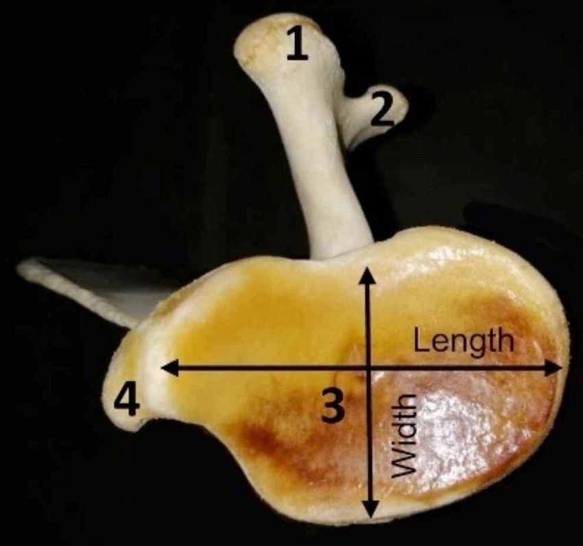
SOEL, M. S. H.; ISLAM, K. N. & RAHMAN, M. L. Anatomical features of some bones of the forelimbs of lions (Panthera leo). Int. J. Morphol., 39(2):378-385, 2021.

                                                                                                Fig. 2. Ventral view of left scapula of lion. 1= Hamate process
                                                                                                (Processus hamatus), 2=Suprahamate process (Processus
                                                                                                suprahamatus), 3= Glenoid cavity (Cavitas glenoidalis) and 4=
                                                                                                Coracoid process (Processus coracoideus).

Fig. 1. Lateral view of left scapula of lion. 1= Cranial angle (Angulus
cranialis), 2= Caudal angle (Angulus caudalis), 3= Cranial margin,
4= Caudal margin, 5= Supraspinous fossa (Fossa supraspinata),
6= Infraspinous fossa (Fossa infraspinata), 7= Scapular spine
(Spina scapulae), 8= Tuberosity of spine, 9=Suprahamate process
(Processus suprahamatus), 10= Hamate process (Processus
hamatus), 11= Scapular notch (Incisura scapulae) and 12=
Coracoids process (Processus coracoideus).

Humerus. The humerus is one of the major bones in the
appendicular skeleton of lion to bear the total body weight.
In the present study, it was a long bone with a spirally twisted
shaft (corpus humeri), which was located obliquely
downward and backward directed. It formed shoulder joint
above by its head with the glenoid cavity of scapula and
elbow joint below by its condyles with the proximal limbs
of radius and ulna. The morphometrical data for different
parameters of humerus of lions are presented in Table I.

        It possessed a cylindrical shaft (corpus humeri) and
two enlarged limbs (epiphysis) such as proximal limb and
distal limb. The head (caput humeri) was rounded (Fig. 4)
                                                                                                Fig. 3. Medial view of left scapula of lion. 1= Cranial angle (Angulus
and had a large, undivided tubercle (tuberculum)- the greater
                                                                                                cranialis), 2= Caudal angle (Angulus caudalis), 3= Caudal ridge,
or major (tuberculum major) and lesser tubercle (tuberculum                                     4= Cranial ridge, 5= Dorsal margin, 6= Cranial margin, 7= Caudal
minus) (Fig. 5). The greater tubercle was large and prominent                                   margin, 8= Scapular notch (Incisura scapulae), 9= Glenoid cavity
on the cranial and lateral surface of proximal end of bone,                                     (Cavitas glenoidalis), 10= Coracoid process (Processus
whereas the later one was smaller, dorsally extended, non-                                      coracoideus) and 11= Subscapular fossa (Fossa subscapularis).

                                                                                                                                                                            381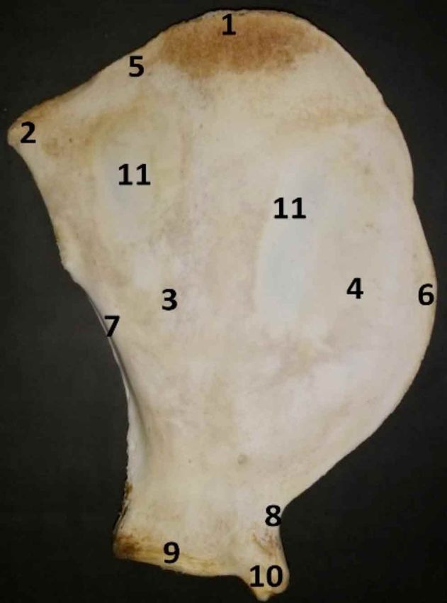
SOEL, M. S. H.; ISLAM, K. N. & RAHMAN, M. L. Anatomical features of some bones of the forelimbs of lions (Panthera leo). Int. J. Morphol., 39(2):378-385, 2021.

articulated just under the head on the medial surface (Fig.                                 Radius and Ulna. The radius and ulna were twin bones
5). Similar findings were observed in Asiatic cheetah                                       of the skeleton of antebrachium which formed elbow joint
(Nazem et al.), but mostly prominent major tubercle was                                     proximally incorporated with the humerus and carpal joint
found in dog (Sisson et al.).                                                               distally with the carpal bones. In lion, the radius bone
                                                                                            was articulated craniolateral to the ulna proximally and
         The shaft (corpus humeri) was compressed                                           craniomedial to the ulna distally. The morphometrical data
craniocaudally in proximal part, rounded to oval in middle                                  for different parameters of scapula of the lions are
part and compressed mediolaterally in distal part (Fig. 6)                                  illustrated in Table I.
as described in lion (Kirberger et al.). This bone had four
surfaces- the lateral, medial, cranial and caudal surface,                                         The radius has a long shaft (corpus radii) and two
but only two surfaces- lateral and medial were observed                                     limbs- the proximal one was smaller and distal one was
in Asiatic cheetah (Nazem et al.). The lateral surface was                                  larger and expanded. The head of the radius (caput radii)
spirally twisted and smooth, whereas the medial one was                                     was very well defined. On the proximal surface of head,
compressed in the proximal part and almost rounded in                                       the concave fovea capitis radii- a triangular articular
the distal part. A shallow, convex musculospiral groove                                     surface was seen which articulated with the lateral condyle
or brachial groove (sulcus musculi brachialis) was present                                  of humerus (Fig. 7). This was in agreement with the
on the lateral surface, which continued until the proximal                                  previous report of Nzalak et al.
half of this bone. The less developed deltoid tuberosity
(tuberositas deltoidei) was noticed at the edge between                                             Immediately below the head, the neck (collum
the lateral and medial surfaces (Fig. 5), whereas well                                      radii) has an irregular surface for the articulation with
developed deltoid tuberosity was noticed in dog (Miller                                     ulna in its caudal part. A rough, prominent eminence- the
et al.; Sisson et al.). On the lateral surface, there is another                            radial tuberosity (tuberositas radii) was present on the
obliquely crest like structure known as tricipital line or                                  medial surface of the proximal limb (Fig. 7) as seen in
deltoid crest was found, which ended in the deltoid                                         tiger (Tomar et al.).
tuberosity. On the cranio-lateral aspect of humerus,
another crest like structure was started from the distal                                            The shaft of radius (corpus radii) was
part of lateral (greater) tuberosity, continued as slightly                                 craniocaudally compressed, which was similar with Asatic
oblique line and finally terminated at teres major                                          cheetah (Nazem et al.), but dissimilar with Asian elephant
tuberosity (tuberositas teres major). On the distal part of                                 (Ahasan et al.). It had four surfaces- anterior, posterior,
the shaft, a supracondyloid crest or ridge (crista                                          lateral and medial. The anterior surface was rough for
supracondylaris lateralis) started just above the lateral                                   the attachment of tendons of muscles, while the posterior
epicondyle (epicondylus laterialis), continued obliquely                                    surface was somewhat concave as reported in dog and
and then ended on its caudal surface (Fig. 4). The nutrient                                 cat (Sisson et al.). The lateral and medial surfaces were a
foramen was observed on the caudal surface of the                                           bit rounded and comparatively smooth. The distal limb
proximal to the middle of the shaft, but Nzalak et al.                                      was the largest part of this bone. It had a medial elongated
observed this foramen on the distal half of the shaft. In                                   projection called styloid process of radius (processus
contrast, two nutrient foramens were observed in Asiatic                                    styloideus radii) (Fig. 8) as reported in tiger (Lucky &
cheetah (Nazem et al.).                                                                     Harshan). An articular surface- ulnar notch for the
                                                                                            attachment of radius with ulna was also present.
        The distal limb of the humerus had two condyles
(condylus), two epicondyles (epicondylus), a                                                       The ulna was the longest bone in the forelimb of
supracondyloid foramen, radial fossa (fossa radialis) and                                   lion. The olecranon of ulna was projected farther than
olecranon fossa (fossa olecrani). A small, shallow radial                                   the radius at the proximal limb (Fig. 7), which was similar
fossa (fossa radialis) was pointed out on the medial surface                                to the cattle (Budras & Habel) and sheep (Sisson et al.),
(Fig. 5). On the contrary, a large and deep olecranon fossa                                 but different from the horse (Sisson et al.). The free end
(fossa olecrani) was present on the other side (Fig. 4).                                    of this olecranon was extended caudolaterally to form
Although these two fossae were shallow in tiger (Tomar et                                   olecranon tuber (tuber olecrani) as observed in dog
al.) and Asiatic cheetah (Nazem et al.). A long, narrow                                     (Sisson et al.), Asiatic cheetah (Nazem et al.) and tiger
open type supracondyloid foramen was found on the medial                                    (Lucky & Harshan). It had three prominences- two were
surface of the distal limb just above the medial epicondyle                                 cranial and the caudal one was the largest and rounded
(epicondylus medialis) (Fig. 6). This foramen didn’t                                        (Fig. 8) as reported in dog (Sisson et al.) and tiger (Lucky
connect the radial fossa with the olecranon fossa as found                                  & Harshan). The trochlear (semilunar) notch (incisura
in dog (Sisson et al.).                                                                     trochlearis) was large and articulated with the trochlea
382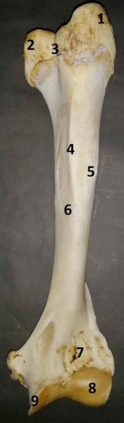
SOEL, M. S. H.; ISLAM, K. N. & RAHMAN, M. L. Anatomical features of some bones of the forelimbs of lions (Panthera leo). Int. J. Morphol., 39(2):378-385, 2021.

           Fig. 4. Caudal view of left humerus of lion. 1= Head of humerus (Caput humerus), 2= Greater or major
           tubercle (Tuberculum majus), 3= Lesser tubercle (Tuberculum minus), 4= Neck of humerus (Collum humeri),
           5= Deltoid tuberosity (Tuberositas deltoidea), 6=Supracondyloid crest or ridge (Crista supracondylaris
           lateralis), 7=Supracondyloid foramen, 8= Olecranon fossa (Fossa olecrani), 9= Lateral epicondyle
           (Epicondylus lateralis) and 10= Medial epicondyle (Epicondylus medialis).

           Fig. 5. Cranial view of left humerus of lion. 1= Greater or major tubercle (Tuberculum majus), 2= Lesser
           tubercle (Tuberculum minus), 3= Intertubercular groove, 4= Teres major tuberosity (Tuberositas teres majus),
           5= Deltoid tuberosity (Tuberositas deltoidea), 6= Shaft of humerus (Corpus humeri), 7= Radial fossa (Fossa
           radialis), 8= Capitulum (Capitulum humeri) and 9= Trochlea (Trochlea humeri).

           Fig. 6. Medial view of left humerus of lion. 1= Lesser tubercle (Tuberculum minus), 2= Head of humerus
           (Caput humerus), 3= Neck of humerus (Collum humeri), 4= Crest of lessser tubercle, 5= Shaft of humerus
           (Corpus humeri) and 6= Medial epicondyle (Epicondylus medialis).

of humerus. It was continued distally by the medial and                                           As like as radius, the shaft of ulna (corpus ulnae)
lateral coronoid processes (processus coronoideus) to                                      was triangular in section and slightly convex cranially.
form a concave surface for articulation, whereas                                           This similar findings were observed in Asiatic cheetah
proximally it was continued with the anconeal process                                      (Nazem et al.). The proximal half of the shaft was thick
(processus anconeus).                                                                      as resembling to the distal part of caudal view. At the
                                                                                                                                                                          383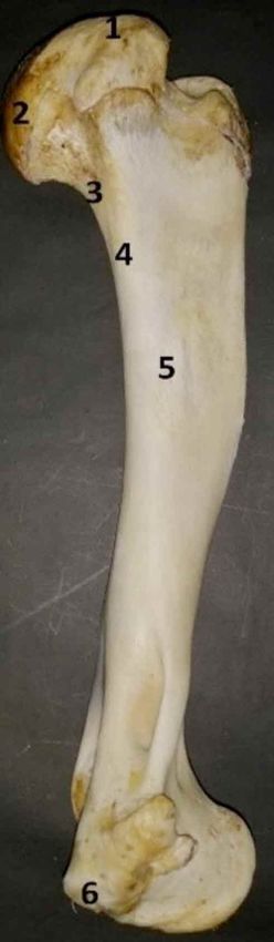
SOEL, M. S. H.; ISLAM, K. N. & RAHMAN, M. L. Anatomical features of some bones of the forelimbs of lions (Panthera leo). Int. J. Morphol., 39(2):378-385, 2021.
distal limb, a distally projected
styloid process (processus styloideus
ulnae) (Fig. 8) was seen which
articulated with the carpal bones as
observed previously by Nzalak et al.
Medially it had a convex facet that
articulated with the radius.
CONCLUSION
The above mentioned
information regarding some unique
anatomical features and their
morphometric measurements can be
helpful for identification, radiographic
interpretation and forensic
investigation of the forelimb bones of
lion. These will also provide the
pathway and guideline for better
understanding the appropriate
anatomical parameters.
SOEL, M. S. H.; ISLAM, K. N. &
RAHMAN, M. L. Características anató-
micas de algunos huesos de los miembros
torácicos de leones (Panthera leo). Int. J.
Morphol., 39(2):378-385, 2021.
RESUMEN: Estudiamos los hue-
sos de las miembros torácicos de cuatro
leones adultos (Panthera leo) de ambos
sexos para registrar las características ana-
tómicas y morfométricas macroscópicas
de la escápula, el húmero, el radio y la
ulna. Se observaron algunas característi-
cas anatómicas únicas que serán útiles
para la interpretación radiográfica y las in-
vestigaciones forenses. La superficie la-
teral de la escápula se dividió de manera
desigual en fosa supraespinosa y fosa
infraspinosa por una columna bien desa-
Fig. 7. Caudomedial view of right radius and ulna of lion. 1= Olecranon tuber (Tuber rrollada (espina de la escápula). El proce-
olecrani), 2= Olecranon process (Processus olecrani), 3= Anconeal process (Processus so del acromion se subdividió en proceso
anconeus), 4= Trochlear notch (Incisura trochlearis), 5= Capitular fovea of radius, 6= suprahamato (processus suprahamatus) y
Radial tuberosity (Tuberositas radii), 7= Shaft of ulna (Corpus ulnae), 8= Shaft of radius proceso hamato (processus hamatus); el
(Corpus radii), 9= Interosseous space, 10=Styloid process of ulna (Processus styloieus tubérculo supraglenoideo (tuberculum
ulnae) and 11=Styloid process of radius (Processus styloieus radii). supraglenoidalis) estaba ausente. La
diáfisis (diafisis) del húmero estaba com-
Fig. 8. Craniomedial view of right radius and ulna of lion. 1= Olecranon tuber (Tuber primida craneocaudalmente en la parte
olecrani), 2= Olecranon process (Processusolecrani), 3= Anconeal process (Processus proximal, redondeada a ovalada en la parte
anconeus), 4= Trochlear notch (Incisura trochlearis), 5= Coronoid process (Processus media y comprimida mediolateralmente
coronoideus), 6= Capitular fovea of radius, 7= Interosseous space, 8=Styloid process of en la parte distal. Se encontró un foramen
radius (Processus styloieus radii) and 9=Styloid process of ulna (Processus styloieus ulnae). supracondileo largo y estrecho en la ex-
384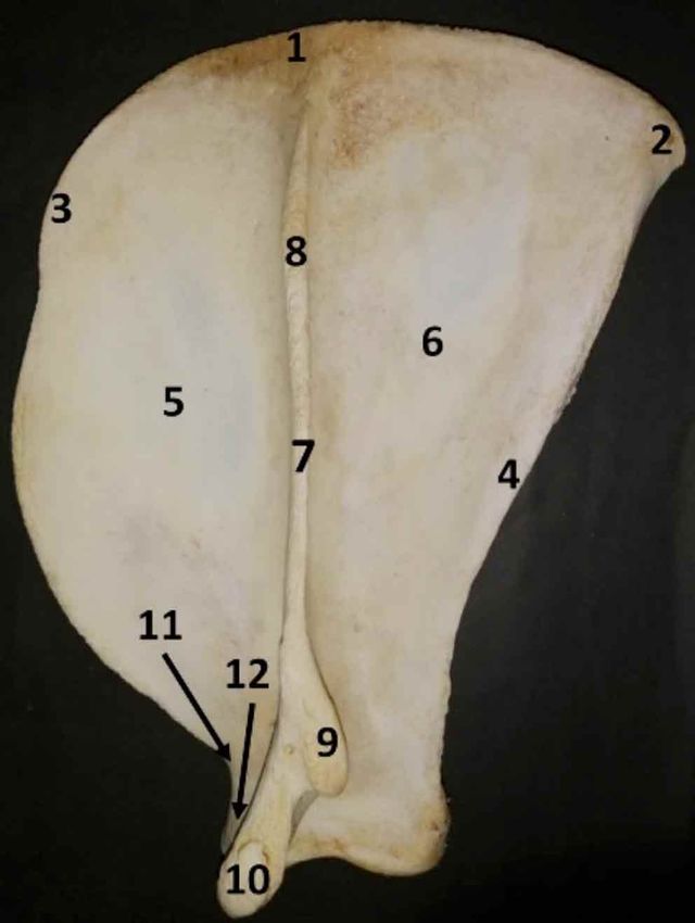
SOEL, M. S. H.; ISLAM, K. N. & RAHMAN, M. L. Anatomical features of some bones of the forelimbs of lions (Panthera leo). Int. J. Morphol., 39(2):378-385, 2021.

tremidad distal, por encima del epicóndilo medial (epicondylus                                Sisson, S.; Grossman, J. D. & Getty, R. Sisson and Grossman's The Anatomy
medilaris) que no conectaba la fosa radial (fosa radial) con la fosa                              of the Domestic Animals. 5th ed. Philadelphia, W. B. Saunders Press,
olecraneana (fossa olecrani). El radio y la ulna eran huesos idén-                                1975.
                                                                                              Tomar, M. P. S.; Taluja, J. S.; Vaish, R.;Shrivastav, A. B.; Shahi, A. &
ticos en los que el radio se articulaba craneolateral a la ulna
                                                                                                  Sumbria, D. Gross anatomy of scapula in tiger (Panthera tigris). Indian
proximalmente, y craneomedial a la ulna distalmente. Sin embar-
                                                                                                  J. Anim. Res., 52(4):547-50, 2018.
go, la ulna era el hueso más largo del miembro torácico del león.
La tuberosidad del olécranon de este hueso tenía tres prominen-
cias: dos eran craneales, mientras que la caudal era la más grande
y redondeada. Se encontraron procesos estiloides proyectados                                  Corresponding author:
distalmente (processus styloideus) en la extremidades distales del                            Md. Shahriar Hasan Sohel
radio y la ulna.                                                                              Laboratory of Veterinary Anatomy
                                                                                              Joint Graduate School of Veterinary Sciences
     PALABRAS CLAVE: León; Anatomía; Escápula;                                                Gifu University, 1-1 Yanagido
Húmero; Radio; Ulna.                                                                          Gifu 501-1193
                                                                                              JAPAN

REFERENCES                                                                                    Email: s.h.sohel08@gmail.com

Ahasan, A. S. M.; Quasem, M.; Rahman, M. L.; Hasan, R. B.; Kibria, A.                         Received: 22-10-2020
    S. M. & Shil, S. K. Macroanatomy of the bones of thoracic limb of                         Accepted: 07-12-2020
    an Asian elephant (Elephas maximus). Int. J. Morphol., 34(3):909-
    17, 2016.
Budras, K. D. & Habel, R. E. Bovine Anatomy: An Illustrated Text.
    Hannover, Schlütersche, 2011.
Hildebrand, M. & Hurley, J. P. Energy of the oscillating legs of a fast-
    moving cheetah, pronghorn, jackrabbit, and elephant. J. Morphol.,
    184(1):23-31, 1985.
Howell, A. B. Speed in Animals, their Specialization for Running and
    Leaping. New York, Hafner Publishing Company Ltd.,1944.
Kirberger, R. M.; du Plessis, W. M. & Turner, P. H. Radiologic anatomy
    of the normal appendicular skeleton of the lion (Panthera leo). Part
    1: thoracic limb. J. Zoo Wildl. Med., 36(1):21-8, 2005.
Lucky, K. M. & Harshan, K. R. Gross anatomy of skeleton antebrachii
    of a tiger (Panthera tigris). Indian J. Anim. Res., 48(3):298-300, 2014.
Miller, M. E.; Christensen, G. & Evans, H. E. Anatomy of the Dog. 9th ed.
    Philadelphia, W. B. Saunders Press,1964.
Nazem, M. N.; Sajjadian, S. M. & Nakhaei, A. Anatomy, functional
    anatomy and morphometrical study of forelimb column in Asiatic
    cheetah (Acinonyx jubatus venaticus). Ital. J. Anat.Embryol.,
    122(3):157-72, 2017.
Nowak, R. M. & Walker, E. P. Walker's Primates of the World. 6th ed.
    Baltimore, Johns Hopkins University Press, 1999.
Nzalak, J. O.; Eki, M. M.; Sulaiman, M. H.; Umosen, A. D.; Salami, S.
    O.; Maidawa, S. M. & Ibe, C. S. Gross anatomical studies of the
    bones of the thoracic limbs of the Lion (Panthera leo). J. Vet. Anat.,
    3(2):65-71, 2010.
Palanisamy, D.; Tomar, M. P. S.; Ankem, P. B.; Ullakula, R. S.;
    Jonnalagadda, N. & Korampalli, V. Gross morphology of scapula in
    Indian wild cat (Felis silvestris ornate: Gray, 1830). Int. J. Curr.
    Microbiol. Appl. Sci., 7(4):2473-7, 2018.
Pandey. S. P.; Bhayani, D. M. & Vyas, Y. L. Gross anatomical study on
    the scapula of Asiatic lion (Panthera persica). Indian J. Vet. Anat.,
    16(1-2):53-6, 2004.
Podhade, D. N. Studies on Characteristic Features of Appendicular
    Skeleton of Leopard as an Aid in Wildlife Forensic. Master of
    Veterinary Science and Animal Husbandry in Wildlife Health and
    Managmenent Thesis. Jabalpur, College of Veterinary Science and
    Animal Husbandry, 2007. Available from: http://
    krishikosh.egranth.ac.in/handle/1/5810125638
Sarma, K.; Sasan, J. S. & Suri, S. Gross and morphometrical studies on
    scapula of civet cat (Viverricula indica). Int. J. Pure Appl. Biosci.,
    5(6):80-5, 2017.

                                                                                                                                                                              385
You can also read