Anomalies in the vertebral column and ilio-sacral articulation of some anuran amphibians
←
→
Page content transcription
If your browser does not render page correctly, please read the page content below
SALAMANDRA 57(1): 53–64
Anomalies in the anuran vertebral column and ilio-sacral articulation
SALAMANDRA
15 February 2021 ISSN 0036–3375 German Journal of Herpetology
Anomalies in the vertebral column and ilio-sacral articulation
of some anuran amphibians
Alexander Haas1, Sophie Schwippert1, Sebastian Büsse2, Thomas Kleinteich3, André Beerlink4,
Jörg U. Hammel5, Stephanie Köhnk1, Stanislav Gorb2 & Karolin Engelkes1
1)
Centrum für Naturkunde, Universität Hamburg, Martin-Luther-King-Platz 3, 20146 Hamburg, Germany
2)
Functional Morphology and Biomechanics, Institute of Zoology, University of Kiel, Am Botanischen Garten 1–9, 24118 Kiel,
Germany
3)
TPW Prüfzentrum GmbH, Xantener Str. 6, 41460 Neuss, Germany
4)
YXLON International GmbH, Essener Bogen 15, 22419 Hamburg
5)
Institute of Materials Research, Helmholtz-Zentrum Geesthacht, Max-Planck-Str. 1, 21502 Geesthacht, Germany
Corresponding author: Alexander Haas, e-mail: alexander.haas@uni-hamburg.de
Manuscript received: 17 August 2020
Accepted: 20 November 2020 by Alexander Kupfer
Abstract. We present accounts of vertebral anomalies in 17 individuals representing 13 species of anuran amphibians.
These cases were detected while perusing a larger survey on the skeleton of frogs, for which µCT scans of a broad range of
species were collected and evaluated. Our data and reports from the literature suggest that malformations, asymmetries,
and irregularities, if present, appear to be particularly prevalent in the posterior region of the axial skeleton in frogs.
Anomalies at the trunk-tail boundary, i.e., at the sacrum and neighbouring segments, were relatively common. Malfor-
mations at the trunk-tail boundary often include sacralization of pre- and postsacral elements with asymmetrically or
symmetrically developed diapophyses, fusion with the posteriormost presacral vertebra, occurrence of postsacral verte-
brae, unusual transverse processes at the proximal end of the urostyle, formation of additional zygapophyses, or fusion of
elements that normally articulate. Vertebral fusion in the anterior vertebral column (Presacral Vertebrae I+II) has been
reported both in evolutionary context and in cases of individual developmental anomalies. Malformations in the middle
section of the vertebral column, such as the case of Epidalea calamita reported herein, are rare.
Key words. Amphibia, Anura, axial skeleton, pelvis, sacrum, malformation.
Introduction es the articulation between the vertebral column and the
pelvis. The sacral vertebra is pivotal for the function of the
The anuran body plan is unique among vertebrates mak- sacro-urostylic complex (Emerson 1982). The tail vertebral
ing members of the monophylum Anura easily recogniz- column is highly reduced in all extant species; only vestiges
able: apart from losing the tail, the truncation of the ver- of the tail vertebrae contribute to the formation of the uro-
tebral column, the reduction in the number of vertebrae, style that is otherwise an evolutionary novelty (Ročkova &
the elongation of the ilia, expanded sacral diapophyses with Roček 2005, Pugener & Maglia 2009a).
ventral articulation to the ilium, and the formation of the The anatomical details and the shape of the ilio-sacral
urostyle are the most obvious synapomorphies in early frog joint vary among the frog lineages and are strongly cor-
evolution (Shubin & Jenkins 1995, Pugener & Maglia related with a species’ specialization in locomotor mode,
2009a). Frogs are the tetrapod group with the fewest verte- such as jumping versus swimming or burrowing, as well
brae. Species of the Ascaphidae and Leiopelmatidae possess as habitat choice (arboreal, fossorial, aquatic, terrestrial)
nine presacral vertebrae, all other anurans have a vertebral (Emerson 1979, Jorgensen & Reilly 2013). For example,
count of eight or less (Duellman & Trueb 1994). There the very wide sacral diapophyses of the sacral vertebra in
is only moderate differentiation into functional groups in pipids allow for a forward sliding of the pelvis (Videler
the presacral vertebral column, for example, in the size of & Jorna 1985, Cundall et al. 2017), whereas the almost
vertebral transverse processes at the level of the suprascap- cylindrical diapophyses in many ranid frogs provide the
ula. Presacral Vertebra I, or Presacral I for short, is the only necessary articular freedom for dorsoventral extension at
neck vertebra and stands out by typically lacking transverse the initial jumping phase (Emerson 1979, 1982, Jenkins &
processes (Gaupp 1896). A single sacral vertebra establish- Shubin 1998).
© 2021 Deutsche Gesellschaft für Herpetologie und Terrarienkunde e.V. (DGHT), Mannheim, Germany
Open access at http://www.salamandra-journal.com 53
Alexander Haas et al.
Considering the crucial functional role of the verte- Sensing & Inspection Technologies GmbH Phoenix Nano
bral column, ilio-sacral articulation, and sacro-urostylic tom S or M, or a GE Sensing & Inspection Technologies
complex, one would expect that mutational malforma- GmbH Phoenix v|tome|x L 450. Depending on the size of
tions of the skeleton were strongly selected against in evo- the specimen, the scanning parameters were set appropri-
lution and should be rare in nature (Zamora-Camacho ately (Supplementary document S1). Volumes were recon-
& Aragón 2019). Congenital skeletal anomalies, however, structed from X-ray projections using the software deliv-
can have many different causes other than genetic muta- ered with the respective scanner. The resulting voxel size
tions. Particularly in organisms that complete a substan- ranged from 11.87–106.01 µm, depending on the specimen
tial part of their skeletal development during larval life and size and CT system used.
metamorphosis, when they can be exposed to teratogens in Amira 6.0.1. software (Thermo Fisher Scientific) was
the environment. Recently, Ouellet (2000) and Henle & used to visualize and inspect µCT volumes. For each avail-
Dubois (2017) have assessed amphibian anomalies in gen- able µCT image stack an Isosurface of the calcified tissues
eral. These hitherto most extensive compilations and eval- (i.e., bones and calcified cartilage) was generated for in-
uations of published reports cover anomalies in colour pat- spection. If the specimen showed anomalies in the verte-
tern and morphology, and review hypotheses about causes bral column or the ilio-sacral articulation, relevant skele-
of such pathologies. Specific skeletal abnormalities, how- tal structures were segmented with Amira’s Segmentation
ever, have been covered only marginally apart from exter- Editor (Brush and Magic Wand tools). As this study fo-
nal features of general limb or body deformations. Henle cuses on the vertebral column and sacral structures, only
& Dubois (2017) provided a glossary, specific cases, and affected skeletal elements were accurately separated dur-
general methodological recommendations. ing segmentation; cruder segmentation methods were ap-
In a different project on skeletal features of the shoulder plied in other areas of the skeleton. Because cartilage does
girdle (Engelkes et al. 2020), we had access to and exam- not visualize reliably in µCT datasets of plain specimens
ined micro-computed tomography (µCT) scans of a broad and because it does not contribute additional aspects to
taxonomic sample of anuran amphibians. During the ex- the phenomena treated herein, cartilage tissue was gener-
amination we recognized several individuals with verte- ally neglected for this study; only calcified cartilage was oc-
bral and sacral malformations or anomalies compared to casionally segmented and visualized in skeletal parts that
conspecifics or congeners. Various categories of vertebral were unaffected by malformations if its grey values fell
anomalies have been described before (Madej 1965). We within the range of bone grey values. Segmented skeletons
classify vertebral malformations herein, first, as any asym- were exported with the mulitExport macro (Engelkes et
metry that was so substantial that it could be detected visu- al. 2018) as polymesh surfaces (.obj format) and, if neces-
ally even without further measurement, such as much in- sary, further processed in MeshLab 1.3.3 (Cignoni et al.
flated or present processes on only one side. In specimens 2008); specifically, Quadric Edge Collapse Decimation and
that could be considered normal, in contrast, asymmetries Taubin Smooth procedures were applied in MeshLab to re-
were not perceivable. A second category of anomalies does duce the number of polygons and to smooth the surfaces
not involve asymmetry but the fusion of consecutive ver- while still preserving the original surface as well as pos-
tebral structures. Third, supernumerary elements can be sible. Next, the surfaces were imported into various ver-
present. Sometimes a combination of these major types of sions of Modo (7–13; The Foundry Visionmongers Ltd.) for
malformations can occur in the same specimen. In the fol- subtle further smoothing (if necessary), topology hole fill-
lowing we describe the osteological malformations of the ing, assigning colours (malformations red; corresponding
vertebral column and ilio-sacral articulation present in structures without indication of malformation green, all
specimens examined. Providing a comprehensive compila- other skeletal parts grey), and final rendering (Dome Light
tion on the topic is beyond the scope of this work, but by and Directional Light). Colour plates were arranged using
reporting the phenomenology of some new cases of axial Graphic 3.1 (Picta Inc.) software.
skeleton anomalies we hope to contribute to the knowledge Abbreviations: AMNH, American Museum of Natural
of anomalies in anurans in general. History; CAS, California Academy of Sciences; CM, Carn-
egie Museum of Natural History; UF, Florida Museum of
Natural History; USNM, Smithsonian, National Museum
Materials and methods of Natural History; ZMH, Zoological Museum Hamburg;
ZSM, Zoologische Staatssammlung München; µCT, micro-
For a study on the shoulder girdles in anuran amphibians computed tomography.
(Engelkes et al. 2020), our group assembled a data set of
125 µCT scans of 74 frog species that could be assessed for
anomalies of the axial skeleton (Supplementary document Results
S1). Among these, 28 datasets were downloaded from Mor-
phoSource (Duke University) online 3D-database (Supple- The comparison of Barbourula busuangensis CAS-SUA
mentary document S1). 21240 (Fig. 1A) and B. busuangensis CAS-SUA 21247
Most of the µCT data were generated on either a Bruker (Fig. 1B; Table 1) in our study revealed severe malforma-
Corp. Skyscan 1172, a YXLON FF20 CT or FF35 CT, a GE tions in CAS-SUA 21247, i.e., it showed: Presacral I and
54
Anomalies in the anuran vertebral column and ilio-sacral articulation
Table 1. Specimens with anomalies in their axial skeleton reported herein. CAS, California Academy of Sciences. FMNH, Field Mu-
seum of Natural History. UF, Florida Museum of Natural History. USNM, National Museum of Natural History. ZMH, Zoologisches
Museum, Hamburg. ZSM, Zoologische Staatssammlung München. See Supplementary Information Table 1 for a list of all specimens
examined.
Species Museum Anomaly
collection number
Barbourula busuangensis CAS-SUA 21247 Fusion Presacrals I+II and Presacrals VII+VIII; supernumerary
Taylor & Noble, 1924 transverse processes at urostyle base.
Bombina bombina (Linnaeus, 1761) ZMH A09674 Supernumerary tenth vertebra with broad left diapophysis; unequal
diapophyses at sacral vertebra.
Bombina bombina (Linnaeus, 1761) ZMH A05619 Wedge vertebra following Presacral VI.
Bombina bombina (Linnaeus, 1761) ZMH A05617 Sacralization of Presacral VIII with left broad diapophysis; sacral
vertebra(?) fused to urostyle and forming a broad diapophysis only on
right side.
Bombina orientalis (Boulenger, 1890) ZMH A14350 Fusion Presacrals I+II.
Bombina variegata (Linnaeus, 1758) ZMH A11873 Unclear segmental pattern; wedge vertebra following Presacral IV.
Sacral vertebra fused to urostyle and possibly representing segment
IX or X; presacral element forming unusually large and flat transverse
processes (sacralization).
Crossodactylus caramaschii USNM 318234 Right side sacralization of Presacral VIII; reduced right diapophysis in
Bastos & Pombal, 1995 sacral vertebra (IX).
Discoglossus montalentii Lanza, ZSM 1300/2006 Sacralization of Presacral VIII, i.e. extended transverse processes.
Nascetti, Capula & Bullini, 1984
Ecnomiohyla miliaria (Cope, 1886) UF Herp 137208 Failed formation of right diapophysis in sacral vertebra; supernumer-
ary tenth vertebra with right side sacralization.
Eleutherodactylus coqui Thomas, 1966 ZMH A10152 Malformed , laterally curved urostyle.
Epidalea calamita (Laurenti, 1768) ZMH A06868 Fusion Presacrals V+VI.
Microhyla nepenthicola ZMH A11645 Reduced vertebral count, seven presacrals; fusion Presacral I+II is
Das & Haas 2010 norm for the species, but Presacral VII (=eighth vert. segment) fused
with sacral vertebra and developing unilateral transverse process.
Microhyla nepenthicola ZMH A11639 Supernumerary postsacral vertebral element with distinct neural arch
Das & Haas 2010 but synostosis with the sacral vertebra at centrum and (possibly)
zygapophyses.
Pelobates fuscus (Laurenti, 1768) UF Herp 36935 Bifurcated left transverse process of Presacral IV.
Pelobates fuscus (Laurenti, 1768) ZMH A07151 Atlas with transverse processes that articulate with transverse
processes of Presacral II.
Pleurodema bibroni Tschudi, 1838 FMNH 132507 Supernumerary ascending neural arch and transverse process on the
right side at Presacral VIII; fused.
Pseudacris triseriata CAS 188145 Reduced left diapophysis in Presacral IX; Presacral IX fused to a
(Wied-Neuwied, 1838) subsequent, supernumerary sacralized vertebra (right diapophysis
broad, left slender); the latter fused to urostyle.
II were fused into one skeletal element (synostosis, block A05619, B. orientalis ZMH A14350, and B. variegata ZMH
vertebra). A second synostosis in the same specimen com- A11873 Fig. 2C) differed in the patterns observed.
prised Presacral VII and VIII. Supernumerary transverse In Bombina bombina ZMH A09674, the count of pre-
processes were present at the base of the urostyle (two pairs sacral vertebrae was normal (eight) and the presacral ver-
instead of one). tebral column was symmetrical. However, the neural arch
Our 23 specimens of Bombina included normally devel- of Presacral I was incompletely ossified mid-dorsally; pos-
oped specimens (such as Bombina bombina ZMH A05110; sibly the ossification process had not been finished in this
Fig. 2A) that possessed eight clearly articulating presacral 25 mm snout–vent-length specimen. The sacral region pos-
vertebrae, symmetrical, broadly expanded sacral diapophy- sessed a duplication of the sacral vertebra. The total count
ses, and a urostyle that had developed a pair of symmetri- of vertebrae was, thus, ten in this specimen. The posterior
cal processes near its base. Within the specimens examined sacral vertebra formed a sacral diapophysis asymmetrical-
for the genus, we found three with anomalies in their ver- ly on the left side. On the right side, there was a slender
tebral columns. Bombina bombina ZMH A09674 (Fig. 2A) transverse process, instead of a dilated sacral diapophysis,
and B. bombina ZMH A05617 (Fig. 2C), B. bombina ZMH in this supernumerary vertebra. The anterior sacral verte-
55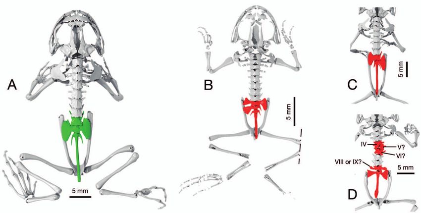
Alexander Haas et al.
bra formed two sacral diapophyses, the right one was larg- relatively short and curved ilia in this specimen. In B. bom
er and longer than the left one. All three diapophyses of the bina ZMH A05619, Presacrals VI and VII were similarly
two vertebrae contributed to the ilio-sacral articulation. malformed as Presacrals IV and V? in B. variegata ZMH
Altogether each of the diapophyses appeared relatively A11873. We further observed the fusion of Presacrals I+II
smaller than the diapophyses in B. bombina ZMH A05110 in B. orientalis ZMH A14350 similar to the occurrence in
that showed no obvious signs of malformations. Barbourula busuangensis CAS-SUA 21247.
In Bombina bombina ZMH A05617 the count of pre- Comparing two specimens of Discoglossus montalentii,
sacral vertebrae was seven and two sacral vertebrae were ZSM 1299/2006 showed no obvious anomalies in the ver-
present, suggesting that the eighth presacral vertebra had tebral column and ilio-sacral joint. In contrast, we detected
developed into a sacral vertebra. Both sacral vertebrae in sacralization of Presacral VIII in specimen ZSM 1300/2006
this specimen bore broad diapophyses laterally, to the left (Figs 3A–B). The sacralization in specimen ZSM 1300/2006
in the anterior and to the right in the posterior sacral ver- was characterized by longer, thicker, and more transversely
tebra, both articulated with the respective ilium. In B. bom oriented processes, reaching the ilium on the right and al-
bina ZMH A05619 the ilio-sacral joint was very similar to most reaching the ilium on the left side (Fig. 3B).
the one in ZMH A05617. The comparison of two individuals from two Crosso
Bombina variegata ZMH A11873 (Fig. 2D) presents with dactylus species (Figs 3A–B) revealed an ilio-sacral de-
multiple interesting malformations: The total count of ver- formity in C. caramaschii USNM 318234. We consider the
tebral elements (urostyle excluded) was ten; however, more perfectly symmetrical formation of Presacral VIII and the
than one interpretation is possible (Fig. 2D). Beginning sacral vertebra in C. trachysomus (CM Herp 2662, Fig. 3D)
with unequivocal position IV, two vertebral “elements” fol- the normal vertebral development. In C. caramaschii
low (V? + VI?) of which V? does not encircle the neural USNM 318234, however, Presacral VIII was sacralized on
canal but is wedged between the anterior IV and posterior the right side: A broad transverse process, reminiscent of
VI elements (wedge vertebra) and restricted to the right a proper sacral diapophysis, was formed and articulated
side of the body. The last vertebra without contact to the with the ilium. On the left side of the same vertebral body,
ilium formed unusually long and flat transverse processes, a normal cylindrical, thin transverse process with antero-
appearing intermediate between normal transverse proc- lateral orientation was formed. The proper sacral vertebra
esses of the vertebra anterior to it and the diapophyses of (ninth vertebra) featured a relatively normal diapophysis
the vertebra posterior to it. The latter established the ilio- on the left side. On its right side, however, a vestigial dia-
sacral articulation and is confluent with the urostyle poste- pophysis, thin and short in dimensions and deflected pos-
riorly. Note also the thickened end of the urostyle and the teriorly, was present.
Figure 1. Comparison of the skeleton in two specimens of Barbourula busuangensis. A) CAS-SUA 21240, specimen with normal ver-
tebral column development; B) CAS-SUA 21247 showing fusions between Presacral I (atlas) and II, as well as Presacrals VII and VIII.
Furthermore, the urostyle of CAS-SUA 21247 possesses four lateral processes. Polymesh surfaces derived from computed tomography
scans, rendered in dorsal views. Dashed lines indicate stop levels of scans from which the polymesh was derived; colours highlight
corresponding skeletal parts, green: normal bone formation, red: anomalous bone elements.
56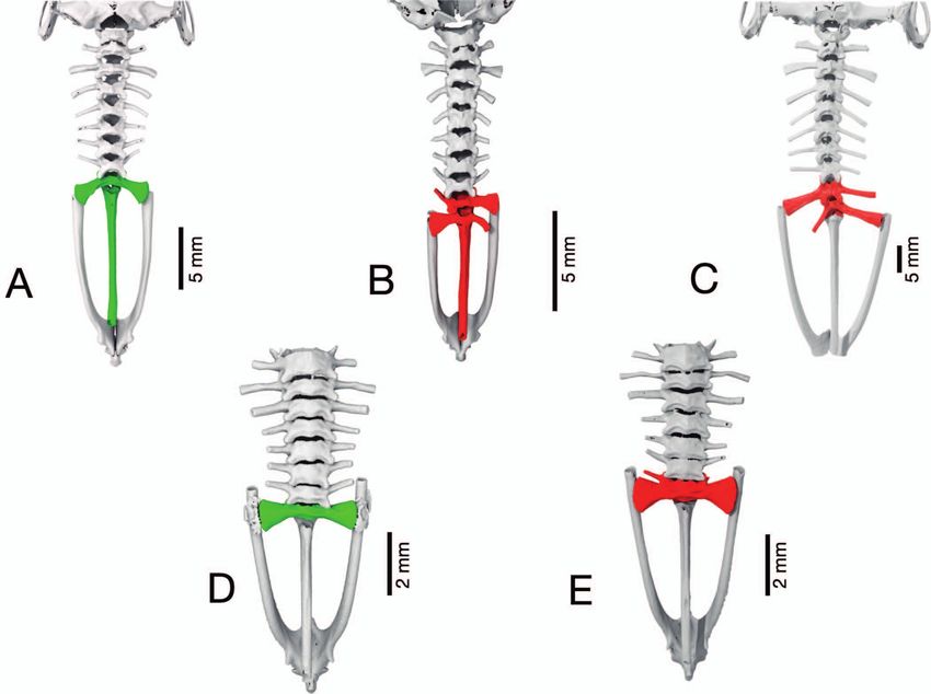
Anomalies in the anuran vertebral column and ilio-sacral articulation
Another category of deformities was detected in Epi tion of the preceding vertebra. The urostyle, however, ap-
dalea calamita ZMH A06868 (Fig. 3E). This specimen pre- peared anomalous as it possessed a small lateral process at
sented with Presacrals V and VI that appeared compressed its base on the left side.
in the antero-posterior dimension, relative to the sizes of The genus Pseudacris was represented in our study by
neighbouring vertebrae. Furthermore, the two elements a seemingly normally, symmetrically developed specimen
were fused at the level of the neural arches, zygapophys- of P. streckeri (AMNH A184936; Fig. 4A). A specimen of
es and centra. In neighbouring vertebrae, these structures P. triseriata (CAS 188145), however, showed clear malfor-
were clearly separate between successive structures so that mations in the sacral vertebra and diapophyses (Fig. 4B).
we can exclude CT artefacts. The malformation of the latter was characterized by a very
In Pleurodema, we had no access to normally developed asymmetrical development of the sacral diapophysis of the
individuals but asymmetric structures in an otherwise bi- actual sacral vertebra (Presacral IX). The left diapophy-
laterally symmetrical organism allowed us to locate de- sis was replaced by a process that was identical in shape
formities in Presacral VIII and the urostyle of P. bibroni and orientation with the transverse process of the preced-
FMNH 132507 (Fig. 3F). This specimen showed a super- ing vertebra; this process did not articulate with the ilium.
numerary vertebral element, specifically a supernumerary What seemed to be the anterior end of the urostyle, howev-
ascending neural arch and transverse process on the right er, showed sacralization and formed a left-side process that
side of the vertebral column only. We detected a bulge at had the length, orientation and shape of a sacral diapophy-
the anterior base of the posterior process, reminiscent of a sis, and articulated with the ilium. On the right side of the
zygapophysis. An intervertebral foramen was present be- same element, yet another process was formed and reached
tween the two right neural arches of Presacral VIII but dor- toward the ilium. It was more cylindrical and less blade-
sal and ventral to this foramen the neural arches, centra like flat than the sacral diapophysis of the preceding sacral
and zygapophyses were ankylosed. Despite the consider- vertebra. Furthermore, in this aberrant specimen, the left
able deviations for the body plan on the right side, the left ilium was shorter than the right ilium, corresponding to the
part of Presacral VIII did not exhibit apparent deformities. respective, broad diapophyseal process. A similar but mir-
The sacral vertebra appeared unaffected by the malforma- rored arrangement was discovered in Ecnomiohyla milia
Figure 2. Vertebral column morphology in Bombina. A) B. bombina ZMH A05110, green colour highlights the normally developed
Presacral VIII, the sacral vertebra with sacral diapophyses, and the urostyle. B) B. bombina ZMH A09674, vertebral column with mal-
formations (red) in the sacral region: presence of two sacral vertebra (IX, X) with a total of three articulating diapophyses. C) B. bom
bina ZMH A05617, vertebral column with malformation (red) in the sacral region. The articulation with the pelvic ilium is established
by Presacral VIII on the left side and a structure on the right side that may include the proper sacral vertebra but is confluent with
the urostyle. D) B. variegata ZMH A11873, vertebral column with several malformations in the vertebral column; unclear segmental
pattern in mid vertebral column. The sacral articulation is formed between the ilium and an element that bears sacral diapophyses but
is confluent with the urostyle posteriorly. The vertebra anterior to that element forms transverse processes that are flat and expanded,
reminiscent of sacral diapophyses. Polymesh surfaces derived from computed tomography scans, rendered in dorsal views; dashed
lines indicate stop level of original scan in B, C and D cropped from original rendered images.
57Alexander Haas et al.
ria UF Herp 137208 (Fig. 4C), in which the sacral vertebra tion is that the last presacral was partially and asymmetri-
(IV) had diapophyseal contact with the ilium only on the cally developed and fused to the sacral vertebra. Further-
left side and a tenth vertebral element was formed and ar- more, in M. nepenthicola A11639 a supernumerary vertebra
ticulated via a diapophysis on the right side with the ilium; was formed posterior to the sacral vertebra. The data sug-
elements were not fused with each other and the urostyle. gested that this element and the preceding sacral vertebra
A more subtle sacral malformation was detected were synostosed at their vertebral bodies and possibly at
in the genus Microhyla. There were eight presacrals in the zygapophyses, however, their neural arches were clear-
M. pulchra USNM 278542 and no obvious anomalies. In all ly distinct (not depicted herein).
four M. nepenthicola examined, however, Presacrals I+II Two specimens of Pelobates fuscus showed some note-
were indistinguishably fused and the resultant fusion el- worthy differences (Fig. 5). Pelobates fuscus ZMH A07151
ement possessed transverse processes. Thus the presacral possessed transverse processes at the atlas (Presacral I) that
count was seven in ZMH A11639, A11647 (Fig. 4D), and articulated with Presacral II; whereas P. fuscus UF Herp
A11650, respectively; we consider this the species’ norm. 36935 had a normal atlas, without transverse processes. The
In M. nepenthicola ZMH A11645 (Fig. 4E), however, there latter specimen, however, formed a bifurcating transverse
were only six free presacrals. A left side transverse process process at Presacral IV, whereas the former seemed normal
was present anterior to the sacral vertebra. Our interpreta- in that respect (no bifurcation).
Figure 3. A–B) Comparison between an apparently normal vertebral element (green) in Discoglossus montalentii ZSM 1299/2006 (A)
and specimen ZSM 1300/2006 (B) of the same species with ilio-sacral anomaly that features expanded transverse processes in Presacral
VIII (red) and slightly narrow sacral diapophyses (red). C–D) Intrageneric comparison of two Crossodactylus species. C) C. caramaschii
USNM 318234 presenting an ilio-sacral anomaly (red) in which Presacral VIII forms a dilated transverse process that articulates with
the ilium on the right side, whereas the sacral vertebra proper has a vestigial right diapophysis and only articulates with the ilium by
its left diapophysis. D) C. trachystomus CM Herp 2662, Presacral VII and sacral vertebra (green) without malformations. E) Epidalea
calamita ZMH A06868. In this specimen, Presacrals V and VI (red) possess anomalous features: both elements seem compressed in
the anteroposterior axis and are ancylosed. F) Pleurodema bibroni FMNH 132507. The specimen has malformations in Presacral VIII
as well as the urostyle (red). Presacral VIII seems serially duplicated but exclusively on the right side. All images are dorsal views of
specimens reconstructed from µCT data sets and rendered as polymesh surface. A and B cropped.
58Anomalies in the anuran vertebral column and ilio-sacral articulation
Finally, Eleutherodactylus coqui ZMH A10152, a F1-gen- available CT scans across major clades of the Anura was
eration captive-bred specimen, among two specimens of large (125 specimens). Knowledge of the morphology of
that species examined, showed a strongly curved urostyle species that were closely related to species in our sample,
(not depicted herein). and literature accounts, were considered sufficient to de-
tect and describe the cases presented herein. We did not
address anomalies of the vertebral column with statistical
Discussion methods at the population level but rather qualitatively at
a higher level of abstraction, that is, the bilateral symme-
Any classification of aberrant, abnormal, deformed or mal- try and segmentation of the amphibian body plan in gen-
formed anatomical structures requires the “norm” as refer- eral. Thus, we focus on presenting observations that clearly
ence. A multifaceted discussion of this topic has been pro- and unambiguously break the patterns of either symmetry
vided by Kovalenko & Kruzhkova (2013a,b,c) in a de- (substantial asymmetry) or seriality (disturbed segmenta-
tailed study on Bufo bufo. In the current study, the number tion pattern) in the vertebral column and ilio-sacral com-
of “normal” specimens for intraspecific reference in a par- plex. Asymmetries, such as unilateral sacralization of ver-
ticular species was small (often n=1), yet the number of tebrae (for example, Peloso 2016), are easily and unam-
Figure 4. A–B) Two species of the genus Pseudacris: A) Pseudacris streckeri AMNH A184936 (cropped). The symmetrical presacral
and sacral vertebrae indicate a normal state, whereas in B) Pseudacris triseriata CAS 188145 (cropped) shows a strongly malformed
sacral vertebra and anterior urostyle. C) Ecnomiohyla miliaria UF Herp 137208 (cropped) unilaterally sacralized Presacral VIII and
incompletely formed left sacral diapophysis. D–E) Comparisons between two specimens of Microhyla nepenthicola: D) Vertebral
column and pelvis of M. nepenthicola ZMH A11647 without obvious anomalies; E) Vertebral column and pelvis of M. nepenthicola
ZMH A11645 with a left side transverse process at the anterior face of the sacral vertebra and vertebral count is only six. The fusion of
Presacral I+II and a total of seven vertebrae was found in four specimens of this species and is considered normal for this miniature
species. All specimen representations are dorsal views of specimens, images rendered from polymesh surfaces derived from computed
tomography datasets; cropped from original data to vertebral column and pelvic regions.
59Alexander Haas et al.
biguously recognized as anomalies. In cases of symmetri- sification (Trueb 1977). Abnormal symmetrical features
cal or serial anomalies (vertebra number), more caution is in single anuran fossils may overlap phenomenologically
in place. The data presented may be considered prelimi- with normal morphologies during the processes of skeletal
nary until larger series are examined. Consultation of the modification in early frog evolution, for example in the re-
literature on the morphology of a species or its close rela- duction of the number of vertebral elements in the trunk
tives is essential to assess the normal condition. and the tail (Estes & Reig 1973, Lynch 1973, Handrigan
The symmetry, serial patterns, and development of the & Wassersug 2007).
vertebral column and ilio-sacral complex in anurans have Unfortunately, the prevalence of axial skeleton anom-
been well-documented in a broad range of studies that can alies in natural populations has not received much atten-
serve as references for the norm of the anuran body plan tion in the literature. Madej (1965) and Kovalenko &
(e.g., Gaupp 1896, Lynch 1973, Wiens 1989, Wild 1997, Kruzhkova (2013b) to our knowledge provided the most
Maglia & Pugener 1998, Haas 1999, Trueb et al. 2000, comprehensive assessment of intraspecific variations and
Sheil & Alamillo 2005, Banbury & Maglia 2006, Han anomalies in anuran species. Madej (1965) examined
drigan & Wassersug 2007, Pugener & Maglia 2009a,b, 555 skeletons of Bombina bombina and 813 skeletons of
Hoyos et al. 2012, Kovalenko & Kruzhkova 2013b, B. variegata, whereas Kovalenko & Kruzhkova (2013b)
Vera & Ponssa 2013, Biton et al. 2016, Soliz & Ponssa assessed anomalies of the vertebral column and ilio-sacral
2016, Senevirathne et al. 2020). Although Shearman & joint in 1633 lab-reared offspring of one mating pair of Bufo
Maglia (2014) concluded that the available accounts on bufo. Both studies categorized the observed phenomena
anuran skeletal morphology cover only a small fraction of and presented quantitative data on the intraspecific prev-
the known number of species (Frost 2020), many of the alence in the respective sample examined. The anomalies
anuran subclades are actually covered by reports of exem- we could detect rather haphazardly in our study with taxa
plar species. Backed by this background knowledge, misi- across the anuran order all were subsets of anomaly catego-
dentification and confusion of a norm character in the ver- ries described in Bombina and Bufo by these authors; new
tebral column for an anomaly, or vice versa, is unlikely in categories were not discovered.
extant frogs (but see Maglia & Pugener (1998) below). The scans downloaded from MorphoSource and our
Caution, however, is advised in general in applying axial own CT-scanning of individuals across the Anura uncov-
skeleton features derived from single specimens in clas- ered 17 individuals (13%) with axial anomalies (Table 1) in
a total sample of 125 examined specimens belonging to 73
species. Madej (1965) detected an overall rate of anoma-
lies of 10% in a large sample of Bombina. Meza-Joya et
al. (2013) reported 22% individuals with anomalies in the
vertebral column in a captivity-reared sample of 58 embry-
os and 96 postembryonic individuals of Eleutherodactylus
johnstonei, a species with direct development. This is, to
our knowledge, the highest known percentage. The axial
malformations they observed included fusion of Presacrals
I+II, fusion of Presacral VIII to the sacral vertebra (IX), de-
velopment of postzygapophyses at the sacral vertebra, and
extra postsacral vertebral elements (half or full).
Dasgupta & Grewal (1970) described vertebral fusion
in Euphlyctis cyanophlyctis and found 8–12% of the popula-
tion affected. Most cases of fusion occurred between Pre-
sacrals I and II, as well as VIII and IX; fewer incidences
were observed in the middle section of the vertebral col-
umn. Pugener & Maglia (2009b) found that more than
8.5% (11 out of 129 Acris creptitans specimens) of the speci-
mens in their study had vertebral anomalies, and that ap-
proximately 50% showed minor variation from the typical
morphology, that, however, were not classified as anoma-
lies.
Figure 5. Two specimens of Pelobates fuscus: A) The atlas of Pelo In the larger scale studies, intraspecific variation of ver-
bates fuscus ZMH A07151 developed transverse processes that tebral number was detected as a relatively rare event: 3.06%
articulate with Presacral II; B) Pelobates fuscus UF Herp 36935
possesses an atlas without transverse processes and Presacral IV
and 2.51% in Bombina and Bufo bufo, respectively (Madej
forms a bifurcated left transverse process. Dorsal views of par- 1965, Kovalenko & Kruzhkova 2013b). Ritland (1955)
tial renditions (skull, vertebral column, pelvis); Presacral VI in B noted less than 3% unilateral sacralization of Presacral IX
separated and shown in anterior view; polymesh surfaces derived in Ascaphus truei, a species with ten vertebrae. Malforma-
from computed tomography datasets; anomalous elements red, tions of variable degree in the coccygeal part of the uro-
normal elements green. style, its lateral processes and zygapophyses, however, were
60Anomalies in the anuran vertebral column and ilio-sacral articulation
common (more than a third of specimens). In her study number of trunk vertebrae, and the concomitant (or pos-
on 58 specimens of Boana lanciformis (then Hyla lanci sibly subsequent) lengthening of the ilia and reduction of
formis), Trueb (1977) reported vertebral anomalies in 9.4% tail vertebra, has been a major process in the evolution of
of the adult specimens. Greer & Byrne (2007) observed modern frogs and their jumping ability (Gans & Parsons
one case (2.4%) of sacralization of the posterior presacral 1966, Shubin & Jenkins 1995, Jenkins & Shubin 1998,
vertebra in their sample of Litoria aurea. Sacralization of Přikryl et al. 2009, Sigurdsen et al. 2012, Ascarrunz et
the presacral vertebra is commonly described in reports we al. 2016). The most recent common ancestor of frogs has
found and has been reported early on (Morgan 1886); our been hypothesized to have possessed nine presacral ver-
findings are in support of this general statement (Figs 2–4). tebrae (Ford & Cannatella 1993). Among extant spe-
Anomalies at the sacrum and its neighbouring seg- cies nine presacrals are only present in the Leiopelmatidae
ments have been described frequently (for example, Rit- and Ascaphidae, followed by a reduction to eight or less in
land 1955, Madej 1965, Kovalenko & Danilevskaya other extant groups (Haas 2003, Frost et al. 2006). Fur-
1994, Kovalenko 1995, Pugener & Maglia 2009b, Kova- ther reductions in vertebral number have been reported in
lenko & Kruzhkova 2013b; Figs. 2–4); anomalies in the a diversity of frog groups, among others in some species
anterior part of the vertebral column, in contrast, appear of the Brachycephalidae, Calyptocephalellidae, Cerato-
to be less frequent. The most anterior anomalies observed phryidae, Craugastoridae, Dendrobatoidea, Pipidae, Bufo-
in our sample involved the atlas: Presacrals I+II were fused nidae, Ptychadenidae, or Myobatrachidae (Stannius 1856,
(synostosis) in Barbourula busuangensis CAS-SUA 21247 Izecksohn 1971, Trueb 1973, Cannatella & Trueb 1988,
(Fig. 1A) and in Bombina orientalis ZMH A14350, while the Baez & Pugener 2003, Guayasamin 2004, Frost et al.
atlas of Pelobates fuscus ZMH A07151 developed transverse 2006, Muzzopappa et al. 2015).
processes (Fig. 5A). The normal vertebral characteristics in Reductions of vertebrae in taxa in crown-group anurans
the two known species of Barbourula (B. busuangensis and have evolved in different ways. For example, the evolution
B. kalimantanensis) have been described in Clarke (1987). of seven presacral vertebrae in Pipa has been explained by
Interestingly, along with the anterior synostosis, B. busu fusion of anterior vertebrae (Trueb et al. 2000) whereas
angensis CAS-SUA 21247 had additional malformations in the integration of the most posterior presacral vertebra
the posterior vertebrae and the urostyle. Multiple malfor- into the sacrum has been suggested to have resulted in sev-
mations in one individual have been reported previously en presacral vertebrae in the closely related Hymenochirus
(for example, Madej 1965, Trueb 1977, Maglia & Puge- and Pseudhymenochirus (Cannatella & Trueb 1988) or
ner 1998). In the study on Bombina (Madej 1965), a fusion the bufonid Dendrophryniscus (Izecksohn 1971). For one
of Presacrals I+II was not explicitly illustrated. Only the to- of the smallest frogs in the world, Microhyla nepenthicola,
tal number of vertebrae (including the sacral vertebra) was we report here the complete fusion of atlas and second ver-
reported to vary from eight to ten. We, however, observed tebra into one element as the species’ norm character. One
fusion of Presacrals I+II in B. orientalis ZMH A14350. In- specimen (ZMH A11645, Fig. 4E) had the last presacral in-
terestingly, Maglia & Pugener (1998) in their work on completely developed and fused to the sacral vertebra as
the skeletal development and adult osteology of B. orienta an anomaly, resulting in only six articulating presacral ele-
lis, depicted the axial skeleton in the adult KU 129703. The ments. In a sample of four specimens, two possessed axial
drawing shows the specimen with fusions of Presacrals anomalies in this highly miniaturized species. The hypoth-
I+II as well as VII+VIII, and an asymmetric development esis that miniaturization might be linked to vertebral count
of the basal lateral processes of the urostyle. This descrip- reduction and developmental instability deserves future at-
tion is also remarkably congruent to the condition in Bar tention. Adolphi (1892) already concluded that events that
bourula busuangensis CAS-SUA 21247 examined herein, we classify as anomalies in some species may correspond
that belongs to the same anuran family (Bombinatoridae). to the norm character in other taxa acquired in the course
Although KU 129703 (in Maglia & Pugener 1998) clear- of evolution (see also Izecksohn 1971).
ly was aberrant from the normal development of the axial The anuran urostyle has been studied thoroughly and its
skeleton in Bombina (Madej 1965), its unusual condition development has been clarified in various taxa (e.g., Hod
was not further expanded on in that paper. Anomalous fu- ler 1949, Branham & List 1979, Wiens 1989, Ročkova &
sions of Presacrals I+II have been reported, among oth- Roček 2005, Handrigan & Wassersug 2007, Pugener &
ers, in some individuals of Denrophryniscus brevipollica Maglia 2009a, Kruzhkova & Kovalenko 2010; Meza-
tus (Izecksohn 1971), Bufotes viridis (Adolphi 1892; then Joya et al. 2013, Muzzopappa et al. 2015, Senevirathne
Bufo variabilis), and Pelobates fuscus (Adolphi 1895). et al. 2020). Variation or externally induced disturbances
The reduction in number of vertebrae has played a ma- (Madej 1965) in the developmental program of the ontoge-
jor role in frog evolution (Lynch 1973), particularly the ev- netically and evolutionarily crucial posterior trunk bound-
olution of the jumping locomotor mode. The stem-group ary (Handrigan & Wassersug 2007) have been hypoth-
fossil Triadobatrachus massinoti possessed more presacral esized to be associated with anomalies in that region that
(trunk) vertebrae and shorter ilia (Rage & Roček 1989, occurred in the single digit range percentage (Kovalenko
Roček & Rage 2000, Ascarrunz et al. 2016) than any & Kruzhkova 2013b). A noteworthy case of perfectly nor-
crown group frogs (Trueb 1973, Haas 2003, Frost et al. mal development of multiple postsacral vertebrae, howev-
2006, Handrigan & Wassersug 2007). A reduction of the er, has been reported in megophryid tadpoles (Haas et al.
61Alexander Haas et al.
2006, Handrigan et al. 2007). Megophryid tadpoles pos- (Zoologische Staatssammlung, Munich). Datasets were accessed at
sess up to 30 ossified centra in pre-metamorphic stages. Morphosource.org (Duke Library Digital Repository) and we are
Axial skeleton anomalies can be induced by external indebted to the curators of this database for kindly providing ac-
cess to resources. Finally, we thank our work-group technician Lisa
agents (see review in Henle et al. 2017). Pesticides have Gottschlich and Alexander Daasch for providing professional
been shown to cause vertebral scoliosis possibly by influ- and reliable support of various kind.
ence on the vitamin D regulated pathways (Alvarez et
al. 1995). Yet, the genetic backgrounds and developmen-
tal mechanisms behind vertebral anomalies remain largely References
unresolved in frogs (Handrigan & Wassersug 2007). It
Adolphi, H. (1892): Über Variationen der Spinalnerven und der
is intriguing to ask, if vertebral developmental instability is Wirbelsäule anurer Amphibien. I. (Bufo variabilis Pall.). – Ge-
linked to phenotypic plasticity in other traits and thereby genbaurs Morphologisches Jahrbuch, 19: 313–375.
(indirectly) positively selected for in certain environments
Adolphi, H. (1895): Über Variationen der Spinalnerven und der
and times (DeWitt et al. 1998). In Euphlyctis cyanophlyc Wirbelsäule anurer Amphibien. II. (Pelobtes fuscus Wagl. und
tis, one dominant gene has been linked to vertebral fusion Rana esculenta L.). – Gegenbaurs Morphologisches Jahrbuch, 22:
and a higher temperature tolerance during development 449–490.
as potential selective advantage for heterozygosity (Das- Alvarez, R., M. P. Honrubia & M. P. Herráez (1995): Skeletal mal-
gupta & Grewal 1968, 1970). The anterior shift of Hox10 formations induced by the insecticides ZZ-Aphox® and Folidol®
gene expression domain defining the trunk-tail boundary during larval development of Rana perezi. – Archives of Environ-
(sacrum) (Handrigan & Wassersug 2007) could play a mental Contamination and Toxicology, 28: 349–356.
role in both the evolutionary reduction of presacral verte- Ascarrunz, E., J.-C.Rage, P. Legreneur, & M. Laurin (2016): Tri
brae in some taxa and the occurrence of anomalies in the adobatrachus massinoti, the earliest known lissamphibian (Ver-
posterior axial region in other taxa if that boundary is per- tebrata: Tetrapoda) re-examined by µCT scan, and the evolution
of trunk length in batrachians. – Contributions to Zoology, 85:
turbed. The surprisingly high number of anomalies in our 201–234.
survey and the data from the literature suggests that verte-
Baez, A. M. & L. A. Pugener (2003): Ontogeny of a new Palaeogene
bral anomalies are not rare in anurans. This could suggest pipid frog from southern South America and xenopodinomorph
that they may have little effect on performance and are not evolution. – Zoological Journal of the Linnean Society, 139: 439–
heavily selected against. 476.
Trunk-tail boundary pertubations probably played a Banbury, B. & A. M. Maglia (2006): Skeletal development of the
role in fossil taxa as well and anomalies have been reported Mexican spadefoot, Spea multiplicata (Anura : Pelobatidae). –
from ancient amphibians (Witzmann 2007, Witzmann et Journal of Morphology, 267: 803–821.
al. 2013). Vertebral anomalies have been well known from Beals, R. K., J. R. Robbins & B. Rolfe (1993): Anomalies associated
other vertebrate groups, such as salamanders (e.g., Pogo- with vertebral malformations. – Spine, 18: 1329–1332.
da & Kupfer 2019; Danto et al. 2020), humans (for exam- Biton, R., R. Boistel, R. Rabinovich, S. Gafny, V. Brumfeld &
ple, Beals et al. 1993) or fishes (Jawad 2017). The phenom- S. Bailon (2016): Osteological observations on the alytid Anura
enologies may be similar across vertebrate groups. Wedge Latonia nigriventer with comments on functional morphology,
vertebrae that develop on one side of the spine only, as ob- biogeography, and evolutionary history. – Journal of Morphol-
served in Bombina variegata ZMH A11873, and sacraliza- ogy, 277: 1131–1145.
tion of the presacral vertebra(e) are relatively common in Branham, A. E. & J.C. List (1979): Development of the urostyle dur-
humans (Werenskiold 1937, Beals et al. 1993, McMas- ing metamorphosis in five species of anurans. – Journal of Mor-
phology, 159: 311–329.
ter 1998).
Cannatella, D. C. & L. Trueb (1988): Evolution of pipoid frogs:
morphology and phylogenetic relationships of Pseudhymenochi
rus. Journal of Herpetology, 22: 439–456.
Acknowledgements
Cignoni, P., M. Callieri, M. Corsini, M. Dellepiane, F. Ga-
First and foremost, we gratefully acknowledge the Wilhelm-Peters- novelli & G. Ranzuglia (2008): MeshLab: an Open-Source
Fonds of the Deutsche Gesellschaft für Herpetologie und Ter- Mesh Processing Tool. – Sixth Eurographics Italian Chapter Con-
rarienkunde e.V (DGHT) for granting funds to make this study pos- ference: 129–136.
sible. Furthermore, work that lead to the discovery of malformed Clarke, B. T. (1987): A description of the skeletal morphology of
frogs was funded by the Deutsche Forschungsgemeinschaft (DFG, Barbourula (Anura: Discoglossidae), with comments on its rela-
German Research Foundation) – 387723284. Alexander Kupfer tionships. – Journal of Natural History, 21: 879–891.
and Mark D. Scherz provided constructive criticism and contrib- Cundall, D., E. Fernandez & F. Irish (2017): The suction mecha-
uted to the quality of the manuscript. We are very grateful to the fol- nism of the pipid frog, Pipa pipa (Linnaeus, 1758). – Journal of
lowing curators and personnel and their respective museums and Morphology, 278: 1229–1240.
collections under their care for specimen loans: David. A. Kiziri-
an (American Museum of Natural History), Jens Vindum (Califor- Danto, M., F. Witzmann & N. B. Fröbisch (2020): Osseous pathol-
nia Academy of Sciences), Ulrich Scheidt and Konrad Kürbis ogies in the lungless salamander Desmognathus fuscus (Pletho-
(Naturkundemuseum Erfurt), Alan Resetar (Field Museum of dontidae). – Acta Zoologica, 101: 324–329.
Natural History), Kevin de Queiroz (National Museum of Natu- Dasgupta, S. & M. S. Grewal (1968): The selective advantage of
ral History), Mark-Oliver Rödel and Frank Tillack (Museum temperature tolerance among the progeny of frogs with vertebral
für Naturkunde Berlin), and Frank Glaw and Michael Franzen fusions. – Evolution, 22: 87–92.
62Anomalies in the anuran vertebral column and ilio-sacral articulation
Dasgupta, S. & M. S. Grewal (1970): Inheritance of vertebral fusion larval megophryids (Anura). – Evolution and Development, 9:
in the Skipper Frog. – Journal of Heredity, 61: 174–176. 190–202.
DeWitt, T. J., A. Sih & D. S. Wilson (1998): Costs and limits of Henle, K. & A. Dubois (2017): Studies on Anomalies in Natural
phenotypic plasticity. – Trends in Ecology & Evolution, 13: 77–81. Populations of Amphibians. – in: Mertensiella 25. – Deutsche
Duellman, W. E. E. & L. Trueb (1994): Biology of Amphibians. – Gesellschaft für Herpetologie und Terrarienkunde (DGHT),
The Johns Hopkins University Press, Baltimore. Mannheim.
Emerson, S. B. (1982): Frog postcranial morphology: identification Henle, K., A. Dubois & V. Vershinin (2017): A review of anom-
of a functional complex. Copeia, 1982: 603–613. alies in natural populations of amphibians and their potential
causes. – pp. 57–164 in: Henle, K. & A. Dubois (eds): Studies on
Emerson, S. B. (1979): The ilio-sacral articulation in frogs – form Anomalies in Natural Populations of Amphibians. Mertensiella
and function. – Biological Journal of the Linnean Society, 11: 153– 25. – Deutsche Gesellschaft für Herpetologie und Terrarienkunde
168. (DGHT), Mannheim.
Engelkes, K., F. Friedrich, J. U. Hammel & A. Haas (2018): A sim- Hodler, F. (1949): Untersuchungen über die Entwicklung von Sac-
ple setup for episcopic microtomy and a digital image process- ralwirbel und Urostyl bei den Anuren. – Revue suisse de Zoolo-
ing workflow to acquire high-quality volume data and 3D surface gie, 56: 747–790.
models of small vertebrates. – Zoomorphology, 137: 213–228.
Hoyos, J. M., M. R. Sánchez-Villagra, A. A. Carlini & C. Mit-
Engelkes, K., L. Kath, T. Kleinteich, J. U. Hammel, A. Beerlink gutsch (2012): Skeletal development and adult osteology of
& A. Haas (2020): Ecomorphology of the pectoral girdle in an- Hypsiboas pulchellus (Anura: Hylidae). – Acta Herpetologica, 7:
urans (Amphibia, Anura): Shape diversity and biomechanical 119–138.
considerations. – Ecology and Evolution 10: 11467–11487.
Izecksohn, E. (1971): Variação no padrão vertebral de Dendrophry
Estes, R. & O. A. Reig (1973): The early fossil record of frogs, a re- niscus brevipollicatus Espada. – Archivos Museu Nacional Rio de
view of the evidence. – pp. 11–63 in: Vial, J. L. (ed.): Evolutionary Janeiro, 54: 129–138.
Biology of the Anurans: Contemporary Research on Major Prob-
lems. – University of Missouri Press, Columbia. Jawad, L. A. (2017): The first record of incidences of ankylosis in
seven triplefin fishes (Pisces: Tripterygiidae) from New Zealand.
Ford, L. S. & D. C. Cannatella (1993): The major clades of frogs. – – The Anatomical Record: Advances in Integrative Anatomy and
Herpetological Monographs, 7: 94–117. Evolutionary Biology, 301: 39–45.
Frost, D. R. (2020): Amphibian Species of the World: an online Jenkins, F. A. & N. H. Shubin (1998): Prosalirus bitis and the an-
reference. Version 6.1. – American Museum of Natural History. uran caudopelvic mechanism. – Journal of Vertebrate Paleonto
Electronic Database accessible at https://amphibiansoftheworld. logy, 18: 495–510.
amnh.org/index.php, accessed on 14. July 2020.
Jorgensen, M. E. & S. M. Reilly (2013): Phylogenetic patterns of
Frost, D. R., T. Grant, J. Faivovich, R. H. Bain, A. Haas, C. Hadd- skeletal morphometrics and pelvic traits in relation to locomotor
ad, R. O. de Sa, A. Channing, M. Wilkinson, S. C. Donnel- mode in frogs. – Journal of Evolutionary Biology, 26: 929–943.
lan, C. J. Raxworthy, J. A. Campbell, B. L. Blotto, P. Moler,
R. C. Drewes, R. A. Nussbaum, J. D. Lynch, D. M. Green & W. Kovalenko, E. E. (1995): On some sacral anomalies in laboratory
C. Wheeler (2006): The amphibian tree of life. – Bulletin of the common platanna (Xenopus laevis). – Russian Journal of Herpe-
American Museum of Natural History, 297: 1–370. tology, 2: 170–173.
Gans, C. & T. S. Parsons (1966): On the origin of the jumping Kovalenko, E. E. & S. E. Danilevskaya (1994): On unique forms
mechanism in frogs. – Evolution, 20: 92–99. of anomalous sacral structure in tailless amphibians. – Russian
Journal of Herpetology, 1: 30–36.
Gaupp, E. (1896): A. Ecker‘s und R. Wiedersheim‘s Anatomie des
Frosches. 1. Abteilung, 3. Auflage. – Vieweg, Braunschweig. Kovalenko, E. E. & Y. I. Kruzhkova (2013a): Individual varia-
tion in the development of the common toad, Bufo bufo (Anura,
Greer, A. E. & M. Byrne (2007): Sex ratio and frequency of os- Bufonidae): 1. Timing of development and anomalies of exter-
teological abnormalities in the australian hylid frog Litoria aurea nal structure. – Russian Journal of Developmental Biology, 44:
from two apparently unpolluted localities in Sydney, New South 173–179.
Wales. – Australian Zoologist, 30: 43–47.
Kovalenko, E. E. & Y. I. Kruzhkova (2013b): Individual variation
Guayasamin, J. M. (2004): The Eleutherodactylus orcesi species in the development of the common toad, Bufo bufo (Anura, Bu-
group (Anura: Leptodactylidae): Comparative osteology and fonidae): 2. Diagnostic characters of the axial skeleton. – Russian
comments on its monophyly. – Herpetological Monographs, 18: Journal of Developmental Biology, 44: 180–193.
142–174.
Kovalenko, E. E. & Y. I. Kruzhkova (2013c): Individual variation
Haas, A. (1999): Larval and metamorphic skeletal development in in the development of the common toad, Bufo bufo (Anura, Bu-
the fast-developing frog Pyxicephalus adspersus (Anura, Rani- fonidae): 3. Limitations of individual variation and their causes.
dae). – Zoomorphology, 119: 23–35. – Russian Journal of Developmental Biology, 44: 194–205.
Haas, A. (2003): Phylogeny of frogs as inferred from primarily lar- Kruzhkova, Y. I. & E. E. Kovalenko (2010): Regularities of mor-
val characters (Amphibia: Anura). – Cladistics, 19: 23–89. phogenesis of the coccygeosacral articulation in Anura. – Rus-
Haas, A., S. Hertwig & I. Das (2006): Extreme tadpoles: The sian Journal of Developmental Biology, 41: 111–121.
morphology of the fossorial megophryid larva, Leptobrachella Lynch, J. (1973): The transition from archaic to advanced frogs. – pp.
mjobergi. – Zoology, 109: 26–42. 133–182 in: Vial, J. L. (ed.): Evolutionary biology of the anurans:
Handrigan, G. R. & R. J. Wassersug (2007): The anuran Bauplan: Contemporary research on major problems. – University of Mis-
a review of the adaptive, developmental, and genetic underpin- souri Press, Columbia.
nings of frog and tadpole morphology. – Biological Reviews of Madej, Z. (1965): Variations in the sacral region of the spine in
the Cambridge Philosophical Society, 82: 1–25. Bombina bombina (Linnaeus, 1761) and Bombina variegata (Lin-
Handrigan, G. R., A. Haas & R. J. Wassersug (2007): Bony-tailed naeus, 1758). – Acta Biologica Cracoviensia Series Zoologia, 8:
tadpoles: the development of supernumerary caudal vertebrae in 186–197.
63Alexander Haas et al.
Maglia, A. M. & L. A. Pugener (1998): Skeletal development and medusinae) and a comparison of this arboreal species with a
adult osteology of Bombina orientalis (Anura: Bombinatoridae). terrestrial member of the genus. – Journal of Morphology, 265:
– Herpetologica, 54: 344–363. 343–368.
McMaster, M. J. (1998): Congenital scoliosis caused by a unilateral Shubin, N. H. & F. A. Jenkins, F.A. (1995): An early jurassic jumping
failure of vertebral segmentation with contralateral hemiverte- frog. – Nature, 377: 49–52.
brae. – Spine, 23: 998–1005. Sigurdsen, T., D. M. Green & P. J. Bishop (2012): Did Triadobatra
Meza-Joya, F. L., E. P. Ramos-Pallares & M. P. RamÍrez-Pinilla, chus jump? Morphology and evolution of the anuran forelimb in
M.P. (2013): Ontogeny of the vertebral column of Eleutherodacty relation to locomotion in early salientians. – Fieldiana Life and
lus johnstonei (Anura: Eleutherodactylidae) reveals heterochro- Earth Sciences, 5: 77–89.
nies relative to metamorphic frogs. – The Anatomical Record: Soliz, M. & M. L. Ponssa (2016): Development and morphological
Advances in Integrative Anatomy and Evolutionary Biology, 296: variation of the axial and appendicular skeleton in Hylidae (Lis-
1019–1030. samphibia, Anura). Journal of Morphology, 277: 786–813
Morgan, C. L. (1886): Abnormalities in the vertebral column of the Stannius, H. (1856): Handbuch der Anatomie der Wirbelthiere. 2.
Common Frog. – Nature, 35: 53–53. Zootomie der Amphibien. – Von Veit & Comp., Berlin.
Muzzopappa, P., L. A. Pugener & A. M. Báez (2015): Postcranial Trueb, L. (1973): Bones, frogs, and evolution. – pp. 65–132 in: Vial,
osteogenesis of the helmeted water toad Calyptocephalella gayi J. L. (ed.): Evolutionary biology of the anurans: Contemporary
(Neobatrachia: Calyptocephalellidae) with comments on the os- research on major problems. – University of Missouri Press, Co-
teology of australobatrachians. – Journal of Morphology, 277: lumbia.
204–230.
Trueb, L. (1977): Osteology and Anuran Systematics: Intrapopula-
Ouellet, M. (2000): Amphibian deformities: Current state of tional variation in Hyla lanciformis. – Systematic Zoology, 26:
knowledge. – pp. 617–661 in: Sparling, D. W., G. Linder & C. A. 165–184.
Bishop (eds): Ecotoxicology of Amphibians and Reptiles. – Soci-
ety of Environmental Toxicology and Chemistry (SETAC) Press, Trueb, L., L. A. Pugener & A. M. Maglia (2000): Ontogeny of the
Pensacola. bizarre: An osteological description of Pipe pipa (Anura: Pipi-
dae), with an account of skeletal development in the species.
Peloso, P. L. V. (2016): Osteological malformation in the tree frog – Journal of Morphology, 243: 75–104.
Hypsiboas geographicus (Anura: Hylidae). Phyllomedusa, 15: 91–
93. Vera, M. C. & M. L. Ponssa (2013): Skeletogenesis in anurans: cra-
nial and postcranial development in metamorphic and postmeta-
Pogoda, P. & A. Kupfer (2019): High osteological variation in a ter- morphic stages of Leptodactylus bufonius (Anura: Leptodactyli-
restrial salamander (genus Salamandrina). – Zoologischer An- dae). – Acta Zoologica, 95: 44–62.
zeiger, 281: 39–43.
Videler, J. J. & J. T. Jorna (1985): Functions of the sliding pelvis in
Přikryl, T., P. Aerts, P. Havelková, A. Herrel & Z. Roček (2009): Xenopus laevis. – Copeia, 1985: 251–254.
Pelvic and thigh musculature in frogs (Anura) and origin of an-
uran jumping locomotion. Journal of Anatomy, 214: 100–139. Werenskiold, B. (1937): Über einen Fall von Wirbelmissbildungen.
Keilwirbel, Spiralwirbel. – Acta Radiologica, 18: 775–797.
Pugener, L. A. & A. M. Maglia (2009a): Developmental evolution
of the anuran sacro-urostylic complex. – South American Journal Wiens, J. J. (1989): Ontogeny of the Skeleton of Spea bombifrons (An-
of Herpetology, 4: 193–209. ura, Pelobatidae). – Journal of Morphology, 202: 29–51.
Pugener, L. A. & A. M. Maglia (2009b): Skeletal morphogenesis Wild, E. R. (1997): Description of the adult skeleton and develop-
of the vertebral column of the miniature hylid frog Acris crepi mental osteology of the hyperossified horned frog, Ceratophrys
tans, with comments on anomalies. Journal of Morphology, 270: cornuta (Anura: Leptodactylidae). – Journal of Morphology, 232:
52–69. 169–206.
Rage, J. C. & Z. Roček (1989): Redescription of Triadobatrachus Witzmann, F. (2007): A hemivertebra in a temnospondyl amphib-
massinoti (Piveteau, 1936) an anuran amphibian from the early ian the oldest record of scoliosis. – Journal of Vertebrate Paleon-
Triassic. – Palaeontographica Abteilung A, 206: 1–16. tology, 27: 1043–1046.
Ritland, R. M. (1955): Studies on the post-cranial morphology of Witzmann, F., B. M. Rothschild, O. Hampe, G. Sobral, Y. M.
Ascaphus truei. I. Skeleton and Spinal nerves. – Journal of Mor- Gubin & P. Asbach (2013): Congenital malformations of the ver-
phology, 97: 119–177. tebral column in ancient amphibians. – Anatomia, Histologia,
Embryologia, 43: 90–102.
Roček, Z. & J.-C. Rage (2000): Proanuran stages (Triadobatra
chus, Czatkobatrachus). – pp. 1283–1294 in: Heatwole, H. & R. Zamora-Camacho, F. J. & P. Aragón (2019): Hindlimb abnormal-
L. Carroll (eds): Amphibian Biology. – Surrey Beatty & Sons, ity reduces locomotor performance in Pelobates cultripes meta-
Chipping Norton. morphs but is not predicted by larval morphometrics. – Herpe-
tozoa, 32: 125–131.
Ročková, H. & Z. Roček (2005): Development of the pelvis and
posterior part of the vertebral column in the Anura. – Journal of
Anatomy, 206: 17–35.
Senevirathne, G., S. Baumgart, N. Shubin, J. Hanken, & N. H. Supplementary data
Shubin (2020): Ontogeny of the anuran urostyle and the devel-
opmental context of evolutionary novelty. – Proceedings of the The following data are available online:
National Academy of Sciences, 117: 3034–3044.
Supplementary document 1. List of specimens / datasets examined
Shearman, R. M. & A. M. Maglia (2014): Osteological develop- for this study.
ment of Cope’s Gray Treefrog, Hyla chrysoscelis. – Acta Zoolog-
ica, 96: 181–198.
Sheil, C. A. & H. Alamillo (2005): Osteology and skeletal de-
velopment of Phyllomedusa vaillanti (Anura: Hylidae: Phyllo
64You can also read