ANTIBODY ESCAPE OF SARS-COV-2 OMICRON BA.4 AND BA.5 FROM VACCINE AND BA.1 SERUM
←
→
Page content transcription
If your browser does not render page correctly, please read the page content below
Article

Antibody escape of SARS-CoV-2 Omicron BA.4 and
BA.5 from vaccine and BA.1 serum
Graphical abstract                                                         Authors
                                                                           Aekkachai Tuekprakhon,
                                                                           Rungtiwa Nutalai,
                                                                           Aiste Dijokaite-Guraliuc, ..., Jingshan Ren,
                                                                           David I. Stuart, Gavin R. Screaton

                                                                           Correspondence
                                                                           liz@strubi.ox.ac.uk (E.E.F.),
                                                                           dongdong.imm.ox.ac.uk@gmail.com
                                                                           (J.H.),
                                                                           juthathip.mongkolsapaya@well.ox.ac.uk
                                                                           (J.M.),
                                                                           ren@strubi.ox.ac.uk (J.R.),
                                                                           dave@strubi.ox.ac.uk (D.I.S.),
                                                                           gavin.screaton@medsci.ox.ac.uk (G.R.S.)

                                                                           In brief
                                                                           SARS-CoV-2 Omicron BA.4 and BA.5
                                                                           sublineages bear mutations that lead to
                                                                           their reduced neutralization by sera from
                                                                           triple-vaccinated individuals when
                                                                           compared with the more recent BA.1 and
                                                                           BA.2. Importantly, sera from individuals
                                                                           with breakthrough BA.1 infections also
Highlights                                                                 show reduced neutralization, suggesting
d   BA.4/5 resist neutralization by triple-dosed vaccinee serum            that repeat Omicron infections are likely
    more than BA.1 and BA.2                                                in the population.

d   BA.1 vaccine breakthrough serum shows reduced
    neutralization of BA.4/5

d   Activity of SARS-CoV-2 therapeutic antibodies against BA.4/
    5 is reduced

d   L452R and F486V mutations both make major contributions
    to BA.4/5 escape

           Tuekprakhon et al., 2022, Cell 185, 2422–2433
           July 7, 2022 ª 2022 The Author(s). Published by Elsevier Inc.
           https://doi.org/10.1016/j.cell.2022.06.005                                                       ll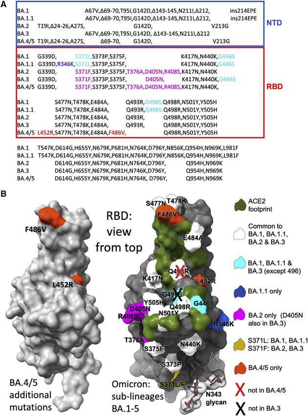
ll
     OPEN ACCESS

Article
Antibody escape of SARS-CoV-2
Omicron BA.4 and BA.5
from vaccine and BA.1 serum
Aekkachai Tuekprakhon,1,16 Rungtiwa Nutalai,1,16 Aiste Dijokaite-Guraliuc,1,16 Daming Zhou,2,3,16 Helen M. Ginn,4
Muneeswaran Selvaraj,1 Chang Liu,1,3 Alexander J. Mentzer,1,5 Piyada Supasa,1 Helen M.E. Duyvesteyn,2 Raksha Das,1
Donal Skelly,5,6,7 Thomas G. Ritter,5 Ali Amini,5,6,8 Sagida Bibi,9 Sandra Adele,5 Sile Ann Johnson,5 Bede Constantinides,10
Hermione Webster,10 Nigel Temperton,11 Paul Klenerman,5,6,8,12 Eleanor Barnes,5,6,8,12 Susanna J. Dunachie,5,6,13,14
Derrick Crook,10 Andrew J. Pollard,9,12 Teresa Lambe,3,9 Philip Goulder,6,15 Neil G. Paterson,4 Mark A. Williams,4
David R. Hall,4 OPTIC Consortium, ISARIC4C Consortium,17 Elizabeth E. Fry,2,* Jiandong Huo,2,16,*
Juthathip Mongkolsapaya,1,3,* Jingshan Ren,2,* David I. Stuart,2,3,4,17,* and Gavin R. Screaton1,3,*
1Wellcome    Centre for Human Genetics, Nuffield Department of Medicine, University of Oxford, Oxford, UK
2Division of Structural Biology, Nuffield Department of Medicine, University of Oxford, The Wellcome Centre for Human Genetics, Oxford, UK
3Chinese Academy of Medical Science (CAMS) Oxford Institute (COI), University of Oxford, Oxford, UK
4Diamond Light Source Ltd, Harwell Science & Innovation Campus, Didcot, UK
5Oxford University Hospitals NHS Foundation Trust, Oxford, UK
6Peter Medawar Building for Pathogen Research, Oxford, UK
7Nuffield Department of Clinical Neurosciences, University of Oxford, Oxford, UK
8Translational Gastroenterology Unit, University of Oxford, Oxford, UK
9Oxford Vaccine Group, Department of Paediatrics, University of Oxford, Oxford, UK
10Nuffield Department of Medicine, University of Oxford, Oxford, UK
11Viral Pseudotype Unit, Medway School of Pharmacy, University of Kent and Greenwich Chatham Maritime, Kent, UK
12NIHR Oxford Biomedical Research Centre, Oxford, UK
13Centre For Tropical Medicine and Global Health, Nuffield Department of Medicine, University of Oxford, Oxford, UK
14Mahidol-Oxford Tropical Medicine Research Unit, Department of Medicine, University of Oxford, Oxford, UK
15Department of Paediatrics, University of Oxford, Oxford, UK
16These authors contributed equally
17Lead contact

*Correspondence: liz@strubi.ox.ac.uk (E.E.F.), dongdong.imm.ox.ac.uk@gmail.com (J.H.), juthathip.mongkolsapaya@well.ox.ac.uk (J.M.),
ren@strubi.ox.ac.uk (J.R.), dave@strubi.ox.ac.uk (D.I.S.), gavin.screaton@medsci.ox.ac.uk (G.R.S.)
 https://doi.org/10.1016/j.cell.2022.06.005

SUMMARY

The Omicron lineage of SARS-CoV-2, which was first described in November 2021, spread rapidly to become
globally dominant and has split into a number of sublineages. BA.1 dominated the initial wave but has been
replaced by BA.2 in many countries. Recent sequencing from South Africa’s Gauteng region uncovered two
new sublineages, BA.4 and BA.5, which are taking over locally, driving a new wave. BA.4 and BA.5 contain
identical spike sequences, and although closely related to BA.2, they contain further mutations in the recep-
tor-binding domain of their spikes. Here, we study the neutralization of BA.4/5 using a range of vaccine and
naturally immune serum and panels of monoclonal antibodies. BA.4/5 shows reduced neutralization by the
serum from individuals vaccinated with triple doses of AstraZeneca or Pfizer vaccine compared with BA.1
and BA.2. Furthermore, using the serum from BA.1 vaccine breakthrough infections, there are, likewise, sig-
nificant reductions in the neutralization of BA.4/5, raising the possibility of repeat Omicron infections.

INTRODUCTION                                                          that all single-point mutations in the large SARS-CoV-2 genome
                                                                      will be generated every day (Sender et al., 2021). Most mutations
SARS-CoV-2 emerged in Wuhan in late 2019 to rapidly cause a           will be silent, deleterious, or of little consequence; however, a
pandemic. It is now estimated to have infected over half a billion    few may give the virus an advantage leading to rapid natural se-
people and caused over 6 million deaths (https://covid19.who.         lection (Domingo, 2010). Many thousands of individual mutations
int/). Although SARS-CoV-2 RNA polymerase possesses some              have been described, and about a year after the outbreak
proofreading ability, there has been a rapid evolution of the viral   started, strains began to emerge containing multiple mutations,
sequence. Because of the scale of the pandemic, it is estimated       particularly in the spike (S) gene. Several of these have been

2422 Cell 185, 2422–2433, July 7, 2022 ª 2022 The Author(s). Published by Elsevier Inc.
This is an open access article under the CC BY license (http://creativecommons.org/licenses/by/4.0/).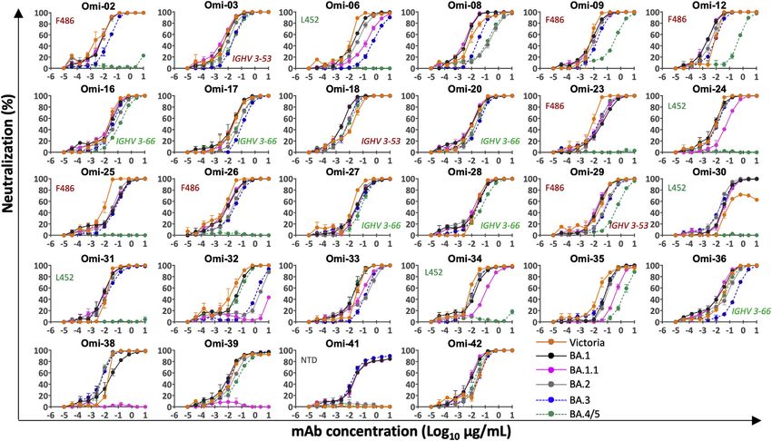
ll
Article                                                                                                                      OPEN ACCESS

designated variants of concern (VoCs) (https://www.cdc.                   BA.1 (Dejnirattisai et al., 2022) and only marginally in BA.2 (Nuta-
gov/coronavirus/2019-ncov/variants/variant-classifications.html)          lai et al., 2022).
and have led to successive waves of infection: first, Alpha (Su-             The initial Omicron wave was caused by the BA.1 strain,
pasa et al., 2021), second, Delta (Liu et al., 2021a), and then Om-       which, compared with ancestral strains, contains 30-aa substitu-
icron (Dejnirattisai et al., 2022) spread globally, becoming the          tions, 6-aa deletions, and 3-aa insertions, which are largely clus-
dominant variants. Alongside these, Beta (Zhou et al., 2021)              tered at the sites of interaction of potently neutralizing anti-
and Gamma (Dejnirattisai et al., 2021b) caused large regional             bodies: the ACE2 interacting surface, around the N343 glycan,
outbreaks in Southern Africa and South America, respectively,             and in the NTD (Dejnirattisai et al., 2022). These changes cause
but did not dominate globally. As of April 29th, over 2.5 million         large reductions in the neutralization titers of vaccine or naturally
cases of Omicron (BA.1 and BA.2) have been reported in                    immune serum, leading to high levels of vaccine breakthrough
the UK alone (https://www.gov.uk/government/publications/                 infections and contributing to the intensity of the Omicron
covid-19-variants-genomically-confirmed-case-numbers/variants-            wave of infection (Dejnirattisai et al., 2022; McCallum
distribution-of-case-data-29-april-2022#omicron), and although            et al., 2022).
the disease is less severe, particularly in vaccinated individuals,          A number of Omicron sublineages have been described. BA.2
the scale of the outbreak has still led to a large number of deaths       and BA.3 were reported at about the same time as BA.1 and
(Nealon and Cowling, 2022).                                               are highly related but contain some unique changes in S (Fig-
   S is the major surface glycoprotein on SARS-CoV-2 and as-              ure 1A), while another sublineage BA.1.1, which contains an addi-
sembles into extended transmembrane anchored trimers                      tional R346K mutation, also emerged (Nutalai et al., 2022). The
(Walls et al., 2020; Wrapp et al., 2020), which give virions their        BA.2 strain, which possesses a small transmission advantage,
characteristic spiky shape. S is divided into N-terminal S1 and           has become globally dominant. BA.3, reported in relatively few
C-terminal S2 regions. S1 contains the N-terminal domain                  sequences compared with BA.1 and BA.2, appears to be a
(NTD) and receptor-binding domain (RBD). A small 25 amino                 mosaic of BA.1 and BA.2 changes (with 3 differences in the
acid (aa) patch at the tip of the RBD is responsible for                  RBD compared with BA.1 and 3 differences compared with
interaction with the cellular receptor angiotensin-converting             BA.2). Cases of BA.2 infection following BA.1 are not thought to
enzyme 2 (ACE2) (Lan et al., 2020). Following ACE2 binding,               be common due to good levels of cross-neutralizing antibodies
S1 is cleaved and detached, whereas S2 undergoes a major                  following vaccination (Nutalai et al, 2022, https://www.who.int/
conformational change to expose the fusion loop, which medi-              news/item/22-02-2022-statement-on-omicron-sublineage-ba.2).
ates the fusion of viral and host membranes, allowing the viral              In early April 2022, two new Omicron lineages were reported
RNA to enter the host cell cytoplasm and commence the repli-              from Gauteng in South Africa and designated BA.4 and BA.5
cative cycle (Walls et al., 2017).                                        (https://assets.publishing.service.gov.uk/government/uploads/
   S is the major target for neutralizing antibodies, and studies by      system/uploads/attachment_data/file/1067672/Technical-Briefing-
a number of groups have isolated panels of monoclonal anti-               40-8April2022.pdf). These have become dominant in Gauteng and
bodies from infected or vaccinated volunteers (Barnes et al.,             look to be fueling a new wave of infection in South Africa, with
2020; Dejnirattisai et al., 2021a; Yuan et al., 2020a). Potently          some international spread. BA.4 and BA.5 (from here on referred
neutralizing antibodies are largely confined to three sets of sites       to as BA.4/5) have identical S sequences and appear to have
on S1. The first is within the NTD (Cerutti et al., 2021; Chi et al.,     evolved from BA.2. They contain additional mutations in the
2020); these antibodies do not block ACE2 interaction, and their          RBD, in particular, the reversion mutation R493Q (Q493 is found
mechanism of action is still not well determined. The second re-          in ancestral strains), together with mutations L452R and F486V
gion of binding is on or in close proximity to the ACE2 binding           (Figure 1A).
surface of the RBD; most potently neutralizing antibodies bind               Here, we report the antigenic characterization of BA.4/5
this region and prevent the interaction of S with ACE2 on the             compared with the other Omicron sublineages (for complete-
host cell, blocking infection (Dejnirattisai et al., 2021a; Yuan          ness, we also report data on BA.3, although this is of less
et al., 2020a). Finally, some potent antibodies bind the RBD              concern). We find that the neutralization of BA.4/5 by triple-dosed
but do not block ACE2 binding, exemplified by mAb S309, which             vaccine serum is reduced compared with BA.1 and BA.2. We also
binds in the region of the N-linked glycan at position 343 (Pinto         see reductions in titers against BA.4/5 compared with BA.1 and
et al., 2020), these antibodies may function to destabilize the           BA.2 in the sera from individuals who had suffered vaccine break-
S-trimer (Huo et al., 2020b; Yuan et al., 2020b; Zhou et al., 2020).      through BA.1 infections. The neutralization of the Omicron line-
   Although mutations in the VoC are spread throughout S, there           age by a panel of recently derived potent Omicron-specific
are particular hotspots in the NTD and RBD, exactly where                 mAbs raised following vaccine breakthrough BA.1 infection (Nu-
potent neutralizing antibodies bind, and they are likely being            talai et al., 2022) is reduced: 10/28 are completely knocked out
driven by escape from the antibody response following natural             against BA.4/5, while several others suffer large reductions in ac-
infection or vaccination. Mutation of the ACE2 interacting sur-           tivity compared with the other Omicron lineages. We corroborate
face may also give an advantage by increasing ACE2 affinity               the neutralization results with a biophysical analysis of binding
for S or by possibly altering receptor tropism (Zahradnı́k et al.,        and provide structure-function explanations for mAb failure
2021). Increased ACE2 affinity has been found in VoC compared             against BA.4/5 with the changes at residues 452 and 486, both
with ancestral strains (Dejnirattisai et al., 2021b; Liu et al., 2021a;   of which cause serious impact. Finally, we measure the affinity
Supasa et al., 2021; Zhou et al., 2021), potentially conferring a         of the BA.4/5 RBD for ACE2 and find that it is higher than earlier
transmission advantage, but affinity is not increased in Omicron          Omicron strains BA.1 and BA.2.

                                                                                                     Cell 185, 2422–2433, July 7, 2022 2423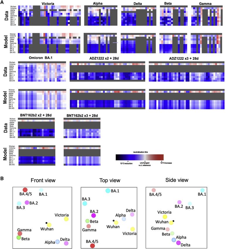
ll
     OPEN ACCESS                                                                                                                 Article
                                                                                            Figure  1. The    Omicron               sublineage
                                                                                            compared with BA.4/5
                                                                                            (A) Comparison of S protein mutations of Omicron
                                                                                            BA.1, BA.1.1, BA.2, BA.3, and BA.4/5 with NTD
                                                                                            and RBD boundaries indicated.
                                                                                            (B) Position of RBD mutations (gray surface with
                                                                                            the ACE2 footprint in dark green). Mutations
                                                                                            common to all Omicron lineages are shown in
                                                                                            white (Q493R, which is reverted in BA.4/5, is
                                                                                            shown with a cross), those common to BA.1 and
                                                                                            BA.1.1 in cyan, those unique to BA.1.1 in blue, and
                                                                                            those unique to BA.2 in magenta. Residue 371
                                                                                            (yellow) is mutated in all Omicron viruses but differs
                                                                                            between BA.1 and BA.2. The N343 glycan is shown
                                                                                            as sticks with a transparent surface.

                                                                                             from Mink early in the pandemic. F486 is
                                                                                             also a site of escape mutations to several
                                                                                             mAbs (Gobeil et al., 2021), and F486I was
                                                                                             noted during SARS-CoV-2 evolution in an
                                                                                             immunocompromised individual (Clark
                                                                                             et al., 2021). The change F486V in BA.4/
                                                                                             5 also causes a reduction in the bulk of
                                                                                             the hydrophobic side chain as in F486L
                                                                                             but is more significant. Both residues
                                                                                             452 and 486 lie close to the edge of the
                                                                                             ACE2 interaction surface (Figure 1B)
                                                                                             and, together with the reversion to ances-
                                                                                             tral sequence Q493, which lies within the
                                                                                             ACE2 footprint, have the potential to
                                                                                             modulate ACE2 affinity and the neutral-
                                                                                             izing capacity of the vaccine or naturally
                                                                                             acquired serum. The L452R and F486V
                                                                                             mutations are likely to cause more anti-
                                                                                             body escape, whereas the reversion at
                                                                                             493 may reduce the escape from the re-
                                                                                             sponses to earlier viruses.
                                                                                                To verify structural inferences, the crys-
                                                                                             tal structure of BA.4/5 RBD was deter-
                                                                                             mined at 1.9 Å as a ternary complex with
                                                                                             a neutralizing Fab and nanobody
                                                                                             (Table S1; Figure S1). This confirmed
RESULTS                                                             that the structure of the BA.4/5 RBD is very similar to that of other
                                                                    variants, although the residue 371–375 region, which is a hotspot
The Omicron lineages BA.4/5                                         of Omicron-specific mutations, is unusually well ordered and the
BA.4 and BA.5 S sequences are identical and closely related to      tip of the arginine side chain of L452R is found in two conforma-
BA.2 (sequence diversity in Omicron S is shown in Figure 1A).       tions (Figure S1).
Compared with BA.2, BA.4/5 has residues 69 and 70 deleted
and contains 2 additional substitutions in the RBD: L452R and       Neutralization of BA.4/5 by vaccine serum
F486V. Finally, BA.4/5 lacks the Q493R change seen in BA.1          We constructed a panel of pseudotyped lentiviruses (Di Genova
and BA.2, reverting to Q493 as in the Victoria/Wuhan strain.        et al., 2020) expressing the S gene from the Omicron sublineages
   The 2 additional mutations in the RBD are of most concern in     BA.1, BA.1.1, BA.2, BA.3, and BA.4/5 together with the early
terms of antibody escape: L452R is a chemically radical change      pandemic Wuhan-related strain, Victoria, used as a control.
and is one of the pair of changes in Delta RBD (the other, T478K,   Neutralization assays were performed using serum obtained
is already found in the Omicron lineage), and L452R is also found   28 days following a third dose of the Oxford-AstraZeneca vac-
in Epsilon and the recently reported Omicron BA.2.11 (https://      cine AZD1222 (n = 41) (Flaxman et al., 2021) or Pfizer-BioNtech
www.who.int/activities/tracking-SARS-CoV-2-variants). Muta-         vaccine BNT162b2 (n = 19) (Cele et al., 2022a; Figures 2A and
tion F486L was found in the sequences of SARS-CoV-2 isolated        2B). For AZD1222, neutralization titers for BA.4/5 were reduced

2424 Cell 185, 2422–2433, July 7, 2022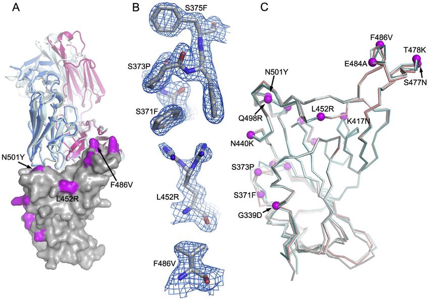
ll
Article                                                                                                                       OPEN ACCESS

                                                                                               Figure 2. Pseudoviral neutralization assays
                                                                                               of BA.4/5 by vaccine and BA.1 immune
                                                                                               serum
                                                                                               (A and B) IC50 values for the indicated viruses
                                                                                               using serum obtained from vaccinees 28 days
                                                                                               following their third dose of vaccine (A)
                                                                                               AstraZeneca AZD1222 (n = 41) or (B) 4 weeks after
                                                                                               the third dose of Pfizer BNT162b2 (n = 19).
                                                                                               (C and D) Serum from volunteers suffering break-
                                                                                               through BA.1 infection taken (C) early, i.e., %
                                                                                               17 days from symptom onset (median 12 days)
                                                                                               n = 12 and (D) late, i.e., R28 days from symptom
                                                                                               onset (median 45 days) n = 14. Comparison is
                                                                                               made with neutralization titers to Victoria an early
                                                                                               pandemic strain, BA.1, BA.1.1, BA.2, and BA.3.
                                                                                               Geometric mean titers are shown above each
                                                                                               column. The Wilcoxon matched-pairs signed-rank
                                                                                               test was used for the analysis, and two-tailed
                                                                                               p values were calculated.

                                                                                              cantly less than those against BA.1 and
                                                                                              BA.2. At the early time point, BA.4/5 titers
                                                                                              were reduced 1.9- (p = 0.0005) and 1.5-
                                                                                              fold (p = 0.0015) compared with BA.1
                                                                                              and BA.2, respectively. At the later point,
                                                                                              BA.4/5 titers were reduced 3.4- (p =
                                                                                              0.0001) and 2-fold (p = 0.0017) compared
                                                                                              with BA.1 and BA.2, respectively.
2.1-fold compared with BA.1 (p < 0.0001) and 1.8-fold compared            Thus, BA.4/5 shows a degree of immune escape from the vac-
with BA.2 (p < 0.0001). For BNT162b2, neutralization titers were        cine/BA.1 response when compared with BA.1 and BA.2. These
reduced 3.1-fold (p < 0.0001) and 3.1-fold (p < 0.0001) compared        samples were all taken reasonably close to the time of infection
with BA.1 and BA.2, respectively. These reductions in titers may        meaning that further waning in the intervening months may
reduce the effectiveness of the vaccines at preventing infection,       render individuals susceptible to reinfection with BA.4/5.
particularly at longer time points, as antibody titers naturally
wane, although it would be expected that protection would               Escape from monoclonal antibodies by BA.4/5
remain against severe disease.                                          We have recently reported a panel of potent human mAb gener-
                                                                        ated from cases of Omicron breakthrough infection (Nutalai
Neutralization of BA.4/5 by serum from breakthrough                     et al., 2022). For the 28 most potent mAbs (BA.1 IC50 titers
BA.1 infection                                                          5-fold reduction in the neutral-
fections were mild. Early samples (n = 12, 9F and 3M; median            ization titer of BA.4/5 compared with BA.2. All of these anti-
age is 26; and median time since vaccine is 141 days) were              bodies interact with the RBD, with the exception of Omi-41,
taken %17 days from symptom onset (median is 12 days),                  which binds the NTD and specifically neutralizes BA.1, BA.1.1,
and later samples (n = 14, 7F and 7M; median age is 23; and             and BA.3 but not BA.2 or BA.4/5 (for unknown reasons, Omi-
median time since vaccine is 111 days) were taken R28 days              41 can neutralize wild-type (WT) Victoria virus but not Victoria
following symptom onset (median is 45 days). All cases had              pseudovirus) (Nutalai et al., 2022).
been vaccinated, all but 2 had received 2 doses, and 3 of the
late convalescent cases received a third dose of vaccine                SENSITIVITY TO L452R
following Omicron infection. Pseudoviral neutralization assays
were performed against the panel of pseudoviruses described             We have previously reported that Omi-24, 30, 31, 34, and 41
above (Figures 2C and 2D).                                              show complete knockout of neutralizing activity against Delta,
   As we have previously described, BA.1 infection following            with Omi-06 showing a severe knockdown of activity (Nutalai
vaccination leads to a broad neutralizing response, with high           et al., 2022). Since BA.1 and BA.2 harbor only one (T478K) of
titers to all the VoC, which is boosted at later time points (Nutalai   the 2 Delta RBD mutations, while BA.4/5 also harbor L452R,
et al., 2022). Neutralization titers against BA.4/5 were signifi-       we would expect all five of these L452-directed mAbs to be

                                                                                                  Cell 185, 2422–2433, July 7, 2022 2425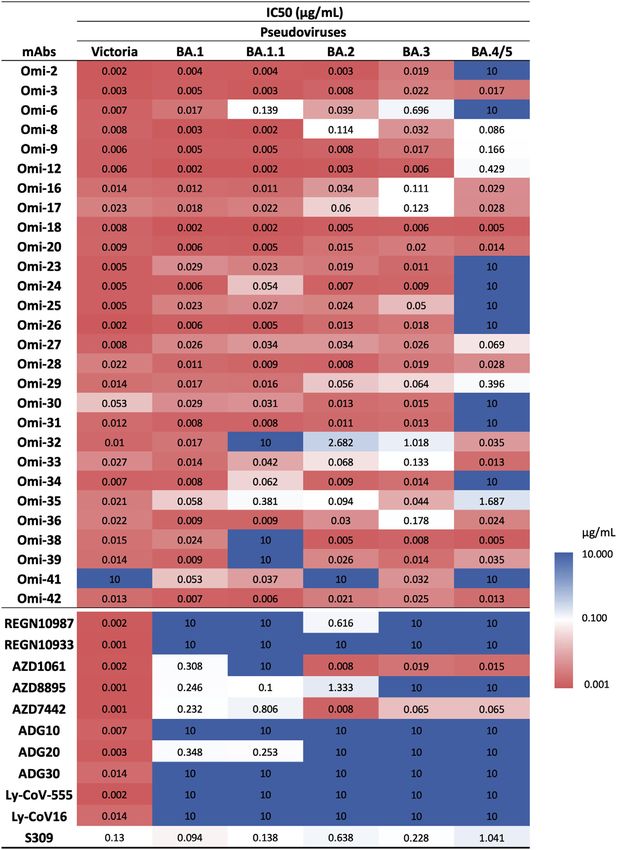
ll
OPEN ACCESS Article
Figure 3. IC50 values for Omicron and com-
mercial mAbs
See also Figures S2, S3, S4, and S5.
shown in Figure 5A, and it reveals that
L452 is tucked neatly into a hydrophobic
pocket, which is unable to accommodate
the larger positively charged arginine in
BA.4/5 and Delta without major confor-
mational changes.
L452R enhancement of binding
Omi-32 shows 77-fold enhanced neutral-
ization of BA.4/5 compared with BA.2. Ki-
netic analysis of Fab binding to the RBDs
suggests that this is mainly achieved by a
5-fold increase in the on rate of binding
(Figures 4B and 4C). This could be ex-
plained by the arginine at 452 making a
salt bridge to residue 99 of the heavy-
chain (HC) CDR3 (Figure 5B). It is possible
that electrostatic changes enhance on
rate by electrostatic steering of the
incoming antibody.
SENSITIVITY TO F486V
Extending the logic used to understand
Delta sensitivity, the remaining antibodies
affected by BA.4/5 > BA.2, but which
retain activity against Delta, namely Omi-
02, 09, 12, 23, 25, 26, and 29, are likely
sensitive to the F486V change. The bind-
ing sensitivity was confirmed by SPR
analysis of Omi-12, a VH1-58 family mem-
ber, which, like AZD 8895 (below), binds
over F486 (Nutalai et al., 2022;
Figures 4D and 4E) and showed an almost
1,000-fold reduction in affinity to BA.4/5.
Another example of the structural basis
of sensitivity to F486V is provided by
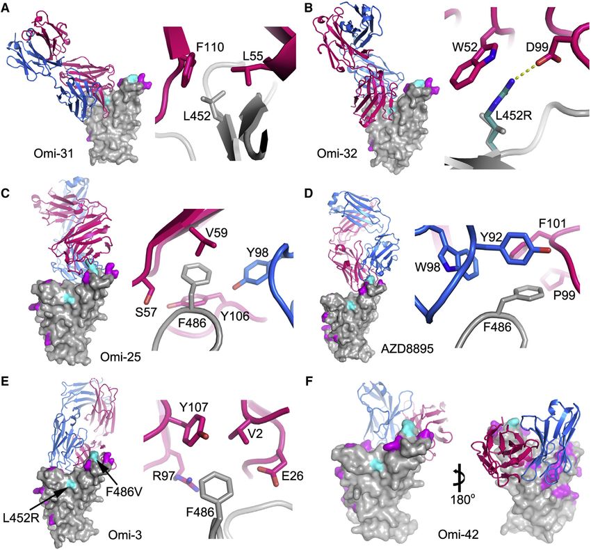
knocked out on BA.4/5. This is indeed observed (Figures 3 and Omi-25, which shows reduced binding and no neutralizing activ-
S2). Omi-41 also fails to neutralize, which is attributed to the dif- ity against BA.4/5 (Figures 3 and S3J). The Omi-25 complex
ferences in mutations in the NTD (Figure 1A). shows that the phenylalanine side chain acts as a binding hot-
To confirm that the neutralization effects observed are directly spot, nestled in a hydrophobic cavity making favorable ring-
attributable to alterations in RBD interactions, we also per- stacking interactions with Y106 of the HC CDR3 (Figure 5C).
formed binding analyses of selected antibodies to BA.4/5 and
BA.2 RBDs by surface plasmon resonance (SPR) (Figures 4 The activity of commercial antibodies against BA.4/5
and S3). Omi-31 was chosen as the representative of the set We tested a panel of antibodies that have been developed for
of L452R sensitive antibodies, and as expected, the binding is therapeutic/prophylactic use against BA.4/5 (Figures 3 and
severely affected (Figure 4A). Since we have detailed informa- S4). Many of these antibodies have already suffered severe re-
tion on the interaction of several Omicron responsive antibodies ductions or knockout of activity against BA.1, BA.1.1, or BA.2.
with the RBD, including Omi-31, we modeled the BA.4/5 RBD For AstraZeneca AZD1061, activity against BA.4/5 was similar
mutations in the context of known structures for Omicron to that against BA.2 (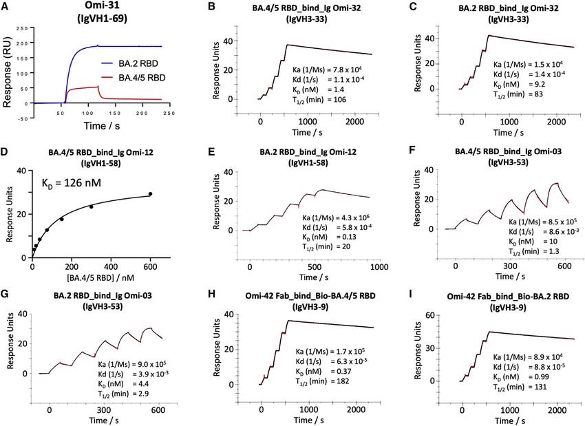
ll
Article                                                                                                                                   OPEN ACCESS

Figure 4. Surface plasmon resonance (SPR) analysis of the interaction between BA.2 or BA.4/5 RBD and selected mAbs
(A) Binding of BA.4/5 RBD is severely reduced compared with that of BA.2, so the binding could not be accurately determined, as shown by a single injection of
200 nM RBD over sample flow cells containing IgG Omi-31.
(B, C, and E–I) Sensorgrams (red: original binding curve; black: fitted curve) showing the interactions between BA.2 or BA.4/5 RBD and selected mAbs, with
kinetics data shown.
(D) Determination of the affinity of BA.4/5 RBD to Omi-12 using a 1:1 binding equilibrium analysis.
See also Figures 3 and S3.

2021) was reduced 8.1-fold compared with BA.2. The residual                     Systematic themes in mAb interactions
activity of REGN10987 (Weinreich et al., 2021) against BA.2                     Both Omi-3 (a representative of the IGVH3-53 gene family) and
was further reduced on BA.4/5; likewise, residual BA.1 neutral-                 AZD8895 (IGVH1-58) make contact with F486. While the F486V
izing activity was knocked out for ADG20 (Yuan et al., 2022) on                 mutation has little effect on Omi-3 (Figures 3, 4F, 4G, and 5E),
BA.4/5. For S309 (VIR-7831/7832) (Sun and Ho, 2020), the activ-                 it seriously reduces the neutralization of AZD8895 and other
ity against BA.4/5 was 1.6-fold reduced compared with BA.2.                     IGVH1-58 mAbs, e.g., Omi-12 (Figures 3, 4D, 4E, and 5D). It is
   These effects can be rationalized by reference to the way                    notable that whereas the numerous Omi series antibodies
the antibodies interact with the RBD; for instance, in the                      belonging to the closely related IGVH3-53 and IGVH3-66 gene
case of AZD8895 (an IGVH1-58 genotype mAb, Figure 5D),                          families (9/28 in total Figure S2) are almost entirely resilient to
F486 forms a hydrophobic interaction hotspot, which will be                     the BA.4/5 changes, the large majority of antibodies from these
abrogated by the mutation to a much smaller valine side chain.                  gene families elicited against earlier variants are knocked out on
Antibody residues involved in the interactions with F486 are                    BA.1 and BA.2 (Nutalai et al., 2022), consistent with selection of a
highly conserved among this genotype of mAbs, including                         subset of antibodies by breakthrough Omicron infection that is
Omi-12, 253, and Beta-47 (Nutalai et al., 2022; Dejnirattisai                   insensitive to the further BA.4/5 mutations.
et al., 2021a; Liu et al., 2021b), explaining the severe effect                    The effects on antibodies with broadly similar epitopes can vary
of the F486V mutation on neutralization of these mAbs (Fig-                     dramatically, and this is equally true for antibodies, which have
ures 3 and S5).                                                                 452 or 486 central to their binding footprint. Thus, Omi-31

                                                                                                               Cell 185, 2422–2433, July 7, 2022 2427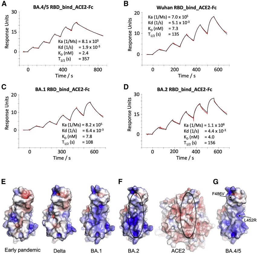
ll
      OPEN ACCESS                                                                                                                                Article

Figure 5. Interactions between mAb and BA.4/5 mutation sites
Overall structure (left panel) and interactions (%4 Å) with BA.4/5 mutation sites (right panel) for (A) BA.1-RBD/Omi-31 (PDB: 7ZFB), (B) BA.1-RBD/Omi-32 (PDB:
7ZFE), (C) BA.1-RBD/Omi-25 (PDB: 7ZFD), (D) Wuhan-RBD/AZD8895 (PDB: 7L7D), (E) BA.1-RBD/Omi-3 (PDB: 7ZF3), and (F) BA.1-RBD/Omi-42 (PDB: 7ZR7)
complexes. In the left panels, RBD is shown as surface representation, with BA.4/5 mutation sites highlighted in magenta and the additional two mutation sites of
BA.4/5 at 452 and 486 in cyan and Fab LC as blue and HC as red ribbons. In the right panel, side chains of RBD, Fab HC, and LC are drawn as gray, red, and blue
sticks, respectively. In (B), the L452R mutation (cyan sticks) is modeled to show that a salt bridge to D99 of CDR-H3 may be formed (yellow broken sticks).
(F) shows that the Fab of Omi-42 does not contact either of the two BA.4/5 mutation sites.
See also Figure S1.

(IGVH1-69) and Omi-32 (IGVH3-33) both bind in front of the right                  compared with the ancestral virus (Wuhan), BA.1, and BA.2
shoulder with their CDR-H3 positioned close to 452; while the ac-                 (approximately 3-, 3-, and 2-fold, respectively [BA.4/5/ACE2
tivity of Omi-31 is abolished by L452R (as detailed above), Omi-32                KD = 2.4 nM]) (Dejnirattisai et al., 2022; Nutalai et al., 2022),
is markedly enhanced (Figures 3, 5A, 5B, and S2). Similarly, Omi-                 which is mainly attributed to an increase in binding half-life.
25 and Omi-42 both belong to the IGVH3-9 gene family, and their                   Modeling of the ACE2/RBD complex suggests that the bulk of
footprints are in the 486 region (Figures 5C and 5F). Omi-25 con-                 this effect comes from the electrostatic complementarity be-
tacts F486, and thus, the neutralization of BA.4/5 is abolished. By               tween ACE2 and the RBD contributed by the L452R mutation
contrast, Omi-42 does not contact either of the mutation sites, and               (Figures 6E–6G).
neutralization is fully retained for BA.4/5 (Figures 3, 4H, 4I, and 5F).
                                                                                  Antigenic cartography
ACE2 RBD affinity                                                                 The neutralization data above has been used to place BA.3 and
We measured the affinity of BA.4/5 RBD for ACE2 by SPR                            BA.4/5 on an antigenic map. We repeated the method used for
(Figures 6A–6D). The affinity of BA.4/5 RBD was increased                         the analysis of the Delta and Omicron variants (Liu et al.,

2428 Cell 185, 2422–2433, July 7, 2022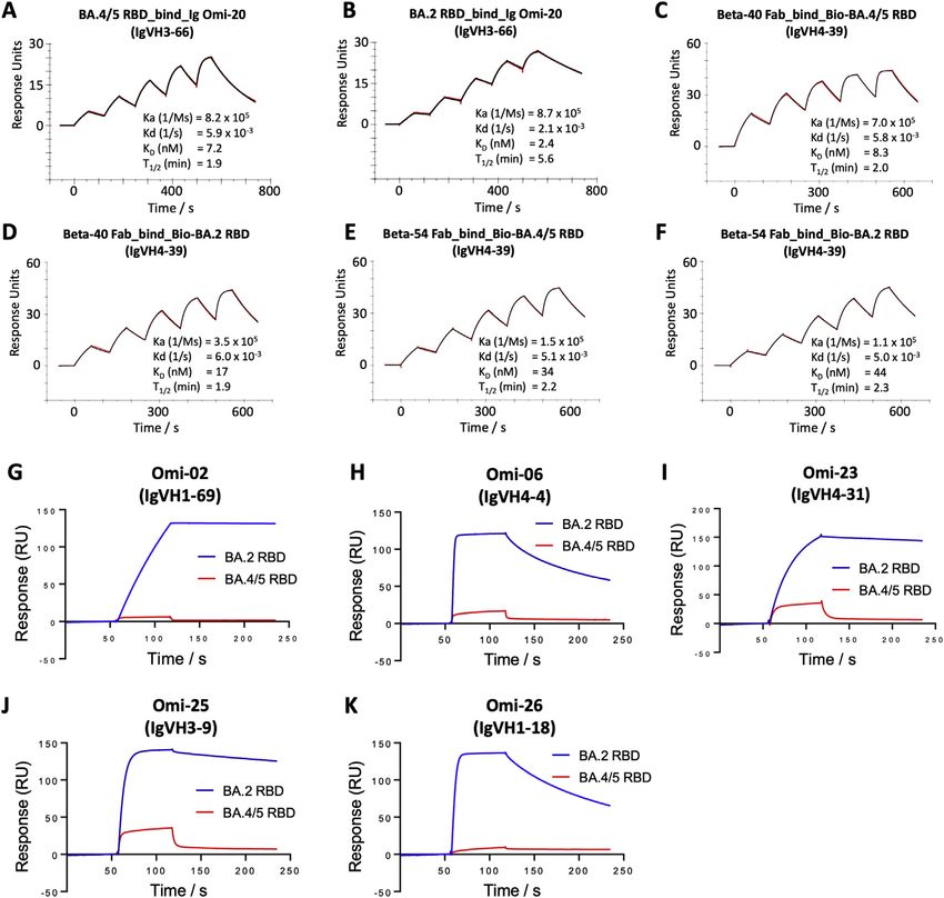
ll
Article                                                                                                                     OPEN ACCESS

                                                                                             Figure 6. ACE2 RBD affinity
                                                                                             (A–D) SPR sensorgrams showing ACE2 binding of
                                                                                             BA.4/5 RBD (A) in comparison with binding to
                                                                                             ancestral (Wuhan) (B), BA.1 (C), and BA.2 RBD (D).
                                                                                             The data for Wuhan, BA.1, and BA.2 have been
                                                                                             reported previously in Nutalai et al. (2022).
                                                                                             (E–G) Electrostatic surfaces, (E) from left to right,
                                                                                             early pandemic, Delta, and BA.1 RBD. (F) Open
                                                                                             book view of BA.2 RBD and ACE2 of the BA.2
                                                                                             RBD/ACE2 complex (PDB: 7ZF7) and (G) BA.4/5
                                                                                             RBD (PDB: 7ZXU). The lozenges on ACE2 and
                                                                                             RBD show the interaction areas.

                                                                                               individual changes in single viruses.
                                                                                               Virus recombination, which was pre-
                                                                                               dicted, is now being detected, allowing
                                                                                               the shuffling of complex genomes,
                                                                                               such as XD (Delta/BA.1) and XE (BA.1/
                                                                                               BA.2), which in the latter case, may
                                                                                               be more transmissible (https://assets.
                                                                                               publishing.service.gov.uk/government/
                                                                                               uploads/system/uploads/attachment_
                                                                                               data/file/1063424/Tech-Briefing-39-25
                                                                                               March2022_FINAL.pdf).
                                                                                                 How such large sequence jumps, such
                                                                                               as that to the Omicron lineage, occur is
                                                                                               not known. It has been suggested that
                                                                                               these may be occurring in immunocom-
                                                                                               promised or HIV-infected cases, where
                                                                                               chronic infections have been docu-
                                                                                               mented to last for many months or in
2021a), where individual viruses were independently modeled al-        some cases over a year. The selection of antibody escape mu-
lowing for serum-specific scaling of the responses (STAR               tations has been documented in such individuals (Cele et al.,
Methods). The measured and modeled responses are shown in              2022b; Karim et al., 2021; Kemp et al., 2021), and successive
Figure 7A (with 1,551 observations and 340 parameters, the re-         rounds of replication, recombination, and perhaps reinfection
sidual error is 23%). The results are best visualized in three         may be responsible for the selection of the constellation of S
dimensions (see Video S1), but 2D projections are shown in Fig-        mutations found in the Omicron lineage.
ure 7B. This shows, as expected, that the Omicron sublineages             BA.4/5, the most recently reported Omicron sublineages,
are clustered together but well separated from early pandemic          seem to be taking hold in South Africa and may spread globally
virus and earlier VoC. Among the Omicron cluster, BA.4/5 is            to replace BA.2. Although highly related to BA.2, BA.4/5 contain
the most distant from the pre-Omicron viruses; the distance be-        the 69-70 deletion in the NTD that was also found in Alpha, BA.1,
tween BA.4/5 and BA.2 is similar to that between BA.2 and BA.1.        and BA.3, together with additional mutations in the RBD (L452R
                                                                       and F486V). Thus, BA.4/5 has assembled mutations at all of the
DISCUSSION                                                             previously described positions in the VoC Alpha (N501Y), Beta
                                                                       (K417N, E484K, N501Y), Gamma (K417T, E484K, N501Y), and
Following the emergence of SARS-CoV-2 in November 2019, a              Delta (L452R, T478K), the only difference being E484A in BA.4/
succession of SARS-CoV-2 viral variants have appeared with             5 rather than E484K found in Beta and Gamma.
increased fitness; these variants have rapidly outcompeted the            Here, we report a greater escape from the neutralization of
preceding strain and spread globally—the most recent, Omi-             BA.4/5 compared with BA.1 and BA.2. Serum from triple vacci-
cron, appearing in late 2021.                                          nated donors has 2- to 3-fold reduction in neutralization titers
   Despite the availability of vaccines, the pandemic has not          compared with the neutralization of BA.1 and BA.2. Additionally,
been brought under control, and through Omicron, infections            serum from breakthrough BA.1 infections in vaccinees shows
are as high as ever. Although vaccines are effective at preventing     2- to 3-fold reduction in neutralization titers to BA.4/5
severe disease, they are less effective at preventing transmis-        compared with BA.1 and BA.2. These reductions are in good
sion, particularly of the Omicron sublineages. The very high level     agreement with the reductions of BA.4 and BA.5 neutralization
of viral replication globally drives the accrual of mutations in the   titers reported following BA.1 vaccine breakthrough infections
viral genome, and we are now seeing the assembly of dozens of          (Khan et al., 2022). These data suggest that a further wave of

                                                                                                Cell 185, 2422–2433, July 7, 2022 2429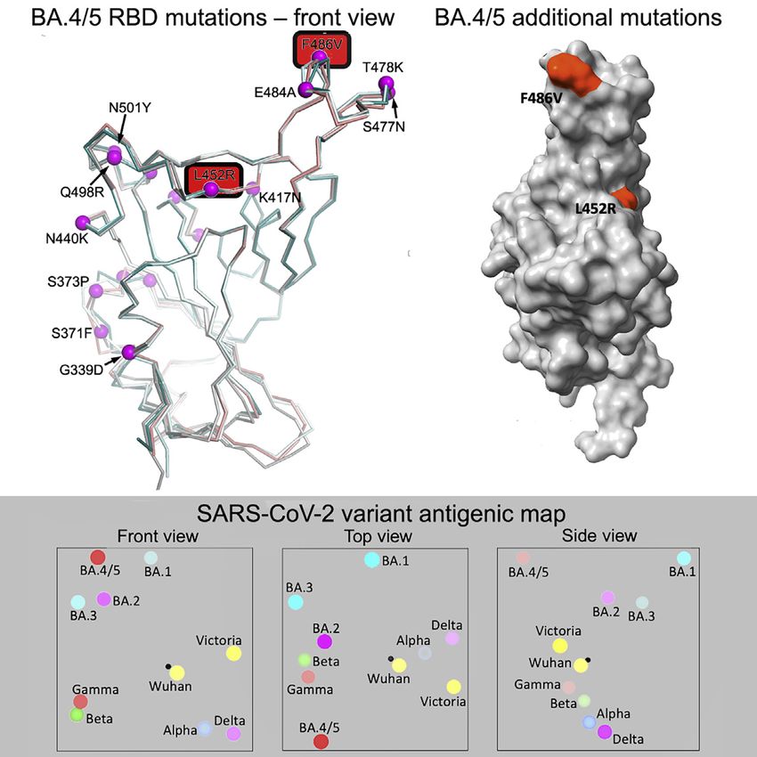
ll
      OPEN ACCESS                                                                                                                                     Article

Figure 7. Antigenic mapping
(A) Neutralization data and model (log titer values) used to calculate antigenic maps in (B). Columns represent sera collected from inoculated volunteers or in-
fected patients. Rows are challenge strains: Victoria, Alpha, Delta, Beta, Gamma, BA.1, BA1.1, BA.2, BA.3, and BA.4/5 in order. Values are colored according to
their deviation from the reference value. The reference value is calculated on a serum-type basis as the average of neutralization titers from the row that gives this
the highest value.
(B) Orthogonal views of the antigenic map showing BA.4/5 in the context of the positions of previous VoC and BA.1, BA.1.1, BA.1, and BA.2, calculated from
pseudovirus neutralization data. Distance between two positions is proportional to the reduction in neutralization titer when one of the corresponding strains is
challenged with a serum derived by infection by the other. No scale is provided since the figures are projections of a three-dimensional distribution; however, the
variation can be calibrated by comparison with (i) BA.1 to BA.2, which is 2.933 reduced, and (ii) BA.2 to BA.4/5, which is 3.033 reduced. The third dimension may
be inferred by fading of the colors with greater distance from the viewer.

Omicron infection, driven by BA.4/5, is likely, partly due to break-                 new RBD mutations in BA.4/5. The activity of many mAbs is
through of vaccine and naturally acquired immunity, although                         either knocked out or severely impaired against BA.4/5
there is no evidence yet of increased disease severity.                              compared with BA.2. From the neutralization data on BA.4/5,
  Using a panel of potent mAbs generated from vaccinated indi-                       compared with that on Delta, we have been able to impute the
viduals infected with BA.1, we show the importance of the two                        contribution of L452R and F486V, and by combining with SPR

2430 Cell 185, 2422–2433, July 7, 2022
ll
Article OPEN ACCESS
data, as well as previous mapping by bio-layer interferometry STAR+METHODS
(BLI) competition matrices and detailed structural data (Nutalai
et al., 2022), we are able to understand the basis of these effects Detailed methods are provided in the online version of this paper
on neutralization and show that the L452R and F486V mutations and include the following:
both make major contributions to BA.4/5 escape.
It is clear that the Omicron lineage, particularly BA.4/5, has d KEY RESOURCES TABLE
escaped or reduced the activity of mAbs developed for clinical d RESOURCE AVAILABILITY
use, with most mAbs showing complete knockout of activity. B Lead contact
AZD7442 still shows activity against BA.4/5 (65 ng/mL), but B Materials availability
B Data and code availability
65-fold less than the activity against Victoria, and S309 activity
against BA.4/5 is 8-fold reduced compared with Wuhan with d EXPERIMENTAL MODEL AND SUBJECT DETAILS
IC50 titers >1,000 ng/mL. The reduction in the neutralizing activ- B Bacterial strains and cell culture
B Plasma from early-pandemic and Alpha cases
ity of S309 reported here using pseudoviruses is less than that for
B Sera from Beta-, Gamma-, and Delta- and BA.1-in-
WT viruses and may be due to differences in the assay format; for
instance, the IC50 for BA.2 using pseudovirus is 638 ng/mL, fected cases
B Sera from BA.1-infected cases, study subjects
while we reported 5,035 ng/mL using a WT virus (Nutalai
B Sera from Pfizer vaccinees
et al., 2022).
New monoclonals and combinations may be needed to plug B AstraZeneca-Oxford vaccine study procedures and
the gap in activity and protect the extremely vulnerable and sample processing
those unable to mount adequate vaccine responses. There is d METHOD DETAILS
also a question about vaccines. All current vaccines use spike B Plasmid construction and pseudotyped lentiviral parti-
derived from the original virus isolated from Wuhan. Vaccines cles production
B Pseudoviral neutralization test
have been remarkably effective at reducing severe disease,
B Cloning of RBDs
and a triple dosing schedule has provided, at least in the
short term, protection against Omicron. However, the preven- B Production of RBDs
B Surface plasmon resonance
tion of transmission may become less effective as viruses
B IgG mAbs and Fabs production
evolve antigenically further from ancestral strains. Some argue
for next-generation vaccines tailored to antigenically distant B Crystallization, X-ray data collection, and structure
strains, such as Omicron, to give better protection, probably determination
B Antigenic mapping
used in combination with boosters containing ancestral
strains. While vaccination is unlikely to eliminate transmission, d QUANTIFICATION AND STATISTICAL ANALYSIS
the combination of vaccines with boosting by natural infection
will probably continue to protect the majority from severe SUPPLEMENTAL INFORMATION
disease.
Supplemental information can be found online at https://doi.org/10.1016/j.cell.
Finally, it is impossible to say where SARS-CoV-2 evolution 2022.06.005.
will go next, but it is certain that the virus will continue to drift anti-
genically. This may be a continuation along the Omicron lineage, CONSORTIA
or we may see a large jump to a completely new lineage, like the
one from Delta to Omicron. The observation that of the 30-aa The full contributor list for the ISARIC4C Consortium is available at https://
substitutions in BA.1, all but one was achieved by a single isaric4c.net/about/authors/. The members of the OPTIC Consortium are Chris-
topher Conlon, Alexandra Deeks, John Frater, Lisa Frending, Siobhan
base change in the codon suggests that there remains plenty
Gardiner, Anni Jämsén, Katie Jeffery, Tom Malone, Eloise Phillips, Lucy Roth-
of antigenic space for SARS-CoV-2 to explore and the capacity well, and Lizzie Stafford.
for recombination, which has so far not been observed to have
breakpoints within the major antigenic sites, could generate a ACKNOWLEDGMENTS
more radical antigenic shift.
This work was supported by the Chinese Academy of Medical Sciences
Limitations of the study (CAMS) Innovation Fund for Medical Science (CIFMS), China (grant number:
2018-I2M-2-002), to D.I.S. and G.R.S. We are also grateful for support from
One of the limitations of this study is that serum was obtained at
Schmidt Futures, the Red Avenue Foundation, and the Oak Foundation.
early time points following vaccination or breakthrough infection, G.R.S. was supported by Wellcome. H.M.E.D. and J.R. are supported by Well-
so titers are likely to wane thereafter. In addition, the true in vivo come (101122/Z/13/Z) and D.I.S. and E.E.F. by the UKRI MRC (MR/N00065X/
protection induced by vaccination may be underestimated using 1). D.I.S. and G.R.S. are Jenner Investigators. This is a contribution from the
in vitro neutralization assays where complement, antibody- UK Instruct-ERIC Centre. A.J.M. is an NIHR-supported Academic Clinical
dependent cell-mediated cytotoxicity and T cell responses are Lecturer. The convalescent sampling was supported by the Medical Research
not operative. It would also be interesting to look at BA.4/5 Council (grant MC_PC_19059, awarded to the ISARIC-4C Consortium, with a
full contributor list available at https://isaric4c.net/about/authors/), the Na-
neutralization using serum from unvaccinated individuals who
tional Institutes for Health, the Oxford Biomedical Research Centre, and an
had suffered primary BA.1 infection where the degree of escape Oxfordshire Health Services Research Committee grant to A.J.M. The Well-
of BA.4/5 may be greater than that seen with the vaccine break- come Centre for Human Genetics is supported by the Wellcome (grant
through BA.1 serum reported here. 090532/Z/09/Z). The computational aspects of this research were supported
Cell 185, 2422–2433, July 7, 2022 2431ll
OPEN ACCESS Article
by the Wellcome Core award grant number 203141/Z/16/Z and the NIHR Ox- REFERENCES
ford BRC. We thank the staff of the MRC Human Immunology Unit for access
to their Biacore Facility. Aricescu, A.R., Lu, W., and Jones, E.Y. (2006 Oct). A time- and cost-efficient sys-
The Oxford Vaccine work was supported by UK Research and Innovation, tem for high-level protein production in mammalian cells. Acta Crystallogr. D Biol.
the Coalition for Epidemic Preparedness Innovations, the National Institute Crystallogr. 62, 1243–1250. https://doi.org/10.1107/S0907444906029799.
for Health Research (NIHR), the NIHR Oxford Biomedical Research Centre, Barnes, C.O., Jette, C.A., Abernathy, M.E., Dam, K.A., Esswein, S.R., Gristick,
and the Thames Valley and South Midland’s NIHR Clinical Research Network. H.B., Malyutin, A.G., Sharaf, N.G., Huey-Tubman, K.E., Lee, Y.E., et al. (2020).
We thank the Oxford Protective T Cell Immunology for COVID-19 (OPTIC) Clin- SARS-CoV-2 neutralizing antibody structures inform therapeutic strategies.
ical team for participant sample collection and the Oxford Immunology Nature 588, 682–687.
Network Covid-19 Response T cell Consortium for laboratory support. We
acknowledge the rapid sharing of Victoria, B.1.1.7, and B.1.351, which was Cele, S., Jackson, L., Khoury, D.S., Khan, K., Moyo-Gwete, T., Tegally, H.,
isolated by scientists within the National Infection Service at PHE Porton San, J.E., Cromer, D., Scheepers, C., Amoako, D.G., et al. (2022a). Omicron
Down, and the B.1.617.2 virus was kindly provided by Wendy Barclay and extensively but incompletely escapes Pfizer BNT162b2 neutralization. Nature
Thushan De Silva. We thank the Secretariat of National Surveillance, Ministry 602, 654–656.
of Health, Brazil, for assistance in obtaining P.1 samples. We acknowledge Cele, S., Karim, F., Lustig, G., San, J.E., Hermanus, T., Tegally, H., Snyman, J.,
Diamond Light Source for time on beamline I03 under Proposal lb27009 for Moyo-Gwete, T., Wilkinson, E., Bernstein, M., et al. (2022b). SARS-CoV-2 pro-
COVID-19 Rapid Access. This work was supported by the UK Department longed infection during advanced HIV disease evolves extensive immune
of Health and Social Care as part of the PITCH (Protective Immunity from escape. Cell Host Microbe 30, 154–162. e5.
T cells to Covid-19 in Health workers) Consortium, the UK Coronavirus Immu- Cerutti, G., Guo, Y., Zhou, T., Gorman, J., Lee, M., Rapp, M., Reddem, E.R.,
nology Consortium (UK-CIC), and the Huo Family Foundation. E.B. and P.K. Yu, J., Bahna, F., Bimela, J., et al. (2021). Potent SARS-CoV-2 neutralizing an-
are NIHR Senior Investigators, and P.K. is funded by WT109965MA and NIH tibodies directed against spike N-terminal domain target a single Supersite.
(U19 I082360). S.J.D. is funded by an NIHR Global Research Professorship Cell Host Microbe 29, 819–833. e7.
(NIHR300791). D.S. is an NIHR Academic Clinical Fellow. The views expressed
Chi, X., Yan, R., Zhang, J., Zhang, G., Zhang, Y., Hao, M., Zhang, Z., Fan, P.,
in this article are those of the authors and not necessarily those of the National
Dong, Y., Yang, Y., et al. (2020). A neutralizing human antibody binds to the
Health Service (NHS), the Department of Health and Social Care (DHSC), the
N-terminal domain of the Spike protein of SARS-CoV-2. Science 369,
National Institutes for Health Research (NIHR), the Medical Research Council
650–655.
(MRC), or Public Health England.
Clark, S.A., Clark, L.E., Pan, J., Coscia, A., McKay, L.G.A., Shankar, S., John-
AUTHOR CONTRIBUTIONS son, R.I., Brusic, V., Choudhary, M.C., Regan, J., et al. (2021). SARS-CoV-2
evolution in an immunocompromised host reveals shared neutralization
J.H. performed the interaction affinity analyses. D.Z. performed the antibody escape mechanisms. Cell 184, 2605–2617. e18.
competition analyses. D.Z., J.H., J.R., D.R.H., M.A.W., and N.G.P. prepared Dejnirattisai, W., Huo, J., Zhou, D., Zahradnı́k, J., Supasa, P., Liu, C., Duyves-
the crystals and enabled and performed the X-ray data collection. J.R., teyn, H.M.E., Ginn, H.M., Mentzer, A.J., Tuekprakhon, A., et al. (2022). SARS-
E.E.F., and D.I.S. analyzed the structural results. G.R.S., J.H., J.M., P.S., CoV-2 Omicron-B.1.1.529 leads to widespread escape from neutralizing anti-
D.Z., R.N., A.T., A.D.-G., M.S., R.D., and C.L. prepared the RBDs, ACE2, body responses. Cell 185, 467–484. e15.
and antibodies. A.T., R.N., A.D.-G., and M.S. performed the neutralization as-
Dejnirattisai, W., Zhou, D., Ginn, H.M., Duyvesteyn, H.M.E., Supasa, P., Case,
says. R.N., A.T., and A.D.-G. constructed and produced the pseudovirus for
J.B., Zhao, Y., Walter, T.S., Mentzer, A.J., Liu, C., et al. (2021a). The antigenic
Omicron variants. D.C., H.W., B.C., and N.T. provided the materials. H.M.G.
anatomy of SARS-CoV-2 receptor binding domain. Cell 184, 2183–2200. e22.
wrote mabscape and performed the mapping and cluster analysis, including
the sequence and antigenic space analyses. A.J.M., D.S., T.G.R., A.A., S.B., Dejnirattisai, W., Zhou, D., Supasa, P., Liu, C., Mentzer, A.J., Ginn, H.M., Zhao,
S.A., S.A.J., P.K., E.B., S.J.D., A.J.P., T.L., and P.G. assisted with the patient Y., Duyvesteyn, H.M.E., Tuekprakhon, A., Nutalai, R., et al. (2021b). Antibody
samples and vaccine trials. E.B., S.J.D., and P.K. conceived the study of vacci- evasion by the P.1 strain of SARS-CoV-2. Cell 184, 2939–2954. e9.
nated healthcare workers and oversaw the OPTIC Healthcare Worker study Di Genova, C., Sampson, A., Scott, S., Cantoni, D., Mayora-Neto, M., Bentley,
and sample collection/processing. G.R.S. and D.I.S. conceived the study E., Mattiuzzo, G., Wright, E., Derveni, M., Auld, B., et al. (2020). Production,
and wrote the initial manuscript draft, with the other authors providing editorial titration, neutralisation and storage of SARS-CoV-2 lentiviral pseudotypes.
comments. All the authors read and approved the manuscript. Bio Protoc 11, e4236.
Domingo, E. (2010). Mechanisms of viral emergence. Vet. Res. 41, 38.
DECLARATION OF INTERESTS
Dong, J., Zost, S.J., Greaney, A.J., Starr, T.N., Dingens, A.S., Chen, E.C.,
G.R.S. sits on the GSK Vaccines Scientific Advisory Board and is a founding Chen, R.E., Case, J.B., Sutton, R.E., Gilchuk, P., et al. (2021). Genetic and
member of RQ Biotechnology. Oxford University holds intellectual property structural basis for recognition of SARS-CoV-2 spike protein by a two-anti-
related to the Oxford-AstraZeneca vaccine and SARS-CoV-2 mAb discovered body cocktail. Nat. Microbiol. 6, 1233–1244.
in G.R.S.’s laboratory. A.J.P. is Chair of UK Dept. health and Social Care’s Emsley, P., Lohkamp, B., Scott, W.G., and Cowtan, K. (2010). Features and
(DHSC) Joint Committee on Vaccination & Immunisation (JCVI) but does not development of coot. Acta Crystallogr. D Biol. Crystallogr. 66, 486–501.
participate in the JCVI COVID-19 committee and is a member of the WHO’s
Flaxman, A., Marchevsky, N.G., Jenkin, D., Aboagye, J., Aley, P.K., Angus, B.,
SAGE. The views expressed in this article do not necessarily represent the
Belij-Rammerstorfer, S., Bibi, S., Bittaye, M., Cappuccini, F., et al. (2021). Re-
views of DHSC, JCVI, or WHO. The University of Oxford has entered into a
actogenicity and immunogenicity after a late second dose or a third dose of
partnership with AstraZeneca on coronavirus vaccine development. T.L. is
ChAdOx1 nCoV-19 in the UK: a substudy of two randomised controlled trials
named as an inventor on a patent application covering this SARS-CoV-2 vac-
(COV001 and COV002). Lancet 398, 981–990.
cine and was a consultant to Vaccitech for an unrelated project while the study
was conducted. S.J.D. is a scientific advisor to the Scottish Parliament on Folegatti, P.M., Ewer, K.J., Aley, P.K., Angus, B., Becker, S., Belij-Rammer-
COVID-19. storfer, S., Bellamy, D., Bibi, S., Bittaye, M., Clutterbuck, E.A., et al. (2020).
Safety and immunogenicity of the ChAdOx1 nCoV-19 vaccine against
Received: May 6, 2022 SARS-CoV-2: a preliminary report of a phase 1/2, single-blind, randomised
Revised: May 23, 2022 controlled trial. Lancet 396, 467–478.
Accepted: June 3, 2022 Gobeil, S.M., Janowska, K., McDowell, S., Mansouri, K., Parks, R., Stalls, V.,
Published: June 9, 2022 Kopp, M.F., Manne, K., Li, D., Wiehe, K., et al. (2021). Effect of natural
2432 Cell 185, 2422–2433, July 7, 2022ll
Article OPEN ACCESS
mutations of SARS-CoV-2 on spike structure, conformation, and antigenicity. Pinto, D., Park, Y.J., Beltramello, M., Walls, A.C., Tortorici, M.A., Bianchi, S.,
Science 373, 6555. Jaconi, S., Culap, K., Zatta, F., De Marco, A., et al. (2020). Cross-neutralization
Huo, J., Le Bas, A., Ruza, R.R., Duyvesteyn, H.M.E., Mikolajek, H., Malinaus- of SARS-CoV-2 by a human monoclonal SARS-CoV antibody. Nature 583,
kas, T., Tan, T.K., Rijal, P., Dumoux, M., Ward, P.N., et al. (2020a). Neutralizing 290–295.
nanobodies bind SARS-CoV-2 spike RBD and block interaction with ACE2. Sender, R., Bar-On, Y.M., Gleizer, S., Bernshtein, B., Flamholz, A., Phillips, R.,
Nat. Struct. Mol. Biol. 27, 846–854. and Milo, R. (2021). The total number and mass of SARS-CoV-2 virions. Proc.
Huo, J., Zhao, Y., Ren, J., Zhou, D., Duyvesteyn, H.M.E., Ginn, H.M., Carrique, Natl. Acad. Sci. USA. 118. e2024815118.
L., Malinauskas, T., Ruza, R.R., Shah, P.N.M., et al. (2020b). Neutralization of Stewart, S.A., Dykxhoorn, D.M., Palliser, D., Mizuno, H., Yu, E.Y., An, D.S., Sa-
SARS-CoV-2 by destruction of the prefusion spike. Cell Host Microbe 28, batini, D.M., Chen, I.S., Hahn, W.C., Sharp, P.A., et al. (2003 Apr). Lentivirus-
445–454. e6. delivered stable gene silencing by RNAi in primary cells. RNA 9, 493–501.
Karim, F., Moosa, M.Y.S., Gosnell, B.I., Cele, S., Giandhari, J., Pillay, S., Te- https://doi.org/10.1261/rna.2192803.
gally, H., Wilkinson, E., San, J.E., Msomi, N., et al. (2021). Persistent SARS- Stuart, D.I., Levine, M., Muirhead, H., and Stammers, D.K. (1979). Crystal
CoV-2 infection and intra-host evolution in association with advanced HIV structure of cat muscle pyruvate kinase at a resolution of 2.6 A. J. Mol. Biol.
nfection. Preprint at medRxiv. https://doi.org/10.1101/2021.06.03.21258228. 134, 109–142.
Kemp, S.A., Collier, D.A., Datir, R.P., Ferreira, I.A.T.M., Gayed, S., Jahun, A., Sun, Y., and Ho, M. (2020). Emerging antibody-based therapeutics against
Hosmillo, M., Rees-Spear, C., Mlcochova, P., Lumb, I.U., et al. (2021). SARS-CoV-2 during the global pandemic. Antib Ther 3, 246–256.
SARS-CoV-2 evolution during treatment of chronic infection. Nature 592, Supasa, P., Zhou, D., Dejnirattisai, W., Liu, C., Mentzer, A.J., Ginn, H.M., Zhao,
277–282. Y., Duyvesteyn, H.M.E., Nutalai, R., Tuekprakhon, A., et al. (2021). Reduced
Khan, K., Karim, F., Ganga, Y., Bernstein, M., Jule, Z., Reedoy, K., Cele, S., neutralization of SARS-CoV-2 B.1.1.7 variant by convalescent and vaccine
Lustig, G., Amoako, D., and Wolter, N. (2022). Omicron sub-lineages BA.4/ sera. Cell 184, 2201–2211. e7.
BA.5 escape BA.1 infection elicited neutralizing immunity. Preprint at medRxiv. Walls, A.C., Park, Y.J., Tortorici, M.A., Wall, A., McGuire, A.T., and Veesler, D.
https://doi.org/10.1101/2022.04.29.22274477. (2020). Structure, function, and antigenicity of the SARS-CoV-2 spike glyco-
Lan, J., Ge, J., Yu, J., Shan, S., Zhou, H., Fan, S., Zhang, Q., Shi, X., Wang, Q., protein. Cell 181, 281–292. e6.
Zhang, L., et al. (2020). Structure of the SARS-CoV-2 spike receptor-binding Walls, A.C., Tortorici, M.A., Snijder, J., Xiong, X., Bosch, B.J., Rey, F.A., and
domain bound to the ACE2 receptor. Nature 581, 215–220. Veesler, D. (2017). Tectonic conformational changes of a coronavirus spike
Libby, R.T., Cosman, D., Cooney, M.K., Merriam, J.E., March, C.J., and Hopp, glycoprotein promote membrane fusion. Proc. Natl. Acad. Sci. USA 114,
T.P. (1988 Aug 23). Human rhinovirus 3C protease: cloning and expression of 11157–11162.
an active form in Escherichia coli. Biochemistry 27, 6262–6268. https://doi. Weinreich, D.M., Sivapalasingam, S., Norton, T., Ali, S., Gao, H., Bhore, R.,
org/10.1021/bi00417a010. Musser, B.J., Soo, Y., Rofail, D., Im, J., et al. (2021). REGN-COV2, a neutral-
Liebschner, D., Afonine, P.V., Baker, M.L., Bunkóczi, G., Chen, V.B., Croll, T.I., izing antibody cocktail, in outpatients with Covid-19. N. Engl. J. Med. 384,
Hintze, B., Hung, L.W., Jain, S., McCoy, A.J., et al. (2019). Macromolecular 238–251.
structure determination using X-rays, neutrons and electrons: recent develop- Winter, G., Waterman, D.G., Parkhurst, J.M., Brewster, A.S., Gildea, R.J., Ger-
ments in Phenix. Acta Crystallogr. D Struct. Biol. 75, 861–877. stel, M., Fuentes-Montero, L., Vollmar, M., Michels-Clark, T., Young, I.D., et al.
Liu, C., Ginn, H.M., Dejnirattisai, W., Supasa, P., Wang, B., Tuekprakhon, A., (2018 Feb 1). DIALS: implementation and evaluation of a new integration pack-
Nutalai, R., Zhou, D., Mentzer, A.J., Zhao, Y., et al. (2021a). Reduced neutral- age. Acta Crystallogr. D Struct. Biol. 74, 85–97. https://doi.org/10.1107/
ization of SARS-CoV-2 B.1.617 by vaccine and convalescent serum. Cell 184, S2059798317017235.
4220–4236.e13. Wrapp, D., Wang, N., Corbett, K.S., Goldsmith, J.A., Hsieh, C.L., Abiona, O.,
Liu, C., Zhou, D., Nutalai, R., Duyvestyn, H., Tuekprakhon, A., Ginn, H., Dejnir- Graham, B.S., and McLellan, J.S. (2020). Cryo-EM structure of the
attisai, W., Supasa, P., Mentzer, A., Wang, B., et al. (2021b). The Beta mAb 2019-nCoV spike in the prefusion conformation. Science 367, 1260–1263.
response underscores the antigenic distance to other SARS-CoV-2 variants. Yuan, M., Liu, H., Wu, N.C., Lee, C.D., Zhu, X., Zhao, F., Huang, D., Yu, W.,
Cell Host Microbe 30, 53–68. Hua, Y., Tien, H., et al. (2020a). Structural basis of a shared antibody response
McCallum, M., Czudnochowski, N., Rosen, L.E., Zepeda, S.K., Bowen, J.E., to SARS-CoV-2. Science 369, 1119–1123.
Walls, A.C., Hauser, K., Joshi, A., Stewart, C., Dillen, J.R., et al. (2022). Struc- Yuan, M., Wu, N.C., Zhu, X., Lee, C.D., So, R.T.Y., Lv, H., Mok, C.K.P., and Wil-
tural basis of SARS-CoV-2 Omicron immune evasion and receptor engage- son, I.A. (2020b). A highly conserved cryptic epitope in the receptor binding
ment. Science 375, 864–868. domains of SARS-CoV-2 and SARS-CoV. Science 368, 630–633.
McCoy, A.J., Grosse-Kunstleve, R.W., Adams, P.D., Winn, M.D., Storoni, L.C., Yuan, M., Zhu, X., He, W.-T., Zhou, P., Kaku, C.I., Capozzola, T., Zhu, C.Y., Yu,
and Read, R.J. (2007). Phaser crystallographic software. J Appl Crystallogr 40, X., Liu, H., Yu, W., et al. (2022). A broad and potent neutralization epitope in
658–674. SARS-related coronaviruses. Preprinta at bioRxiv. https://doi.org/10.1101/
Nealon, J., and Cowling, B.J. (2022). Omicron severity: milder but not mild. 2022.03.13.484037.
Lancet 399, 412–413. Zahradnı́k, J., Marciano, S., Shemesh, M., Zoler, E., Harari, D., Chiaravalli, J.,
Nettleship, J.E., Ren, J., Rahman, N., Berrow, N.S., Hatherley, D., Barclay, Meyer, B., Rudich, Y., Li, C., Marton, I., et al. (2021). SARS-CoV-2 variant pre-
A.N., and Owens, R.J. (2008 Nov). A pipeline for the production of antibody diction and antiviral drug design are enabled by RBD in vitro evolution. Nat. Mi-
fragments for structural studies using transient expression in HEK 293T cells. crobiol. 6, 1188–1198.
Protein Expr. Purif. 62, 83–89. https://doi.org/10.1016/j.pep.2008.06.017. Zhou, D., Dejnirattisai, W., Supasa, P., Liu, C., Mentzer, A.J., Ginn, H.M., Zhao,
Nie, J., Li, Q., Wu, J., Zhao, C., Hao, H., Liu, H., Zhang, L., Nie, L., Qin, H., Y., Duyvesteyn, H.M.E., Tuekprakhon, A., Nutalai, R., et al. (2021). Evidence of
Wang, M., et al. (2020). Establishment and validation of a Pseudovirus neutral- escape of SARS-CoV-2 variant B.1.351 from natural and vaccine-induced
ization assay for SARS-CoV-2. Emerg. Microbes Infect 9, 680–686. sera. Cell 184, 2348–2361. e6.
Nutalai, R., Zhou, D., Tuekprakhon, A., Ginn, H.M., Supasa, P., Liu, C., Huo, J., Zhou, D., Duyvesteyn, H.M.E., Chen, C.P., Huang, C.G., Chen, T.H., Shih,
Mentzer, A.J., Duyvesteyn, H.M.E., Dijokaite-Guraliuc, A., et al. (2022). Potent S.R., Lin, Y.C., Cheng, C.Y., Cheng, S.H., Huang, Y.C., et al. (2020). Structural
cross-reactive antibodies following Omicron breakthrough in vaccinees. Cell. basis for the neutralization of SARS-CoV-2 by an antibody from a convalescent
https://doi.org/10.1016/j.cell.2022.05.014. patient. Nat. Struct. Mol. Biol. 27, 950–958.
Cell 185, 2422–2433, July 7, 2022 2433ll
OPEN ACCESS Article
STAR+METHODS
KEY RESOURCES TABLE
REAGENT or RESOURCE SOURCE IDENTIFIER
Antibodies
Nanobody C1 Huo et al., 2020a N/A
Fab Dejnirattisai et al., 2021a N/A
IgG Dejnirattisai et al., 2021a; N/A
Liu et al., 2021b
EY6A mAb Zhou et al., 2020 N/A
Regeneron mAbs AstraZeneca Cat#REGN10933, and REGN10987
AstraZeneca mAbs AstraZeneca Cat#AZD1061, AZD8895, and AZD7442
Vir mAbs Adagio Cat#S309
Lilly mAbs Adagio Cat#Ly-CoV555, and Cat#Ly-CoV16
Adagio mAbs Adagio Cat#ADG10, Cat#ADG20, and Cat#ADG30
28 mAbs generated from cases of Omicron Nutalai et al., 2022 N/A
breakthrough infection
Anti-c-Myc 9E10 antibody Biolegend Catt#626872
Bacterial, virus strains, and yeast
DH5a bacteria InVitrogen Cat#18263012
Saccharomyces cerevisiae EBY100 ATCC Cat#MYA-4941
E. coli clone 10G cells Lucigen, USA Cat#60117-1
Biological samples
Serum from Pfizer-vaccinated individuals University of Oxford N/A
Serum from AstraZeneca-Oxford-vaccinated University of Oxford N/A
individuals
Plasma from SARS-CoV-2 patients John Radcliffe Hospital in N/A
Oxford UK, South Africa,
and FIOCRUZ (WHO) Brazil
Chemicals, peptides, and recombinant proteins
His-tagged SARS-CoV-2 RBD Dejnirattisai et al., 2021a N/A
His-tagged SARS-CoV-2/Omicron RBD This paper N/A
His-tagged SARS-CoV-2/Omicron BA.4 RBD This paper N/A
His-tagged SARS-CoV-2/Omicron BA.5 RBD This paper N/A
His-tagged SARS-CoV-2 RBD-62 Zahradnı́k et al., 2021 N/A
His-tagged SARS-CoV-2 RBD N501Y Supasa et al., 2021 N/A
His-tagged SARS-CoV-2 RBD K417N, E484K, N501Y Zhou et al., 2021 N/A
His-tagged SARS-CoV-2 RBD K417T, E484K, N501Y Dejnirattisai et al., 2021b N/A
His-tagged SARS-CoV-2 RBD L452R, T478K Liu et al., 2021a N/A
His-tagged human ACE2 Liu et al., 2021a N/A
Human ACE2-hIgG1Fc Liu et al., 2021a N/A
His-tagged 3C protease Libby et al., 1988 N/A
Phosphate buffered saline tablets Sigma-Aldrich Cat#P4417
Dulbecco’s Modified Eagle Medium, high glucose Sigma-Aldrich Cat#D5796
Dulbecco’s Modified Eagle Medium, low glucose Sigma-Aldrich Cat#D6046
FreeStyle 293 Expression Medium Gibco Cat#12338018
L-Glutamine–Penicillin–Streptomycin solution Sigma-Aldrich Cat#G1146
GlutaMAX Supplement Gibco Cat#35050061
Opti-MEM Gibco Cat#11058021
Fetal Bovine Serum Gibco Cat#12676029
(Continued on next page)
e1 Cell 185, 2422–2433.e1–e8, July 7, 2022You can also read