An implantable human stem cell-derived tissue-engineered rostral migratory stream for directed neuronal replacement
←
→
Page content transcription
If your browser does not render page correctly, please read the page content below
ARTICLE
                  https://doi.org/10.1038/s42003-021-02392-8                OPEN

                  An implantable human stem cell-derived tissue-
                  engineered rostral migratory stream for directed
                  neuronal replacement
                  John C. O’Donnell 1,2,7, Erin M. Purvis1,2,3,7, Kaila V. T. Helm1,2, Dayo O. Adewole1,2,4, Qunzhou Zhang5,
                  Anh D. Le5,6 & D. Kacy Cullen 1,2,4 ✉

                  The rostral migratory stream (RMS) facilitates neuroblast migration from the subventricular
                  zone to the olfactory bulb throughout adulthood. Brain lesions attract neuroblast migration
1234567890():,;

                  out of the RMS, but resultant regeneration is insufficient. Increasing neuroblast migration into
                  lesions has improved recovery in rodent studies. We previously developed techniques for
                  fabricating an astrocyte-based Tissue-Engineered RMS (TE-RMS) intended to redirect
                  endogenous neuroblasts into distal brain lesions for sustained neuronal replacement. Here,
                  we demonstrate that astrocyte-like-cells can be derived from adult human gingiva
                  mesenchymal stem cells and used for TE-RMS fabrication. We report that key proteins
                  enriched in the RMS are enriched in TE-RMSs. Furthermore, the human TE-RMS facilitates
                  directed migration of immature neurons in vitro. Finally, human TE-RMSs implanted in
                  athymic rat brains redirect migration of neuroblasts out of the endogenous RMS. By emu-
                  lating the brain’s most efficient means for directing neuroblast migration, the TE-RMS offers a
                  promising new approach to neuroregenerative medicine.

                  1 Center for Brain Injury & Repair, Department of Neurosurgery, Perelman School of Medicine, University of Pennsylvania, Philadelphia, PA, USA. 2 Center for

                  Neurotrauma, Neurodegeneration & Restoration, Corporal Michael J. Crescenz Veterans Affairs Medical Center, Philadelphia, PA, USA. 3 Department of
                  Neuroscience, Perelman School of Medicine, University of Pennsylvania, Philadelphia, PA, USA. 4 Department of Bioengineering, School of Engineering and
                  Applied Science, University of Pennsylvania, Philadelphia, PA, USA. 5 Department of Oral and Maxillofacial Surgery & Pharmacology, University of
                  Pennsylvania School of Dental Medicine, Philadelphia, PA, USA. 6 Department of Oral & Maxillofacial Surgery, Penn Medicine Hospital of the University of
                  Pennsylvania, Perelman Center for Advanced Medicine, Philadelphia, PA, USA. 7These authors contributed equally: John C. O’Donnell, Erin M. Purvis.
                  ✉email: dkacy@pennmedicine.upenn.edu

                  COMMUNICATIONS BIOLOGY | (2021)4:879 | https://doi.org/10.1038/s42003-021-02392-8 | www.nature.com/commsbio                                               1
ARTICLE                                                                 COMMUNICATIONS BIOLOGY | https://doi.org/10.1038/s42003-021-02392-8

A
        dult neurogenesis continues in the mammalian brain in                 improve functional recovery at physiological levels26,36. There is a
        the subgranular zone of the dentate gyrus and the sub-                plethora of preclinical research demonstrating that enhancing the
        ventricular zone (SVZ) surrounding the lateral                        redirection of neuroblasts from the SVZ into regions of injury
ventricles1,2. Neural precursor cells (NPCs) in the SVZ can dif-              with experimental intervention can induce functional recovery
ferentiate into neuroblasts and migrate through the rostral                   following injury14,21,37–48. For example, overexpression of Slit1 in
migratory stream (RMS) to the olfactory bulb (OB) where they                  neuroblasts enhanced SVZ neuroblast migration into a stroke-
mature into interneurons and integrate into existing circuitry3–6.            induced lesion, maturation into striatal neurons, integration into
Neuroblasts migrate in chain formation at a rate between 30–70                the circuitry, and improved functional recovery following
µm/h (0.72–1.68 mm/day)7–10 along with astrocytes that com-                   experimental stroke in rodents14.
prise the RMS (Fig. 1b). Various directional cues guide SVZ                      Neural tissue engineering has introduced the possibility of
neuroblasts on their journey through the RMS, critical in reg-                developing customized therapies to enhance neuronal regenera-
ulating rapid and unidirectional neuroblast migration3,11,12. For             tion following traumatic brain injury. A variety of biomaterial
example, the diffusible protein Slit1 is released by migrating                and tissue engineering technologies have been developed to
neuroblasts and its corresponding Robo2 receptor is expressed on              enhance the neurogenic potential of the SVZ and redirect the
RMS astrocytes3,11,13. Via Slit1 release, migrating neuroblasts               migration of SVZ neuroblasts to neuron-deficient brain regions
tunnel through the astrocytes of the RMS through a chemor-                    following various experimental brain injuries (for a recent review,
epellent interaction with astrocytic Robo2 receptors, forming the             see Purvis et al., 202049). The evidence from tissue engineering
glial tube that enables proper migration through the RMS11,14.                techniques, along with that of pharmacological and genetic
Additionally, the membrane-cytoskeletal linking protein ezrin is              approaches, collectively demonstrates that experimental inter-
expressed at high levels in RMS astrocytes, hypothesized to reg-              vention to enhance the brain’s intrinsic repair mechanism to
ulate migration via two-way communication with migrating                      replace lost or damaged neurons with endogenous SVZ NPCs can
neuroblasts15.                                                                improve recovery after acquired brain injury. However, while
   Endogenous neurogenesis is upregulated in the SVZ following                promising, these interventions have thus far only afforded tran-
brain injury16–18. Increased neurogenesis has been reported in the            sient re-direction of neuroblasts, while a sustained influx of new
rodent SVZ following multiple experimental models of acquired                 neurons is likely required for meaningful functional improve-
brain injury, including but not limited to stroke19–28, controlled            ments across a spectrum of brain injury severities.
cortical impact brain injury29–31, and lateral fluid percussion                   Our laboratory fabricates three-dimensional tissue-engineered
brain injury32,33. Following brain injury, these newly formed                 “living scaffolds” that replicate specific neuroanatomical features
NPCs can mature into neuroblasts, divert from the SVZ/RMS,                    of neural architecture and/or circuitry. Implantation of these fully
and migrate toward injured brain regions27,28,34,35 (Fig. 1c).                formed, living microtissue scaffolds in vivo has allowed us to
However, the quantity of SVZ-derived cells that mature into                   successfully facilitate nervous system repair by replacing and/or
functional neurons in injured regions appears insufficient to                  augmenting lost circuitry50–54 and facilitating axonal

Fig. 1 Physiological inspiration and potential therapeutic application of the tissue-engineered rostral migratory stream (TE-RMS). Sagittal view of a
rodent brain (a) depicting the endogenous rostral migratory stream (b). neural precursor cells continue to be produced in the subventricular zone of most
adult mammals. These cells can mature into neuroblasts and migrate in chains along the pathway of aligned astrocytes that comprise the rostral migratory
stream to arrive at the olfactory bulb. In the presence of a lesion, neuroblasts divert from the endogenous SVZ/RMS and migrate toward the lesion, but
their numbers are not sufficient to improve functional recovery (c). The TE-RMS is comprised of tight bundles of longitudinally aligned astrocytes within a
hydrogel microcolumn. Immature neurons seeded on one end of the TE-RMS migrate as chains through the TE-RMS in vitro (d). Migrating neurons release
Slit1, which is recognized by the Robo2 receptors that are expressed by the astrocytes comprising the TE-RMS (e). This chemorepellent communication
allows the neuroblasts to efficiently migrate through the aligned astrocyte network and serves as one example of the dynamic two-way communication
that occurs in the endogenous RMS. The TE-RMS can be extracted from its hydrogel microcolumn and implanted into the rodent brain to span the distance
between the SVZ/RMS and the lesion (f). Proof-of-principle evidence suggests that neuroblasts will divert from the SVZ/RMS and migrate in chain
formation through the implanted TE-RMS (g). Based on existing literature, we predict that over time redirected neuroblasts will mature into phenotype-
relevant mature neurons in lesioned regions and integrate into existing circuitry (h). This diagram was created with BioRender.com.

2                           COMMUNICATIONS BIOLOGY | (2021)4:879 | https://doi.org/10.1038/s42003-021-02392-8 | www.nature.com/commsbio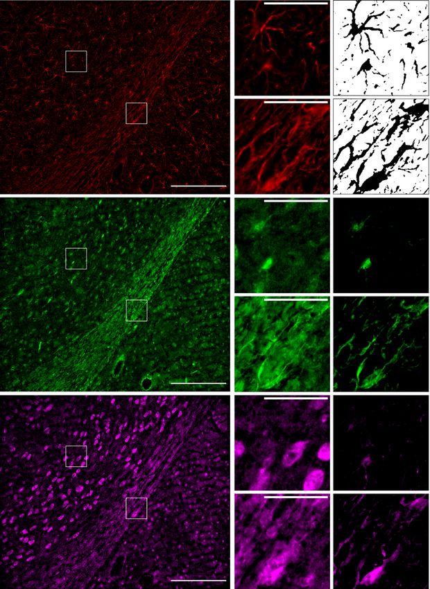
COMMUNICATIONS BIOLOGY | https://doi.org/10.1038/s42003-021-02392-8                                                                  ARTICLE

regeneration and pathfinding55. In addition, we have recently                   GFAP, Ezrin, and Robo2 (Fig. 2; Supplementary Data 1). We
developed the first tissue-engineered rostral migratory stream                  captured images of each individual label in the RMS and sur-
(TE-RMS), which is an implantable scaffold designed to replicate               rounding tissue via epifluorescence microscopy. Standardized
the endogenous RMS56–58. This engineered neuronal replacement                  ROIs containing glial tube astrocytes of the RMS and proto-
strategy replicates the only known mechanism for continual,                    plasmic astrocytes from the surrounding area were used for
long-distance neuroblast redirection that occurs intrinsically                 pairwise comparisons of labeling intensities. Since these proteins
within the adult brain via the RMS. By recapitulating the struc-               can be expressed by other cell types, the GFAP channel was used
ture and function of the glial tube at the core of the RMS, the TE-            to spatially isolate astrocytic signals from each channel for
RMS is designed to promote the sustained delivery of neuroblasts               quantification. Mean intensities were calculated for each ROI to
to neuron-deficient regions following injury or neurodegenerative               remove the influence of differences in the astrocytic area. Com-
disease. We anticipate that stable, long-term neuroblast redirec-              paring mean astrocytic intensities via two-tailed, paired Student’s
tion via this engineered living scaffold will set this technology              t-tests revealed that astrocytes of the RMS were significantly
apart from previous strategies that have typically induced tran-               enriched in GFAP (Fig. 2f; t = 3.770, df = 4, p = 0.02), Ezrin
sient neuronal redirection.                                                    (Fig. 2l; t = 3.642, df = 4, p = 0.02), and Robo2 (Fig. 2r; t = 3.890,
   The TE-RMS is fabricated within a small-diameter agarose                    df = 4, p = 0.02), compared with surrounding protoplasmic
microcolumn that promotes astrocyte bundling with extracellular                astrocytes. It should also be noted that the astrocytic intensity of
matrix and self-assembly into long, longitudinally aligned cables              GFAP, Ezrin, and Robo2 labeling in the endogenous rat RMS was
(Fig. 1d). Previous experiments to date have demonstrated that                 higher than that of the surrounding protoplasmic astrocytes in
the basic structure of the TE-RMS (tight astrocytic bundles with               every brain analyzed, though there was some variability in the size
bidirectional morphology) recapitulate the cell type and basic                 of those differences between brains.
morphology of the endogenous RMS56,57. We hypothesize that
the living astrocytes of the TE-RMS will also engage in dynamic,
                                                                               Astrocytes of the rat TE-RMS are enriched in Robo2 and Ezrin.
two-way communication with neuroblasts as they migrate
                                                                               We have previously reported on the development of fabrication
through the scaffold (Fig. 1e), made possible by emulating the
                                                                               techniques for the TE-RMS, in which optimal microcolumn
specific protein expression that facilitates neuroblast migration
                                                                               diameter, collagen concentration, media constituents, seeding
within the endogenous RMS. Here, the TE-RMS has the potential
                                                                               density, and other factors were optimized to facilitate astrocyte
to serve as an anatomically relevant testbed to study the inter-
                                                                               self-assembly into longitudinally aligned, tightly bundled cords
action between neuroblasts and the RMS in vitro. However, our
                                                                               over a period of just 8 h. We have also provided evidence for
ultimate goal for the TE-RMS is to enable the redirection of
                                                                               similarities in the morphology and structural arrangement of rat
endogenous neuroblasts from the SVZ/RMS to neuron-deficient
                                                                               TE-RMS astrocytes compared with astrocytes of the endogenous
brain regions in vivo. Following focal brain injury (Fig. 1c), the
                                                                               RMS56–58. In this study, we sought to test the hypothesis that TE-
TE-RMS could be implanted into the brain spanning from the
                                                                               RMS astrocytes also recapitulate the enhanced expression of
SVZ/RMS into the injured brain region (Fig. 1f). We hypothesize
                                                                               GFAP, Ezrin, and Robo2 observed in the endogenous RMS (as
that neuroblasts will divert from the SVZ/RMS and migrate in
                                                                               verified in the experiments of Fig. 2). We tested for the enrich-
chain formation through the TE-RMS and into the lesion
                                                                               ment of these proteins in the TE-RMS by comparing fluorescence
(Fig. 1g). Future studies will test whether gradual, sustained
                                                                               immunocytochemistry (ICC) labeling intensities in TE-RMS
introduction facilitates neuroblast survival and maturation fol-
                                                                               astrocytes with those of protoplasmic astrocytes from planar
lowing arrival at their new location (Fig. 1h), thereby efficiently
                                                                               sister cultures imaged via epifluorescence microscopy (Fig. 3;
repopulating injured regions.
                                                                               Supplementary Data 2). While seeding from the same sources
   In the current study, we compare protein expression in
                                                                               provided excellent control for any unforeseen culture variability
astrocytes of the TE-RMS to that of the glial tube in the endo-
                                                                               that could have interfered with our ability to test differences
genous RMS. In an exciting recent development, we also report
                                                                               between planar vs. TE-RMS groups, the fabrication and culturing
the ability to fabricate the TE-RMS from a readily available source
                                                                               process after seeding led us to consider each sample as inde-
of adult human gingiva mesenchymal stem cells (GMSCs) from
                                                                               pendent. Therefore, we could not take advantage of paired means
which we can derive astrocyte-like cells within one week using
                                                                               testing as in the brain sections of Fig. 2, and instead employed
non-genetic techniques without the need for dedifferentiation.
                                                                               two-tailed Student’s t-tests to compare labeling intensities in the
This enhances the translational potential of this technology by
                                                                               planar cultures (n = 6) and TE-RMSs (n = 9) seeded from pri-
introducing the possibility that, with further development, human
                                                                               mary cortical rat astrocyte cultures. Due to the close proximity of
autologous TE-RMS implants can be created. We also demon-
                                                                               cells in TE-RMSs as compared to planar sister cultures, Hoechst-
strate that the human TE-RMS facilitates immature neuronal
                                                                               stained nuclei were often in overlapping visual fields in TE-RMS
migration in vitro. Finally, we report that implantation of the
                                                                               images, making automated cell counting unreliable. Therefore, we
human TE-RMS into the athymic rat brain facilitates migration of
                                                                               measured the total nuclear area from the Hoechst channel for
endogenous neuroblasts out of the native RMS and throughout
                                                                               each image to allow for an unbiased, automated calculation of cell
the TE-RMS, providing surgical feasibility and proof-of-concept
                                                                               amount in each image for normalization. This revealed that
evidence for this nascent technology.
                                                                               Hoechst intensities of the planar and TE-RMS groups were not
                                                                               different (Fig. 3g). Also, as observed in the endogenous RMS,
                                                                               astrocytes of the TE-RMS were significantly enriched in Ezrin
Results
                                                                               (Fig. 3j; t = 3.845, df = 13, p = 0.002), GFAP (Fig. 3m; t = 3.086,
Astrocytes of the endogenous rat RMS are enriched in Robo2
                                                                               df = 13, p = 0.009), and Robo2 (Fig. 3p; t = 4.855, df = 13, p =
and Ezrin. Previous studies have characterized the enrichment of
                                                                               0.0003), as compared with the astrocytes in the planar sister
protein markers in the glial tube astrocytes of the RMS as com-
                                                                               cultures.
pared to surrounding protoplasmic astrocytes. As such, we
compared the expression and distribution of these enriched
proteins in astrocytes of the native RMS versus astrocytes of the              Astrocyte-like cells can be derived from adult human GMSCs
TE-RMS. We applied fluorescence immunohistochemistry (IHC)                      and used for TE-RMS fabrication. In pursuit of a clinically
in sagittally sectioned FFPE adult rat brains (n = 5 brains) to label          relevant starting biomass for TE-RMS fabrication, we evaluated

COMMUNICATIONS BIOLOGY | (2021)4:879 | https://doi.org/10.1038/s42003-021-02392-8 | www.nature.com/commsbio                                        3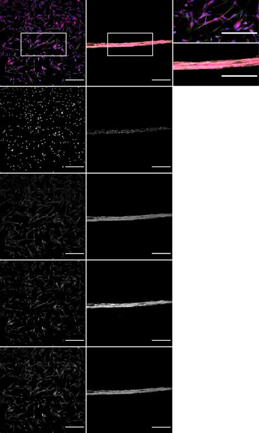
ARTICLE COMMUNICATIONS BIOLOGY | https://doi.org/10.1038/s42003-021-02392-8
a GFAP b d f GFAP
35
30
b
% Proto Intensity
25
Proto 20 *
c 15
c e
10
RMS
5
0
Proto RMS
g Ezrin h j l Astrocytic Ezrin
35
30
h
% Proto Intensity
25
*
20
i 15
i k
10
5
0
Proto RMS
m Robo2 n p r Astrocytic Robo2
35
30 *
n
% Proto Intensity
25
20
o 15
o q 10
5
0
Proto RMS
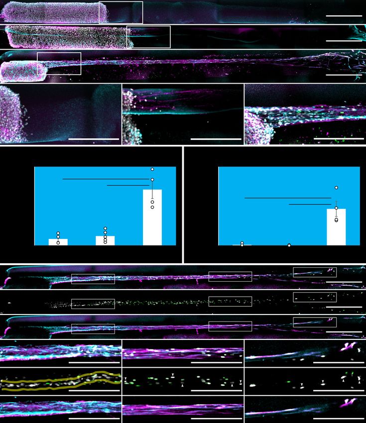
Fig. 2 Characteristic protein enrichment in RMS astrocytes relative to surrounding protoplasmic astrocytes in the rat brain. Formalin-fixed, paraffin-
embedded (FFPE) rat brains were sagittally sectioned and immunostained for GFAP (a–f), Ezrin (g–l), and Robo2 (m–r). GFAP, Ezrin, and Robo2 channels
from a representative image are displayed in a wide view containing a portion of RMS and surrounding brain (a, g, and m), with standardized regions of
interest (ROIs) annotated within for both RMS and protoplasmic (Proto) astrocytes. Enlarged ROIs are provided for each channel (b, c, h, i, n, o).
Automated binary masks were generated from GFAP ROIs (d, e) to allow for the isolation of astrocytic signals from the ROIs of each channel. Images
resulting from the application of these astrocytic GFAP masks to ROIs are provided for the Ezrin (j, k) and Robo2 (p, q) channels. Mean intensities were
quantified from ROIs after astrocytic signal isolation, and intensity values for the Proto/RMS ROI pairs from each image were compared by paired Student’s
t-test for GFAP (f), Ezrin (l), and Robo2 (r). Intensity values normalized to the Proto measurements for each pair are displayed for all five animals. *p <
0.05. Scale bars: 200 microns (a, g, m), 50 microns (b, c, h, i, n, o).
the efficacy of a novel differentiation protocol to derive astrocytes starting biomass for TE-RMS fabrication, they rapidly self-
from GMSCs. Of note, this protocol was adapted from a pre- assembled into cables of longitudinally aligned, bidirectional
viously published method for astrocyte derivation from oral astrocyte-like cells within the same 8 h timeframe observed when
mucosal stem cells59. Here, we successfully applied this non- fabricating with primary astrocytes from the rat cortex. This rapid
genetic derivation protocol to GMSCs from three deidentified remodeling/bundling appears to be unique to astrocytes, as when
adult human patients obtained via minimally invasive punch we apply the same fabrication methods using Schwann cells,
biopsy. After the derivation process—which takes less than a bundling and alignment take several days60. These human TE-
week—the cultured cells from each subject expressed astrocytic RMSs stained positive for Ezrin and Robo2, which can be seen
proteins glutamine synthetase (GS), glutamate aspartate trans- localized to the plasma membrane in the single high magnifica-
porter (GLAST), GFAP, and S100-β and were negative for the tion z plane confocal images of Fig. 4l–n.
endothelial marker CD31 (Fig. 4a–j). Western blot analyses
confirmed that GMSCs from three de-identified donors did not
express GFAP or GS prior to derivation, but the astrocyte-like Astrocyte-like cells of the human TE-RMS are enriched in
cells derived from GMSCs did express GFAP and GS (Fig. 4k; Robo2 and Ezrin. We used fluorescence ICC with laser confocal
Supplementary Data 3). The morphology of these cells was also microscopy to confirm that TE-RMSs fabricated from human
consistent with astrocytes in planar culture, and they thrived GMSC-derived astrocytes express GFAP, Ezrin, and Robo2
under astrocytic culture conditions. These cells were also com- (Fig. 4l–n), the combination of which is characteristic of glial tube
patible with passaging techniques for astrocyte culture purifica- astrocytes of the RMS. Then, to test whether the human GMSC-
tion, including vigorous mechanical perturbation prior to derived TE-RMS is enriched in GFAP, Ezrin, and Robo2 as
trypsinization that is commonly applied to detach non-astrocytic observed in the endogenous rat RMS and the cortical rat astrocyte
cells from culture flasks for removal prior to passaging. Fur- TE-RMS, we employed the same experimental techniques and
thermore, when we used the human GMSC-derived astrocytes as analyses used to investigate the rat TE-RMS (see Fig. 3). Hoechst
4 COMMUNICATIONS BIOLOGY | (2021)4:879 | https://doi.org/10.1038/s42003-021-02392-8 | www.nature.com/commsbio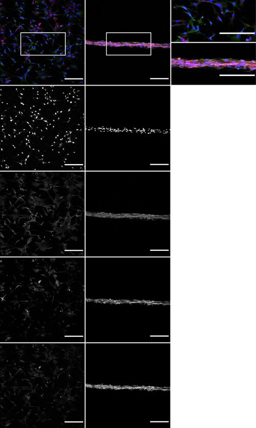
COMMUNICATIONS BIOLOGY | https://doi.org/10.1038/s42003-021-02392-8 ARTICLE
a Hoechst b c
Ezrin
GFAP
Robo2
c d
d
e Hoechst f g 12000
Hoechst Intensity / Cell
10000
8000
6000
4000
2000
0
Planar TE-RMS
h Ezrin i j 12000 **
Ezrin Intensity / Cell
10000
8000
6000
4000
2000
0
Planar TE-RMS
k GFAP l m 12000 **
GFAP Intensity / Cell
10000
8000
6000
4000
2000
0
Planar TE-RMS
n Robo2 o p 12000
***
Robo2 Intensity / Cell
10000
8000
6000
4000
2000
0
Planar TE-RMS
Fig. 3 TE-RMSs fabricated from rat astrocytes are enriched in GFAP, Ezrin, and Robo2 relative to planar sister cultures. Primary rat astrocytes were
passaged, split, and either plated in a planar collagen matrix or used for TE-RMS fabrication. Cell nuclei were labeled with Hoechst stain, and cells were
immunostained for GFAP, Ezrin, and Robo2. Representative wide views of merged fluorescent channels are provided for planar sister cultures (a) and TE-
RMS (b), with call-out boxes providing magnified views of planar (c) and TE-RMS (d) organization, morphology, and relative protein. Maximum contrast
white-on-black single-channel images along with quantification of normalized intensities are provided for Hoechst (e–g), Ezrin (h–j), GFAP (k–m), and
Robo2 (n–p). Values are displayed as mean ± SEM. Means were compared by Student’s t-test. **p < 0.01, ***p < 0.005. Scale bars: 250 microns.
COMMUNICATIONS BIOLOGY | (2021)4:879 | https://doi.org/10.1038/s42003-021-02392-8 | www.nature.com/commsbio 5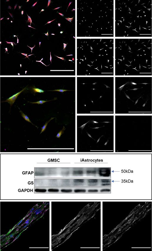
ARTICLE                                                                  COMMUNICATIONS BIOLOGY | https://doi.org/10.1038/s42003-021-02392-8

                            a Hoechst                                         b Hoechst                c GS
                                GS
                                GLAST
                                GFAP

                                                                              d GLAST                  e GFAP

                            f   Hoechst                                       g Hoechst                h S100-B
                                S100-B
                                GFAP
                                CD31

                                                                              i   GFAP                 j   CD31

                            k
                                          Donor D1         D2       D3      D1      D2       D3

                            l   Single z plane                m Ezrin                          n Robo2
                                Hoechst
                                Ezrin
                                Robo2

Fig. 4 Astrocyte-like cells can be derived from adult human gingiva mesenchymal stem cells (GMSC) and used for TE-RMS fabrication. A
representative image of human GMSC-derived astrocyte-like cells in planar culture is provided with merged fluorescent channels (a), and maximum
contrast white-on-black single-channel images are provided for Hoechst staining of nuclei (b) and immunostaining that demonstrates expression of
astrocytic proteins glutamine synthetase (GS), glutamate/aspartate transporter (GLAST) (d), and GFAP (e). A merged fluorescent image is also provided
at higher magnification with alternative staining targets (f), with maximum contrast white-on-black single-channel images for Hoechst staining of nuclei
(g) and immunostaining that demonstrates expression of astrocytic proteins S100B (h) and GFAP (i), but not endothelial marker CD31 (j). Western blot
analysis from three donors before and after astrocyte induction, demonstrating increased expression of astrocytic proteins GFAP and GS, with GAPDH
loading control (k). A representative TE-RMS fabricated using the human GMSC-derived astrocytes as starting biomass was labeled with Hoechst nuclear
stain, immunostained for Ezrin and Robo2, and imaged via laser confocal microscopy (l–n). Single z plane overlay illustrating the bidirectional morphology
and longitudinal alignment of astrocytes comprising the human TE-RMS. Maximum contrast white-on-black single z plane images of individual channels at
high magnification demonstrate the presence and plasma membrane localization of Ezrin (m) and Robo2 (n) proteins known to be enriched in glial tube
astrocytes. Scale bars: 200 microns (a–j), 50 microns (l–n).

6                           COMMUNICATIONS BIOLOGY | (2021)4:879 | https://doi.org/10.1038/s42003-021-02392-8 | www.nature.com/commsbio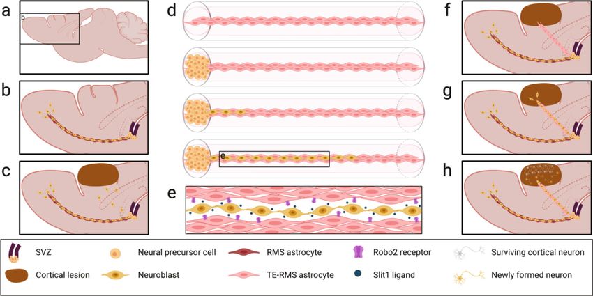
COMMUNICATIONS BIOLOGY | https://doi.org/10.1038/s42003-021-02392-8                                                                                              ARTICLE

                         a   Hoechst                            b                                      c
                             Ezrin
                             GFAP
                             Robo2

                                  c                                      d
                                                                                                       d

                         e   Hoechst                            f                                      g                           5000

                                                                                                        Hoechst Intensity / Cell
                                                                                                                                   4000

                                                                                                                                   3000

                                                                                                                                   2000

                                                                                                                                   1000

                                                                                                                                      0

                                                                                                                                          Planar        TE-RMS

                         h   Ezrin                              i                                       j                          5000
                                                                                                                                                   **

                                                                                                        Ezrin Intensity / Cell
                                                                                                                                   4000

                                                                                                                                   3000

                                                                                                                                   2000

                                                                                                                                   1000

                                                                                                                                     0

                                                                                                                                          Planar        TE-RMS

                         k   GFAP                               l                                      m                           5000            **
                                                                                                        GFAP Intensity / Cell

                                                                                                                                   4000

                                                                                                                                   3000

                                                                                                                                   2000

                                                                                                                                   1000

                                                                                                                                      0

                                                                                                                                          Planar        TE-RMS

                         n   Robo2                              o                                      p                           5000
                                                                                                        Robo2 Intensity / Cell

                                                                                                                                   4000            *

                                                                                                                                   3000

                                                                                                                                   2000

                                                                                                                                   1000

                                                                                                                                      0

                                                                                                                                          Planar        TE-RMS

Fig. 5 TE-RMSs fabricated from adult human gingiva-derived astrocytes are enriched in GFAP, Ezrin, and Robo2 relative to planar sister cultures.
Astrocytes derived from adult human gingiva stem cells were passaged, split, and either plated in a planar collagen matrix or used for TE-RMS fabrication. Cell nuclei
were labeled with Hoechst stain, and cells were immunostained for GFAP, Ezrin, and Robo2. Representative wide views of merged fluorescent channels are provided
for planar sister cultures (a) and TE-RMS (b), with call-out boxes providing magnified views of planar (c) and TE-RMS (d) organization, morphology, and relative
protein. Maximum contrast white-on-black single-channel images along with quantification of normalized intensities are provided for Hoechst (e–g), Ezrin (h–j), GFAP
(k–m), and Robo2 (n–p). Values are displayed as mean ± SEM. Means were compared by Student’s t-test. *p < 0.05, **p < 0.01. Scale bars: 250 microns.

COMMUNICATIONS BIOLOGY | (2021)4:879 | https://doi.org/10.1038/s42003-021-02392-8 | www.nature.com/commsbio                                                            7
ARTICLE COMMUNICATIONS BIOLOGY | https://doi.org/10.1038/s42003-021-02392-8
intensities of the planar (n = 6) and TE-RMS (n = 5) groups were processes ran parallel to—but did not overlap—the GFAP-positive
not different (Fig. 5g). As observed in the endogenous RMS and processes of the human TE-RMS (Fig. 6h′, h†, and h‡).
rat TE-RMS, human GMSC-derived TE-RMSs were significantly
enriched in Ezrin (Fig. 5j; t = 4.720, df = 9, p = 0.001), GFAP
The human TE-RMS redirects migration of neuroblasts from
(Fig. 5m; t = 4.350, df = 9, p = 0.002), and Robo2 (Fig. 5p; t =
the rat RMS in vivo. Finally, we performed in vivo experiments
2.639, df = 9, p = 0.027), compared with the astrocytes of their
with stereotactic implantation of human TE-RMSs into the brains
planar sister cultures (Supplementary Data 4).
of athymic rats to test surgical feasibility and proof-of-principal
for redirecting neuroblast migration away from the endogenous
RMS (Fig. 7). In athymic rats (n = 6), we bilaterally implanted
The human TE-RMS facilitates directed migration of imma-
pairs of 4 mm TE-RMSs and acellular collagen control micro-
ture rat neurons in vitro. Looking beyond replicating the mor-
columns to span RMS to the motor cortex. Animals were
phology, arrangement, and protein expression of the endogenous
euthanized 6 days later, and their FFPE brains were sagittally
RMS, the human TE-RMS must ultimately replicate the function of
sectioned for IHC analyses. By injecting implants 2.5 mm rostral
the RMS to be used for clinical application or as an in vitro testbed.
from bregma and 1 mm from the midline in either direction at a
We adapted techniques from our previously reported migration assay
depth of 5 mm, we were able to reproducibly contact the endo-
in the rat TE-RMS58, and assessed migration of immature rat cortical
genous RMS as verified by gross pathology and epifluorescence
neurons through the human TE-RMS as compared to acellular
microscopy (Fig. 7d, e). During the 6 days following implantation,
collagen-coated or collagen + laminin-coated control columns (Fig. 6;
we did not observe alterations in behavior and there was minimal
Supplementary Data 5). Immature cortical neuronal aggregates were
disruption of surrounding areas by gross pathology. Doublecortin
placed at one end of a microcolumn containing a fully formed human
(DCX) positive cells were observed near the ends of the con-
TE-RMS, an acellular collagen control, or an acellular collagen +
tralateral acellular collagen control implants but were absent from
laminin control, and migration from the aggregate to the opposite
central regions (Fig. 7f, g). However, we observed doublecortin-
side of the microcolumn was assessed at 72 h. For ICC analyses, we
positive, human-negative cells—indicative of migrating endo-
applied Hoechst nuclear stain and immunostained for Tuj1 (Beta-III-
genous rat neuroblasts—throughout the human TE-RMS
tubulin) to label neuronal processes. We also co-stained for Human
implants (Fig. 7h, i), suggesting that host cells were migrating
Nuclei and GFAP in the TE-RMSs, collagen in the collagen controls,
through the TE-RMS while only incidental infiltration of host
and laminin in the collagen + laminin controls. Under these condi-
cells was taking place near the ends of the acellular control col-
tions, we observed little if any migration into the acellular collagen
umns. However, given the techniques employed for these feasi-
control columns (n = 4) or into the acellular collagen + laminin
bility experiments we were unable to distinguish between DCX+
columns (n = 5), while immature rat neurons (Hoechst-positive/
cells from the RMS and those that may have somehow entered the
Human-negative nuclei with Tuj1-positive processes) migrated
TE-RMS from the cortex, so future studies will be needed to
through the entire 4 mm length of human TE-RMS within 72 h (n =
provide greater specificity. Combined with the in vitro experi-
4). Neuronal aggregates exhibited notably different behavior when
ments of Fig. 6, this implantation study provides proof-of-
seeded into the collagen + laminin control columns, in which they
principal evidence for redirection of neuroblast migration via the
exhibited little migration out of the aggregate but instead extended
human GMSC-derived TE-RMS and surgical feasibility for its
neurites into the ECM to an average length of 353.6 µm (SEM = 71.0)
implantation into the brain to span the endogenous RMS and
at 72 h (Fig. 6b′). Neuronal aggregates did not exhibit any measurable
cortex.
neurite extension in the collagen-only control columns. To compare
migration quantitatively, we measured the area of Hoechst-positive,
Human-negative nuclei (nuclei from the immature neuronal aggre- Discussion
gate) in a zone proximal to the aggregate (within 1 mm) and a zone The TE-RMS is the first biomimetic implantable microtissue
more distal (1–3.5 mm from aggregate). The amount of migrating designed to redirect the migration of endogenous neuroblasts out
neurons within 1 mm of the aggregate (Fig. 6d) in the TE-RMS was of the RMS and into distal lesions, intended to provide sustained
significantly greater than in the acellular collagen (t = 5.223, df = 10, delivery to replace lost neurons and improve functional recovery.
p = 0.001) or collagen + laminin controls (t = 4.460, df = 10, p = In pursuit of this goal, we have designed the TE-RMS to emulate
0.004). Beyond 1 mm there were essentially no migrating neurons in the brain’s only existing method for transporting neuroblasts to a
the controls whereas migrating neurons were found throughout the distal area for neuronal replacement. Whereas prior studies have
entire length of the TE-RMSs, so unsurprisingly the amount of transplanted exogenous fetal grafts, single-cell suspensions, or
migrating neurons between 1 and 3.5 mm of the aggregate (Fig. 6e) in cells in 3-D matrices, our method is considerably different in that
the TE-RMS was significantly greater than in the acellular collagen the living cytoarchitecture of the TE-RMS—mimicking the
(t = 5.626, df = 10, p = 0.0007) or collagen + laminin controls (t = architecture of the RMS—is fully fabricated in vitro and then
6.375, df = 10, p = 0.0002). A single z plane view of neuronal precisely delivered to unlock the regenerative potential of the
migration through the TE-RMS (Fig. 6f–h‡) allows for the visuali- brain’s own endogenous neuroblasts. In the current study, we
zation of cell-to-cell interactions in greater detail, though in some confirmed that the TE-RMS is enriched in Ezrin and Robo2, both
cases components may be out of a plane (e.g. nuclear signal with of which are similarly enriched in the endogenous RMS and
processes out of the visible z plane). Completing a 4 mm journey important for facilitating neuroblast migration. We also report a
through the TE-RMS construct within 72 h indicates an average new method for deriving astrocyte-like cells from adult human
migration rate of at least 56 µm/h, placing them within the reported GMSCs adapted from a previous protocol applied to the oral
range of 30–70 µm/h for neuroblast migration in the endogenous mucosa, potentially providing a minimally invasive autologous
RMS7,10,61. Hoechst-positive/Human-negative nuclei from the rat starting biomass for patients. Indeed, utilizing these cells for
cortical aggregate were densest near the aggregate, where a narrow fabrication produces a human TE-RMS consisting of tightly
“follow-the-leader” path can be most easily visualized (yellow lines, bundled, bidirectional, longitudinally aligned, astrocyte-like cells
Fig. 6g′). Hoechst-positive/Human-negative nuclei were also observed enriched in GFAP, Ezrin, and Robo2. Furthermore, the human
throughout the length of the human TE-RMS (white arrows, Fig. 6g† TE-RMS facilitates in vitro migration of immature neurons at
and g‡). These migrating cells were Tuj1-positive, consistent with an rates within the range observed for migration of neuroblasts in
immature neuronal phenotype (Fig. 6h), and their Tuj1-positive the endogenous RMS. Finally, we provide in vivo evidence of
8 COMMUNICATIONS BIOLOGY | (2021)4:879 | https://doi.org/10.1038/s42003-021-02392-8 | www.nature.com/commsbioCOMMUNICATIONS BIOLOGY | https://doi.org/10.1038/s42003-021-02392-8 ARTICLE
a a′
Neuronal Aggregate
Hoechst Tuj1 Collagen
b b′
Hoechst Tuj1 Laminin
c c′
Human Hoechst Tuj1 GFAP
a′ b′ c′
Hoechst Tuj1 Collagen Hoechst Tuj1 Laminin Human Hoechst Tuj1 GFAP
d e
Migraon Density 0 - 1 mm from Aggregate Migraon Density 1 - 3.5 mm from Aggregate
21000 21000
**
Hoechst Area (μm2)
Hoechst Area (μm2)
18000 18000
**
15000 15000
***
12000 12000 ***
9000 9000
6000 6000
3000 3000
0 0
Coll Lam+Coll TE-RMS Coll Lam+Coll TE-RMS
f Hoechst Tuj1 f†
f‡
Human GFAP f′
(Single Z Plane)
g Hoechst g†
g‡
Human g′
h Tuj1 h‡
h′ h†
GFAP
f′ f† f‡
g′ g† g‡
h′ h† h‡
Fig. 6 Migration of immature rat neurons is facilitated by human TE-RMSs in vitro. Immature neuronal aggregates prepared from the rat cortex were
inserted in one end of human GMSC-derived TE-RMSs and acellular collagen controls, and these assembled in vitro migration assays were then fixed 72 h
later for immunolabeling and analyses. Compressed z stacks of stitched confocal images are displayed in a wide view with all channels merged, consisting
of Hoechst (nuclei) and Tuj1 (neurites) channels, along with either Collagen in the representative acellular collagen control (a), Laminin in the
representative acellular collagen/laminin control (b), or Human nuclei and GFAP in the representative human TE-RMS (c). Call-out boxes provide
magnified views proximal to the aggregate in each column (a′–c′). Quantification of the area of Hoechst-positive, Human-negative nuclei (nuclei from the
immature neuronal aggregate) is provided for all groups for 0–1 mm from the aggregate (d) and 1–3.5 mm from the aggregate (e). Data are displayed as
mean ± SEM with points to indicate individual sample values; n = 4, 5, and 4 for Coll, Lam + Coll, and TE-RMS, respectively (**p < 0.005, ***p < 0.001 with
Bonferroni correction for multiple comparisons). A single z plane of a stitched confocal image from a representative human TE-RMS containing Hoechst,
Human Nuclei, Tuj1, and GFAP channels is displayed in a wide view with all channels merged (f), with just the nuclear labels (g), and with just the astrocyte
and neuron-specific cytoskeleton labels (h). Call-out boxes provide magnified views along the TE-RMS proximal to the aggregate (f′–h′), ~2.5 mm from the
aggregate (f†–h†), and ~3.5 mm from the aggregate (f‡–h‡). Opaque yellow outlines in g′ highlight the narrow path for chain migration forged by immature
neurons through the TE-RMS. White arrows in g† and g‡ indicate the Hoechst+/Human- nuclei of immature neurons migrating the length of the TE-RMS.
Scale bars: 500 microns (a–c; f–h), 250 microns (a′–c′; f′–h‡).
COMMUNICATIONS BIOLOGY | (2021)4:879 | https://doi.org/10.1038/s42003-021-02392-8 | www.nature.com/commsbio 9ARTICLE COMMUNICATIONS BIOLOGY | https://doi.org/10.1038/s42003-021-02392-8
a d h Human
DCX
b e Rat GFAP
lant
SVZ
Imp
c
RMS
f Collagen
DCX
i Human
DCX
g Collagen
DCX
Fig. 7 Implantation of human TE-RMSs in the brains of athymic rats demonstrates surgical feasibility and proof of principle for redirecting neuroblast
migration. Pairs of human GMSC-derived TE-RMSs and acellular collagen controls were bilaterally implanted into the brains of athymic rats using precise
stereotaxic coordinates to span RMS and cortex. Images captured during (a) and after (b) bi-lateral stereotactic implantation of TE-RMS. Gross pathology
of the formalin-fixed brain from the top (c) and side (d) view (note that d is blocked to show the implant trajectory). Immunolabelling rat GFAP
demonstrating accurate placement of TE-RMS contacting the RMS (e). Immunolabelling showing labelling of collagen within the acellular control implant
and DCX positive host cells present in the surrounding tissue but absent from the collagen implant midway through the column (f; ~3 mm from RMS), and
present in the collagen at the interface with the endogenous RMS (g). Immunolabelling showing non-overlapping colabeling of human nuclei of the TE-RMS
astrocytes and DCX positive (Human negative) host neuroblasts migrating through the TE-RMS midway through the implant (h; ~3 mm from RMS; white
arrows indicate DCX+/Human− cells), and at the interface between TE-RMS and host RMS (i). Scale bars: 500 microns.
10 COMMUNICATIONS BIOLOGY | (2021)4:879 | https://doi.org/10.1038/s42003-021-02392-8 | www.nature.com/commsbioCOMMUNICATIONS BIOLOGY | https://doi.org/10.1038/s42003-021-02392-8 ARTICLE surgical feasibility for human TE-RMS implantation into athymic combinations of neurotrophic factors including epidermal growth rat brains spanning from the RMS to the cortex, and proof-of- factor, erythropoietin, fibroblast growth factor, vascular endo- principle evidence that the human TE-RMS can redirect migra- thelial growth factor, and others has been shown to improve tion of endogenous rat neuroblasts out of the RMS and into the short-term functional recovery by enhancing proliferation in the cortex with no overt negative histological or behavioral effects on SVZ after injury, in turn increasing the number of neuroblasts test subjects. migrating into lesions by virtue of increasing the overall number While the direct implantation of neural stem cells into injury of neuroblasts39. There is extensive evidence in humans and sites has been shown to improve outcomes by providing neuro- animal models for exercise-induced improvements in functional trophic factors, and recent work has demonstrated that integra- recovery after brain injury, and the effects are largely attributed to tion of human-induced pluripotent stem cell (iPSC)-derived increased neuronal plasticity and proliferation of endogenous neurons in rat cortex is possible62, appropriate maturation and NPCs in response to exercise-induced increases in brain-derived integration of these exogenous cells to functionally replace lost neurotrophic factor68,69. While neurotrophic factors appear to neurons remains a significant challenge63–65. Furthermore, each exert their effects on neuroblasts primarily through increasing delivery of exogenous neural stem cells requires invasive surgery, proliferation, implantation of acellular permissive substrates has therefore these proposed treatment strategies typically involve been employed to directly facilitate the migration of neuroblasts only a single bolus delivery of exogenous stem cells. In contrast, a into lesions. Several acellular scaffolds consisting of extracellular single surgical procedure to implant the TE-RMS could, in theory, matrix proteins often infused with neurotrophic factors have provide sustained delivery of endogenous NPCs by emulating the demonstrated feasibility for redirecting migration of endogenous brain’s own strategy for relocating and integrating new neurons. neuroblasts away from the SVZ/RMS46,70,71. A series of studies In addition, strategies for direct implantation of exogenous stem utilizing cryogenic cortical injury in mice has shown improved cells often rely on iPSCs that must be dedifferentiated from a neurological recovery with a laminin-based scaffold, with more mature cell source and therefore carry risks related to the refinements achieved through incorporating additional features retention of epigenetic memory from the original cell source (i.e. like N-cadherin that have been shown to play a role in neuroblast leading to de-differentiation)66. In contrast, the TE-RMS provides migration in vivo48,72,73. In the current study, we observed the brain’s own endogenous NPCs that do not require any prior in vitro chain migration of immature cortical neurons through de-differentiation, and therefore do not suffer from the pheno- the TE-RMS, whereas columns loaded with ECM only (1 mg/ml typic abnormalities sometimes associated with epigenetic memory collagen + 1 mg/ml laminin) promoted neurite outgrowth of in iPSCs. Endogenous neuroblasts from the SVZ have been these immature cells and did not promote migratory behavior. observed migrating into injured striatum and forming mature, This is distinct from previous research demonstrating neuroblast synaptically integrated neurons, and when this injury response migration along with planar laminin and collagen in 2D culture was experimentally enhanced by overexpressing Slit1 in neuro- in vitro73. This difference in cell behavior is likely due to differ- blasts it significantly improved functional recovery in a rodent ences in immature neuronal phenotype (SVZ-derived versus model of stroke14,24. Genetic modification of neuroblasts may not cortical) as well as ECM preparation across in vitro studies represent a translational therapeutic strategy, but it does provide leading to differential binding of immature neurons to laminin. compelling evidence for the feasibility of enhancing neuroblast Indeed, migration and maturation of neuroblasts along the RMS migration into lesions to facilitate neuroregeneration. The TE- relies on complex, dynamic signaling between astrocytes and RMS can be precisely implanted to span SVZ to the lesion, neuroblasts74–76. Unlike extracellular-matrix-based constructs providing a migratory pathway to augment and amplify this that offer a permissive, acellular substrate70,72,73, the TE-RMS natural regenerative response of endogenous SVZ neuroblasts possesses a unique, living astrocytic microtissue makeup that can after injury without the need for genetic manipulation. provide directional, structural, and neurotrophic support, making Vasculature plays an important role in neuroblast migration it capable of sending as well as responding to complex signals along the endogenous RMS. Blood vessels surround and support with migrating neuroblasts and the local micro-environment. the structure of the RMS, and neuroblasts occasionally migrate Relying on an acellular substrate infused with a handful of sig- along astrocytic processes enwrapping these blood vessels that naling molecules is akin to trying to coordinate a complex project run parallel to the RMS11. Vascular remodeling occurs following in a foreign language of which you speak only a few words and neuronal injury11, and we expect significant vascular remodeling cannot hear or respond to anything anyone else is saying, versus following TE-RMS transplantation such that blood vessels will the living microtissue TE-RMS that provides total fluency in the grow to surround and support the transplanted TE-RMS. Of note, language and engagement in dynamic, collaborative conversa- while the TE-RMS may be fabricated to be centimeters in length, tions. To test this hypothesis, future studies testing the efficacy of the relatively narrow diameter of the resulting microtissue (e.g., the TE-RMS for facilitating regenerative rehabilitation after focal typically < 200 µm) should allow adequate diffusion-based mass brain injury will include direct comparisons to the most pro- transport of oxygen and other nutrients to support implant sur- mising acellular biomaterial approaches. vival while vascular remodeling ensues. Future studies will test the Glial tube astrocytes possess several features that make them hypothesis that angiogenesis will occur surrounding the TE-RMS unique among the widely heterogeneous astrocyte milieu. They implant tract, aiding in the recruitment of new blood vessels to possess a bidirectional morphology, extending processes in form this vascular scaffolding surrounding the TE-RMS. Such opposite directions along the glial tube in parallel with each other vascular remodeling could support the long-term stability of the to form a cord-like bundle. We have previously established that transplanted TE-RMS, and trophic factors secreted by these these structural features are recapitulated in the TE-RMS56–58. recruited vascular endothelial cells could serve as chemoattractant Glial tube astrocytes are also enriched in several proteins factors for neuroblasts much as they do in the endogenous important for facilitating neuroblast migration. Ezrin—a member RMS11. of the cytoskeleton-membrane linking ERM (Ezrin, radixin, Increasing delivery of neuroblasts into lesions after an injury moesin) protein family—is enriched in the astrocytes of the glial has also been approached through pharmacological strategies and tubes, while its cousin radixin is enriched in migrating implantation of acellular permissive substrates49,67. Pharmaco- neuroblasts15,77. In the endogenous RMS, astrocytic Robo2 logical approaches have focused mainly on the administration of receptors detect Slit1 protein released by neuroblasts, and this neurotrophic factors. Intraventricular infusion of various signaling results in tunnel formation in the astrocytic meshwork COMMUNICATIONS BIOLOGY | (2021)4:879 | https://doi.org/10.1038/s42003-021-02392-8 | www.nature.com/commsbio 11
ARTICLE                                                               COMMUNICATIONS BIOLOGY | https://doi.org/10.1038/s42003-021-02392-8

of the glial tubes to facilitate neuroblast migration78. In this study,      redirecting neuroblasts to various discrete brain areas, and we will
we verified previous reports of enrichment of GFAP, Ezrin, and                investigate both in vivo as well as in vitro by utilizing the TE-RMS
Robo2 in the glial tube astrocytes of the endogenous rat RMS.                as a biofidelic testbed for efficient and precise mechanistic studies.
Using the same methods, we also expanded on our previous                     As we consider the discovery and translational paths now ahead
morphological and structural analyses to report that the TE-RMS              of us, the human TE-RMS has opened new avenues for potential
—fabricated from either primary rat astrocytes or human GMSC-                study, and a promising novel approach to leveraging the endo-
derived astrocytes—is also enriched in GFAP, Ezrin, and Robo2.               genous regenerative potential of the brain.
   Building on this concept, mimicking the RMS may also facil-
itate the maturation of neuroblasts during migration along the
                                                                             Methods
TE-RMS, allowing the new neurons to functionally replace lost                Cell culture. All procedures adhered to the National Institutes of Health Guide for
neurons after degeneration has occurred instead of merely acting             the Care and Use of Laboratory Animals and were approved by the Institutional
as neurotrophic “factories” in the acute and sub-acute time per-             Animal Care and Use Committee at the University of Pennsylvania. For TE-RMS
iods. This strategy to enable the replacement of lost neurons well           fabrication, primary astrocytes were harvested from the cortices of postnatal day
                                                                             0–1 Sprague-Dawley rat pups (Charles River, Wilmington, MA). Cells were
after acquired brain injury or neurodegeneration is highly unique            dissociated57 and cultured in DMEM/F12 medium supplemented with 10% FBS
and presents an innovative approach to repairing currently                   and 1% Penicillin–Streptomycin in a 37 °C/5% CO2 cell culture incubator. Over
untreatable injuries affecting millions of patients. Moreover, their         several weeks, astrocyte cultures were maintained in tissue culture flasks and
miniature form factor allows for minimally invasive, stereotactic            passaged at 80% confluency to purify the astrocyte population. Astrocytes between
                                                                             passages 4–10 were utilized for all in vitro experiments. For in vitro migration of
delivery into the brain. In rat models, we have previously
                                                                             immature neurons through the TE-RMS, primary rat neurons were dissociated
demonstrated the ability to precisely microinject similar allo-              from cortices of embryonic day 18 (E18) rats57. Following tissue dissociation, with
geneic neuronal microtissue constructs, which maintain their pre-            trypsin–EDTA and DNAse I, a cell solution with a density of 1.0–2.0 × 10⁶ cells/ml
transplant architecture, survive, and integrate into the native              was prepared. 12 µl of this solution was transferred to each well in the pyramidal
nervous system52–54,79,80, and our TE-RMS implant experiments                micro-well array. The plate containing these micro-wells was centrifuged to pro-
                                                                             duce cell aggregates.
included in this study further demonstrate the surgical feasibility
of this strategy.
   In this study, we report the successful derivation of astrocyte-          Fabrication of hydrogel micro-columns. Hydrogel micro-columns composed of
like cells from adult human GMSCs, their suitability as starting             3% agarose were utilized to induce the alignment of astrocytes and create the TE-
                                                                             RMS57. Agarose was dissolved and heated in Dulbecco’s phosphate-buffered saline
biomass for TE-RMS fabrication, and proof-of-concept evidence                (DPBS). An acupuncture needle (diameter = 300 µm) was inserted into the bottom
that these human TE-RMSs facilitate neuroblast migration                     opening of a bulb dispenser. A glass capillary tube (inner diameter = 701 µm) was
in vitro and in vivo. Like iPSCs, the GMSCs offer minimally                  inserted over the needle external to the bulb and secured to the rubber section of
invasive access to a patient-specific autologous starting biomass.            the bulb dispenser. Warm agarose was drawn into a capillary tube with the needle
                                                                             in the center. After allowing the agarose to cool, the capillary tube was carefully
However, the one-week GMSC-to-astrocyte derivation process                   removed, and the micro-columns were gently pushed off the needle into DPBS and
takes only a fraction of the time of iPSC derivations, due in part           sterilized by UV light for 30 min. Micro-columns had an outer diameter of 701 µm
to the fact that dedifferentiation is unnecessary in the case of             and an inner diameter of 300 µm. Optimal micro-column dimensions for inducing
GMSCs. That lack of a dedifferentiation step also means that                 alignment and bundling of astrocytes were determined based on previous
                                                                             experiments56.
GMSCs do not carry risks associated with epigenetic memory66.
The appeal of a patient-specific starting biomass is primarily due
to avoiding the dangers of immune rejection. However, the                    Fabrication of tissue-engineered RMSs. Hydrogel micro-columns were cut with
clinical application of a product with a de-centralized biomass              angled forceps to a length of 4 mm. The inner lumen of the micro-columns was
                                                                             loaded with 1 mg/ml rat tail type 1 collagen diluted in a cell culture medium. The
source could pose challenges. Human embryonic stem cells from                collagen-loaded constructs were incubated for 3 h at 37 °C/5% CO2 to allow for
an established cell line could potentially offer a more streamlined          collagen polymerization and dehydration yielding in a hollow microcolumn with
and centralized quality control process by virtue of having a                the surface of the inner lumen coated in collagen. After the complete collagen
single source for the starting biomass. and they have also been              polymerization, the inner lumen of the micro-columns was seeded with astrocytes
                                                                             in serum-free co-culture media at a density of ~1 million cells/ml (optimal seeding
shown to elicit minimal immunoreactivity81. Though they would                density confirmed by Winter et al., 201656). Co-culture media consisting of neu-
likely require immunosuppression after transplantation, we will              robasal medium supplemented with 2% B-27, 1% G-5, 0.25% L-glutamine, and 1%
consider human embryonic stem cell lines along with the auto-                penicillin–streptomycin induced the astrocytes into a process-bearing phenotype.
logous GMSC source when evaluating potential translational                   Columns were seeded twice with astrocytes to ensure that the entire interior of each
                                                                             micro-column was filled with cells. Following astrocyte seeding, columns were
strategies.
                                                                             incubated at 37 °C/5% CO2 for one hour and subsequently reinforced with 1 mg/ml
   While this study provides feasibility and proof-of-concept                collagen. Collagen reinforcement provided more ECM to the astrocytes, helping to
evidence for the first human TE-RMS, future work will focus on                prevent collapse during astrocyte bundling. Following reinforcement, astrocyte-
directly testing the effects of TE-RMS implantation on regen-                loaded columns were incubated for another hour at 37 °C/5% CO2, flooded with
eration and functional recovery after brain injury. The TE-RMS               warm co-culture media, and returned to the cell culture incubator. Over a relatively
                                                                             short time period of ~8 h, the astrocytes extend processes to gather collagen and
strategy is not intended for neuroprotection, but rather neuronal            self-assemble into a bundled cord of longitudinally aligned astrocytes with bidir-
replacement and functional regeneration; therefore, we will                  ectional processes, effectively forming TE-RMSs. For experimental purposes, TE-
implant after the acute period to allow the so-called “hostile”              RMSs were utilized 24 h after astrocyte seeding. Acellular collagen control columns
environment—marked by dysregulated interstitial tissue, ongoing              for in vitro migration assays were prepared as above but with no addition of cells.
                                                                             For acellular collagen/laminin columns, a mixture of 1 mg/ml collagen and 1 mg/
cell death, and widespread inflammation—to dissipate. This                    ml mouse laminin diluted in cell culture medium was loaded into micro-columns.
strategy is also intended to serve as a means for gradual, sustained         Following extracellular matrix polymerization at 37 °C/5% CO2 (~3 h), columns
delivery of neuroblasts into injury sites, so long-term studies              were flooded with warm co-culture media and returned to the cell culture
assessing survival and maturity of redirected neuroblasts, mor-              incubator.
phological changes in transplanted TE-RMS astrocytes, vascular
remodeling, and functional recovery will be pursued, which will              TE-RMS extraction from microcolumns for ICC. Following overnight bundling of
have the added benefit of investigating the long-term con-                    astrocytes and formation of the TE-RMS, astrocytic bundles were extracted from
sequences of a microtissue implant in the brain. Iterations                  hydrogel micro-columns onto glass coverslips using surgical forceps and a ste-
                                                                             reoscope for visual guidance. TE-RMSs were slowly drawn out of the micro-
refining secondary encasement and even incorporating supple-                  columns into a bead of collagen diluted in culture media and left to dry for 15 min
mental neurotrophic factors may be pursued. We are also inter-               at 37 °C/5% CO2 to facilitate coverslip adhesion prior to fixation. Extraction was
ested in determining the potential spectrum of cell fates when               not performed with migration assay columns as it would disrupt the neuronal

12                         COMMUNICATIONS BIOLOGY | (2021)4:879 | https://doi.org/10.1038/s42003-021-02392-8 | www.nature.com/commsbio
You can also read