Parkin is an E3 ligase for the ubiquitin-like modifier FAT10, which inhibits Parkin activation and mitophagy
←
→
Page content transcription
If your browser does not render page correctly, please read the page content below
Article

Parkin is an E3 ligase for the ubiquitin-like modifier
FAT10, which inhibits Parkin activation and
mitophagy
Graphical Abstract                                                          Authors
                                                                            Nicola D. Roverato, Carolin Sailer,
                                                                            Nicola Catone, Annette Aichem,
                                                                            Florian Stengel, Marcus Groettrup

                                                                            Correspondence
                                                                            marcus.groettrup@uni-konstanz.de

                                                                            In brief
                                                                            Roverato et al. identify Parkin as an E3
                                                                            ligase for the ubiquitin-like modifier
                                                                            FAT10. FAT10ylation of Parkin leads to its
                                                                            proteasomal degradation, to its
                                                                            inhibition, and to impairment of its
                                                                            translocation to damaged mitochondria
                                                                            resulting in a delay of mitophagy. FAT10
                                                                            enhances the sensitivity of neuronal cells
                                                                            to mitochondrial damage.

Highlights
d   Parkin is an E3 ligase for the ubiquitin-like modifier FAT10

d   Parkin mediates FAT10ylation and proteasomal degradation
    of mitofusin2

d   FAT10 inhibits Parkin activation, translocation to damaged
    mitochondria, and mitophagy

d   Death induced by rotenone in dopaminergic neuronal cells is
    elevated by FAT10

           Roverato et al., 2021, Cell Reports 34, 108857
           March 16, 2021 ª 2021 The Author(s).
           https://doi.org/10.1016/j.celrep.2021.108857                                                     ll
                                       Konstanzer Online-Publikations-System (KOPS)
                               URL: http://nbn-resolving.de/urn:nbn:de:bsz:352-2-jpy86fkp1vqj3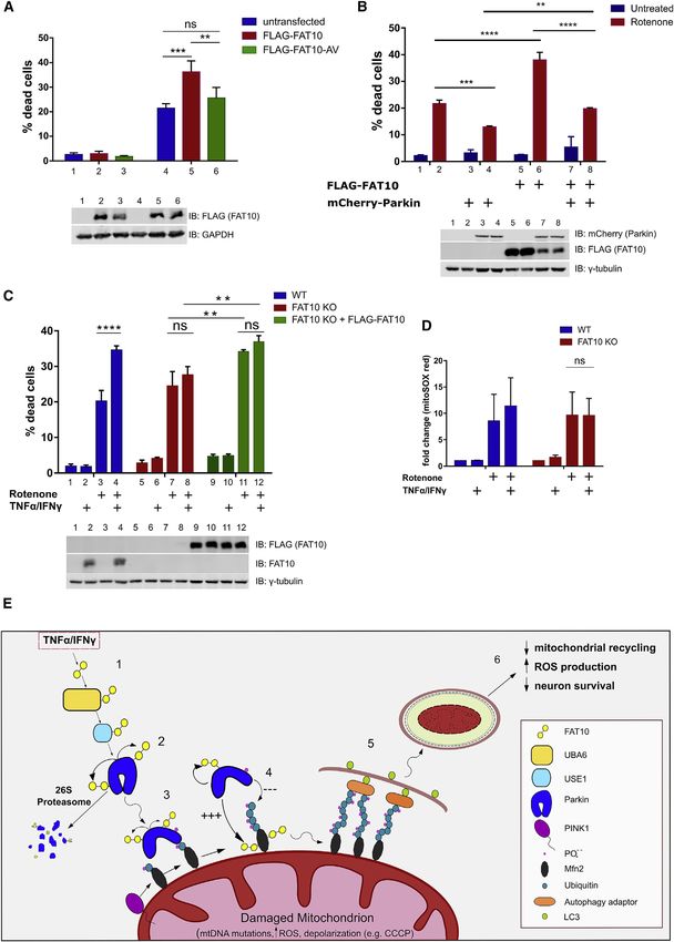
ll
                                                                                                                         OPEN ACCESS

Article
Parkin is an E3 ligase
for the ubiquitin-like modifier FAT10,
which inhibits Parkin activation and mitophagy
Nicola D. Roverato,1 Carolin Sailer,2 Nicola Catone,3 Annette Aichem,1,3 Florian Stengel,2 and Marcus Groettrup1,3,4,*
1Department    of Biology, Division of Immunology, University of Konstanz, 78457 Konstanz, Germany
2Department    of Biology, University of Konstanz, 78457 Konstanz, Germany
3Biotechnology Institute Thurgau at the University of Konstanz, 8280 Kreuzlingen, Switzerland
4Lead contact

*Correspondence: marcus.groettrup@uni-konstanz.de
 https://doi.org/10.1016/j.celrep.2021.108857

SUMMARY

Parkin is an E3 ubiquitin ligase belonging to the RING-between-RING family. Mutations in the Parkin-encod-
ing gene PARK2 are associated with familial Parkinson’s disease. Here, we investigate the interplay between
Parkin and the inflammatory cytokine-induced ubiquitin-like modifier FAT10. FAT10 targets hundreds of pro-
teins for degradation by the 26S proteasome. We show that FAT10 gets conjugated to Parkin and mediates its
degradation in a proteasome-dependent manner. Parkin binds to the E2 enzyme of FAT10 (USE1), auto-FA-
T10ylates itself, and facilitates FAT10ylation of the Parkin substrate Mitofusin2 in vitro and in cells, thus
identifying Parkin as a FAT10 E3 ligase. On mitochondrial depolarization, FAT10ylation of Parkin inhibits
its activation and ubiquitin-ligase activity causing impairment of mitophagy progression and aggravation
of rotenone-mediated death of dopaminergic neuronal cells. In conclusion, FAT10ylation inhibits Parkin
and mitophagy rendering FAT10 a likely inflammation-induced exacerbating factor and potential drug target
for Parkinson’s disease.

INTRODUCTION                                                           the damaged mitochondria (Dai et al., 2015; Wang et al., 2020).
                                                                       The efficient elimination of depolarized mitochondria that are a
Autosomal recessive Parkinson’s disease (PD) is characterized          prominent source of reactive oxygen species (ROS) protects
by a selective loss of dopaminergic neurons of the substantia ni-      neurons from severe ROS-mediated damage and consequent
gra pars compacta that leads to the early onset of parkinsonism.       apoptosis, thereby explaining the link between functional inacti-
Inherited familial PD is frequently caused by loss of function         vation of Parkin and early onset of PD (Gao et al., 2017). Given its
mutations in the PARK2 gene which encodes the ubiquitin E3             neuroprotective effect, efforts to elucidate the dysregulation of
ligase Parkin. Parkin is a protein that acts as a protective agent     Parkin’s enzymatic activity are crucial to understand the onset
in a broad variety of cellular stresses, and this pro-survival         and progression of Parkinsonism. The function of Parkin is
activity is generally linked to its function as an E3 ligase (Dawson   fine-tuned by post-translational modifications such as phos-
and Dawson, 2010). In fact, Parkin is mainly known as a key            phorylation, SUMOylation, ISGylation, and NEDDylation (Chak-
component of the PINK1/Parkin axis that, after mitochondrial de-       raborty et al., 2017). Nonetheless, the role of the inflammatory
polarization, triggers the selective degradation of damaged mito-      response in the striatum of PD patients in the regulation of Parkin
chondria by autophagy in a process known as ‘‘mitophagy.’’             function is poorly understood. Inflammation in the central ner-
Here, the loss of mitochondrial membrane potential leads to            vous system (CNS) during pathogenesis of PD is accompanied
the localization of the kinase PINK1 on the outer mitochondrial        by microglial activation and massive production of pro-inflam-
membrane, where it phosphorylates several targets including            matory cytokines (Mogi et al., 1994). In particular, high levels of
the ubiquitin-like (UBL) domain of Parkin (Jin et al., 2010; Konda-    tumor necrosis factor alpha (TNF-a) and interferon (IFN)g are ex-
palli et al., 2012; Shiba-Fukushima et al., 2012). This modifica-      pressed in the substantia nigra of PD patients, that stimulate
tion, in concert with the phosphorylation of ubiquitin, triggers       dopaminergic neurons (McGuire et al., 2001) and are known to
the conformational opening of Parkin and the exposure of the           strongly and synergistically induce the ubiquitin-like modifier
catalytic domain, resulting in Parkin activation and subsequent        HLA-F adjacent transcript 10 (FAT10) (Liu et al., 1999).
polyubiquitylation of several substrate proteins on the mitochon-         FAT10 (also known as ubiquitin D [UBD]) is an 18-kDa ubiqui-
drial outer membrane (Gladkova et al., 2018). These ubiquitin          tin-like modifier (UBL) that consists of two ubiquitin-like domains
conjugates enable recruitment of autophagy adaptors, driving           (UBDs) that are joined by a flexible linker (Aichem et al., 2018).
mitophagosome formation and lysosomal degradation of                   FAT10 is encoded in the major histocompatibility complex

                                                                       Cell Reports 34, 108857, March 16, 2021 ª 2021 The Author(s). 1
                                    This is an open access article under the CC BY license (http://creativecommons.org/licenses/by/4.0/).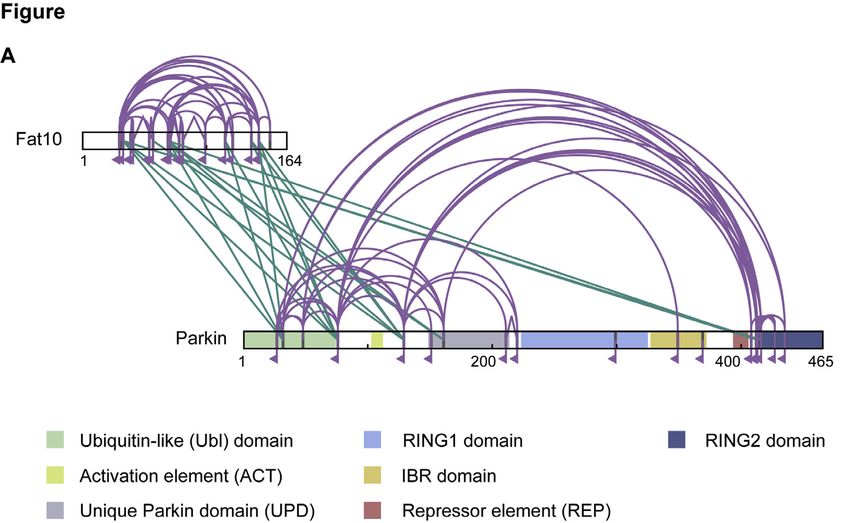
ll
OPEN ACCESS Article
A 1h CCCP
B
1h CCCP
Myc-Parkin + ++++ Myc-Parkin + ++++
FLAG-FAT10 + + + FLAG-FAT10 + + +
FLAG-FAT10-AV + + + FLAG-FAT10-AV + + +
1 2 3 4 5 6 7 8 1 2 3 4 5 6 7 8
KDa KDa
170 IP: FLAG 170
130
130
IP: Myc 100 Parkin-FAT10 IB: Myc 100 Parkin-FAT10
70
IB: FLAG 70 (high contrast)
55
Myc-Parkin
(low contrast) 55
IP: FLAG 25
IB: FLAG
25
FLAG-FAT10
IB: FLAG (FAT10)
IP: Myc
IB: Myc 55 Load IB: Myc (Parkin)
IB: FLAG (FAT10) IB: γ-Tubulin
Load IB: Myc (Parkin)
IB: γ-Tubulin
C IP: IP: D
IgG control Parkin KO KO
B A6 E1
2 2 U US
13 13 Myc-Parkin ++++
MG MG
TNFα/IFNγ + ++ + FLAG-FAT10 + +++
1 2 3 4 5 6
KDa 1 2 3 4 5 6
KDa IP: FLAG 170
130
170 IB: Myc 100 Parkin-FAT10
130 (high contrast)
Parkin-FAT10 70
100
Myc-Parkin
IB: FAT10 70
(low contrast) 55
Myc-Parkin
IP: FLAG FLAG-FAT10
25
IB: FLAG
FAT10
IB: FLAG (FAT10)
IB: Parkin IB: Myc (Parkin)
Load IB: USE1
IB: FAT10
IB: UBA6
Load IB: Parkin
IB: GAPDH
IB: GAPDH
E
(legend on next page)
2 Cell Reports 34, 108857, March 16, 2021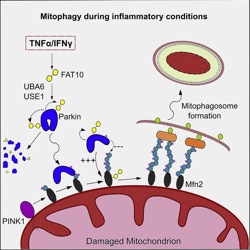
ll
Article                                                                                                                                          OPEN ACCESS

(MHC) class I locus (Fan et al., 1996) and is expressed mainly in                    myc-tagged Parkin with an inactive variant of FLAG-FAT10 in
organs and cells of the immune system (Lukasiak et al., 2008;                        which the C-terminal GG motif was replaced by AV (Figures 1A
Schregle et al., 2018). FAT10 gets isopeptide-linked via its C-ter-                  and 1B). Because FAT10-Parkin conjugates did not form with
minal diglycine motif to hundreds of substrate proteins that label                   FLAG-FAT10AV, and because FAT10 does not form chains,
them for rapid degradation by the 26S proteasome indepen-                            we conclude that Parkin is mono-FAT10ylated at two sites under
dently of ubiquitin attachment (Aichem et al., 2012; Hipp et al.,                    overexpression conditions. As a further confirmation, the
2005; Schmidtke et al., 2009). In contrast to ubiquitin, FAT10                       immunoprecipitation was performed in the other direction that
does not get cleaved from its substrates at the 26S proteasome                       validated our conclusion (Figures 1B and S1C). To further
but is most likely degraded along with its substrates thus ex-                       corroborate these results, we used SH-SY5Y cells to study the
plaining its short half-life of 1 h (Hipp et al., 2005). Similar to other            interaction between Parkin and FAT10 under fully endogenous
ubiquitin-like modifiers, FAT10 possesses its private activation                     conditions. SH-SY5Y is a dopaminergic neuroblastoma-derived
and conjugation cascade, which consists of the E1 activating                         cell line expressing endogenous Parkin and is commonly used to
enzyme UBA6 (Jin et al., 2007; Chiu et al., 2007; Pelzer et al.,                     study neuronal function (Xicoy et al., 2017). Because these cells
2007), the E2 conjugating enzyme USE1 (Aichem et al., 2010),                         also express high amounts of endogenous FAT10 on 24 h of
and putative, but so far unknown, E3 ligases. Here, we identify                      stimulation with TNF-a and IFNg, we performed a co-immuno-
Parkin as an E3 ligase for FAT10 and the mitochondrial fusion                        precipitation of FAT10 with endogenous Parkin using an anti-
protein mitofusin2 (Mfn2) as a Parkin-dependent FAT10ylation                         Parkin antibody, followed by FAT10 western blot. We confirmed
substrate in neuronal cells. Moreover, we show that FAT10 in-                        the formation of covalent FAT10-Parkin conjugates under
duction inhibits the ubiquitin-ligase activity of Parkin and the                     endogenous conditions (Figure 1C). Curiously, in presence of
localization of Parkin to depolarized mitochondria leading to an                     TNF-a/IFNg, we detected additional FAT10ylated forms of
impairment of mitophagy with likely adverse consequences for                         Parkin, suggesting that cytokine-induced factors might exist
the pathogenesis of PD.                                                              that enhance the formation of FAT10-Parkin conjugates. Such
                                                                                     a discrepancy between overexpression and endogenous condi-
RESULTS                                                                              tions was previously noticed also for the formation of FAT10-p62
                                                                                     conjugates (Aichem et al., 2012). Next, we asked whether the so
Parkin is a substrate for FAT10ylation                                               far known conjugation machinery for FAT10 is involved in FAT10-
A previous proteomic analysis has described endogenous                               Parkin conjugate formation (Chiu et al., 2007). To this aim, we
FAT10 to interact with 569 proteins and to covalently modify                         investigated Parkin FAT10ylation in Hek293-UBA6 KO cells (Ai-
176 of them. Unexpectedly, among these FAT10 binding part-                           chem et al., 2019b) or in Hek293-Use1 KO cells (Aichem et al.,
ners there were various mitochondrial proteins (Aichem et al.,                       2018) and confirmed that UBA6 and USE1 are essential for the
2012). Accordingly, we speculated that E3 ligases involved in                        formation of Parkin-FAT10 conjugates. Interestingly, we
the maintenance of mitochondrial homeostasis could promote                           observed that FAT10 has a poor capacity to non-covalently
the FAT10ylation of mitochondrial substrates. Therefore, we                          bind Parkin in the absence of USE1 (Figure 1D). Finally, we
tested whether FAT10 interacts with one of these E3s. We over-                       analyzed the interaction between Parkin and FAT10 by chemical
expressed FLAG-tagged FAT10 in Hek293 cells together with li-                        crosslinking coupled to mass spectrometry (XL-MS). FAT10
gases known to be involved in mitochondrial dynamics, such as                        mainly formed inter-protein crosslinks with the ubiquitin-like
MUL-1, MARCH5, and Parkin. After immunoprecipitating FLAG-                           (Ubl) domain of Parkin (Figure 1E; Table S1). Additionally, some
FAT10, we aimed to detect putative interactors using antibodies                      inter-protein crosslinks with a region around the unique Parkin
against the co-expressed ligases. In addition, we treated the                        domain (UPD) and the catalytic RING2 domain were detected.
cells with the mitochondrial uncoupler CCCP (carbonyl cyanide                        Overall, these results describe Parkin as an interaction partner
m-chlorophenyl hydrazine) for 1 h to create a state of mitochon-                     for FAT10 and as a substrate for oligo-FAT10ylation.
drial dysfunction. Among the tested E3 ligases, we found a
unique interaction between FAT10 and Parkin (Figures 1A,                             FAT10ylated Parkin can localize in both cytosol and
S1A, and S1B), but the conjugate formation was independent                           mitochondria and is degraded by the proteasome
of the mitochondrial membrane potential (Figure 1A). To confirm                      FAT10 is, besides ubiquitin, the only ubiquitin-like modifier that
the covalent nature of this interaction, we also co-expressed                        directly guides its substrates to proteasomal degradation (Hipp

Figure 1. Parkin is a FAT10ylation substrate
(A and B) Immunoblot (IB) analysis of the co-immunoprecipitation (coIP) of Myc-Parkin with 3xFLAG-FAT10 or 3xFLAG-FAT10-AV from whole cell lysates of
Hek293 cells transiently expressing the indicated constructs. Cells were treated with DMSO or, where specified, with 10 mM CCCP for 1 h prior to lysis.
(C) Total cell extracts of untreated or TNF-a/IFNg-stimulated SH-SY5Y cells were used to immunoprecipitate endogenous Parkin. An IgG with unrelated
specificity was used as control. Immunoblotting was then performed with a polyclonal anti-FAT10 antibody to detect endogenous Parkin-FAT10 conjugate
formation. Cells were treated with DMSO or, where specified, with MG132 prior to lysis.
(D) Immunoblot analysis of the co-immunoprecipitation of transiently expressed Myc-Parkin with 3xFLAG-FAT10 from whole cell lysates from wild-type Hek293
cells, Hek293 UBA6 knockout (KO) cells, or Hek293 USE1 KO cells, after lysis in denaturing conditions (4% SDS lysis buffer).
(E) Interaction of Parkin and Fat10 was analyzed by chemical cross-linking and mass spectrometry (XL-MS). Overall crosslinking pattern of mixtures of Parkin and
FAT10 are shown (n = 3). Lysine residues, as potential targets of the crosslinking agent, are highlighted in gray. Parkin domains are indicated in different colors as
indicated. Inter-protein crosslinks are shown in green and intra-protein crosslinks in purple (uxID n = 2, LD score R25, false discovery rate [FDR] %0.05).
Shown results are representatives of at least three independent experiments with similar outcomes.

                                                                                                                Cell Reports 34, 108857, March 16, 2021 3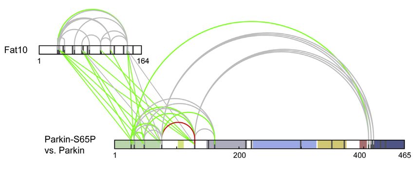
ll
OPEN ACCESS Article
et al., 2005). In fact, its intrinsic instability leads to a rapid degra- Lys 63 (Chew et al., 2011; Cunningham et al., 2015; Durcan
dation of both, FAT10 and its substrates, without the need for de- et al., 2014). Considering that the FAT10-Parkin conjugate is
conjugation (Aichem et al., 2018). Therefore, we investigated the formed both in the cytosol and at mitochondria (Figures 1A and
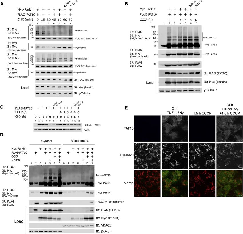
degradation of the FAT10-Parkin conjugate. We found that the 2D), we asked whether Parkin might serve as an E3 ligase for
FAT10-Parkin conjugate rapidly disappeared after 30 min of FAT10. We first confirmed the formation of the FAT10-Parkin con-
cycloheximide (CHX) treatment. Because monomeric FAT10 jugate in vitro using immunopurified myc-tagged Parkin that was
was degraded with the reported half-life of 1 h (Hipp et al., previously overexpressed in Hek293 cells (Figure 3A). When
2005), Parkin-FAT10 was degraded even faster than monomeric FAT10 was added to the in vitro reaction in the presence of
FAT10 (Figure 2A). However, FAT10 did not significantly alter UBA6, we could detect the formation of a FAT10-Parkin conjugate
the steady-state level of Parkin during the CHX treatment (Figures (Figure 3A, lane 5). The addition of USE1 slightly increased the FA-
2A and S2A). The proteasome inhibitor MG132 prevented degra- T10ylation of Parkin, whereas the replacement of GG by AV at the
dation of the FAT10-Parkin conjugate whereas the autophagy-in- C terminus of FAT10 abolished the covalent FAT10 attachment
hibitor bafilomycin A1 did not stabilize it (Figure 2A). We then (Figure 3A). To investigate the effect of Parkin activation on the
asked whether the FAT10-Parkin conjugate was accumulating Parkin-FAT10 conjugate formation, we compared the in vitro FA-
as an insoluble aggregate and found that the level of FAT10ylated T10ylation of the unmodified recombinant Parkin protein to a Par-
Parkin was not significantly altered over time in the insoluble frac- kin variant in which the Serine65 residue was phosphorylated
tion (Figure 2A). Because Parkin is a protein involved in mitoph- (Figure S3A), a post-translational modification that opens and ac-
agy, we examined whether mitochondrial uncoupling by CCCP tivates Parkin (Sauvé et al., 2018). Interestingly, we observed that
has an effect on the degradation rate of the FAT10-Parkin conju- phosphorylated Parkin undergoes enhanced FAT10ylation (Fig-
gate. Strikingly, we found that the prolonged treatment of the cells ure 3B). To confirm this finding, we performed a similar experiment
with CCCP lead to a significant reduction of the steady-state level as in Figure 3B but comparing a recombinant unmodified GST-
of FAT10, whereas the Parkin-FAT10 conjugate, after an initial tagged Parkin to its catalytically inactive recombinant C431A
reduction (1 h), was accumulating over 6 h of CCCP treatment mutant. At the same time, we compared these proteins to a
(Figure 2B). Interestingly, the degradation kinetic of FAT10 was form that was previously phosphorylated in vitro by PINK1 (Fig-
not significantly altered by CCCP on CHX treatment (Figure 2C). ure S3A). Remarkably, the exchange of the active site cysteine
Next we investigated where the FAT10-Parkin conjugate is of Parkin to alanine strongly reduced FAT10-Parkin conjugate for-
formed in cells during steady-state and mitochondrial depolariza- mation, whereas Parkin phosphorylation enhanced the formation
tion conditions. We overexpressed myc-Parkin and FLAG-FAT10 of the FAT10-Parkin conjugates, demonstrating that Parkin uses
in Hek293 cells, followed by 1 h CCCP and/or 5 h MG132 its E3 ligase activity to auto-FAT10ylate itself (Figures 3C, 3D,
treatment to induce mitochondrial depolarization and inhibition and S3B). Notably, the FAT10ylation of C431A GST-Parkin was
of the proteasomal degradation of FAT10ylated Parkin, respec- not completely abolished, and this effect is likely due to the fact
tively. Subsequently, we separated the mitochondrial from that the FAT10 E1/E2 enzymes (UBA6/USE1) are able to directly
the cytosolic fraction, and the FAT10-Parkin conjugate was conjugate FAT10 to several substrates under in vitro conditions
immunopurified and visualized. The FAT10-Parkin conjugate (Aichem et al., 2012; Bialas et al., 2015, 2019). In addition, our
was mainly found in the cytosol, and proteasome inhibition quantitative XL-MS analysis also suggests an intensified interac-
lead to its accumulation (Figure 2D, lanes 4–7). Interestingly, tion of FAT10 with the UBL domain of phosphorylated Parkin, as
FAT10 and FAT10ylated Parkin were detectable in the mitochon- indicated by significantly upregulated inter-protein crosslinks
drial fraction on treatment with CCCP and MG132, demonstrating in samples containing Parkin-S65P/FAT10 compared to non-
that the FAT10-Parkin conjugate could also be formed at phosphorylated Parkin/FAT10 complexes (Figure 3E; Table S2),
mitochondria on their depolarization (Figures 2D, lane 14, and in line with the results in Figures 3C and 3D. Finally, we evaluated
S4A). By immunostaining of FAT10 in TNF-a/IFNg-treated SH- the interaction between Parkin and USE1, the conjugating enzyme
SY5Y cells (validated in Figure S2B), we found no detectable for FAT10. Interestingly, we found that in Hek293 cells endoge-
FAT10 translocation to mitochondria on CCCP treatment (Fig- nous USE1 non-covalently interacted with either EGFP- or myc-
ure 2E), but it was mainly cytosolic, in line with the results in tagged Parkin (Figures 3F and 3G). These results describe
Figure 2D. Parkin as a multi-functional ligase that is able to auto-FAT10ylate
itself.
Parkin auto-FAT10ylates itself in vitro
Parkin is a primarily cytosolic protein and is known to ubiquitylate Parkin is a FAT10 E3 ligase that enhances the formation
cytosolic substrates independently of PINK1 activation. For of the FAT10-Mfn2 conjugate
instance, Parkin promotes the clearance of cytosolic proteins Next, we asked whether there are proteins undergoing Parkin-
such as tubulin a and b (Stevens et al., 2015). Furthermore, it ubiq- dependent FAT10ylation. Mitofusin2 (Mfn2) is a GTPase involved
uitylates Bax in a PINK1-independent manner, whereas its activity in mitochondrial dynamics as an essential component of the mito-
in mediating K63-ubiquitylation of M. tuberculosis mediates resis- chondrial fusion machinery (Santel and Fuller, 2001). Moreover,
tance to intracellular pathogens (Johnson et al., 2012; Manzanillo Mfn2 is a well-known PINK1/Parkin-dependent phospho-ubiqui-
et al., 2013). However, the exact mechanism regulating Parkin ac- tylation substrate and is degraded in a proteasome-dependent
tivity in the cytosol is still poorly understood. Parkin is a versatile manner after mitochondrial stress (McLelland et al., 2018). In a pre-
E3 ligase and mediates Lys48 poly-ubiquitylation as well as vious quantitative mass spectrometry analysis of FLAG-FAT10
mono-ubiquitylation or poly-ubiquitylations at Lys 6, Lys 11, and conjugates, we identified Mfn2 as an interaction partner for
4 Cell Reports 34, 108857, March 16, 2021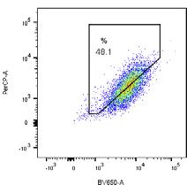
ll
Article                                                                                                                                     OPEN ACCESS

Figure 2. FAT10ylated Parkin can localize in both cytosol and mitochondria and is degraded by the proteasome
(A) Immunoblot analysis of the co-immunoprecipitation of Myc-Parkin with 3xFLAG-FAT10 in whole cell lysate from transiently transfected Hek293 cells ex-
pressing the indicated constructs. Cells were treated with DMSO or, where specified, with the protein synthesis inhibitor cycloheximide (CHX) at the indicated
time points with or without autophagy inhibitor bafilomycin A1 (Baf-A1) or proteasome inhibitor MG132.
(B) Immunoblot analysis of the co-immunoprecipitation (coIP) of Myc-Parkin with 3xFLAG-FAT10 in whole cell lysate from Hek293 cells expressing the indicated
constructs. Cells were treated with DMSO or, where specified, with CCCP (10 mM) for the indicated time periods with or without bafilomycin A1 (Baf-A1, 100 nM)
or MG132 (5 mM).
(C) Immunoblot analysis of the degradation rate of overexpressed FLAG-FAT10 in Hek293 cells after treatment with 50 mg/mL CHX in presence or absence of the
mitochondrial uncoupler CCCP (10 mM).
(D) Untransfected Hek293 cells or Hek293 cells stably expressing Myc-Parkin were transiently transfected for 24 h with a plasmid expressing FLAG-FAT10 where
indicated. DMSO or, where indicated, CCCP (10 mM) and/or MG132 (5 mM) were added for 1 h or 5 h prior to lysis, respectively. Cell lysates were fractionated into
mitochondria and cytoplasm enriched samples followed by immunoprecipitation of FLAG-tagged FAT10, SDS-PAGE, and immunoblot with the indicated antibodies.
(E) SH-SY5Y cells were treated for 24 h with TNF-a/IFNg to induce FAT10 expression, followed by 2 h DMSO or CCCP treatment where indicated. Cells were
immunostained with antibodies against TOM20 (red) and FAT10 (green). Scale bars, 10 mm.
Shown results are representatives of at least three independent experiments with similar outcomes.

                                                                                                             Cell Reports 34, 108857, March 16, 2021 5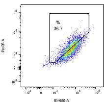
ll
     OPEN ACCESS                                              Article

                   A                        B

                   C

                                                    D

                   E

                   F                            G

                                                        (legend on next page)

6 Cell Reports 34, 108857, March 16, 2021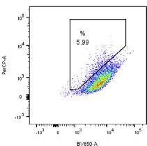
ll
Article                                                                                                                                          OPEN ACCESS

FAT10 (data not shown). In fact, when expressed together in                          S4C) abolished the formation of the CCCP-dependent Mfn2-
Hek293 cells, we detected a prominent stable conjugate when                          FAT10 conjugate in SH-SY5Y cells, confirming Parkin as a
myc-tagged Mfn2 was transiently co-expressed with FLAG-                              FAT10 E3 ligase and Mfn2 as a Parkin-dependent FAT10ylation
tagged WT FAT10, but not with FLAG-FAT10-AV (Figure 4A). To                          substrate under endogenous conditions (Figure 4F). To further
confirm these data under endogenous conditions, we treated                           confirm this data, we performed an in vitro FAT10ylation experi-
Hek293 wild-type (WT) or Hek293 FAT10 knockout (KO) cells (Ai-                       ment with myc-tagged Mfn2 as substrate that was immuno-puri-
chem et al., 2019a) with TNF-a/IFNg for 24 h to induce FAT10 pro-                    fied from Hek293 cells after overexpression (Figure 4G). We
tein expression, followed by an immunoprecipitation with the                         detected the strongest FAT10-Mfn2 conjugate formation when
monoclonal anti-FAT10 antibody 4F1 and a western blot with an                        the complete conjugation machinery UBA6, USE1, and active
anti-Mfn2 antibody. We confirmed that the KO of FAT10 in                             pS65-Parkin was present in the in vitro reaction. Overall, we
Hek293 cells led to the complete disappearance of the Mfn2 inter-                    confirmed Parkin to act as a FAT10 E3 ligase and discovered
action and conjugate formation (Figure 4B). We subsequently                          Mfn2 as a Parkin-dependent FAT10ylation substrate.
investigated the dynamics of the FAT10-Mfn2 conjugate degrada-
tion in a CHX chase experiment. In contrast to the Parkin-FAT10                      FAT10 inhibits the activation of Parkin and the Parkin-
conjugate, the FAT10-Mfn2 conjugate followed a kinetic of protea-                    dependent Mfn2 ubiquitylation
some-dependent degradation that resembled the one of mono-                           Parkin interactors such as BAG5 and PICK1 inhibit the ubiquityla-
meric FAT10 (Figure 4C). To investigate whether mitochondrial                        tion activity of Parkin, whereas the ubiquitin-like modifiers NEDD8
depolarization stress, and the consequent Parkin activation, could                   and SUMO-1 have been described to activate Parkin (He et al.,
influence the formation of the Mfn2-FAT10 conjugate, we exam-                        2018; Kalia et al., 2004; Um and Chung, 2006; Um et al., 2012).
ined the formation of this complex in SH-SY5Y cells. Interestingly,                  Hence, we investigated whether the FAT10ylation of Parkin has
mitochondrial depolarization lead to a decrease in FAT10 steady-                     an effect on its functions. As an assay to assess Parkin activity,
state level (Figure 4D), as we have reported for Hek293 cells (Fig-                  we investigated its CCCP-induced auto-ubiquitylation (Chung
ure 2B). We could not detect significant levels of FAT10-Mfn2                        et al., 2004). When we treated a stably transfected Hek293 clone
conjugates in TNF-a/IFNg-stimulated SH-SY5Y cells (Figure 4D),                       expressing GFP-Parkin with CCCP, we observed Parkin poly-
in contrast to what we had previously observed in Hek293 cells                       ubiquitylation after immunoprecipitation of overexpressed HA-
(Figure 4B), suggesting that the efficiency of the formation of the                  ubiquitin in Parkin western blots (Figure 5A). Strikingly, when cells
Mfn2-FAT10 conjugate is cell-type-specific. Nevertheless, the in-                    were transiently co-transfected with FLAG-tagged FAT10 WT, the
hibition of proteasome activity with MG132 allowed us to detect                      CCCP-dependent auto-ubiquitylation of Parkin was strongly
the FAT10-Mfn2 conjugate (Figure 4D, lane 3). Strikingly, 2 h of                     reduced, whereas this inhibitory effect was absent when
mitochondrial depolarization with CCCP lead to an increase in                        FAT10-AV was co-expressed (Figure 5A). To investigate whether
the formation of the FAT10-Mfn2 conjugate, and this complex                          the same effect was visible under endogenous conditions, we
further accumulated after proteasome inhibition (Figure 4D, lanes                    evaluated the CCCP-induced auto-ubiquitylation of Parkin with
4 and 5). Moreover, we performed a co-immunoprecipitation anal-                      or without pre-stimulation by TNF-a/IFNg. Additionally, we
ysis that validated that FAT10-Mfn2 interaction and conjugate for-                   compared Parkin auto-ubiquitylation in SH-SY5Y WT cells to
mation takes place at mitochondria after treatment of SH-SY5Y                        SH-SY5Y FAT10 KO cells (validated in Figure S5A). To achieve
cells with MG132 and CCCP (Figure S4A). Given that in Hek293                         that, we treated the cells with 10 mM CCCP for 1.5 h, followed
cells the co-expression of EGFP-tagged Parkin followed by 1 h                        by immunoprecipitation of endogenous ubiquitin and western
CCCP treatment led to significant enhancement of the FAT10yla-                       blot analysis using a monoclonal anti-Parkin antibody. Strikingly,
tion of Mfn2 (Figure 4E), we investigated whether the depletion of                   in SH-SY5Y WT cells, the cytokine pre-treatment led to reduced
cellular Parkin in SHSY5 leads to a reduction of the Mfn2-FAT10                      auto-ubiquitylation of Parkin, and this negative effect was abol-
complex. Remarkably, knocking down Parkin (Figures S4B and                           ished by the knock out of the Fat10 gene (Figure 5B). Accordingly,

Figure 3. Parkin auto-FAT10ylates itself
(A) Myc-Parkin was immunopurified from total lysates of transiently transfected Hek293 cells in which Myc-Parkin was overexpressed. Purified Myc-Parkin was
then used in an in vitro FAT10ylation assay in presence of recombinant FLAG-UBA6, His-USE1, FAT10, or FAT10-AV as indicated. Proteins were incubated in
reaction buffer at 37 C for 45 min, separated by SDS-PAGE under reducing conditions and immunoblotted as described.
(B) Recombinant Parkin and recombinant Parkin pSer65 were subjected to an in vitro FAT10ylation assay as described in (A).
(C) Recombinant GST-Parkin, GST-pSer65-Parkin and GST-Parkin C431A were subject to an in vitro FAT10ylation as described in (A).
D) Quantification of the intensity of signals of experiment (C).
(E) Influence of the phosphorylation at serine 65 on the crosslinking pattern of Parkin and FAT10. Shown are changes in crosslink abundances of Parkin and FAT10
in presence or absence of the phosphorylation at serine 65. Only crosslinks that could be reproducibly quantified from the pool of identified high-confidence
crosslinks in both samples (n = 3) are shown (violation = 0, p value %0.05, LD score R25, see STAR Methods). Depicted in green are crosslinks that were
significantly upregulated (R2-fold) in samples containing phosphorylated Parkin, while red links indicate significant downregulation (%2-fold). Crosslinks ex-
hibiting no significant change in abundance in both samples are depicted in gray.
(F) Immunoblot analysis of the coIP of EGFP-Parkin with endogenous USE1 in presence or absence of overexpressed FLAG-FAT10. Proteins were purified from
lysates of Hek293 cells transiently expressing the indicated constructs. Cells were treated with DMSO or, where indicated, with 10 mM CCCP for 1 h prior to lysis.
(G) Immunoblot analysis of the coIP of Myc-Parkin with endogenous USE1 as described in (D).
Shown results are representatives of at least three independent experiments with similar outcomes. Error bars in (D) indicate SD (n = 4). *p < 0.05 (Student’s t test),
n.s., not significant.

                                                                                                                 Cell Reports 34, 108857, March 16, 2021 7
ll
      OPEN ACCESS                                                                                                                           Article

Figure 4. Parkin FAT10ylates Mitofusin2
(A) Immunoblot analysis of the coIP of Myc-Mfn2 with 3xFLAG-FAT10 or 3xFLAG-FAT10-AV from whole cell lysates of Hek293 cells transiently expressing the
indicated constructs.
(B) Total cell extracts of untreated or TNF-a/IFNg-stimulated Hek293 WT or HEK293 FAT10 KO cells were used to immunoprecipitate endogenous FAT10.
Immunoblotting was then performed with an anti-Mfn2 antibody to detect endogenous Mfn2-FAT10 conjugate formation.
(C) Immunoblot analysis of the coIP of Myc-Mfn2 with 3xFLAG-FAT10 from whole cell lysates of Hek293 cells expressing the indicated constructs. Cells were
treated with DMSO or, where specified, with cycloheximide (CHX) for the indicated time periods. Where indicated, MG132 or bafilomycin A1 (Baf A1) have been
added for the entire duration of the CHX-chase experiment.
(D) Total cell extracts of untreated or TNF-a/IFNg-stimulated SH-SY5Y cells were used to immunoprecipitate endogenous FAT10. Immunoblotting was then
performed with a polyclonal anti-Mfn2 antibody to detect endogenous Mfn2-FAT10 conjugate formation. Where indicated, cells were treated for 2 h with CCCP
(10 mM) and/or with MG132 (5 mM) prior to lysis.
                                                                                                                         (legend continued on next page)

8 Cell Reports 34, 108857, March 16, 2021
ll
Article                                                                                                                                 OPEN ACCESS

we assessed the capacity of Parkin to ubiquitylate the mitochon-               et al., 2013; Zheng and Hunter, 2013). These studies have re-
drial substrate Mfn2. We detected a prominent reduction of                     ported that EGFP-Parkin requires 1 h to localize to the surface
Parkin-related Mfn2 ubiquitylation after CCCP treatment in the                 of mitochondria on CCCP treatment, and the E3 ligase activity
presence of FAT10 but not FAT10-AV (Figure 5C). Moreover, in                   of Parkin is required for the translocation. Hence, we first
a co-immunoprecipitation assay co-expression of WT FAT10                       ensured that the treatment of Hek293 cells stably expressing
(but not FAT10-AV) with EGFP-Parkin abolished the interaction                  EGFP-Parkin with low concentrations of tet was not altering
of the E3 ligase with Mfn2 on mitochondrial uncoupling (Fig-                   the translocation of the E3 ligase to the mitochondria (data not
ure 5D). To further confirm that the inhibitory effect of FAT10 is             shown). Subsequently, we used the same time-lapse live cell im-
dependent on its conjugation, we tested whether FAT10 could                    aging-based approach to evaluate the effect of overexpressed
inhibit Parkin activation in Hek293 UBA6 KO cells. Remarkably,                 FLAG-FAT10 on Parkin translocation. Although we could not
the absence of UBA6 completely abolished the inhibitory effect                 identify any significant difference after 1 h of CCCP treatment
of FAT10 on Parkin activation (Figure 5E). Finally, we reconstituted           (Figure 6B), we noticed that the kinetics of Parkin translocation
an in vitro system for Parkin auto-ubiquitylation and combined it              was slowed down in the presence of FAT10 (Figures 6C and
with an in vitro FAT10ylation system to investigate whether the                6D). To further validate the inhibitory effect of FAT10 on the pro-
overall inhibitory effect of FAT10 was directly mediated by the                gression of Parkin-dependent mitophagy, we tested the conse-
impairment of Parkin activation. We first performed an in vitro FA-            quence of tet-induced FAT10 overexpression on mitochondrial
T10ylation of Parkin, followed by the addition to the in vitro reac-           turnover by evaluating the shift of mtKeima fluorescence excita-
tion of ubiquitin, UBE1, and Ubc7 as indicated in Figure 5F.                   tion when the mitochondria are acidified in the autophagolysoso-
pSer65 Parkin was able to auto-ubiquitylate itself in vitro as previ-          mal compartment subsequent to mitophagy (Katayama et al.,
ously reported, whereas the unmodified form was only poorly un-                2011). We could detect a progressive increase of mitochondrial
dergoing self-modification (Figure 5F, lane 7) (Ordureau et al.,               acidification over 12 h post CCCP treatment by exciting
2014). Of note, our in vitro data were in accordance with our in cel-          mtKeima at a wavelength of 561 nm (Figure 6E). Next, we treated
lulo data: a pre-incubation of Parkin with FAT10 alone was not                 the cells with CCCP for 6 h and assessed the ratio of acidified
affecting its activation (Figure 5F, lane 8), whereas a reduction              mtKeima in single cells by flow cytometry as a quantitative
of its auto-ubiquitylation and accumulation of unmodified Parkin               marker for mitophagy progression (Katayama et al., 2011).
was evident when Parkin was pre-incubated with FAT10 and                       When cells were pre-treated for 24 h with tet to induce FAT10
UBA6 or with a combination of UBA6 and USE1 (Figure 5F, lanes                  expression, we detected a significant decrease in acidified
9 and 10, respectively). Furthermore, FAT10-AV was unable to                   mtKeima-positive cells on 6 h of CCCP treatment (Figures 6F
inhibit Parkin auto-ubiquitylation (Figure 5F, lane 11), suggesting            and 6G). To validate this data under endogenous conditions,
that FAT10 directly impairs Parkin activation and that this effect             we established two stable cell lines based on SH-SY5Y WT
requires FAT10 conjugation.                                                    and SH-SY5Y FAT10 KO cells both stably expressing mtKeima.
                                                                               In this context, we could investigate the effect of TNF-a and
FAT10 delays Parkin translocation to mitochondria and                          IFNg-induced endogenous FAT10 expression on mitophagy-
inhibits mitophagy progression                                                 dependent mitochondrial turnover. Although a previous report
To investigate the biological consequences of FAT10-dependent                  has shown that TNF-a is responsible for the induction of mitoph-
inhibition of Parkin activity, we established a cellular expression            agy in macrophages (Bell et al., 2013), we could observe the
system based on the T-Rex-293 cell line in which EGFP-Parkin is                opposite effect in our neuronal cell line model. In fact, when
stably expressed and FLAG-FAT10 expression can be induced                      SH-SY5Y cells were pre-stimulated for 24 h with a combination
by tetracycline (tet) (Figure 6A). In addition, this transfectant              of TNF-a and IFNg, we found a significant reduction in the total
also stably expresses mtKeima, a fluorescent protein that is                   number of acidified mitochondria after 18 h of CCCP treatment
localizing to mitochondria and that is widely used to quantify mi-             (Figures 6H and 6I, bars 3 and 4). However, pre-stimulations
tophagy (Figures 6B and S6A) (Sun et al., 2017). We first tested               with TNF-a and IFNg did not lead to a significant decrease in
whether FAT10 expression has an effect on Parkin translocation                 acidified mitochondria after 18 h of CCCP treatment in FAT10-
to mitochondria on exertion of mitochondrial stress. Several                   deficient cells (Figures 6H and 6I, bars 7 and 8). These data
studies have demonstrated that the integrity of the RING2                      demonstrate that under inflammatory conditions, FAT10 plays
domain of Parkin is essential for Parkin ligase activity, and the              a role in inhibiting depolarization-induced mitophagy. Moreover,
loss of function mutation of the C431 active site cysteine abol-               these results are in line with our previous experiments showing
ishes the translocation of Parkin to mitochondria (Trempe                      that FAT10 impedes the CCCP-induced activation of Parkin

(E) Immunoblot analysis of the coIP of Myc-Mfn2 with 3xFLAG-FAT10 or 3xFLAG-FAT10-AV in cell lysates of WT Hek293 cells or of Hek293 cells stably ex-
pressing EGFP-Parkin. Cells were treated with DMSO or, where indicated, with CCCP (10 mM) for 1 h prior to lysis.
(F) Total cell extracts of untreated or TNF-a/IFNg-stimulated SH-SY5Y cells were used to immunoprecipitate endogenous FAT10. Immunoblotting was then
performed with a polyclonal anti-Mfn2 antibody to detect endogenous Mfn2-FAT10 conjugate formation. Where indicated, endogenous Parkin was depleted
from cells using a small interfering RNA (siRNA)-based strategy. Cells were treated with DMSO or, where indicated, with CCCP (10 mM) for 2 h prior to lysis.
(G) Myc-Mfn2 was immunopurified from total lysates of Hek293 cells in which the construct was overexpressed for 24 h. Purified Myc-Mfn2 was used for an
in vitro FAT10ylation assay in presence of recombinant FLAG-UBA6, His-USE1, pSer65-Parkin, or FAT10 as indicated. Proteins were incubated in reaction buffer
at 37 C for 45 min, separated by SDS-PAGE under reducing conditions and immunoblotted as indicated.
Shown results are representatives of at least three independent experiments with similar outcomes.

                                                                                                         Cell Reports 34, 108857, March 16, 2021 9
ll
OPEN ACCESS Article
(legend on next page)
10 Cell Reports 34, 108857, March 16, 2021ll
Article OPEN ACCESS
(Figure 5) and the recruitment of Parkin to mitochondria (Figures ing cells significantly counteracted its pro-survival effect (Fig-
6C and 6D). ure 7B, lines 4 and 8). To assess the effect of endogenous
FAT10 expression on the susceptibility of SH-SY5Y cells to rote-
FAT10 promotes neuronal cell death in vitro none treatment, we compared SH-SY5Y WT cells to SH-SY5Y
A pertinent question raised by our data so far is whether the FAT10 KO cells (Figure S2B).The cells were pretreated with
inhibitory effect of FAT10 on mitophagy in neuronal cells will TNF-a/IFNg for 18 h to induce FAT10, followed by a 32-h treat-
affect their susceptibility to agents known to cause mitochon- ment with rotenone, TNF-a/IFNg, or the combination of both
drial depolarization and the induction of oxygen radicals. stimuli. Then, we analyzed cell viability and mitochondrial ROS
Accordingly, we investigated whether FAT10 influences the production using mitoSOX red (i.e., a fluorescent probe detect-
viability of SH-SY5Y cells (Xicoy et al., 2017). We treated SH- ing superoxide radicals). The combination of rotenone and pro-
SY5Y cells for 32 h with 5 mM rotenone that is an inhibitor of mito- inflammatory cytokines lead to a significantly stronger reduction
chondrial complex I and impairs the electron transport chain, in the viability of SH-SY5Y WT cells as compared to treatment
with consequent over-production of ROS (Li et al., 2003; New- with rotenone alone (Figure 7C, lanes 3 and 4). Interestingly,
house et al., 2004). Subsequently, we analyzed cell viability to the lack of FAT10 in the gene targeted SH-SY5Y cells (Figures
investigate how the expression of Parkin and FAT10 influences S2B and S5A) abolished the TNF-a/IFNg-mediated negative
the rotenone-induced cell death. First, we established by lentivi- effect on cell viability (Figure 7C, lanes 7 and 8). Thus, endoge-
ral transduction two SH-SY5Y cell lines that stably express nously expressed FAT10 was essential for this cytokine-depen-
FLAG-FAT10 WT or FLAG-FAT10-AV, followed by evaluation of dent effect in accordance with FAT10-mediated mitophagy inhi-
cell viability after 32 h of rotenone treatment. Remarkably, cells bition described in Figures 6H and 6I. Using mitoSOX red as a
overexpressing WT FLAG-FAT10 underwent a significant in- detection system for mitochondrial ROS we confirmed the ex-
crease of cell death (Figure 7A, lanes 4 and 5). This toxic effect pected rotenone-mediated enhancement of ROS in mitochon-
was not apparent when SH-SY5Y cells expressed the FLAG- dria but this was only slightly but not significantly further
FAT10-AV variant, implying that the toxic role of FAT10 is corre- enhanced by the cytokine treatment (Figure 7D, left side) and
lated to the FAT10-mediated inhibition of Parkin ubiquitin-ligase the latter tendency was not observed in FAT10-deficient cells
activity, which requires FAT10 conjugation (Figure 7A, lanes 5 (Figure 7D, right side). To confirm that the observed effect on
and 6). Several works have described Parkin as an enzyme cell viability is due to the lack of FAT10 expression, we reconsti-
with cytoprotective effect in the neuronal cell line SH-SY5Y (Ca- tuted FLAG-FAT10 by stable expression in SH-SY5Y FAT10 KO
€ller-Rischart et al., 2013).
sarejos et al., 2006; Dai et al., 2015; Mu cells (Figure 7C, lanes 9–12). Remarkably, the ectopic expres-
Other investigations describe autophagy as a key cellular pro- sion of FAT10 led to a significant increase in cell death, in line
cess counteracting rotenone-induced production of ROS and with Figures 7A and 7B. Taken together, we conclude that the
cell death in the SH-SY5Y model (Deng et al., 2013; Zhang negative effect of FAT10 on the viability of rotenone-treated
et al., 2018), with the degradation of mitochondria by PINK1/Par- SH-SY5Y cells is likely due to its inhibitory effect on Parkin-medi-
kin-dependent mitophagy exerting an essential pro-survival ac- ated mitophagy (Figure 7E).
tivity (Pan et al., 2009; Peng et al., 2019). In the light of these
studies, we investigated whether Parkin overexpression has a DISCUSSION
cytoprotective effect in our model and whether FAT10 is involved
in the modulation of Parkin function. To achieve that, we estab- Previous studies described several post-translational modifica-
lished a SH-SY5Y cell line which stably expresses mCherry- tions to modulate Parkin activity, including SUMOylation and
tagged Parkin and one which express both mCherrry-Parkin NEDDylation (Um and Chung, 2006). Here, we show that Parkin
and FLAG-FAT10. We found that the overexpression of Parkin is a substrate for FAT10 on stimulation with TNF-a and IFNg (Fig-
leads to reduced cell death after rotenone treatment (Figure 7B, ure 1). The Parkin-FAT10 conjugate is formed in the cytosol and
lines 2 and 4), confirming the cytoprotective role of the Parkin E3 is rapidly degraded by the proteasome as reported for other
ligase. Strikingly, the co-expression of FAT10 in Parkin-express- FAT10 substrates (Aichem et al., 2012; Bialas et al., 2019; Hipp
Figure 5. FAT10 inhibits the ubiquitin-E3 ligase activity of Parkin
(A) Immunoblot analysis of the coIP of HA-Ubiquitin with EGFP-Parkin in lysates from Hek293 cells. Where indicated, the constructs for 3xFLAG-FAT10 or
3xFLAG-FAT10-AV were transiently co-expressed. Cells were treated with DMSO or, where indicated, with CCCP (10 mM) for 1 h prior to lysis to induce Parkin
activation and auto-ubiquitylation.
(B) Immunoblot analysis of the coIP of ubiquitin with Parkin in lysates from SH-SY5Y cells and SH-SY5Y FAT10 KO cells. Where indicated, cells were treated with
24 h TNF-a/IFNg and/or 1.5 h CCCP (10 mM) prior to lysis. DMSO treatment was used as a control.
(C) Immunoblot analysis of the coIP of HA-Ubiquitin with endogenous Mfn2 from lysates of Hek293 cells. Where indicated, the constructs for EGFP-Parkin,
3xFLAG-FAT10, or 3xFLAG-FAT10-AV were transiently co-expressed. Cells were treated with DMSO or, where specified, with CCCP (10 mM) for 1 h prior to lysis
to induce Parkin-dependent ubiquitylation of Mfn2.
(D) Immunoblot analysis of the coIP of EGFP-Parkin with endogenous Mfn2 in lysates of Hek293 cells. Where indicated, the constructs for 3xFLAG-FAT10 or
3xFLAG-FAT10-AV have been co-expressed. Cells were treated with DMSO or, where specified, with CCCP (10 mM) for 1 h prior to lysis to induce Parkin
activation.
(E) Immunoblot analysis as in (A) including the Hek293 UBA6 KO control.
F) Immunoblot of the in vitro ubiquitylation of Parkin or pSer65-Parkin in presence of the indicated proteins.
Shown results are representatives of at least three independent experiments with similar outcomes.
Cell Reports 34, 108857, March 16, 2021 11ll
OPEN ACCESS Article
A B CCCP treatment (min)
EGFP-Parkin + + 0 60 0 60
Tet +
1 2
KDa
IB: FLAG (FAT10) mtKeima
(405 nm excitation)
FAT10 conjugates
EGFP-Parkin
25
FLAG-FAT10
100
IB: Parkin
Merged
IB: GAPDH
+ Tet
C CCCP treatment (min) D
20 25 30 35 40 - Tet
% cells with GFP-Parkin on mitochondria
+ Tet
100
80
*
EGFP-Parkin
*
60
40 *
EGFP-Parkin 20
+ Tet
0
0
10
20
30
40
50
60
t (min)
E mtKeima mtKeima
F
pH7 (405 nm) pH4 (561 nm) EGFP-Parkin WT
CCCP
5.99 % 48.1 % G - Tet
Untreated + Tet
mtKeima pH 4 (561 nm)
- Tet 60 *
acidified mtKeima (6h)
% cells with high ratio
40
6.11 % 36.7 %
+ Tet
12h 20 ns
CCCP
0
mtKeima pH 7 (405 nm)
H SH-SY5Y WT SH-SY5Y FAT10 KO I WT
FAT10 KO
CCCP CCCP 60 ** *
ns
acidified mtKeima (18h)
% cells with high ratio
mtKeima pH 4 (561 nm)
40
20
0
24h 1 2 3 4 5 6 7 8
TNFα/IFNγ
CCCP + + + +
TNFα/IFNγ + + + +
1 2 3 4 5 6 7 8
mtKeima pH 4 (561 nm) IB: FAT10
IB: γ-tubulin
(legend on next page)
12 Cell Reports 34, 108857, March 16, 2021ll
Article OPEN ACCESS
et al., 2005). Furthermore, we found that a fraction of FAT10y- ure 1E shows that FAT10 actively interacts with the Ubl domain
lated Parkin is present on depolarized mitochondria (Figure 2D). of Parkin, a region that is strongly involved in the regulation of
In the light of the extreme versatility of Parkin as an E3 enzyme Parkin activation. In fact, several works have shown that Parkin
(Chew et al., 2011; Cunningham et al., 2015; Durcan et al., activity is negatively regulated by its UBL domain, and the pertur-
2014), we investigated whether it could act as a FAT10 E3 ligase. bation at this level influences Parkin function, leading to the acti-
As criteria to identify Parkin as a ligase, we investigated its auto- vation of the E3 ligase by the disruption of its auto-inhibited
FAT10ylation and its capacity to FAT10ylate a substrate protein conformation (Burchell et al., 2012; Chaugule et al., 2011).
(Huibregtse et al., 1995; Lee et al., 2017). Strikingly, we found Accordingly, we speculate that the strong interaction between
that Parkin contributes to its own in vitro FAT10ylation and that FAT10 and the UBL domain of Parkin could be involved in the
it FAT10ylates Mfn2 in SH-SY5Y cells (Figures 3 and 4). Interest- perturbation of the structure of the N terminus of Parkin leading
ingly, phosphorylated Parkin could auto-FAT10ylate itself more to a transient activation of cytosolic Parkin that can explain how
efficiently than the WT protein, suggesting that the regulation Parkin is able to FAT10ylate itself independently of its phosphor-
of Parkin activity by FAT10 could take place at two distinct levels: ylation status (Figures 3C and 3D). At mitochondria, activated
in the cytosol and at mitochondria. In the first case, we observed Parkin possesses a high affinity for FAT10 (Figure 3E) and it
that Parkin contributes to its own FAT10ylation while being in its accordingly auto-FAT10ylates itself and the mitochondrial sub-
‘‘inactive’’ (cytosolic) form (Figures 3C and 3D). Here, a pertinent strate Mfn2 (Figures 2D and 4). Overall, we propose that Parkin
question would be how Parkin is activated in the cytosol. In fact, contributes to its FAT10ylation at both cytosolic and mitochon-
several works showed that cytosolic Parkin is in a repressed drial level. At the cytosolic level, this modification sequesters a
state and possesses no capacity to interact with cognate E2s fraction of Parkin in the cytosol where it is degraded by the pro-
through the RING1 domain, while also the catalytic RING2 teasome. At the mitochondrial level, activated Parkin FAT10y-
domain is hindered and cannot perform the ubiquitin transthiola- lates itself and mitofusin2. Simultaneously, Parkin FAT10ylation
tion reaction (Trempe et al., 2013; Wauer and Komander, 2013). leads to a reduction in the ubiquitylation of Parkin itself and in
This notion led to the assumption that Parkin can act as a ligase the ubiquitylation of Mfn2 (Figure 5), suggesting that there exists
exclusively at mitochondria upon their depolarization and PINK1 a negative-regulatory mechanism by which the FAT10-ligase ac-
activation. However, these studies have been performed in the tivity of Parkin directly alters its ubiquitin-ligase activity. Interest-
absence of TNF/IFNg stimulation that is required for FAT10 ingly, this effect is dependent on FAT10 conjugation, and the
expression. Moreover, several reports suggest that numerous FAT10 activating enzyme UBA6 is essential for the FAT10-
Parkin substrates are not mitochondrial and do not require mito- dependent hindrance of Parkin activity (Figure 5), suggesting
chondrial depolarization in order to become ubiquitylated by that the pharmacological inhibition of UBA6 could rescue the
Parkin. For instance, Parkin exhibits ubiquitin-ligase activity to- biological negative consequences that FAT10 has on Parkin acti-
ward FBP1 and Hsp70 in SH-SY5Y cells (Ko et al., 2006; Moore vation. Remarkably, we found that UBA6 alone was not sufficient
et al., 2008) and toward RanBP2 and RIPK1 in Hek293 cells (Um to FAT10ylate Mfn2 in vitro, whereas the combination of UBA6
et al., 2006; Wang et al., 2018) that adds to the evidence of mi- and Parkin can exert this function, in line with previous work
tophagy-unrelated functions of Parkin (Johnson et al., 2012; describing Parkin as a unique ligase that is able to catalyze
Manzanillo et al., 2013). Apparently, other so far unknown factors in vitro mono-ubiquitylation of substrate proteins independently
could transiently activate Parkin in the cytosol. For instance, Fig- of E2 conjugating enzymes (Chew et al., 2011). Furthermore, we
Figure 6. FAT10 delays Parkin translocation to mitochondria and inhibits mitophagy progression
(A) Immunoblot analysis of the total lysate of T-REx Hek293 cells stably expressing EGFP-Parkin and transiently expressing FLAG-FAT10 in a tetracycline (Tet)-
dependent manner (200 ng/mL for 24 h).
(B) T-REx Hek293 cells described in (A) stably express the mitochondria-localizing protein mtKeima. Cells were treated with DMSO or, where specified, with
10 mM CCCP for 1 h to induce Parkin activation and translocation to mitochondria. Cells were visualized by time lapse microscopy. Where indicated, cells were
pre-incubated with 200 ng/mL tetracycline (Tet) for 24 h prior to CCCP treatment. Scale bars, 20 mm.
(C) Time lapse microscopy analysis of EGFP-Parkin recruitment to mitochondria as in (B) showing the time interval between 20 and 40 min after CCCP treatment.
Scale bars, 20 mm.
(D) Quantification of EGFP-Parkin recruitment to mitochondria on 1 h CCCP treatment (10 mM). The percentage of cells with EGFP-Parkin localized to mito-
chondria was analyzed every 5 min.
(E) T-REx Hek293 cells described in (A) stably express the mitochondria-localizing protein mtKeima. Cells were treated with DMSO or, where indicated, with
10 mM CCCP for 12 h to induce mitophagy. Cells were analyzed by time lapse microscopy and mitochondria in the cytosol (neutral pH) and in the lysosomes
(acidic pH) were visualized at different mtKeima excitation wavelength as indicated. Scale bars, 20 mm.
(F) Cells described in (B) were treated with DMSO or, where indicated, with CCCP (10 mM) for 6 h. mtKeima fluorescence in EGFP-Parkin expressing cells was
measured using flow cytometry by excitation at 405 nm and 561 nm. Representative dot plots of fluorescence emission from excitation at both wavelengths are
shown. Where indicated, cells were treated with 200 ng/mL tetracycline (tet) for 24 h prior to CCCP treatment to induce FLAG-FAT10 expression.
(G) Quantification of mitophagy-positive cells from (F).
(H) Flow cytometry analysis was performed as in (F) to compare the progression of mitophagy in SH-SY5Y WT and SH-SY5Y FAT10 KO cells both stably ex-
pressing mtKeima. Cells were treated with DMSO or, where indicated, with 18 h of CCCP (10 mM). Where indicated, cells were pre-treated with TNF-a/IFNg to
induce FAT10 expression.
(I) Quantification of mitophagy-positive cells from (H). TNF-a/IFNg-induced FAT10 expression is shown in the WB at the bottom using g-tubulin as loading control.
Shown results are representatives of at least three independent experiments with similar outcomes. Error bars in (D) indicate SEM (n = 3). Error bars in (F) and (I)
indicate SD (n = 3).*p < 0.05 (Student’s t test), n.s., not significant.
Cell Reports 34, 108857, March 16, 2021 13ll
OPEN ACCESS Article
(legend on next page)
14 Cell Reports 34, 108857, March 16, 2021ll
Article OPEN ACCESS
found that the combination of TNF-a and IFNg that leads to B In vitro FAT10ylation and ubiquitylation assay
FAT10 expression impairs the progression of mitophagy (Fig- B Parkin in vitro phosphorylation and Phos-tag gel
ure 6) and it aggravates the rotenone-induced ROS production, B Plasmids
with consequent augmentation of cell death (Figure 7). This ef- B Transfection of plasmids
fect is counteracted by silencing the FAT10 gene, confirming B siRNA-mediated gene silencing of the Parkin gene
that FAT10 plays an anti-survival role in this experimental setting B Cell extracts, immunoprecipitation and cycloheximide
by directly counteracting Parkin ubiquitin-ligase activity. More- (CHX) experiments
over, although our study focused on the effect of FAT10 on Par- B Triton X-100-based soluble/insoluble fractionation
kin-dependent mitophagy, we do not exclude that FAT10 might B Preparation of crude mitochondrial fractions
interfere with the other pleiotropic functions of Parkin, such as B Confocal microscopy and live cell imaging
the modulation of TNF-a signaling or its role as a putative tumor B Chemical crosslinking coupled to mass spectrometry
suppressor (Henn et al., 2007; Liu et al., 2018). (XL-MS)
In conclusion, we report that during inflammation, FAT10 B Quantitative chemical crosslinking coupled to mass
expression is induced and Parkin FAT10ylates itself and Mfn2 spectrometry (q-XL-MS)
(see schematic in Figure 7E). Thereby, Parkin is inhibited and B Enrichment of crosslinked peptides by size exclusion
has a reduced capacity to trigger mitophagy. This inflamma- chromatography (SEC)
tion-induced reduction of Parkin activity causes a decrease in B LC-MS/MS analysis
its cytoprotective effect and, accordingly, leads to neuronal B Flow cytometry and mtKeima measurement
death. Hence, we identify FAT10 and its activation and conjuga- B Rotenone treatment, detection of ROS, and cell
tion cascade as likely exacerbating factors in the pathogenesis viability assay
of PD that are druggable (e.g., by UBA6 inhibitors) (Hyer et al., d QUANTIFICATION AND STATISTICAL ANALYSIS
2018) and hold further promise as potential drug targets.
SUPPLEMENTAL INFORMATION
STAR+METHODS
Supplemental Information can be found online at https://doi.org/10.1016/j.
celrep.2021.108857.
Detailed methods are provided in the online version of this paper
and include the following:
ACKNOWLEDGMENTS
d KEY RESOURCES TABLE
We thank Macel Leist for providing the cell line SH-SY5Y, the staff of the Flow
d RESOURCE AVAILABILITY Cytometry Facility of the University of Konstanz (FlowKon) for cell sorting, and
B Lead contact the Bioimaging Center of the University of Konstanz (BIC) for help with micro-
B Materials availability scopy work. This work was funded by the German Research Foundation (DFG)
B Data and code availability Collaborative Research Center (CRC)969 project C01 and DFG grant GR 1517/
d EXPERIMENTAL MODEL AND SUBJECT DETAILS 25-1 to M.G. and by CRC969 to A.A. F.S. acknowledges funding by the Emmy
B Cell culture and cell lines Noether-Program of the DFG (STE 2517/1-1). N.R. is a doctoral student of the
Konstanz Research School Chemical Biology and was supported by a stipend
B Generation of a stable cell lines
by the German Academic Exchange Service (DAAD).
B Generation of CRISPR/Cas9 knockout mutants
B Induction of endogenous FAT10
AUTHOR CONTRIBUTIONS
d METHOD DETAILS
B Expression and purification of 6HIS-GST-Parkin / 6his- N.D.R. performed all experiments except the experiments in Figures 1E and 3E
HIS-Parkin-C431A (performed by C.S and F.S). N.D.R. evaluated data and wrote the manuscript.
Figure 7. FAT10 sensitizes neuronal cells for death caused by mitochondrial ROS
(A) SH-SY5Y WT cells, SH-SY5Y FLAG-FAT10 cells, and SH-SY5Y FLAG-FAT10 AV cells were cultured in low glucose medium (1 g/L) and treated with DMSO or,
where indicated, with 5 mM rotenone. After 32 h, cells were harvested and stained with SYTOX Blue dead cell dye to assess cell viability by fluorescence-activated
cell sorting (FACS).
(B) SH-SY5Y WT cells, SH-SY5Y FLAG-FAT10 cells, SH-SY5Y mCherry-Parkin cells, and SH-SY5Y mCherry-Parkin FLAG-FAT10 cells were treated as in (A) and
the cell viability has been quantified by FACS using the SYTOX Blue dead cell dye.
(C) SH-SY5Y WT cells, SH-SY5Y FAT10 KO, and SH-SY5Y FAT10 KO FLAG-FAT10 cells were pre-treated, where indicated, with TNF-a/IFNg, followed by the
replacement with fresh low glucose medium (1 g/L) containing DMSO or, where indicated, 5 mM rotenone and/or TNF-a/IFNg as specified. After 32 h, cells where
harvested, stained with SYTOX Blue dead cell dye as described in (A).
(D) SH-SY5Y WT cells and SH-SY5Y FAT10 KO were treated as in (C) and the intracellular ROS levels have been detected by flow cytometry after incubation of the
cells with the MitoSOX dye for 10 min at room temperature.
(E) Schematic representation of the effect of FAT10 on mitophagy and neuron survival. (1) FAT10 is expressed on TNF-a/IFNg stimulation, is activated by UBA6,
and conjugated by USE1. (2) Parkin interacts with USE1, auto-FAT10ylates itself and is degraded by the 26S proteasome. (3) Parkin’s ability to translocate to
depolarized mitochondria is reduced by the expression of FAT10. (4) At mitochondria, active Parkin gains FAT10-ligase activity and auto-FAT10ylates itself and
the mitochondrial substrate Mfn2. Simultaneously, the ubiquitin-ligase activity of Parkin is impaired by FAT10 leading to reduced ubiquitylation of the mito-
chondrial substrate Mfn2. (5) FAT10 hinders the overall progression of mitophagy and autolysosomal degradation of damaged mitochondria and (6) contributes to
accelerated neuronal cell death.
Error bars in (A)–(D) indicate SD (n = 3). *p < 0.05 (Student’s t test), n.s., not significant.
Cell Reports 34, 108857, March 16, 2021 15You can also read