Surfactant protein C mutation links postnatal type 2 cell dysfunction to adult disease
←
→
Page content transcription
If your browser does not render page correctly, please read the page content below
Surfactant protein C mutation links postnatal type 2 cell
dysfunction to adult disease
Sneha Sitaraman, … , Yan Xu, Timothy E. Weaver
JCI Insight. 2021. https://doi.org/10.1172/jci.insight.142501.

 Research   In-Press Preview Cell biology      Pulmonology

Mutations in the gene (SFTPC) encoding surfactant protein C (SP-C) are associated with interstitial lung disease in
children and adults. To assess the natural history of disease, we knocked-in a familial, disease-associated SFTPC
mutation, L188Q [L184Q (LQ) in mice], into the mouse Sftpc locus. Translation of the mutant proprotein, proSP-CLQ,
exceeded that of proSP-CWT in neonatal alveolar type 2 epithelial (AT2) cells and was associated with transient activation
of oxidative stress and apoptosis leading to impaired expansion of AT2 cells during postnatal alveolarization.
Differentiation of AT2 to AT1 cells was also inhibited in ex vivo organoid culture of AT2 cells isolated from LQ mice;
importantly, treatment with antioxidant promoted alveolar differentiation. Upon completion of alveolarization, SftpcLQ
expression was downregulated leading to resolution of chronic stress responses; however, the failure to restore AT2 cell
numbers resulted in a permanent loss of AT2 cells that was linked to decreased regenerative capacity in the adult lung.
Collectively, these data support the hypothesis that susceptibility to disease in adult LQ mice is established during
postnatal lung development and provide a potential explanation for the delayed onset of disease in patients with familial
pulmonary fibrosis.

Find the latest version:
https://jci.me/142501/pdf
Surfactant protein C mutation links postnatal type 2 cell dysfunction to adult disease

Sneha Sitaraman1†, Emily P. Martin1, Cheng-Lun Na1, Shuyang Zhao2, Jenna Green1, Hitesh Deshmukh1, 4,

Anne-Karina T. Perl1, 4, James P. Bridges3, Yan Xu1, 2, 4, Timothy E. Weaver*1, 4

1
    Divisions of Neonatology and Pulmonary Biology, Cincinnati Children’s Hospital Medical Center,

Cincinnati, Ohio 45229
2
    Division of Biomedical Informatics, Cincinnati Children’s Hospital Medical Center, Cincinnati, Ohio

45229
3
    Division of Pulmonary, Critical Care and Sleep Medicine, National Jewish Health, Denver, Colorado

80206
4
    Department of Pediatrics, University of Cincinnati College of Medicine, Cincinnati, Ohio 45229
†
    Present address: Section on Vascular Cell Biology, Joslin Diabetes Center, and Department of Medicine,

Harvard Medical School, Boston, Massachusetts 02215

*Correspondence

Timothy E. Weaver, PhD

Division of Pulmonary Biology, MLC7029

Cincinnati Children's Hospital Medical Center

3333 Burnet Avenue

Cincinnati, OH 45229-3039

Phone: 513-636-7223

Email: tim.weaver@cchmc.org

The authors have declared that no conflict of interest exists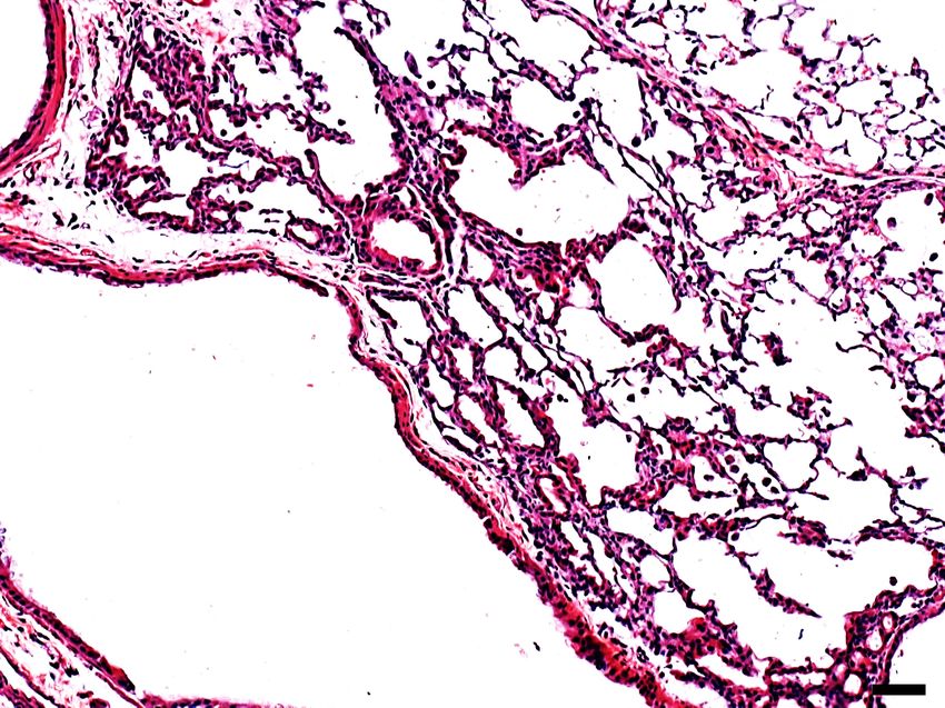
Abstract.

Mutations in the gene (SFTPC) encoding surfactant protein C (SP-C) are associated with interstitial lung

disease in children and adults. To assess the natural history of disease, we knocked-in a familial, disease-

associated SFTPC mutation, L188Q [L184Q (LQ) in mice], into the mouse Sftpc locus. Translation of the

mutant proprotein, proSP-CLQ, exceeded that of proSP-CWT in neonatal alveolar type 2 epithelial (AT2)

cells and was associated with transient activation of oxidative stress and apoptosis leading to impaired

expansion of AT2 cells during postnatal alveolarization. Differentiation of AT2 to AT1 cells was also

inhibited in ex vivo organoid culture of AT2 cells isolated from LQ mice; importantly, treatment with

antioxidant promoted alveolar differentiation. Upon completion of alveolarization, SftpcLQ expression was

downregulated leading to resolution of chronic stress responses; however, the failure to restore AT2 cell

numbers resulted in a permanent loss of AT2 cells that was linked to decreased regenerative capacity in the

adult lung. Collectively, these data support the hypothesis that susceptibility to disease in adult LQ mice is

established during postnatal lung development and provide a potential explanation for the delayed onset of

disease in patients with familial pulmonary fibrosis.

Introduction.

Alveolar type 2 epithelial (AT2) cells synthesize and secrete pulmonary surfactant which is essential for

respiration. Surfactant is a complex mixture of lipids and proteins that forms a bioactive film at the alveolar

air-liquid interface and reduces surface tension at end expiration. The protein components of surfactant,

particularly the hydrophobic peptides surfactant protein B (SP-B) and surfactant protein C (SP-C), promote

the formation and maintenance of a stable surfactant film (1). Expression of SP-B is absolutely required for

adaptation to air breathing at birth and postnatal survival whereas SP-C is dispensable for postnatal lung

function. However, although the role of SP-C in surfactant homeostasis is not well understood, mutations

in the gene encoding SP-C (SFTPC in humans; Sftpc in mice) have been linked to development of interstitial

lung disease.

Human SP-C is synthesized as a 197 amino acid integral membrane precursor protein consisting of a

cytosolic N-terminal domain (residues 1-23) that directs intracellular trafficking of the proprotein, a

hydrophobic membrane spanning domain that comprises most of the biophysically active, secreted mature

peptide (residues 24-58), and a luminal linker region (residues 59-89) followed by the C-terminal

BRICHOS domain (residues 90-197). The luminal BRICHOS domain functions as an intramolecular

chaperone that promotes proper folding and insertion of the transmembrane α-helix (2, 3). The SP-C

proprotein (proSP-C) traverses the secretory pathway from the endoplasmic reticulum (ER) to the

multivesicular body (MVB)/late endosome where Nedd4-2 mediated ubiquitination of a PY motif in the

cytosolic domain allows for internalization of proSP-C from the limiting membrane of the MVB to internal

vesicles, followed by proteolytic removal of the N- and C- terminal domains to generate the mature SP-C

peptide (4, 5). Subsequent fusion of the MVB with a lamellar body (LB) leads to SP-B-mediated

incorporation of vesicles and SP-C into surfactant membranes (lamellae) of the LB. The fully assembled

lipid-protein surfactant complex is stored in the LB until its contents are released into the alveolar airspaces

via exocytosis. SFTPC mutations leading to degradation or mis-trafficking of proSP-C are linked to

pathogenesis in humans.

Monoallelic mutations in the SFTPC gene are associated with development of diffuse parenchymal lung

disease in both children and adults (6). Significant variability exists in the age of onset and clinical

presentation of disease, which can range from non-specific interstitial pneumonitis in infancy (7) to

fulminant pulmonary fibrosis in adults (8). Adults with SFTPC mutations develop fibrotic lung disease as

early as 30 years of age, significantly earlier than patients with non-familial disease. Mutations mapping to

the BRICHOS domain result in formation of a misfolded proprotein leading to ER retention, accumulation

of toxic, misfolded proSP-C, and activation of the ER stress and unfolded protein response (UPR) pathways

(9, 10).

The exon 5+128 T®A BRICHOS mutation, resulting in substitution of a highly conserved leucine (L) by

glutamine (Q) at position 188, is one of the most well-characterized familial SFTPC mutations (11, 12).

The L188Q mutation was identified in a large kindred of over 300 members with a long history of lung

disease and is inherited in an autosomal dominant mode with incomplete penetrance and expressivity (13,

14). Several children in the kindred presented with pneumonitis associated with viral infections at the time

of diagnosis whereas adults presented with usual interstitial pneumonia, the histological correlate of

idiopathic pulmonary fibrosis. In adult transgenic mice, doxycycline-induced expression of L188Q resulted

in activation of ER stress and augmented lung injury associated with AT2 cell apoptosis following

bleomycin challenge (15). L188Q transgenic mice that were also deficient for Chop were protected against

bleomycin-induced injury supporting the hypothesis that AT2 cell loss is associated with dysregulated lung

repair (16). Stress responses observed in transgenic mice were consistent with acute exacerbations

associated with sudden activation of transgene expression and an increase in SP-C proprotein levels.

Consistent with this hypothesis, transgenic overexpression of SftpcΔexon4 (9) or tamoxifen-mediated increase

in SftpcC121G expression (17) resulted in rapid induction of ER stress, apoptosis of AT2 cells and/or lethality

associated with inefficient epithelial regeneration. The severity of the phenotype correlated with expression

levels of the mutant Sftpc allele supporting the hypothesis that misfolded proSP-C exerts a dose-dependent,

cytotoxic effect on AT2 cells.

SFTPC mutations are not typically associated with postnatal lethality and symptomatic disease in

infants/children may be relieved following therapeutic intervention. Since the majority of mouse models

over-expressed or induced acute expression of mutant Sftpc, the observed phenotypes likely model the

phase of disease typified by acute exacerbations. Importantly, ER stress pathways were not activated in cell

lines with constitutive expression of BRICHOS domain mutations suggesting that cells adapted to chronic

stress imposed by misfolded proSP-C (18, 19). In this study we generated a knock-in mouse model of the

L188Q mutation (L184Q in mice) to follow the natural history of disease associated with mutant proSP-C

expression during postnatal alveolarization. We demonstrate that cell stress, arising from a transient

neonatal increase in SP-C proprotein levels, impairs postnatal AT2 cell endowment. Upon completion of

alveolarization, Sftpc expression is downregulated leading to resolution of stress responses; however, the

postnatal loss of AT2 cells significantly compromises the regenerative capacity of the adult lung.

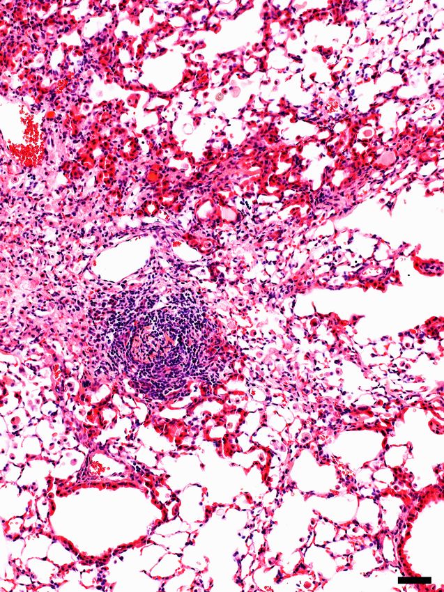
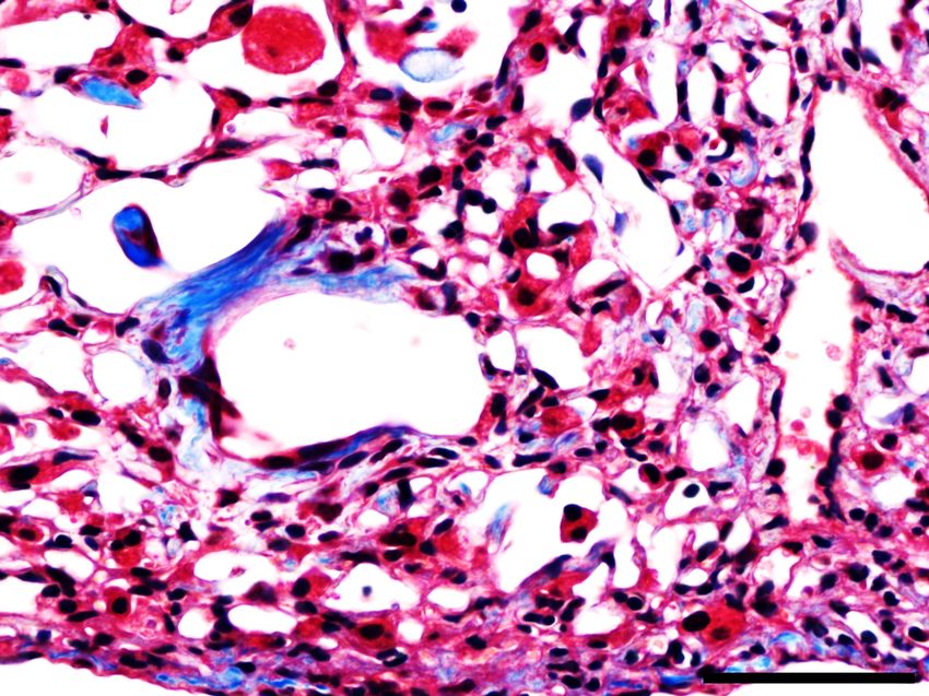
Results.

Constitutive expression of the L184Q allele is associated with impaired lung repair following injury.

A mouse cDNA construct encoding the equivalent of the human exon 5+128 T®A (L188Q) mutation was

knocked into the mouse Sftpc locus in-frame with exon 1, resulting in an amino acid substitution at position

184 (L184Q, LQ) of the mouse SP-C proprotein (mouse proSP-C is 4 amino acids shorter than human

proSP-C) (supplemental figure 1A). The floxed neomycin (neo) resistance cassette was excised by breeding

mice carrying the targeted allele to an EIIA-Cre transgenic line resulting in germline deletion of the cassette

(supplemental figure 1A). No significant changes were observed in Sftpc gene expression in LQ mice with

or without the neo-resistance cassette and all experiments were performed with ‘neo-free’ mice.

Gross lung structure was unremarkable in mice homozygous for the LQ allele (LQ/LQ) up to 18-months of

age in the absence of an exogenous stressor (supplemental figure 1B), suggesting that constitutive

expression of the LQ allele was insufficient to initiate a pathological response. To determine if expression

of the LQ allele impacted the repair capacity of the adult lung, 10-12-week-old WT and LQ/LQ mice were

injured with the fibroproliferative agent bleomycin. Both WT and LQ/LQ mice demonstrated dose-

dependent responses to single bleomycin challenge with an augmented response in LQ/LQ mice compared

to WT mice (supplemental figure 2A). A single high dose of 3U/kg bleomycin resulted in mortality in

LQ/LQ but not WT mice: 67% of LQ/LQ mice (4/6) died in the first 35 days of the study (supplemental

figure 2B). Six months after bleomycin challenge, WT mice demonstrated variable lung repair ranging from

nearly normal lung structure to sustained lung injury; in contrast, surviving LQ/LQ mice demonstrated a 2-

fold increase in lung injury compared to WT (supplemental figure 2 C-D).

To assess lung repair following chronic injury, 10-12-week-old WT and LQ/LQ mice were challenged with

a low dose of bleomycin (1U/kg) once every 2 weeks for 8 weeks and analyzed 1 month after the fourth

and final dose (day 71, figure 1A). On day 71 of the study, bleomycin challenged LQ/LQ mice demonstrated

significant lung injury marked by inflammation (figure 1B arrows & supplemental figure 3A), lymphocytic

aggregates (figure 1B arrowhead), and proximalization of the distal airspaces, as indicated by ectopic

expression of the airway epithelial marker SOX2 (figure 1B boxed region & figure 1C). LQ/LQ mice
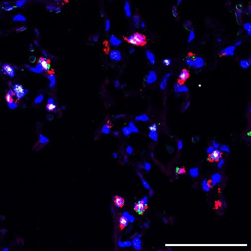
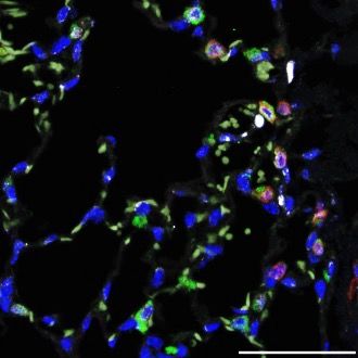
demonstrated a 3.5-fold increase in lung injury compared to WT mice in response to bleomycin challenge

(figure 1D). LQ/LQ mice demonstrated an exacerbated fibrotic response with a 2.8-fold increase in collagen

content measured by second harmonic imaging (figure 1 B & E) and 1.3-fold increase in hydroxyproline

concentration (figure 1F) compared to bleomycin challenged WT mice. Consistent with increased lung

collagen, bleomycin challenged LQ/LQ mice developed restrictive lung disease as indicated by a 35%

decrease in lung compliance (figure 1G) and 1.5-fold increase in lung elastance (supplemental figure 3B);

in contrast, lung function in bleomycin challenged WT mice was similar to their unchallenged counterparts.

Surfactant homeostasis was altered in bleomycin challenged LQ/LQ mice with a 1.5-fold increase in both

total phospholipids and saturated phosphatidylcholine levels in bronchoalveolar lavage fluid compared to

bleomycin challenged WT mice (supplemental figure 3 C-D). Collectively, these data indicate that lung

repair capacity following repeated injury is significantly compromised in LQ/LQ mice and is associated

with the risk of persistent disease.

Constitutive expression of the LQ allele is associated with impaired AT2 cell expansion in the

postnatal lung.

To determine the cause of increased susceptibility of LQ/LQ mice to injury, lung sections from

unchallenged adult LQ/LQ mice were stained with proSP-C, ABCA3 and NKX2.1 to identify AT2 cells,

the site of SP-C expression; antibodies directed to both N- and C-terminal peptides of proSP-C were used

to verify results in LQ/LQ mice. Confocal imaging demonstrated largely peripheral, punctate localization

of proSP-C in WT AT2 cells (figure 2A box) compared to perinuclear proSP-C accumulation in LQ/LQ

AT2 cells (figure 2A box), consistent with retention of proSP-CLQ in the ER. In WT mice, 100% of AT2

cells were NKX2.1+ proSP-C+ and ABCA3+ in adult lung and at all postnatal time points analyzed (figure

2B). Importantly, in LQ/LQ mice, a minor subset of NKX2.1+ ABCA3+ cells with undetectable levels of

proSP-C was observed at P4 (figure 2B). This population of SP-C- AT2 cells increased at P14 and

comprised 55.3±16.6% of total AT2 cells in adult lung; therefore, for morphometric analyses, AT2 cells

were defined as NKX2.1+, proSP-C+ and/or ABCA3+. The frequency of AT2 cells in adult LQ/LQ mice

decreased by 48.4% compared to age-matched WT controls (figure 2C), indicating that impaired lung repair

in adult LQ/LQ mice was associated with an antecedent reduction in AT2 cell numbers.

To determine the onset of reduced AT2 cell frequency, morphometric analyses were performed on lung

sections obtained from WT and LQ/LQ mice during postnatal alveolarization between P4 and P30.

Postnatal expansion of WT AT2 cells occurred between P4 and P21, with the frequency of AT2 cells

increasing 1.3-fold between P4 and P7 followed by a further 1.3-fold increase between P14 and P21 (figure

2D). By P30 the frequency of WT AT2 cells was similar to that in adult lung with an overall increase of

1.9-fold between P4 and P30 (figure 2D). In contrast, no increase in AT2 cell frequency was detected in

LQ/LQ mice between P4 and P14 (figure 2D). LQ/LQ AT2 cells increased by 1.2-fold between P14 and

P21; however, the frequency of AT2 cells at P30 was not significantly different from that at P4, resulting

in an overall 42% decrease compared to WT mice (figure 2D). Notably, the number of nuclei was

comparable between WT and LQ between P4 and P14 and was significantly decreased in LQ/LQ lungs at

P21 and P30, indicating that AT2 cell frequency was not skewed by normalization to frequency of nuclei

(supplemental figure 4A). Overall, constitutive expression of the LQ allele was associated with failure of

AT2 cell expansion during alveolarization.

Postnatal expansion of AT2 cells stems from active proliferation supported by mesenchymal signaling,

which commenced at P4 in WT lungs (supplemental figure 4 B-C). AT2 cell proliferation was maintained

at P7, decreased by 65% at P14 and ceased at P21 (0%) in WT lungs. In contrast, the proliferative index of

LQ/LQ AT2 cells was relatively low at P4 (decreased by 66% compared to WT) and peaked at P7

(supplemental figure 4C). Proliferation of LQ/LQ AT2 cells decreased modestly at P14 and was barely

detectable at P21. Comparable proliferative indices for WT and LQ/LQ AT2 cells between P7 and P21

suggest that both continual cell loss and decreased proliferation at P4 contribute to fewer AT2 cells in

LQ/LQ mice throughout postnatal alveolarization. Collectively, these data indicate that expression of the

LQ allele impairs postnatal AT2 cell expansion resulting in a significant and permanent reduction in AT2

cell numbers in adult mice; further, expression of the mutant allele was associated with delayed onset of

AT2 cell proliferation.
Impaired postnatal AT2 expansion is associated with a transient neonatal increase in proSP-CLQ

protein accumulation.

To determine if altered postnatal expansion of LQ/LQ AT2 cells was associated with changes in proSP-CLQ

expression, western blot analyses were performed on equal numbers of AT2 cells isolated at timepoints

bracketing the postnatal proliferative phase. At P4 SP-C proprotein in LQ/LQ AT2 cells was increased by

2.2-fold compared to WT proSP-C (figure 3 A-B); SP-CLQ was detected predominantly as a single

proprotein band, consistent with trapping in the ER and failure of post-ER modifications including

proteolytic processing. The increase in proSP-CLQ protein was observed as early as P1, where LQ/LQ AT2

cells demonstrated a 2.9-fold increase in proSP-C levels compared to WT cells (supplemental figure 5 A-

B). At P21 proSP-CLQ protein levels in LQ/LQ AT2 cells were unchanged compared to P4; in contrast, SP-

C proprotein in WT AT2 cells increased by 3.3-fold between P4 and P21 resulting in significantly higher

levels compared to LQ/LQ AT2 cells (figure 3 A-B). Processing of proSP-C to its mature peptide was

detected at all timepoints in WT but was inhibited in LQ/LQ AT2 cells (figure 3A and supplemental figure

5A). Importantly, in the absence of a challenge, neither the lack of mature peptide or the loss of AT2 cells

was associated with changes in lung function or surfactant homeostasis in LQ/LQ lungs (control data in

figure 1G & supplemental figure 3 B-D).

Unexpectedly, the neonatal increase in proSP-CLQ protein occurred independently of mRNA levels: Sftpc

expression in LQ/LQ AT2 cells was only 24% of WT levels at P4 (figure 3C). At P21 there was a 2.8-fold

increase in WT Sftpc expression that corresponded with the temporal increase in WT proSP-C protein levels

(figure 3C); however, a similar increase in Sftpc expression was not observed in LQ/LQ AT2 cells (figure

3C). These data indicate a discordance between abundance and subsequent translation of the SftpcLQ

mRNA.

In the absence of elevated Sftpc mRNA levels, increased proSP-CLQ protein could arise from altered

translation and/or degradation of SP-C in the P1-P4 developmental window. To assess synthesis of the SP-

C proprotein, lung slices from P3 mice were metabolically labelled with [35S] methionine/cysteine followed

by immunoprecipitation with an antibody that detects both the proprotein and mature peptide. Even though
Sftpc expression was significantly lower in LQ AT2 cells, levels of newly synthesized proSP-C were higher

in LQ/LQ lung slices compared to WT (figure 3D). The mature peptide was absent from radiolabeled

LQ/LQ samples, consistent with the degradation of misfolded proSP-CLQ and consequent inhibition of

mature peptide generation. To determine if increased synthesis of proSP-CLQ at P4 occurred due to an

increase in overall protein synthesis, isolated AT2 cells were labelled with the amino acid analog L-

azidohomoalanine followed by ligation with biotin alkyne to detect newly synthesized proteins. Western

blot analyses of biotinylated proteins indicated levels of nascent proteins in LQ/LQ AT2 cells were

comparable to WT AT2 cells at both P4 and P21 (supplemental figure 6). These results indicate that global

protein synthesis in P4 LQ/LQ AT2 cells cannot fully account for increased proSP-CLQ synthesis suggesting

additional, and as yet unidentified, post-transcriptional regulation of SftpcLQ; the unique, transient neonatal

increase in proSP-CLQ protein is associated with increased translation of the LQ allele that occurs prior to

impaired postnatal AT2 cell expansion.

Increased neonatal accumulation of proSP-CLQ is associated with activation of oxidative stress.

To identify differences in gene expression and cell-type composition associated with increased proSP-CLQ

translation, single cell RNA sequencing (scRNAseq) was performed on P4 lungs using the 10X genomics

platform. A total of 12,652 single cells were analyzed from P4 lungs (supplemental figure 7A) and

unsupervised clustering revealed 52 cell clusters and 15 major cell types (supplemental figure 7B). A total

of 415 and 540 AT2 cells were identified from WT and LQ/LQ P4 lungs, respectively (figure 4A). AT2

cell-specific differentially expressed genes were identified using a binomial based differential expression

test. Genes expressed higher in WT (n=1032) were functionally associated with lipid metabolism,

principally cholesterol biosynthesis and SREBP signaling (supplemental figure 7C). In contrast, genes

induced in LQ/LQ AT2 cells (n=191) were functionally associated with oxidative stress (particularly

glutathione metabolism), NF-κB signaling, and apoptosis (figure 4B). To determine if the robust oxidative

stress signature was associated with increased levels of reactive oxygen species (ROS), isolated AT2 cells

were incubated with 2′,7′-dichlorofluorescin diacetate (DCFDA). DCFDA is deactelyated by cellular

esterases and subsequently oxidized to the highly fluorogenic 2′,7′-dichlorofluorescein (DCF) by ROS;
fluorescence emitted by DCF is directly proportional to the amount of cellular ROS. P4 LQ/LQ AT2 cells

demonstrated a 1.7-fold increase in ROS levels compared to WT AT2 cells at baseline (figure 4C).

Furthermore, LQ/LQ AT2 cells were exquisitely sensitive to addition of tert-Butyl hydroperoxide (TBHP),

an inducer of oxidative stress, resulting in a 2-fold increase in ROS levels compared to TBHP-treated WT

AT2 cells (figure 4C). Sensitivity to TBHP treatment was maintained in P21 LQ/LQ AT2 cells, with a 2-

fold increase in ROS levels compared to WT AT2 cells (supplemental figure 7D); however, no significant

differences in ROS levels were observed at baseline for P21 WT and LQ/LQ AT2 cells. Consistent with

increased oxidative stress, P4 LQ/LQ AT2 cells demonstrated a 3.2- and 9.1-fold increase in GSH and

GSSG levels, respectively (figure 4D), decreasing the GSH/GSSG ratio by 66% in LQ/LQ AT2 cells

compared to WT. The increase in GSH and GSSG levels was maintained in LQ/LQ AT2 cells at P21 with

a 4-fold and 2.8-fold increase in GSH and GSSG levels, respectively, compared to WT (supplemental figure

7E); however, GSH/GSSG ratio in LQ/LQ AT2 cells was comparable to that of WT at P21, consistent with

resolution of oxidative stress. Overall, increased neonatal translation of proSP-CLQ protein was associated

with a strong oxidative stress response that transiently altered redox balance in postnatal AT2 cells.

Oxidative stress impacts AT2 cell survival and differentiation.

Augmented production of ROS and imbalanced redox states are associated with activation of apoptosis.

Single cell explorer (20), a recently developed algorithm for scRNAseq analysis and visualization of

enriched bioprocess and signaling pathways on t-SNE or UMAP plots, identified a strong stress and cell

death signature in the P4 LQ/LQ AT2 cell cluster, including the ER associated degradation (ERAD),

unfolded protein response (UPR), proteasome, and apoptosis pathways compared to the WT AT2 cell

cluster (supplemental figure 8). Quantitative PCR demonstrated a 1.8-fold increase in the expression of the

pro-apoptotic gene Bax in P4 LQ/LQ AT2 cells that resolved to WT levels by P21 (figure 5A). Western

blotting of isolated AT2 cells demonstrated a 1.9-fold increase in BAX protein levels in P4 LQ/LQ AT2

cells compared to WT (figure 5 B-C). Although BAX protein decreased by 45% in P21 LQ/LQ AT2 cells

compared to P4, levels remained elevated by 1.3-fold compared to P21 WT suggesting that apoptotic

signaling may be sustained.
To determine if oxidative stress negatively impacted AT2 cell differentiation, ex vivo organoids were

generated from AT2 cells isolated from P4 lungs co-cultured with adult mouse lung fibroblasts (figure 5D).

Consistent with previous studies (21), P4 WT AT2 cells differentiated into AT1 cells in organoid cultures;

however, organoids generated from P4 LQ/LQ AT2 cells demonstrated relatively little expression of AT1

markers, HOPX and AGER (figure 5D). Importantly, treatment of LQ/LQ organoids with the antioxidant

butylated hydroxyanisole (BHA) resulted in a 7.4-fold and 4.8-fold increase in AGER+ and HOPX+ cells,

respectively (figure 5 E-F). Expression of proSP-C increased by 3.4-fold in BHA-treated LQ/LQ organoids

(figure 5G). Notably, number and size of organoids generated from WT and LQ/LQ AT2 cells remained

unchanged with or without BHA treatment (supplemental figure 9 A-C). Collectively, these results suggest

that oxidative stress arising from a transient neonatal increase in proSP-CLQ protein levels inhibits postnatal

survival, expansion and differentiation of AT2 cells. Importantly, expression of proximal epithelial marker,

CCSP, remained unchanged with BHA treatment (figure 5H) indicating that antioxidant treatment of

LQ/LQ organoids selectively restored key aspects of distal alveolar epithelial cell maturation.

AT2 cell stress is associated with impaired macrophage development and M2 polarization.

To determine if activation of apoptotic signaling in AT2 cells at P4 impacted development and

differentiation of other cell types during postnatal alveolarization, scRNAseq was performed on P21 lungs

using the 10X genomics platform. A total of 15,218 cells and 14 major cell clusters were identified, which

included 1,229 WT and 443 LQ/LQ AT2 cells. scRNAseq datasets obtained from P4 and P21 lungs were

integrated to analyze potential temporal changes in cell-type composition and gene expressions

(supplemental figure 10A); analyses were focused to immune cells, particularly macrophages

(supplemental figure 10 B-D), since the development and maturation of alveolar macrophages occurs

coincident to AT2 cell expansion during postnatal alveolarization (22, 23). Gene expression analyses of

markers associated with the alveolar and interstitial macrophage subsets demonstrated reduced expression

in P4 LQ/LQ lungs compared to WT; expression of marker genes resolved to WT levels in P21 LQ/LQ

lungs (supplemental figure 10E). Restoration of marker expression was observed as early as P14, where

flow cytometric analyses demonstrated comparable numbers of alveolar macrophages (supplemental figure
11A). Consistent with scRNAseq analyses, comparable expression of CD163 and CD206 (encoded by

MRC1) was identified in alveolar macrophages from P14 LQ/LQ mice, compared to WT mice. Finally, the

frequency of Arginase1+ alveolar macrophages was increased by 5-fold and 7.1-fold in the lungs and BALF

of P14 LQ/LQ mice, respectively, compared to WT mice (supplemental figure 11 B-C), suggesting

alternative activation of macrophages and induction of an anti-inflammatory response. Collectively, these

data suggest that oxidative stress in neonatal AT2 cells impacts crosstalk with the immune cells resulting

in suppression of macrophage development; resolution of development occurs concurrent with M2

polarization of alveolar macrophages.

Chronic stress promotes adaptation in P21 LQ/LQ AT2 cells.

We hypothesized that activation of stress responses at P4 promoted adaptation, thereby allowing for

survival of LQ/LQ AT2 cells at and after P21. Consistent with the scRNAseq dataset, proteomic analyses

of isolated P4 AT2 cells confirmed activation of a robust stress signature marked by an abundance of

proteins associated with catabolism and, in particular, the proteasome pathway (n=726 proteins) in P4

LQ/LQ AT2 cells (figure 6A & supplemental figure 12A); in contrast, proteins associated with metabolic

processes (n= 391 proteins) were more abundant in P4 WT AT2 cells (supplemental figure 12B). Consistent

with an adaptive response, analyses of the scRNAseq dataset at P21 revealed minimal differences between

the WT and LQ/LQ AT2 population, indicating that the P21 LQ/LQ AT2 cells shared a similar

transcriptome to that of WT AT2 cells. Furthermore, transcriptomic similarity was observed between P21

LQ/LQ AT2 cells and P4 WT AT2 cells (figure 6B), suggesting delayed maturation of LQ/LQ AT2 cells.

P21 LQ/LQ AT2 cell population was characterized by genes (n=723) primarily associated with chromatin

and RNA modification (figure 6C); in addition, P21 LQ/LQ and P4 WT AT2 cells were commonly

characterized by genes (n=710) associated with chromatin modification, PI3 kinase, PDGF and FOXO

signaling pathways and lipid biosynthesis compared to P4 LQ/LQ AT2 cells (figure 6D).

To confirm resolution of stress responses and adaptation in LQ/LQ AT2 cells, western blot analyses of

select stress markers were performed in AT2 cells isolated at P4 and P21. Given the strong ERAD signature

observed by scRNAseq of P4 LQ/LQ AT2 cells (supplemental figure 8), markers associated with the UPR,
and proteasome pathways were chosen for validation. Western blot analyses of the master ER chaperone

BiP indicated a 1.8-fold increase in P4 LQ/LQ AT2 cells compared to WT (figure 7A & supplemental

figure 13A), confirming activation of ER stress. Importantly, BiP levels were similar in P1 WT and LQ/LQ

AT2 cells (supplemental figure 5 A & C), indicating that further activation of UPR occurred after this

timepoint. A significant increase in spliced Xbp1 (sXbp1) expression in P4 LQ/LQ AT2 cells compared to

WT cells confirmed activation of the IRE1a branch of the UPR (figure 7B). Western blot analyses also

demonstrated a 2.7- and 2.3-fold increase in phosphorylated eIF2α and GADD34 levels, respectively, in

LQ/LQ AT2 cells compared to WT AT2 cells at P4, consistent with activation of the PERK branch of the

UPR and the integrated stress response (ISR) (figure 7 C-D & supplemental figure 13 B-C). LQ/LQ AT2

cells demonstrated augmented expression of proteasome subunits (figure 7 E-F & supplemental figure

13D), validating activation of the ERAD pathway and proteasome-mediated clearance of the misfolded

proSP-C at P4.

By P21 there were significantly fewer AT2 cells in LQ/LQ lungs (figure 2D); further, more than half of

surviving LQ/LQ AT2 expressed little or no detectable SP-C whereas all WT AT2 robustly expressed SP-

C (figure 2B). These results suggest that adaptation involves loss of SP-C expressing AT2 cells as well as

diminished translation and/or increased degradation of mutant proprotein. Consistent with enhanced

clearance of misfolded SP-C, there was a significant increase in the expression of proteasome subunits

RPT5 and α (1-7), compared to both P4 LQ/LQ and P21 WT AT2 cells (figure 7 E-F & supplemental figure

13D). Importantly, GADD34 levels remained elevated at P21 (figure 7D & supplemental figure 13C),

consistent with sustained PERK activation and delayed recovery of general translation in cells that survive

chronic ER stress (24). BiP and peIF2α levels remained elevated at P21 but were now similar in WT and

LQ/LQ AT2 (figure 7 A & C & supplemental figure 13 A-B), likely related to increased surfactant lipid

synthesis associated with AT2 cell maturation. Interestingly, sXbp1 expression decreased by 30% in P21

LQ/LQ AT2 cells compared to P4 LQ/LQ AT2 cells (figure 7B), consistent with a switch to an adaptive

response where IRE1a signaling was attenuated during persistent ER stress (25). Collectively, these results
suggest that both increased degradation and decreased translation may contribute to diminished proSP-C

load in LQ/LQ AT2 cells at P21. The varied temporal course of activation of the PERK and IRE1a branches

of the UPR influences the fate of AT2 cells in the face of chronic ER stress; a subpopulation of LQ/LQ

AT2 cells emerge at P21 that survive and adapt to the stress imposed by expression of mutant proSP-CLQ.
Discussion.
Constitutive expression of a disease-associated allele encoding a misfolded form of proSP-C (L184Q)

resulted in activation of oxidative stress that was linked to a transient increase in Sftpc mRNA translation

in neonatal mice. Subsequent induction of multiple cell stress pathways was associated with impaired

expansion of AT2 cells during postnatal alveolarization resulting in a permanent loss of AT2 cells and,

ultimately, decreased regenerative capacity in the adult lung.

Expression of allele variants encoding SP-C have been linked to cytotoxicity and pathogenesis in vivo.

Prenatal expression of a Δexon4 SFTPC transgene (9) or the C121G Sftpc allele (17) resulted in AT2 cell

death, disrupted lung morphogenesis and neonatal lethality. These mouse models confirmed in vitro

findings of mutant SP-C toxicity but failed to model the human disease, which is not typically associated

with postnatal lethality. Acute induction of expression of the I73T (26) or C121G Sftpc allele in adult lung

resulted in interstitial lung disease marked by alveolitis, aberrant remodeling and restrictive lung disease.

These mouse models strongly linked expression of the mutant Sftpc allele to development of fibrotic lung

disease and model the acute exacerbation phase of disease. Further, acute induction of an L188Q SFTPC

transgene in adult lung was associated with decreased repair capacity following lung injury. In the current

study we confirm that constitutive (as opposed to induced) expression of the L184Q Sftpc allele is also

associated with impaired lung regeneration and link the adult phenotype to permanent loss of AT2 cells

during postnatal alveolarization. The current mouse model suggests that adult susceptibility to injury is

established in the postnatal period and provides a potential explanation for the delayed onset of disease in

patients with familial pulmonary fibrosis.

Sftpc mRNA in LQ/LQ mice was expressed at one fourth of WT levels in both postnatal and adult AT2

cells. Similarly, in all mouse models to date, mutant Sftpc alleles were consistently expressed at lower

levels than WT Sftpc despite significant differences in targeting strategies and construct design (15, 17, 26).

These results suggest that mutations in this locus might suppress Sftpc expression, although the mechanism

underlying putative allele suppression and whether allele suppression occurs in human patients with SFTPC

mutations are unknown. Importantly, there was significant discordance between Sftpc mRNA and protein
levels in postnatal LQ/LQ AT2 cells, such that misfolded proSP-CLQ accumulated to significantly higher

levels than in WT AT2 cells; this observation is consistent with increasing findings that mRNA abundance

is a poor predictor of protein abundance and that translational control is a critical step in the regulation of

post-transcriptional gene expression (27).

Consistent with perinatal maturation of the surfactant system, AT2 and AT1/AT2 bipotent cells experience

transient lipid stress and UPR activation at P1 (28); cell stress in this critical developmental window is

further augmented by expression of the LQ allele. Increased neonatal synthesis of mutant SP-C proprotein

resulted in activation of multiple stress pathways, including ER stress and UPR, oxidative stress and the

ISR, coincident with commencement of postnatal alveolarization at P4. Resolution of stress responses

towards the culmination of alveolarization was accompanied by decreased frequency of surviving AT2 cells

which, in turn, was associated with impaired lung repair in adult LQ/LQ mice. Overall, these data are

consistent with the hypothesis that stress arising from postnatal expression of the LQ allele, superimposed

on stress associated with normal AT2 cell maturation, leads to a transient increase in proSP-CLQ synthesis

and permanent loss of AT2 cells. Whether these cell stress events are unique to the developing lung or are

recapitulated during lung regeneration remains an important and unanswered question.

Increased neonatal synthesis of proSP-CLQ was associated with generation of ROS and altered glutathione

levels. Previous studies have demonstrated that defective folding of proteins in the ER lumen resulted in

activation of the UPR and oxidative stress leading to apoptosis (29, 30). Dysregulated translation in mice

with disrupted eIF2a phosphorylation resulted in increased ROS generation and b-cell failure (31).

Furthermore, increased protein synthesis downstream of the eIF2a pathway was identified as a critical step

required for recovery of cells from acute stress (32, 33); however, increased protein synthesis without

restoration of homeostasis resulted in generation of ROS. In the present study, global protein synthesis was

comparable between LQ/LQ and WT AT2 cells and increased translation of proSP-CLQ was associated with

oxidative stress and apoptosis. Given that proSP-C is an extraordinarily hydrophobic and highly abundant

secretory protein, increased translation of the misfolded proprotein likely rapidly overwhelms the ER
protein folding and ERAD machinery in AT2 cells. Importantly, all AT2 cells in LQ/LQ mice expressed

high levels of SP-C proprotein at P4; in contrast, there was a significant increase in the frequency of proSP-

C- ABCA3+ AT2 cells at P21 and beyond, with resolution of oxidative stress and normalization of

GSH/GSSG ratio at P21 in LQ/LQ AT2 cells. These results identify the temporal emergence of distinct

AT2 cell populations in which a subpopulation suppress proSP-CLQ expression (proSP-Clow) and a separate

subpopulation maintains high levels of proSP-CLQ (proSP-Chi). We propose that proSP-Chi AT2 cells fail

to adapt to chronic stress resulting from constitutive SftpcLQ expression and are eventually eliminated by

apoptosis.

Surviving proSP-Clow AT2 cells were characterized by genes associated with lipid biosynthetic pathways,

suggesting that adaptation to elevated postnatal cell stress promoted AT2 cell maturation and suppression

of proSP-CLQ expression. Similar adaptation to chronic ER stress involving an increase in lipid synthesis

and metabolism was recently reported in thyrocytes expressing misfolded thyroglobulin (34). Oxidative

and ER stress are known to promote formation of stress granules and p-bodies mediated in part by

phosphorylation of eIF2a (35); stress granules sequester untranslated mRNA and are involved in mRNA

storage and degradation (36). A recent study demonstrated the coordinated involvement of VCP/p97 and

the proteasome in ribosome release and partitioning of mRNA into stress granules under conditions of stress

(37); whether similar stress granules form and sequester SftpcLQ in P21 LQ/LQ AT2 cells remains an

important unanswered question.

Unresolved ER and oxidative stress disrupt cellular physiology and survival (38). Notably, ER stress

associated with insulin mutations resulted in reduced b-cell mass with no apparent apoptosis of b-cells (39,

40); exposure to ER stress during the postnatal period considerably reduced expansion, differentiation and

functional maturation of b-cells in the Akita mouse model (40). LQ/LQ AT2 cells demonstrated a strong

apoptosis signature by scRNAseq analyses and decreased proliferation at P4. Although proliferation

normalized in LQ/LQ AT2 cells by P7, reduced frequency of AT2 cells between P7 and P21 indicate that

proliferation was insufficient to compensate for AT2 cell loss. Expression of a pro-apoptotic marker was
elevated in LQ/LQ AT2 cells; however, we were unable to identify apoptotic AT2 cells in LQ/LQ lungs

likely due to rapid clearance of dead cells by macrophages.

Limited AT2 to AT1 cell differentiation was observed in organoids derived from P4 LQ/LQ lungs,

consistent with the scRNAseq data which demonstrated that processes associated with AT2 cell maturation

and development were underrepresented in P4 LQ/LQ AT2 cells compared to WT. Antioxidant treatment

of LQ/LQ organoids restored significant AT1 differentiation and AT2 maturation, suggesting that oxidative

stress impairs alveolar epithelial differentiation. The precise mechanisms underlying defective

differentiation in P4 LQ/LQ organoids and whether differentiation is similarly impaired in adult LQ/LQ

AT2 cells merit further investigation.

L184Q mice provide a physiological model of chronic stress response with temporally distinct molecular

and cellular phenotypes directly proportional to misfolded proSP-CLQ levels and activation of oxidative

stress. Interestingly, neonatal mice challenged with hyperoxia between P0 and P4 demonstrated a 50%

reduction in Sftpc+ AT2 cells in adulthood with minimal effect on alveolar homeostasis similar to L184Q

mice (41-43). Further, lung regeneration was significantly impaired in response to bleomycin challenge

(42) or IAV infection (44) resulting in enhanced recruitment of airway progenitors. Collectively, these

results support the hypothesis that neonatal exposure to intrinsic or extrinsic oxidative stress impairs

postnatal AT2 cell expansion leading to permanent loss of AT2 cells and dysfunctional lung repair in adult

mice. These findings raise the intriguing possibility that postnatal antioxidant therapy may restore AT2

endowment and enhance lung repair in patients carrying BRICHOS mutations.
Methods.

Mice.

The 551 TàA point mutation (L184Q substitution) was generated by site-directed mutagenesis

(QuikChange, Stratagene) of the mouse Surfactant Protein C (Sftpc) cDNA. Three stop codons, bovine

growth hormone polyadenylation signal (BGH-pA), and a 3’ AscI site were subsequently incorporated into

the cassette encoding the cDNA. To generate the 5’ arm, 3314 base pairs of the Sftpc promoter were

amplified from WT ES cell genomic DNA (Taffy SvEvTAC 129 substrain), cloned downstream of a 5’

AscI site and subsequently built onto the mutated cDNA. To generate the 3’ arm, Sftpc sequence from the

beginning of intron 1 to exon 6+3361 nucleotides flanked by NotI sites was cloned from WT ES cell

genomic DNA. The 3’ and 5’ arms were subcloned into the NotI and AscI sites of the OSdupdel2 vector (a

gift of Dr. Oliver Smithies, University of North Carolina, Chapel Hill), respectively. MLE12 cells were

transfected with the complete construct and Sftpc mRNA levels were examined by PCR electrophoresis.

Linearized plasmid DNA (AclI site) was submitted to the University of Cincinnati ES Cell Core for

electroporation. Clones were selected for resistance to neomycin and recombination was verified by loss

of PGK-TK cassette. Positive ES cell clones were injected into C57Bl/6 blastocysts and implanted into

pseudo-pregnant female mice. The resulting chimeras were mated to C57Bl/6 mice to generate founder

mice which were screened for the presence of a single (heterozygous) ‘knock-in’ allele. Subsequently, the

neomycin resistance cassette (neo) was removed in vivo by crossing founder mice to the EIIa-Cre mice

(Jackson Labs). Excision of the cassette was confirmed by PCR using primers P1 (exon 1): TGG ACA

TGA GTA GCA AAG AGG TC, P3 (neomycin cassette): AGT TCT TCT GAG GGG ATC AAT TC and

P4 (intron 1): ATC CTA AAA GCC CAA TCC TAA GC. F1 ‘neo-free’ progeny were back-crossed to

C57Bl/6n mice (Charles River) to remove the EIIA-Cre cassette. Genotyping of ‘neo-free’ mice was

performed with primers P1, P4 and P2-625 (BGH-pA): CCA TCT GTT GTT TGC CCC TC. Mice were

backcrossed for 9 generations to C57Bl6/n (Charles River) and all experiments were performed with

animals in the C57Bl6/n genetic background.

Bleomcyin challenge, hydroxyproline measurements and lung mechanics.
Mice were lightly anesthetized with isoflurane and 50 µl of 1U/kg, 2U/kg or 3U/kg bleomycin sulfate (15U,

Hospira NDC 61703-332-18) or vehicle (50 µl of 0.9% saline) administered via oropharyngeal aspiration

using a micropipette. Mice were hung vertically by the upper two incisors on a horizontally extended string,

the tongue was extended, and liquid was placed onto the distal part of the oropharynx while the nose was

gently closed. After 2-3 deep inhalations, mice were placed in the cage for recovery and monitored. At the

end of the study, mice were euthanized, and left lung was inflated with paraformaldehyde as described

below and right lung lobes were partitioned for biochemical analyses (right upper and accessory lobe was

frozen for western blotting, right middle lobe was hydrolyzed for hydroxyproline assay, and right lower

lobe was frozen for RNA). Total lung collagen was measured by assaying lung hydroxyproline content after

hydrolysis of right middle lung lobe with 6 N HCl exactly as previously described (45). Lung mechanics

were assessed on anesthetized mice using a computerized flexiVent system (SCIREQ) exactly as previously

described (45).

Histology, immunofluorescence, and second harmonic imaging.

Lungs were inflated with 4% paraformaldehyde under 25 cm pressure and immersed in the same fixative

overnight at 4°C. Right lung lobes and left lung were sub-dissected and embedded in paraffin after

dehydration in an ethanol series. Lung pieces were sectioned at 5 µm using a Leica RM2235 microtome for

hematoxylin and eosin (H&E) staining. Images in supplemental figure 1 were obtained at 10X with Zeiss

Axio A2 microscope equipped with an AxioCam MRc5 camera. Tile scans of H&E-stained sections were

obtained at 4X magnification using a Nikon NiE upright microscope system. Total area of the lung lobe

and area of bleomycin injured lung were calculated using Nikon Elements by drawing a user-defined region

of interest (ROI). Percentage of lung injured was defined as: (injured ROI/lung ROI) x 100. For

immunofluorescence analyses, standard procedures were followed, and sections were stained with primary

antibodies (supplemental table 1) with sodium citrate antigen retrieval as required. All Alexa-fluor

conjugated secondary antibodies (supplemental table 2) were used at a dilution of 1:200. High

magnification images were taken at 60X with Nyquist magnification or 100X using a Nikon A1 LUNA
inverted microscope. For second harmonic imaging, 5 µm thick paraffin sections were dewaxed in xylene,

rehydrated in an ethanol gradient series and placed in PBS. Tile scans at 10X magnification (air objective,

plan apochromat) were obtained using Nikon A1R multiphoton upright microscope. Spectral unmixing was

performed to separate the second harmonic signal for collagen and auto fluorescence of the lung tissue.

Threshold function on general analysis program (Nikon Elements) was used to determine the area of signals

from second harmonic and auto-fluorescence channels.

Morphometric analyses.

Morphometric analyses for adult mice (P30 and older) were performed exactly as previously described (46).

For neonatal mice, 5-8 random fields of immunofluorescent stained lung sections were imaged at 60X with

Nyquist magnification or 100X using a Nikon A1 LUNA inverted microscope. Images contained at least

eight individual 1 μm optical sections. A binary channel for DAPI was created using general analysis

program on Nikon Elements to obtain clear nuclear boundaries between cells in clusters. Processed images

with a ‘DAPI binary’ channel were rendered in 3D and cells were manually counted on Imaris (Bitplane

version 9.3.0). The binary channel for DAPI was masked with a surface and object was split with seed

points (diameter: P1 to P4= 3.2 µm, P7= 3.5 µm, P14= 3.8 µm and P21= 4 µm) to determine total nuclei

per image field. A total of 1200-1500, 500-700, 400-650, 200-350, 180-300 nuclei per mouse were counted

for P1, P4, P7, P14 and P21 lungs, respectively. Orthogonal projections of confocal z-stacks were analyzed

manually to verify Ki67 positivity of AT2 cells.

Alveolar type 2 epithelial (AT2) cell isolation and organoid assay.

Lungs were inflated with 1 ml, 2 ml and 2.5 ml dispase (BD Biosciences 354235) for P4 pups, P21 pups

and adult mice, respectively. Lungs were perfused with 0.9% saline prior to dispase instillation only in adult

mice. Single cell suspensions were prepared in C-tubes (Miltenyi Biotec 130-096-334) using the

gentleMACS dissociator (Miltenyi Biotec) with 120 U/ml DNaseI (Sigma D4527). Suspensions were

filtered through 40 µm strainers and incubated in RBC lysis buffer (BioLegend 420301) for 4 minutes at

4°C (RBC lysis was performed only for P4 and P21 cell suspensions). Cells were washed with complete
RPMI (with 10% FBS) and resuspended in MACS buffer (1X PBS+2 mM EDTA+0.05% BSA) and

incubated with the following anti-mouse biotinylated antibodies: CD45 (BioLegend 103104, clone 30-F11),

CD16/32 (BD Pharmingen 553143, clone 2.462), CD31 (BioLegend 102503, clone MEC13.3), CD90.2

(BioLegend 105304, clone 30-H12) and Ter119 (BioLegend 116203, clone Ter119). Suspensions were

subsequently incubated with anti-biotin microbeads (Miltenyi Biotec 130-097-046) and purified over a LS

column (Miltenyi Biotec 130-042-401) attached to a QuadroMACS separator (Miltenyi Biotec). Eluates

from the column (CD45- CD16/32- CD31- CD90- Ter119-) were stored at -80°C as a dry pellet for western

blot analysis or processed immediately for RNA isolation and biochemical analyses. MACS isolation

resulted in AT2 cell yields in the range of 5-8 x 10⁶ cells/mouse with a purity of 80-90% (determined by

flow cytometry).

For generation of organoids, WT mixed background mice (n=6) were subjected to pneumonectomy and

fibroblasts were harvested 5 days post-surgery (47). Single cell suspensions were prepared with dispase and

DNaseI as above. Cell suspensions were incubated with Fc receptor block in MACS buffer for 10 minutes

followed by incubation with PDGFRa (CD140a+) microbeads (Miltenyi Biotec 130-101-502) for 15

minutes. Cells were purified over LS columns attached to a QuadroMACS magnet. CD140a+ fibroblasts

were co-cultured with P4 AT2 cells in a 10:1 ratio, combined with matrigel (Trevigen 3445-005-01) in a

1:1 ratio and seeded on an air liquid interface transwell system in a 24-well plate (Corning 3495). The co-

cultures were incubated in MTEC plus media [DMEM-Ham’s F-12 (Invitrogen 11330-032), HEPES,

penicillin & streptomycin, fungizone, insulin (Sigma I-6634), transferrin (Sigma T1147), cholera toxin

(Sigma C8052), EGF (Sigma E4127) and bovine pituitary extract (Invitrogen 13028-014)] for 21 days.

Rock inhibitor Y27632 (Enzo ALX-270-333) was included in the medium for the first two days (48). ROCK

inhibitor was removed after 48 hours of culture and media was supplemented with or without 10 µM BHA

(Sigma B1253). Medium was changed every 48 hours and fresh BHA was included at each change.

Organoids were cultured for 3 weeks, fixed in 4% PFA and processed for immunofluorescence analysis.
Images were obtained on a Nikon A1R inverted microscope and analysis of differentiation markers was

completed using general analysis program on Nikon Elements.

Characterization of organoid size.

Organoid formation was quantified for both number and size using the Cytation 5 primary cellular analysis

function. The parameters for each brightfield image were set as follows: channel- brightfield; threshold-

auto; background- light; minimum object size- 20 µm, maximum object size- 1000 µm, fill holes in mask,

include primary edge objects, split touching objects. Size and circularity criteria were used to eliminate

non-spheroidal objects in addition to objects smaller than the minimal size. Every image that was collected

for each transwell of organoid culture was run through the analysis program with set parameters. The

number of organoids between 20 µm-500 µm as well as 500 µm-1000 µm for each treatment group was

input into Prism for further statistical analysis.

Metabolic labelling, SDS PAGE and western blotting.

One mm thick lung slices were incubated for 1 hour in methionine- and cysteine-free DMEM (Sigma

21013-024) supplemented with 5% dialyzed FBS (Gibco 26400). Slices were pulse-labeled with 0.5 mCi/ml

[35S] methionine/cysteine (Perkin Elmer NEG772002MC) for 30 minutes. Labelled slices were

homogenized in 190 mM NaCl, 6 mM EDTA, 60 mM Tris, pH 7.4, 4% SDS, proteins precipitated by

trichloroacetic acid and incorporated [35S] counts per minute (cpm) determined with a liquid scintillation

counter (Perkin Elmer Tricarb 2910TR). Samples containing equal cpm were immunoprecipitated with 5

µl of rabbit mature SP-C antibody (Seven Hills Bioreagents 76694) immobilized on 30 µl of pre-cleared

recombinant Sepahrose G beads (Invitrogen 101242) overnight. Beads were washed 4 times with 150 mM

NaCl, 50 mM Tris, pH 7.5, 5 mM EDTA, 0.1% Triton X-100, 0.02% SDS, followed by 2 washes with 150

mM NaCl, 50 mM Tris, pH 7.5, 5 mM EDTA and resuspended in 6X Laemmli sample buffer containing

BME for SDS-PAGE. Gels were fixed in methanol/acetic acid, dried and exposed to a phosphor screen

which was imaged at various intervals to detect radioactivity using a Typhoon FLA 9500 system (GE

Healthcare).
For SDS-PAGE/western blotting, AT2 cells were harvested and lysed by sonication at a concentration of

1×10⁶ cells/80 µl in 1X PBS containing 1% mammalian protease inhibitor cocktail (Sigma Aldrich P8340)

and 1X PhosSTOP (Sigma Aldrich 4906837001). Protein concentration in the supernatants was assessed

with the Pierce Micro BCA kit (Thermo Fisher Scientific 23235). Equal amounts of protein were separated

on 10-20% tris-tricine (Thermo Fisher Scientific EC66252BOX, figure 3A & supplemental figures 5A, 6

13A & 13D) or 10-20% tris-glycine gels (Thermo Fisher Scientific XP10202BOX, figure 3D &

supplemental figure 11 B-C), under reducing electrophoretic conditions at 125V for 1.5 hours and

transferred to 0.1 µm nitrocellulose membranes (GE Amersham 10600000) at 180 mA for 1 hour using a

semi-dry apparatus. Membranes were blocked in 5% non-fat dry milk and subsequently incubated with

primary antibodies (supplemental table 1) overnight at 4°C. Membranes were subsequently washed with

1X TBS containing 0.05-0.1% Tween-20, incubated with the appropriate HRP-conjugated secondary

antibodies (supplemental table 2), developed using Immobilon forte Western HRP substrate (EMD

Millipore WBLUF0100) or Pierce SuperSignal West Pico PLUS Chemiluminescent Substrate (Thermo

Scientific 34580) and analyzed on a ChemiDoc Touch Imaging System (BioRad). Membranes were

stripped with Restore Western blot stripping buffer (Thermo Fisher Scientific 21063) prior to re-probing.

Total protein normalization was performed using the ImageLab software (BioRad) with gels stained with

Expedeon instant blue (Sigma ISB1L) after transfer.

RNA isolation and quantitative real time PCR.

Total RNA was isolated from 1-2×10⁶ AT2 cells using the Quick-RNA miniprep kit (Zymo Research

R1054), and cDNA was synthesized using iScript cDNA Synthesis Kit (BioRad 1708891). Quantitative

RT-PCR was performed with 20 ng of cDNA per reaction on QuantStudio 3 (Thermo Fisher Scientific)

with Taqman assays (Integrated DNA Technologies) for mouse Sftpc (IDT Mm.PT.58.9922071, exon 1-2),

and Bax (IDT Mm.PT.58.14012210, exon 3-4). Multiple housekeeping genes were used to identify stable

expression across developmental ages (P4 and P21) and data were normalized to mouse Ppia (IDT

Mm.PT.39a.2.gs).
Single cell RNA sequencing (scRNAseq) and data analysis.

P4 and P21 lungs were inflated with 1 ml dispase, resected and incubated in dispase for 6 minutes at 37°C.

Lung lobes were separated and placed in C-tubes containing 5 ml RPMI (Gibco 11875-093) supplemented

with 10% FBS (Sigma F4135), 1M HEPES and 1% Penicillin/Streptomycin. Single cell suspensions were

prepared using the gentleMACS dissociator with 120 U/ml DNaseI and filtered through a 40 µm strainer,

washed with media and incubated in RBC lysis buffer for 4 minutes at 4°C. Suspensions were washed with

media, cell counts were performed and cells were resuspended in RPMI at a final concentration of 100

cells/µl. GEM generation and barcoding, and cDNA library preparation were completed by the CCHMC

Gene Expression Core using the Chromium Single Cell 3’ Reagent Kit (10X Genomics version 2.0 and

version 3.0). Sequencing was performed on NovaSeq 6000 at a depth of approximately 300-450 million

reads. Read alignment and gene-expression quantification of data were performed using the CellRanger

pipeline (10X Genomics version 2.0.0 and version 3.0.2). Cell Ranger R kit version 2.0.0 (10X Genomics)

was used to process raw sequencing data and paired-end sequence alignment to mouse genome (mm10).

Cells with at least 500 expressed genes (UMI>0) and less than 10% of UMIs mapping to mitochondrial

genes were included for downstream analysis. Genes expressed in at least two cells in each dataset were

included. Datasets were integrated with Harmony (49) and downstream analyses were performed in R

(version 3.5.0) using custom scripts, SINCERA (50, 51), and Seurat (versions 2.3.4) (52, 53). The

expression of a gene in a cell was measured by its UMI counts in the cell normalized by the total number

of UMIs in the cell. Principal-component analysis was performed for dimension reduction. Reduced

dimensions were used for cell cluster identification using the Jaccard-Louvain clustering algorithm (54).

Clusters were mapped to cell types based on the expression of known marker genes. Cluster-specific

differentially expressed genes were identified using a binomial based differential expression test

implemented in the SINCERA pipeline (49, 50). Genes with p value 2 expressed in

>20% of the cells in a given cluster were considered significant and functional enrichment analyses were

performed using Toppfun in ToppGene suite (55). All Venn diagrams were generated on BioVenn (56).
You can also read