Applying grazing incidence X-ray reflectometry (XRR) to characterising nanofilms on mica
←
→
Page content transcription
If your browser does not render page correctly, please read the page content below
Journal of Colloid and Interface Science 306 (2007) 459–463
                                                                                                                              www.elsevier.com/locate/jcis

                                                                            Note

                      Applying grazing incidence X-ray reflectometry (XRR)
                               to characterising nanofilms on mica
          Wuge H. Briscoe a,∗,1 , Meng Chen a , Iain E. Dunlop a,2 , Jacob Klein a,3 , Jeffrey Penfold b ,
                                          Robert M.J. Jacobs a,∗
                  a Physical and Theoretical Chemistry Laboratory, University of Oxford, South Parks Road, Oxford OX1 3QZ, United Kingdom
                                        b Rutherford Appleton Laboratory, Chilton, Didcot OX11 0QX, United Kingdom

                                                    Received 29 September 2006; accepted 15 October 2006
                                                               Available online 21 October 2006

Abstract
   Molecularly smooth mica has hitherto not been widely used as a substrate for the X-ray reflectometry (XRR) technique. That is largely due to
the difficulty of achieving flatness over a sufficiently large area of mica. Here we show that this difficulty can be overcome by slightly bending the
mica substrate over an underlying cylinder; the enhanced rigidity of the bent mica sheet along the axis of the cylinder provides sufficient flatness
along this axis for XRR measurements. To test this method, we have employed it to characterise three types of nanofilms on mica in air: (A) Cr–Au
thin films; (B) a surface-grown zwitterionic polymer brush; and (C) a Langmuir–Blodgett (LB) phospholipid monolayer, using a table-top X-ray
reflectometer. Fitting the obtained reflectivity curves with the standard Parratt algorithm allows us to extract the structural information of the
nanofilms (both thickness and apparent roughness). Our simple method points to how XRR may be exploited as a useful characterisation tool for
nanofilms on mica.
© 2006 Elsevier Inc. All rights reserved.

Keywords: XRR; Thin films; Nanofilms; X-ray reflectometry; Mica; Surface-grown polymer brushes; Surface characterisation

1. Introduction                                                                  available to unravel such structural information, such as ellip-
                                                                                 sometry and Fourier transform infrared spectroscopy (FTIR).
    Polymers and amphiphilic molecules (e.g., surfactants or                     Due to its readily achievable molecular smoothness, mica is
lipids) can be attached onto solid surfaces to form films of                     the preferred model substrate in many techniques such as the
nanometer thickness, as an effective strategy to modify sur-                     surface force apparatus/balance (SFA/B) [1–3] and the atomic
face properties and to mediate desirable surface interactions.                   force microscope (AFM) [4], as well as the electron micro-
The efficacy of such processes hinges on our ability to tai-                     scope (EM) [5]. Techniques to probe the corresponding struc-
lor, and thus on our understanding of, the structures of these                   ture of the mica-surface-adsorbed films would be highly com-
soft nanofilms. There are a range of optical methods that are                    plementary to such force or imaging methods. However, due
                                                                                 to mica’s intrinsic birefringence, the optical characterisation
 * Corresponding authors. Fax: 44 1865 275410.                                   methods routinely employed for other substrates such as sili-
   E-mail addresses: wuge.briscoe@chem.ox.ac.uk, wuge.briscoe@kau.se             con, silica, quartz and sapphire have not been widely applied
(W.H. Briscoe), robert.jacobs@chem.ox.ac.uk (R.M.J. Jacobs).                     to nanofilms on mica [6]. This is partly because reliable data
 1 Visiting scientist at Fakulteten för teknik- och naturvetenskap, Karlstad
                                                                                 is hard to obtain, as the optical axes of the mica substrate prior
University, Universitetsgatan 1, SE65188 Karlstad, Sweden.                       and subsequent to the film deposition must be in exactly the
 2 Current address: Department of New Materials and Biosystems, Max-
                                                                                 same azimuthal directions. Otherwise, any slight change in the
Planck-Institute for Metals Research, Heisenbergstr. 3, 70569 Stuttgart, Ger-
many.                                                                            axis directions due to sample misalignment will significantly
 3 Also at Department of Materials and Interfaces, Weizmann Institute of Sci-    affect the measurement. A second difficulty is that even once
ence, Rehovot 76100, Israel.                                                     reliable data is recorded, the modelling of it, although theoret-
0021-9797/$ – see front matter © 2006 Elsevier Inc. All rights reserved.
doi:10.1016/j.jcis.2006.10.031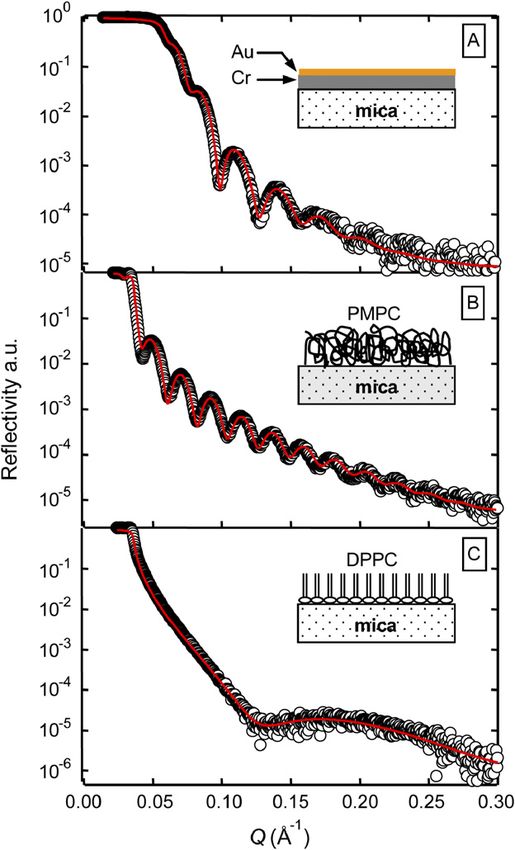
460 W.H. Briscoe et al. / Journal of Colloid and Interface Science 306 (2007) 459–463
ically understood [7,8], is significantly more complex than for
an isotropic material such as silicon. An alternative solution to
this alignment issue in these optical methods would be to study
the film deposition in situ, but the related practical difficulties
have led to the common compromise in some previous stud-
ies, of using, e.g., a separate silicon substrate for ellipsometric
characterisation of thin films while using mica for other types
of analysis [9].
Over the past decades, grazing angle incidence X-ray reflec-
tometry (XRR) [10–19] has been applied effectively to obtain
information about nanofilm structures, and is a promising tech-
nique for mica substrates since its measurement is insensitive
to mica birefringence. However, mica has hitherto been con-
sidered as a non-ideal substrate for the XRR technique [13]
as explained below. In this letter, we report a simple “bending
mica” method which enables the application of XRR to char-
acterising different nanofilms on mica in air, using a table top
X-ray reflectometer. Fitting the X-ray reflectivity curves with a
simple layer-model using the standard Parratt algorithm allows
us to extract the thickness and the apparent roughness of the
nanofilms. Our method indicates how XRR may be extended as
a useful characterisation tool for a wide range of nanofilms on
mica.
2. Materials and methods
In our XRR measurements, which were performed us-
ing a Bruker® D8 reflectometer, a CuKα X-ray beam (flux
∼106 photons/s) of approximately 8 mm by 100 µm in cross
section and wavelength λ = 1.54 Å is incident upon a sample
at a grazing angle θi . This requires the sample to be flat over a
few cm2 , and mica has been traditionally considered non-ideal
for such measurements for two reasons. Firstly, a major diffi-
culty arises from the inability to achieve sufficient flatness—
a cleaved mica sheet undulates over such a large area. This
results in the specular reflection (θi = θr ) being spread over
a range of detector angles (the range of which changes with
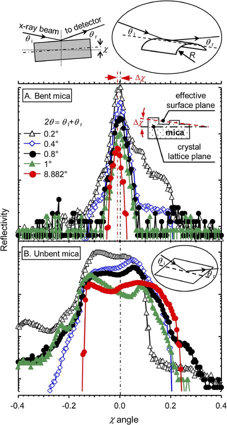
incident angle), making it impossible to interpret the data. Fig. 1. Rocking curves (reflectivity in arbitrary units vs χ angle in degree on
We have overcome this difficulty using a simple method, in a log-liner scale) on bare mica in air using the bending mica method (A) in
which a freshly cleaved mica sheet of thickness h = 30–100 µm contrast to those using unbent mica (B), collected at different fixed total incident
and ∼4 × 4 cm (width b × length L) in size is gently bent and reflection angles, 2θ = θi + θf . The reflectivity for each 2θ is obtained
and clamped onto an underlying cylinder (R ∼ 7.5 cm; the by normalizing the reflection counts with the maximum count; and thus the
maximum reflectivity in each curve is 1 but an offset of 0.1 is added to different
geometry of the set-up is shown schematically at the top of curves sequentially for clarity. The schematics at the top of the figure show the
Fig. 1). The enhanced rigidity of the mica sheet along the axis experimental set-up and the geometry of the bending mica method. The inset
of bending is due to the very large stretching energy along in (A) illustrates schematically the interpretation of the effective surface plane
this axis [20]. The Searle parameter for our mica sheets is of reflection due to the presence of macroscopic domains on the mica surface,
μ = b2 /Rh ∼ (210–710), and detailed analysis in the field which gives rise to a small shift χ in the rocking curve taken at 2θ close to
the Bragg angle.
of elasticity shows that bending deformation for such μ val-
ues falls into the “plate regime” [21], i.e., anticlastic bending
(which would make the sheets bend into a saddle shape) is of Soller slits on the detector side cuts out scattering that is
precluded along the apex of the axis, ensuring the flatness more than 2◦ from the specular plane. The effectiveness of
along this axis. Concomitantly, possible associated curvature this method may be examined from the reflectivity “rocking
effects can be alleviated by confining the detector cross sec- curves” collected while fixing the total of the incident and
tion to 100 µm in height and 300–800 µm in width.4 A set reflection angles (i.e., θi + θr = 2θ ) and rocking the sam-
ple (i.e., varying the angle χ ). Fig. 1A shows examples of
4 Thus, the incident and reflected beams are matched in size (100 µm) in the such rocking curves measured on freshly cleaved bare mica
plane of specular reflection. in air, and the coincidence of the relative reflectivity peaksW.H. Briscoe et al. / Journal of Colloid and Interface Science 306 (2007) 459–463 461
at different 2θ (0.2◦ –1◦ ) indicates that the bent mica sheet is
sufficiently flat to perform the grazing angle incidence XRR
measurements.5 This is in contrast to the poorly defined and
broad peaks obtained on unbent mica shown in Fig. 1B—
it would be impossible to interpret or analyse the XRR data
from such an unbent mica substrate to generate any useful in-
formation.
A second reason why mica has been considered unsuitable
for laboratory-based XRR measurements is that steps or ter-
races of height of order micrometer and lateral size of order
millimeter to centimeter are inevitable over a large area on a
cleaved mica sheet, giving rise to macroscopic domains—this
is the so-called “mosaic effect” [13]. In theory, this may be tol-
erated because the large size of the domains, compared to the
nanometer length scale of the nanofilms of interest, means they
will not contribute coherently to specular reflection. In practice,
however, it is as if the reflection takes place at an effective pla-
nar surface whose position is obtained by averaging over all the
domains illuminated, and this effective plane is not necessarily
parallel to the underlying mica lattices (as shown schematically
in the inset to Fig. 1A). The rocking curve shown in red circles
in Fig. 1A is collected at 2θ = 8.882◦ , where the reflectivity is
dominated by the Bragg diffraction from the underlying mica
crystal lattices. The small discrepancy in the reflectivity peak
positions, χ ∼ 0.01◦ in this case, is interpreted to be the angle
between the effective surface plane of reflection and the crys-
tal lattice plane. Such an interpretation could be further tested
by examining the rocking curves collected at other 2θ angles
(e.g., 2◦ –7◦ ); however, due to the relatively low flux of our
lab-based X-ray source, to obtain reliable reflectivity data at
such high incident angles would have required extremely pro-
longed integration times. It should be noted that the presence Fig. 2. Reflectivity vs momentum transfer Q = (4π/λ) sin θ for three nanofilms
of large numbers of small domains of these terraces could ex- on mica in air, as schematically illustrated in the insets: (A) ∼5 nm Au
aggerate the contribution of diffuse scattering to the specular and ∼20 nm Cr thermally evaporated on plasma treated mica in high vac-
reflection, an effect that could be exacerbated by the curved uum; (B) ∼28 nm surface grown zwitterionic polymer brush pMPC; and (C)
a ∼2.5 nm DPPC phospholipid monolayer Langmuir–Blodgett deposited on
substrate geometry. In implementing our bending mica method, mica. The open circles are the experimental data, and the red curves are com-
we have found it sufficient to avoid using mica sheets with too puted using the fitting parameters listed in Table 1. The corresponding AFM
many small domains as judged by visual observations of re- images for these three nanofilms are shown in Fig. S2 in the supplementary
flected monochromatic light, and to keep the radius of curvature material section.
considerably larger than the beam footprint at the smallest an-
gles studied. 3. Results and discussion
Using this bending mica method, we have carried out proof-
of-principle XRR measurements on three very different sys- Fig. 2 shows reflectivity (open circles) vs Q = (4π/λ) sin θ
tems: (A) a Cr–Au nanofilm thermally evaporated in high vac- for these three nanofilms. Note the intensity fluctuations, known
uum on oxygen plasma-cleaned mica, (B) a surface-grown as Kiessig fringes, that arise from multiple beam interference
zwitterionic polymer brush made from poly(2-methacryloyl- in the presence of the nanofilms. In obtaining these reflectiv-
oxyethyl phosphorylcholine) (pMPC) via atomic transfer rad- ity curves, we have first collected the specular reflection (i.e.,
ical polymerization from mica pre-initiated with an adsorbed θi = θr ) and then subtracted from it the diffuse reflection (i.e.,
random copolymer [22,23] and (C) a Langmuir–Blodgett (LB) θr = θi − θoff ) obtained over the same integration time to ac-
monolayer of the phospholipid 1,2-dipalmitoylphosphatidyl- count for background scattering, with θoff determined from
rocking curves (see Fig. S1 in the supplementary material). The
choline (DPPC). The details for sample preparation and the
total acquisition time for both specular and off specular scans is
AFM images of the samples are described in the supplemen-
typically around 24 h as 2θ varies between 0.1◦ and 5◦ . In ad-
tary material.
dition, area corrections have been made at very small θi (below
the critical edge θc ∼ (2δ)1/2 ) where the illumination area ex-
5 The absence of Kiessig fringes in the diffuse reflectivity curves provides ceeds the sample size, knowing its relative reflectivity must be
further evidence for the substrate flatness. 1 due to total external reflection.462 W.H. Briscoe et al. / Journal of Colloid and Interface Science 306 (2007) 459–463
Table 1
Fitting parameters for different nanofilms using the Parratt algorithm: thickness t , real (dispersion) and imaginary (absorption) parts of the scattering length density
SLD ρr and ρm , and apparent roughness σ
Samples (A) Cr–Au (B) pMPC (C) DPPC
Model 2 layer model 2 layer model 1 layer model
Layers Au Cr Bulk pMPC Macro-initiator Bulk Lipid Bulk
mica layer under-layer mica layer mica
t (Å) 50.2 193.4 260.4 16.0 25.7
ρr (Å−2 ) 8.918e–05 5.922e–05 2.449e–05 1.125e–05 5.958e–06 2.579e–05 9.829e–06 2.539e–05
ρm (Å−2 ) 2.602e–06 7.649e–06 7.293e–07 5.329e–08 2.421e–07 7.368e–07 1.12e–07 9.94e–07
σ (Å) 25.0 13.8 6.0 9.3 1.8 4.4 5.0 5.7
In order to fit the Kiessig fringes in the reflectivity curves ter scale surface roughness. The fitted ρr values for the Au, Cr,
to extract structural information, we have used the simple layer and the pMPC layers are physically reasonable in comparison
model to describe the density profile of the nanofilms and the to the theoretically calculated values, and consistent with the
standard Parratt algorithm [10] to compute the reflectivity. The above interpretation of the nanofilm structures.
number of layers in the model is assigned first, biased by our The Kiessig fringe from the thin LB DPPC monolayer spans
knowledge of the nanofilm composition. Each layer is charac- over a larger Q range (Fig. 2C) and is more sensitive to back-
terised by a complex index of refraction, n = 1 − δ + iβ, with ground diffuse scattering. AFM imaging revealed the pres-
δ and β respectively the dispersion and absorption terms, de- ence of pin holes in the lipid layer (Fig. S2C), which should
termined by the atomic composition of the layer and related to lead to a reduced effective δ value. However, the fitted ρr of
more commonly used complex scattering length density (SLD) 9.829 × 10⁻⁶ Å−2 in Table 1 is higher than the theoretical value
ρ = ρr + iρm with ρr = (2π/λ2 )δ and ρm = −(2π/λ2 )β. The of 8.49 × 10⁻⁶ Å−2 for DPPC. This could be related to the
reflectivity is then computed recursively by varying the layer Debye–Waller treatment of roughness which may be less ap-
thickness t as well as n to fit the measured reflectivity curve. propriate in the case of the thin DPPC layer than other thicker
In the Parratt algorithm, the roughness amplitude σ at an inter- films. It is possible that a more sophisticated model is needed
face is treated as the full width at half maximum (FWHM) of a to improve the fit [15,16], highlighting the intrinsic difficulty
Gaussian electron density variation across the interface, which (i.e., on any substrate) with the XRR measurement on very thin
causes a Debye–Waller-like damping of the reflection ampli- nanofilms.
tude. Table 1 lists the four fitting parameters t, σ , ρr and ρm The fitted ρr values for mica for the three systems differ
for each layer that we have used to compute the red curves in slightly from one another. This may be attributed to the active
Fig. 2. area correction and also the underlying errors in the measure-
For the Cr–Au system (Fig. 2A), the fitted layer thicknesses ment of the angle θ (thus within the scatter of the data).
are very close to those registered by the quartz crystal moni- In a pioneering study by Cheng et al. [26], synchrotron XRR
tor during the sample preparation. The relatively large σ values was employed to examine the density oscillations of water adja-
at the air–Au and Au–Cr interfaces are consistent with the ac- cent to mica, and the authors acknowledged that such measure-
cepted picture of thin metallic film growth kinetics, according ments on mica were nontrivial and required appropriate sample
to which thermally evaporated metals should form islands at selection and mounting without elaborating on the details. The
such a small thickness [24], and complementary atomic force important features in their reflectivity curves were in a rela-
microscope (AFM) imaging confirms the presence of island- tively high Q range (up to 5.6 Å−1 ), implying a small X-ray
like Au structures on the underlying Cr layer (Fig. S2). In beam footprint which could alleviate the demanding require-
the case of the surface-grown pMPC brush (Fig. 2B), we have ment for flatness in their mica sample. In a separate synchrotron
found that a two-layer model gives a better fit than a one-layer study, Lee et al. [12] found it necessary to model the waviness
model, with a surface layer accounting for the adsorbed initi- in their free standing polymer films as a random distribution of
ating polymer layer underneath the pMPC brush. The fitted ρr lens shaped islands, in order to fit their XRR curves. We have
is close to the theoretically predicted value based on the known demonstrated here that the bending mica method is effective in
SLD for pure MPC, suggesting a dense polymer layer and a ensuring the flatness as required by the XRR measurement, and
collapsed brush conformation in air.6 The corresponding AFM that the obtained reflectivity curves can be satisfactorily fitted
image (Fig. S2B) shows good surface coverage and nanome- using the simple layer-model to yield the film thickness and ap-
parent roughness, at least in the case of the Cr–Au and pMPC
nanofilms.
6 It is likely that water has been incorporated in the pMPC layer. The scatter-
In summary, our main aim here is to demonstrate the practi-
ing length densities of water and MPC are very similar, making it difficult to cal usefulness of our simple bending mica method to facilitate
distinguish between them. We have ascertained the growth of the pMPC brush
on mica by varying the polymerization time, which has resulted in different
XRR measurements of nanofilms. In doing so, we have cho-
layer thicknesses. Further supporting evidence comes from our surface force sen not to over-interpret our data, and hence used as simple
measurements using an SFB [25]. a model as possible and fitted our data with the standard Par-W.H. Briscoe et al. / Journal of Colloid and Interface Science 306 (2007) 459–463 463
rott algorithm that is readily available. We believe that such a References
simple layer model has satisfactorily described our data, and to
our knowledge, this is the first report on XRR characterisation [1] J.N. Israelachvili, G.E. Adams, Nature 262 (1976) 774.
[2] J. Klein, Nature 288 (1980) 248.
of nanofilms with mica as a substrate. Structural information
[3] W.H. Briscoe, S. Titmuss, F. Tiberg, R.K. Thomas, D.J. McGillivary, J.
of the type that we have obtained is particularly relevant to Klein, Nature 444 (2006) 191.
the direct force measurement techniques such as SFA/B and [4] W.A. Ducker, T.J. Senden, R.M. Pashley, Nature 353 (1991) 239.
AFM that commonly employ mica as a model substrate. For [5] P.I. Hanson, R. Roth, H. Morisaki, R. Jahn, J.E. Heuser, Cell 90 (1997)
instance, we have measured the surface forces and friction be- 523.
[6] R.P. Richter, A.R. Brisson, Biophys. J. 88 (2005) 3422.
tween pMPC brushes on mica using an SFA [23,25], and when [7] J. Lekner, Theory of Reflection, Martinus Nijhoff Publishers, Dordrecht,
compared with the dry pMPC brush thickness measured using 1987.
XRR (Fig. 2B), our measurements give clear evidence that the [8] M. Born, E. Wolf, Principles of Optics Electromagnetic Theory of Prop-
brush swells significantly in aqueous media. The measurements agation Interference and Diffraction of Light, sixth ed., Cambridge Univ.
presented here have been carried out using a laboratory-based Press, Cambridge, 1999.
[9] P.M. Claesson, E. Blomberg, O. Paulson, M. Malmsten, Colloids Surf.
X-ray reflectometer, the relatively low flux of which limits the A 112 (1996) 131.
surrounding medium to air. The much higher flux of a syn- [10] L.G. Parratt, Phys. Rev. 95 (1954) 359.
chrotron X-ray source, and concomitantly its higher dynamic [11] M. Schalke, M. Losche, Adv. Colloid Interface Sci. 88 (2000) 243.
range, will enable such measurements to be extended to aque- [12] D.R. Lee, K. Shin, O.H. Seeck, H. Kim, Y.S. Seo, M. Tolan, M.H. Ra-
failovich, J. Sokolov, S.K. Sinha, Phys. Rev. Lett. 90 (2003).
ous or organic liquid media.
[13] P. Fenter, in: P. Fenter, M. Rivers, N.C. Sturchio, S. Sutton (Eds.), Ap-
plications of Synchrotron Radiation in Low-Temperature Geochemistry
Acknowledgments and Environmental Science, in: Reviews in Mineralogy and Geochemistry,
vol. 49, Geochemical Society, 2002, p. 149.
[14] R.K. Chew, S.F. Yoon, H.K. Chan, C.F. Ng, Q. Zhang, J. Ahn, Int. J. Mod.
The MPC monomer was kindly provided by Andrew Lewis Phys. B 16 (2002) 1072.
(Biocompatibles®), and the ATRP macro-initiator by Steve [15] P. Poloucek, U. Pietsch, T. Geue, Ch. Symietz, G. Brezesinski, J. Phys. D
Armes. The sample stage of the X-ray reflectometer was de- Appl. Phys. 34 (2001) 450.
[16] E. Politsch, G. Cevc, J. Appl. Crystallogr. 35 (2002) 347.
signed in part by Frank Schreiber. We would like to thank [17] A.S. Brown, S.A. Holt, P.M. Saville, J.W. White, Aust. J. Phys. 50 (1997)
Simon Titmuss for his comments on our manuscript, and 391.
Susan Perkin and Robert Thomas for helpful discussions. [18] J. Als-Nielsen, D. McMorrow, Elements of Modern X-Ray Physics, Wiley,
Financial support from the EPSRC is gratefully acknowl- West Sussex, UK, 2001.
[19] M. Tolan, X-Ray Scattering from Soft-Matter Thin Films, Springer, Hei-
edged.
delberg, 1999.
[20] A. Boudaoud, P. Patricio, Y. Couder, M. Ben Amar, Nature 407 (2000)
Supplementary material 718.
[21] S.K. Kaldor, I.C. Noyan, Appl. Phys. Lett. 80 (2002) 2284.
[22] X.Y. Chen, S.P. Armes, Adv. Mater. 15 (2003) 1558.
(1) Sample preparations for the XRR measurement, (2) XRR [23] M. Chen, W.H. Briscoe, S.P. Armes, H. Cohen, J. Klein, J. Am. Chem.
rocking curves for the three nanofilm systems on mica, and Soc. (2006), submitted for publication.
(3) complementary AFM images of the three nanofilms. This [24] D.W. Pashley, M.J. Stowell, M.H. Jacobs, T.J. Law, Philos. Mag. 10 (1964)
127.
material is available free of charge via the Internet at http:// [25] M. Chen, W.H. Briscoe, J. Klein, 2006, in preparation.
www.elsevier.com/. [26] L. Cheng, P. Fenter, K.L. Nagy, M.L. Schlegel, N.C. Sturchio, Phys. Rev.
DOI:10.1016/j.jcis.2006.10.031. Lett. 8715 (2001).You can also read