Biomechanical finite element analysis of vertebral column resection and posterior unilateral vertebral resection and reconstruction osteotomy
←
→
Page content transcription
If your browser does not render page correctly, please read the page content below
Han et al. Journal of Orthopaedic Surgery and Research              (2021) 16:88
https://doi.org/10.1186/s13018-021-02237-4

 RESEARCH ARTICLE                                                                                                                                  Open Access

Biomechanical finite element analysis of
vertebral column resection and posterior
unilateral vertebral resection and
reconstruction osteotomy
Ye Han1†, Xiaodong Wang1†, Jincheng Wu1†, Hanpeng Xu1, Zepei Zhang2, Kepeng Li1, Yang Song1 and
Jun Miao2*

  Abstract
  Background: Regarding the repair of vertebral compression fractures, there is a lack of adequate biomechanical
  verification as to whether only half of the vertebral body and the upper and lower intervertebral discs affect spinal
  biomechanics; there also remains debate as to the appropriate length of fixation.
  Methods: A model of old vertebral compression fractures with kyphosis was established based on CT data.
  Vertebral column resection (VCR) and posterior unilateral vertebral resection and reconstruction (PUVCR) were
  performed at T12; long- and short-segment fixation methods were applied, and we analyzed biomechanical
  changes after surgery.
  Results: Range of motion (ROM) decreased in all fixed models, with lumbar VCR decreasing the most and short
  posterior unilateral vertebral resection and reconstruction (SPUVCR) decreasing the least; in the long posterior unilateral
  vertebral resection and reconstruction (LPUVCR) model, the internal fixation system produced the maximum VMS stress
  of 213.25 mPa in a lateral bending motion and minimum stress of 40.22 mPa in a lateral bending motion in the SVCR.
  Conclusion: There was little difference in thoracolumbar ROM between PUVCR and VCR models, while thoracolumbar
  ROM was smaller in long-segment fixation than in short-segment fixation. In all models, the VMS was most significant
  at the screw-rod junction and greatest at the ribcage–vertebral body interface, partly explaining the high probability of
  internal fixation failure and prosthesis migration in these two positions.
  Keywords: Old vertebral compression fracture, Biomechanics, Finite element analysis, Spinal osteotomy

Introduction                                                                          fashion, they may develop into old fractures, accompan-
Thoracolumbar vertebral compression fractures are                                     ied by kyphotic deformities. The symptoms of old verte-
common in orthopedics; these fractures include osteo-                                 bral fractures with kyphotic deformities include
porotic fractures in the elderly and fractures resulting                              persistent low back pain, sometimes accompanied by
from falls or car accidents. If not treated in a timely                               spinal cord compression symptoms. Conservative treat-
                                                                                      ment of old vertebral compression fractures with
                                                                                      kyphotic deformities is often not effective; therefore,
* Correspondence: 86094310@qq.com
†
 Ye Han, Xiaodong Wang and Jincheng Wu contributed equally to this work.              treatment is often surgical [1]. For severe old vertebral
2
 Department of Orthopaedics, Tianjin Hospital, No. 406, Jiefang South Road,           compression fractures with kyphotic deformities, verte-
Hexi District, Tianjin, China                                                         bral column resection (VCR) surgery can relieve spinal
Full list of author information is available at the end of the article

                                       © The Author(s). 2021 Open Access This article is licensed under a Creative Commons Attribution 4.0 International License,
                                       which permits use, sharing, adaptation, distribution and reproduction in any medium or format, as long as you give
                                       appropriate credit to the original author(s) and the source, provide a link to the Creative Commons licence, and indicate if
                                       changes were made. The images or other third party material in this article are included in the article's Creative Commons
                                       licence, unless indicated otherwise in a credit line to the material. If material is not included in the article's Creative Commons
                                       licence and your intended use is not permitted by statutory regulation or exceeds the permitted use, you will need to obtain
                                       permission directly from the copyright holder. To view a copy of this licence, visit http://creativecommons.org/licenses/by/4.0/.
                                       The Creative Commons Public Domain Dedication waiver (http://creativecommons.org/publicdomain/zero/1.0/) applies to the
                                       data made available in this article, unless otherwise stated in a credit line to the data.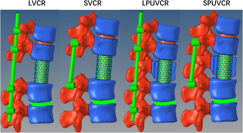
Han et al. Journal of Orthopaedic Surgery and Research   (2021) 16:88                                                                        Page 2 of 7

cord compression and restore vector sequence balance.                   bone, and ligament structures, in which the interver-
This has been the classical therapeutic goal. However,                  tebral disc was composed of annulus fibrosus matrix,
traditional VCR requires long operation times and sub-                  nucleus pulposus, annulus fibrosus fibers, and upper
stantial surgical trauma, both of which pose substantial                and lower endplates; the nucleus pulposus accounted
challenges to surgeons. For these reasons, researchers                  for 43% of the total intervertebral disc [2]. The surgi-
proposed posterior unilateral vertebral resection and                   cal model was processed in Hypermesh, and the
reconstruction (PUVCR) to correct kyphotic deformities                  screw-rod system was made using Pro/Engineer PTC,
using a unilateral approach and partial osteotomy. We                   MA, USA) and assembled in Hypermesh. Abaqus
used finite element analysis to simulate T12 VCR and                    (Hibbitt, Karlsson, and Sorensen, Inc., Providence, RI,
PUVCR for bone cutting. Long-segment or short-                          USA) was used for material property definition, model
segment fixation was performed to measure biomechan-                    assembly, loading, and finite element analysis. This
ical stability after surgery.                                           study’s material properties were validated based on
  Computed tomography (CT) images of normal                             published finite element models and thoracolumbar
subjects were imported into computer modeling soft-                     spines of human cadavers (Table 1) [3–5].
ware to establish a model, thereby simulating various
surgical osteotomy modes, restoring vector balance, and                 Design of the kyphotic cutoff model
installing a screw-rod system. Finally, we used finite                  We fabricated four fixation models using various screw
element analysis software to calculate and compare bio-                 combinations. The pedicle screws inserted into T11 and
mechanical parameters.                                                  12 measured 6.0 × 40 mm, and the pedicle screws inserted
                                                                        into L1 and two vertebral bodies measured 6.5 × 45 mm.
Methods                                                                 The LVCR model refers to the removal of the whole
Design, location, and timing                                            T12 vertebral body and T11/12, T12/L1 intervertebral
We performed a finite element analysis of patients                      disc, the application of a cylinder of 18-mm diameter
treated between 1 January 2020 and 1 June 2020 at the                   and 1-mm thickness to simulate a titanium cage fixed
Department of Spinal Surgery, Tianjin Hospital, Tianjin                 in the vacant position of the vertebral body, with can-
University.                                                             cellous bone placed in the middle. Pedicle screw
                                                                        fixation was applied at T10, T11, L1, and L2. SVCR
Participants                                                            refers to the removal of pedicle screws at T10 and L2
Volunteers were recruited from the Department of Spinal                 based on LVCR. LPUVCR refers to removing T12
Surgery, Tianjin Hospital, Tianjin University. A 40-year-old            right vertebral plate, facet joint, and vertebral body,
man, height 176 cm, and weight 72 kg underwent spinal
CT scan with three-dimensional reconstruction and MRI
                                                                        Table 1 Material properties of spine tissues used in the finite
examination to rule out other spinal diseases. After obtain-
                                                                        element model
ing consent from the patients and their families, informed
                                                                        The material properties of the finite element model
consent forms were signed following the relevant regula-
                                                                        Component                Young’s                Poisson’s        Cross-section
tions and submitted to the Ethics Committee for approval.               name                     modulus(MPa)           ratio            area(mm2)
                                                                        Cortical bone            12,000                 0.3              -
Acquisition and reconstruction of CT tomography images
                                                                        Cancellous bone          100                    0.3              -
We obtained 128-row, 256-slice GE spiral CT thin-
section scans (Sensation 16 Siemens, Germany) with a                    Cartilage                10                     0.4              -
slice thickness of 1 mm and resolution of 512 × 512 to                  Bony endplate            1000                   0.4              -
obtain sagittal two-dimensional tomographic images of                   Nucleus pulposus         1                      0.499            -
the T8–L3 vertebral body, and files were saved in                       Annulus fibrosus         450                    0.3              -
DICOM format.
                                                                        ALL                      20                     0.3              63.7
   DICOM images were imported into Mimics 20.0
                                                                        PLL                      20                     0.3              20
(Materials Company, Leuven, Belgium) to create a 3-
dimensional (3D) vertebral surface model from T10 to                    LF                       19.5                   0.3              40
L2 that were stored as STL format files; 3-Matic 12.0                   ISL                      11.6                   0.3              40
software (Materialise, LA, USA) was used after wrapping,                SSL                      15                     0.3              30
smoothing, and removal of excess triangulars. Data were                 TL                       58.7                   0.3              3.6
imported into Geomagic Studio 12.0 (Geomagic, Cary,
                                                                        CL                       32.9                   0.3              60
NC, USA) for solidification.
                                                                        ALL anterior longitudinal ligament, PLL posterior longitudinal ligament, ISL
   Hypermesh (Altair Engineering, Troy, MI, USA) was                    interspinous ligament, SSL supraspinal ligament, CL capsular ligament, LF
used to grid and construct the intervertebral disc,                     ligamantumflavum, TL transverse ligaments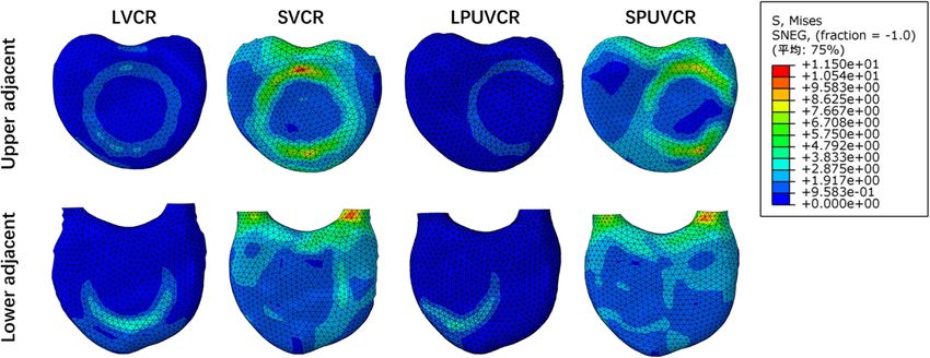
Han et al. Journal of Orthopaedic Surgery and Research      (2021) 16:88                                                               Page 3 of 7

T11/12, T12/L1 right intervertebral disc. A cylinder                       overall fixation system. Because only one subject was
with a diameter of 10 mm and a thickness of 1 mm                           modeled, statistical analysis was not performed in this
was used to simulate the titanium cage. The cage was                       study.
fixed at the vacant position of the vertebral body, and
the cancellous bone is placed in the middle for test-                      Results
ing. Pedicle screw fixation was applied at T10, T11,                       Validation of the model
L1, and L2. SPUVCR refers to the removal of pedicle                        The model was determined to be reasonable by compari-
screws at T10 and L2 on top of LPUVCR (Fig. 1).                            son with biomechanical experiments and finite element
                                                                           models. Using the same loading conditions, the ROM
Evaluate loading conditions and spinal motion simulation                   values of the complete model at the moment of 7.5 N
Abaqus was used to assess the boundary and loading                         were consistent with those of previous studies [6–8].
conditions and to simulate spinal motion. The L2 verte-                    This finding suggests that this study’s finite element
bral body was assumed to be fixed, and its substructure                    model is effective for further simulation of old fractures
was set to the boundaries without displacement or rota-                    of the thoracolumbar spine.
tion in all directions. Spinal motion in the sagittal, cor-
onal, and transverse planes was defined as flexion-                        ROMs for the thoracolumbar segment
extension, lateral bending, and rotation, respectively. Ac-                Compared with the intact model, ROMs decreased in
cording to the human body’s bearing capacity and the                       all fixed models under loading, with LVCR decreasing
previously published literature, an axial load of 200 N                    the most, forward flexion decreasing by 88.8%, exten-
and an additional torque load of 7.5 Nm were applied to                    sion decreasing by 97.3%, left lateral bending decreas-
simulate flexion and extension, lateral bending, and rota-                 ing by 89.6%, right lateral bending decreasing by
tion of the spine. An axial load was applied to the T10                    90.8%, left lateral rotation decreasing by 88.8%, and
vertebral body’s superior surface, and a torsional load                    right lateral rotation decreasing by 88.9%. SPUVCR
was applied to the center of the T10 vertebral body.                       decreased the least, with 51.2% decrease in forward
                                                                           flexion, 33.8% decrease in extension, 17.6% decrease
Evaluation indicators                                                      in left lateral curve, 14.6% decrease in right lateral
Three metrics were used to assess the mechanical                           curve, 78.8% decrease in left rotation, and 79.1% de-
performance of the constructs: (1) ROM of the overall                      crease in right rotation (Fig. 2).
fixation (T10–L2); (2) von Mises stress of the instrumen-
tation system; and (3) stress magnitude and distribution                   Von Mises stress in the internal fixation system
of the inferior endplate of T11 and superior endplate of                   Regardless of the fixation mode, the screw-rod system's
L1. These measures were chosen to assess the role of                       stress concentration point was located at the screw-rod
various vertebral body reconstruction methods in the                       junction position. Compared with the short-segment

 Fig. 1 Finite element models of the fixation constructs:LVCR:long vertebral column resection, SVCR:short vertebral column resection, LPUVCR:long
 posterior unilateral vertebral resection and reconstruction, SPUVCR:short posterior unilateral vertebral resection and reconstruction
Han et al. Journal of Orthopaedic Surgery and Research (2021) 16:88 Page 4 of 7
Fig. 2 Angular rom of thoracolumbar junction (T10–L2)
fixation, the stress of the screw-rod system in the long- and the upper and lower endplates, especially in front of
segment fixation was within the bearing range of the contact area between the titanium mesh and the
internal fixation, in which the maximum stress generated endplates, producing more significant stress in both the
the lateral bending motion in the LPUVCR model, which upper and lower endplates. The maximum stress in the
was 213.25 mPa; the minimum stress generated the inferior endplate of T11 was produced in the lateral
lateral bending motion in the SVCR, which was 40.22 bending movement of the SPUVCR model, which was
mPa. In addition to lateral bending motion, the rod’s max- 36.25 mPa, while the minimum stress was produced in
imum stress was greater for PVCR than for VCR (Fig. 3). the extension movement of the LPVCR, which was 3.87
mPa. The minimum stress in the upper endplate of L1
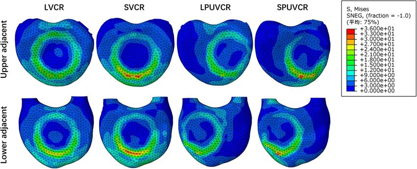
Stress distribution in T11 inferior endplate and L1 was produced in the extension movement of the LPVC
superior endplate R, which was 1.91 mPa, while the stress was the largest
In the four models, the stress concentration area was in the lateral bending in the SVCR, which was 44.77
located in the contact area between the titanium mesh mPa. See Figs. 4 and 5 for specific stresses.
Fig. 3 Von Mises stress on rods and pedicle screwsHan et al. Journal of Orthopaedic Surgery and Research (2021) 16:88 Page 5 of 7 Fig. 4 The stress in the flexion movement in the inferior endplate of T11 and the upper endplate of L1 Discussion PUVCR, a modified modification of VCR, exposes Compression fractures often cause old vertebral compres- the vertebral body through a unilateral approach, sion fractures after trauma or in elderly patients who are removes half of the vertebral body and the upper and not diagnosed and treated in a timely fashion. The lower intervertebral discs, shortens the operation kyphotic deformity often accompanies these fractures. time, and reduces the blood loss while restoring the The clinical symptoms are primarily persistent low back sagittal sequence of the spine [13]. Some studies sug- pain and neurological symptoms caused by spinal cord gest that PUVCR can completely decompress and compression; these include lower limb numbness and effectively restore deformity in the treatment of old decreased muscle strength. When conservative treatment thoracolumbar fractures [1, 13, 14]. Nevertheless, is ineffective, spinal cord compression can be relieved by there is a lack of effective biomechanical verification surgery to restore the spine’s sagittal balance; this can as to whether only half of the vertebral body and the effectively improve clinical symptoms and correct the upper and lower intervertebral discs affect spinal bio- kyphotic deformity [9–12]. mechanics; there also remains debate as to the appro- VCR is a traditional procedure to treat spinal osteot- priate length of fixation. For these reasons, in this omy. Through posterior resection of the spinous process study, we analyzed the effect of VCR and PUVCR on vertebral plate, pedicle screws are placed on the upper vertebral stability using finite element analysis. and lower two vertebrae; then, through resection of the Finite element analysis reconstructs the shape of the entire vertebral body and upper and lower intervertebral spine using computer simulation. It then directly inter- discs, titanium mesh is placed and fixed, followed by prets spinal biomechanics changes caused by internal screw-rod fixation. VCR surgery effectively corrects fixation by loading its material parameters and loading severe kyphotic deformity and achieves reconstruction of conditions and guiding the surgical plan [15]. Finite the vertebral body; however, due to the difficulty of sur- element analysis can be repeatedly applied, saving med- gery, there are often problems such as longer operation ical resources while safely and efficiently simulating time, larger amounts of blood loss, and greater degree of complicated surgical procedures. For these reasons, it trauma. has become a hot area of research. Fig. 5 The stress in the extension movement in the inferior endplate of T11 and the upper endplate of L1
Han et al. Journal of Orthopaedic Surgery and Research   (2021) 16:88                                                    Page 6 of 7

   Studies have analyzed lumbar ROM after Ponte osteot-                 terms of titanium cages, although the maximum stress
omy and showed that this technique leads to 20%                         site varied in various models due to the length of fix-
instability, while total discectomy leads to further                    ation, the maximum value of VMS was at the contact
instability [16]. Due to decreased stability, additional                surface site between the titanium cage and the vertebral
fixation is required to enhance stability, and pedicle                  body, suggesting that the risk of prosthesis displacement
screw fixation is the most common fixation method in                    is the greatest at the contact surface between the titan-
spinal fixation. Liu et al. analyzed the stress of the screw            ium cage and the vertebral body. Compared with VCR,
rod and found that the VMS of the pedicle screw was                     the PUVCR titanium cage’s stress increased by 41.9, 5.2,
the smallest when the directional pedicle screw was ap-                 3.2, and 2.2% in forward flexion, extension, lateral bend-
plied (136.9 mPa), and the VMS of the pedicle screw                     ing, and rotation, respectively.
was the largest with the hybrid application (382.6 mPa)                    In contrast, in long-segment fixation, the PUVCR
[17]. VMS of the rod was the largest when a polyaxial                   titanium cage’s stress increased by 4.5%, 1.2%, and 25.2%
pedicle screw was applied (439.9 mPa), and the VMS of                   in forward flexion, extension, and rotation, respectively,
the rod was the smallest when a directional pedicle                     compared with VCR. In the position of the superior and
screw was applied (341.7 mPa). Because most patients                    inferior endplates, the stress in the short segment was
with old vertebral compression fractures and kyphotic                   significantly greater than the stress in the long segment,
deformities are elderly and have low relative bone min-                 and this may explain why the short segment is more
eral density, we applied directional pedicle screws for                 prone to subsidence of the anterior prosthesis. Neverthe-
simulated fixation, and this can reduce the stress on the               less, the amount of vertebral body removal did not ap-
screw-rod and prevent its pullout. Natarajan et al.                     pear to affect the change of the area of stress
showed that the application of a 6-mm screw-rod system                  concentration, whether it was the VCR model (LVCR,
resulted in stronger fixation than a 5-mm screw-rod sys-                SVCR) or the PUVCR model (LPUVCR, SPUVCR). The
tem, increasing the size of the system from 5 mm to 6                   site with higher stress was located in the contact pos-
mm, increasing the flexion/extension moment by 8%,                      ition between the titanium cage and the endplate, sug-
the torsion moment by 14%, and the lateral bending mo-                  gesting that the prosthesis settlement could occur at the
ment by 24% [18]. We applied a 6-mm screw-rod system                    contact site.
to simulate two surgical modalities in the hope of in-                     When performing surgery, clinicians should pay more
creasing fixation strength.                                             attention to the screw-rod junction and the contact pos-
   Fusion fixation reduces thoracolumbar motion, as                     ition between the titanium cage and the endplate to pre-
shown in previous studies [15, 19]. Lu and Lu found that                vent the fracture of the screw-rod and the settlement of
the short-segment fusion fixed model decreased by 52–                   the titanium cage. Simultaneously, a long-segment fix-
94% compared with the intact model [20].                                ation can be applied to enhance the stability of the spine.
   There are few studies on ROM values after VCR sur-                   In terms of surgical methods, both VCR and PUVCR
gery. In our test, the ROM of all models decreased rela-                have similar fixation strengths and can achieve adequate
tive to the intact model, with the LVCR model showing                   fixation. However, PUVCR is relatively more straightfor-
the largest decrease of 97.28% in extension and the                     ward to operate than VCR; the operation time is shorter,
SVCR model showing the smallest decrease of 14.90% in                   and there is less blood loss.
lateral bending. The thoracolumbar ROMs of long-                           This study has some limitations. First, we used CT
segment fixed models (LVCR and LPUVCR) were                             scans of normal subjects to construct the old thoracol-
significantly smaller than those of short-segment fixed                 umbar fracture model, which may have impacted the
models (SVCR and SPUVCR), while the ROM of the                          test. Second, we only used one person to establish the
VCR model was relatively smaller than that of the                       model. Due to individual differences, there may be struc-
PUVCR model; however, the difference was not signifi-                   tural variations in vertebral bodies and ligaments. Third,
cant. This finding suggests that the thoracolumbar seg-                 our test was not verified using cadaver testing. In future
ment's ROM is greatly affected by fixation; however, it                 studies, model reconstruction using several patients can
has little relative relationship with the amount of verte-              be further performed, resulting in more objective results.
bral body and soft tissue removed.                                      The biomechanics of cadaveric specimens can also be
   We found that the site with the largest VMS in                       used as a supplement to the finite element analysis.
internal fixation was at the screw-rod connection site in
all models, explaining why internal fixation is prone to                Conclusion
failure at the screw-rod connection site. Similar findings              Compared with the VCR model, the thoracolumbar
were reported previously [21–23]. PUVCR showed                          ROM of PUVCR was not significantly different, while
slightly higher screw and rod stresses than VCR in all                  the thoracolumbar ROM was less than that of short-
loading cases except in short-segment lateral bending. In               segment fixation after long fixation. Except for short-
Han et al. Journal of Orthopaedic Surgery and Research                (2021) 16:88                                                                         Page 7 of 7

segment rotations, the stresses were more remarkable                                 6.    Alizadeh M, Kadir MR, Fadhli MM, Fallahiarezoodar A, Azmi B, Murali MR,
for the PUVCR instrumentation systems than for the                                         et al. The use of X-shaped cross-link in posterior spinal constructs improves
                                                                                           stability in thoracolumbar burst fracture: a finite element analysis. J Orthop
VCR. In the stress distribution, the screw-rod junction                                    Res. 2013;31(9):1447–54.
had the largest VMS. In contrast, the cage and vertebral                             7.    Panjabi MM, Oxland TR, Lin RM, McGowen TW. Thoracolumbar burst
body interface had the largest VMS, and this partly                                        fracture. A biomechanical investigation of its multidirectional flexibility.
                                                                                           Spine. 1994;19(5):578–85.
explained the high probability of internal fixation failure                          8.    Elmasry S, Asfour S, Travascio F. Effectiveness of pedicle screw inclusion at
and prosthesis migration in these two positions.                                           the fracture level in short-segment fixation constructs for the treatment of
                                                                                           thoracolumbar burst fractures: a computational biomechanics analysis.
Abbreviations                                                                              Comput Methods Biomech Biomed Engin. 2017;20(13):1412–20.
ROM: Range of motion; VCR: Vertebral column resection; PUVCR: Posterior              9.    Riaz-ur-R, Azmatullah, Azam F, Mushtaq, Shah M. Treatment of traumatic
unilateral vertebral resection and reconstruction; LVCR: Long vertebral                    unstable thoracolumbar junction fractures with transpedicular screw
column resection; SVCR: Short vertebral column resection; LPUVCR: Long                     fixation. J Pak Med Assoc. 2011;61(10):1005–8.
posterior unilateral vertebral resection and reconstruction; SPUVCR: Short           10.   Xu JG, Zeng BF, Zhou W, Kong WQ, Fu YS, Zhao BZ, et al. Anterior Z-plate
posterior unilateral vertebral resection and reconstruction; VMS: Von Mises                and titanic mesh fixation for acute burst thoracolumbar fracture. Spine.
stress                                                                                     2011;36(7):E498–504.
                                                                                     11.   Marco RA, Kushwaha VP. Thoracolumbar burst fractures treated with
Acknowledgements                                                                           posterior decompression and pedicle screw instrumentation supplemented
Not applicable                                                                             with balloon-assisted vertebroplasty and calcium phosphate reconstruction.
                                                                                           J Bone Joint Surg Am. 2009;91(1):20–8.
                                                                                     12.   Marco RA, Meyer BC, Kushwaha VP. Thoracolumbar burst fractures treated
Authors’ contributions
                                                                                           with posterior decompression and pedicle screw instrumentation
Miao Jun contributed to the data collection. Han Ye studied the design.
                                                                                           supplemented with balloon-assisted vertebroplasty and calcium phosphate
Wang Xiaodong wrote the manuscript. Wu Jincheng analyzed the data. All
                                                                                           reconstruction. surgical technique. J Bone Joint Surg Am. 2010;92(Suppl 1 Pt
authors read and approved the final manuscript.
                                                                                           1):67–76.
                                                                                     13.   Wang H, Ding W. Posterior vertebral column resection through unilateral
Funding                                                                                    osteotomy approach for old lumbar fracture combined with kummell
Not applicable                                                                             disease. World Neurosurg. 2018;109:147–51.
                                                                                     14.   Liu FY, Huo LS, Liu S, Wang H, Zhang LJ, Yang DL, et al. Modified posterior
Availability of data and materials                                                         vertebral column resection for Kummell disease: case report. Medicine.
Please contact the corresponding author for data requests.                                 2017;96(5):e5955.
                                                                                     15.   Demir E, Eltes P, Castro AP, Lacroix D, Toktas I. Finite element modelling of
Ethics approval and consent to participate                                                 hybrid stabilization systems for the human lumbar spine. Proc Inst Mech
The ethical committee approved the study of the participating hospitals. All               Eng H. 2020;34(12):1409–20.
subjects signed informed consent by each patient. All clinical investigations        16.   Wang C, Bell K, McClincy M, Jacobs L, Dede O, Roach J, et al. Biomechanical
had been conducted according to the principles expressed in the                            comparison of ponte osteotomy and discectomy. Spine. 2015;40(3):E141–5.
Declaration of Helsinki.                                                             17.   Liu H, Wang H, Liu J, Li C, Zhou Y, Xiang L. Biomechanical comparison of
                                                                                           posterior intermediate screw fixation techniques with hybrid monoaxial and
Consent for publication                                                                    polyaxial pedicle screws in the treatment of thoracolumbar burst fracture: a
Written informed consent for publication was obtained from all participants.               finite element study. J Orthop Surg Res. 2019;14(1):122.
                                                                                     18.   Natarajan RN, Watanabe K, Hasegawa K. Biomechanical analysis of a long-
                                                                                           segment fusion in a lumbar spine-a finite element model study. J Biomech
Competing interests
                                                                                           Eng. 2018;140(9):091011.
The authors declare that they have no competing interests.
                                                                                     19.   Niosi CA, Zhu QA, Wilson DC, Keynan O, Wilson DR, Oxland TR.
                                                                                           Biomechanical characterization of the three-dimensional kinematic
Author details
1                                                                                          behaviour of the Dynesys dynamic stabilization system: an in vitro study.
 Graduate School, Tianjin Medical University, Tianjin, China. 2Department of
                                                                                           Eur Spine J. 2006;15(6):913–22.
Orthopaedics, Tianjin Hospital, No. 406, Jiefang South Road, Hexi District,
                                                                                     20.   Lu T, Lu Y. Interlaminar stabilization offers greater biomechanical advantage
Tianjin, China.
                                                                                           compared to interspinous stabilization after lumbar decompression: a finite
                                                                                           element analysis. J Orthop Surg Res. 2020;15(1):291.
Received: 14 December 2020 Accepted: 15 January 2021
                                                                                     21.   Su Y, Wang X, Ren D, Liu Y, Liu S, Wang P. A finite element study on
                                                                                           posterior short segment fixation combined with unilateral fixation using
                                                                                           pedicle screws for stable thoracolumbar fracture. Medicine. 2018;97(34):
References                                                                                 e12046.
1. Tang HZ, Xu H, Yao XD, Lin SQ. Single-stage posterior vertebral column            22.   Elmasry SS, Asfour SS, Travascio F. Finite element study to evaluate the
    resection and internal fixation for old fracture-dislocations of thoracolumbar         biomechanical performance of the spine after augmenting percutaneous
    spine: a case series and systematic review. Eur Spine J. 2016;25(8):2497–513.          pedicle screw fixation with kyphoplasty in the treatment of burst fractures. J
2. Polikeit A, Ferguson SJ, Nolte LP, Orr TE. Factors influencing stresses in the          Biomech Eng. 2018;140(6):061005.
    lumbar spine after the insertion of intervertebral cages: finite element         23.   Wang TN, Wu BL, Duan RM, Yuan YS, Qu MJ, Zhang S, et al. Treatment of
    analysis. Eur Spine J. 2003;12(4):413–20.                                              thoracolumbar fractures through different short segment pedicle screw
3. Zhong ZC, Wei SH, Wang JP, Feng CK, Chen CS, Yu CH. Finite element                      fixation techniques: a finite element analysis. Orthop Surg. 2020;12(2):601–8.
    analysis of the lumbar spine with a new cage using a topology
    optimization method. Med Eng Phys. 2006;28(1):90–8.
4. Baroud G, Nemes J, Ferguson SJ, Steffen T. Material changes in osteoporotic       Publisher’s Note
    human cancellous bone following infiltration with acrylic bone cement for        Springer Nature remains neutral with regard to jurisdictional claims in
    a vertebral cement augmentation. Comput Methods Biomech Biomed                   published maps and institutional affiliations.
    Engin. 2003;6(2):133–9.
5. Zhang L, Yang G, Wu L, Yu B. The biomechanical effects of osteoporosis
    vertebral augmentation with cancellous bone granules or bone cement on
    treated and adjacent non-treated vertebral bodies: a finite element
    evaluation. Clin Biomech. 2010;25(2):166–72.
You can also read