BIRTHMARKS, BUMPS AND BEYOND - ELENA B. HAWRYLUK, MD, PHD APRIL 2021
←
→
Page content transcription
If your browser does not render page correctly, please read the page content below
Birthmarks, Bumps and Beyond

    Elena B. Hawryluk, MD, PhD
             April 2021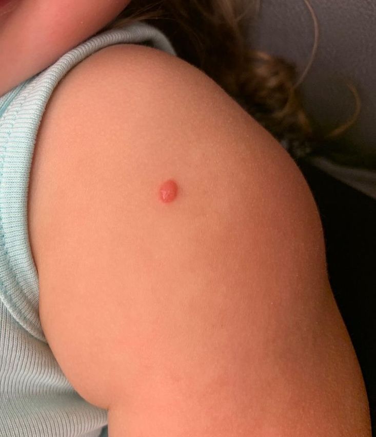
Disclosures
   My spouse/partner and I have the following
relevant financial relationship with a commercial
               interest to disclose:

           Gritstone Oncology (salary, stock)
                     Path AI (stock)
                  UpToDate (royalty)
               Purity Brands (consultant)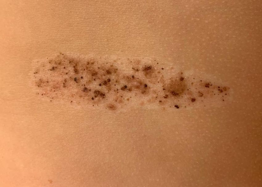
Infantile Hemangioma
▪   Not present at birth
▪   Appears after few weeks of life
▪   Maximum size reached by 3-6 months
▪   Majority regress by 5-7 yrs. of age
▪   Most common tumor of infancy - 4 % of all children
Risk Dictates Management

High Risk                     Intermediate Risk
▪ > 5 cm on face,             ▪ Lateral face, scalp, hands,
  lumbosacral area               feet
▪ Bulky lesion on face        ▪ Body folds
▪ Early white discoloration   ▪ >5 cm trunk, arms, legs
▪ Central face
▪ Periorbital, perinasal,     Low Risk
  perioral                    ▪ Trunk, arms, legs
Multifocal Hemangiomas
▪ Established association between multiple
  cutaneous IH with hepatic hemangiomas
▪ Mortality 11-18%
▪ Screen for 5+ cutaneous IH
▪ If large burden - Thyroid function test
  —increased levels of a catalyst of a thyroid-
   inactivating enzyme (iodothyronine deiodinase)
   have been detected in cutaneous hemangioma
   tissues, large hepatic hemangiomas
Workup/Considerations
• Early diagnosis – maximize options for management

• Multiple hemangiomas
   – Abdominal ultrasound for hepatic involvement, thyroid
     testing
• Large regional hemangiomas
   – PHACE: cardiac/aortic echo, MRI/MRA brain, ophtho eval
   – LUMBAR: CT abdomen, renal/urologic workup
   – Airway: ENT/scope

• Later considerations: surgery, laser of residual lesions
Treatment: Early Discussion Is KEY ▪ Propranolol ▪ Oral corticosteroids ▪ Timolol ▪ Intralesional corticosteroids ▪ Wound care, pain control for ulcerated hemangiomas ▪ Pulsed dye laser ▪ Excision
Topical Timolol
Timolol 0.5% gel forming solution (off label use)
▪ 1-2 drops TOP BID
▪ Do not use on ulcerated hemangioma
  (absorption)
▪ Most common complaints: dryness, white
  peeling of medication on skin
▪ Effective for superficial hemangiomas (not
  absorbed well)
Pulsed Dye Laser
• Reduces redness
• 595 nm targets blood vessels, set pulse duration
  according to vessel width
• Series of treatments every 6-8 weeks. Can
  perform under local anesthesia (eye protection
  needed)
• Controversy re: use during ulceration
• No sun exposure/tanning
• Each treatment causes “bruising” – appears more
  purple, redness fades over 6-8 weeks
Capillary Malformation
My Favorite Pediatric
           Pigmented Lesions!
•   Congenital nevus
•   Atypical or Dysplastic nevus
•   Halo nevus
•   Nevus spilus
•   Spitz nevus
Nevi – Congenital nevus • Nevus is present at birth • Slightly increased risk of melanoma (skin or CNS) depending on number of lesions, size • Small: 40 cm
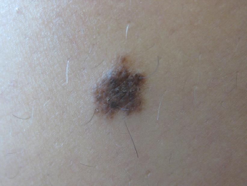
Nevi – Atypical or Dysplastic nevus
• Abnormal features clinically or on pathology

• Melanoma risk:
  – single dysplastic nevus increases risk by 2X
  – having ≥10 increases risk by 12X - Tucker et al,
    1997
Nevi – Atypical or Dysplastic nevus • NOT a “pre-melanoma” • However, these nevi are markers for increased risk of developing melanoma!
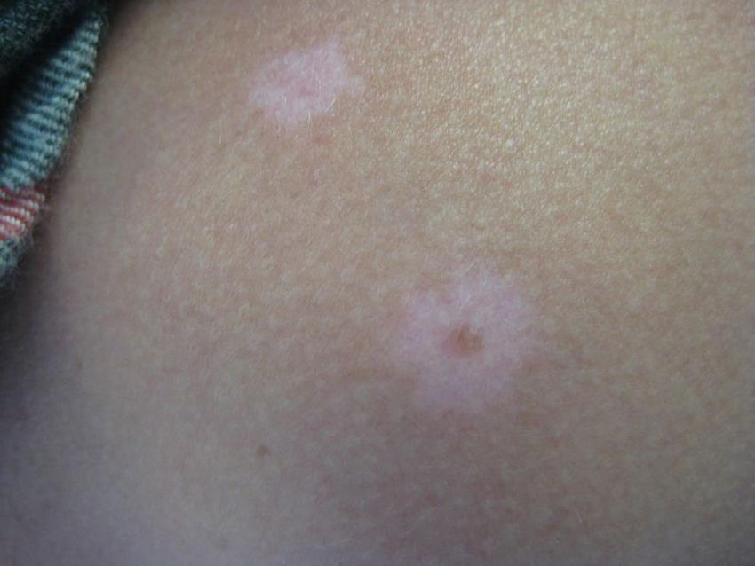
Nevi – Halo nevus
• Immunological destruction of
  melanocytes and nevus cells

• Multiple halo nevi confer a
  higher risk of vitiligo and
  other autoimmune diseases

• Halo typically can persist an
  average of 7.8 years, with
  eventual involution and return
  to normal-appearing skin
Nevi – Nevus spilus
• Presents in early childhood
  like a café-au-lait patch,
  with development of
  brown papules and
  macules within
Nevi – Spitz nevus
• “melanoma of childhood”

• Common Spitz nevi may
  be monitored clinically

• Those with clinically
  unusual, changing, or
  concerning features are
  biopsied
ABCD Criteria for Melanoma Detection

Traditional MM        Pediatric MM
• Asymmetry           • Amelanotic
• Border              • Bump, Bleeding
• Color Variegation   • Color uniformity
• Diameter > 6mm      • De novo, any Diameter
• Evolution/Change
                      Cordoro K et al. JAAD June 2013
CUP Criteria for Pediatric Melanoma
Standard ABCDE criteria plus:
• Color that is pink/red, Changing
• Ulceration, Upward thickening
• Pyogenic granuloma-like, Pop-up of new lesions
Silverberg NB, McCuaig CC. Cutis 2013Evolution Is Biggest Clue!
PATH: Spitzoid melanoma, 3.5mm, level IV, ulcerated, 14 mitoses/mm2
Bartenstein et al, 2017You can also read