BMK AEP, AN ANTI-EPILEPTIC PEPTIDE DISTINCTLY AFFECTS THE GATING OF BRAIN SUBTYPES OF VOLTAGE-GATED SODIUM CHANNELS - MDPI
←
→
Page content transcription
If your browser does not render page correctly, please read the page content below
International Journal of
           Molecular Sciences

Article
BmK AEP, an Anti-Epileptic Peptide Distinctly
Affects the Gating of Brain Subtypes of
Voltage-Gated Sodium Channels
Fan Zhang † , Ying Wu † , Xiaohan Zou, Qinglian Tang, Fang Zhao and Zhengyu Cao *
 State Key Laboratory of Natural Medicines and Jiangsu Provincial Key Laboratory for TCM Evaluation and
 Translational Development, School of Traditional Chinese Pharmacy, China Pharmaceutical University,
 Nanjing 211198, China; zhangfan20111112@126.com (F.Z.); wingwycpu@126.com (Y.W.);
 imzouxiaohan@163.com (X.Z.); cputangqinglian@163.com (Q.T.); zhaofang180930@163.com (F.Z.)
 * Correspondence: zycao1999@hotmail.com; Tel.: +86-25-8618-5955
 † These authors contributed equally to this work.

 Received: 25 December 2018; Accepted: 31 January 2019; Published: 8 February 2019

 Abstract: BmK AEP, a scorpion peptide purified form the venom of Buthus martensii Karsch, has been
 reported to display anti-epileptic activity. Voltage-gated sodium channels (VGSCs) are responsible
 for the rising phase of action potentials (APs) in neurons and, therefore, controlling neuronal
 excitability. To elucidate the potential molecular mechanisms responsible for its anti-epileptic activity,
 we examined the influence of BmK AEP on AP firing in cortical neurons and how BmK AEP influences
 brain subtypes of VGSCs (Nav 1.1–1.3 and Nav 1.6). BmK AEP concentration-dependently suppresses
 neuronal excitability (AP firing) in primary cultured cortical neurons. Consistent with its inhibitory
 effect on AP generation, BmK AEP inhibits Na+ peak current in cortical neurons with an IC50 value
 of 2.12 µM by shifting the half-maximal voltage of activation of VGSC to hyperpolarized direction
 by ~7.83 mV without affecting the steady-state inactivation. Similar to its action on Na+ currents
 in cortical neurons, BmK AEP concentration-dependently suppresses the Na+ currents of Nav 1.1,
 Nav 1.3, and Nav 1.6, which were heterologously expressed in HEK-293 cells, with IC50 values of
 3.20, 1.46, and 0.39 µM with maximum inhibition of 82%, 56%, and 93%, respectively. BmK AEP
 shifts the voltage-dependent activation in the hyperpolarized direction by ~15.60 mV, ~9.97 mV,
 and ~6.73 mV in Nav 1.1, Nav 1.3, and Nav 1.6, respectively, with minimal effect on steady-state
 inactivation. In contrast, BmK AEP minimally suppresses Nav 1.2 currents (~15%) but delays the
 inactivation of the channel with an IC50 value of 1.69 µM. Considered together, these data demonstrate
 that BmK AEP is a relatively selective Nav 1.6 gating modifier which distinctly affects the gating of
 brain subtypes of VGSCs.

 Keywords: BmK AEP; anti-epilepsy; action potentials; voltage-gated sodium channels

1. Introduction
     Scorpion venoms are rich sources of bioactive peptides that modulate ion channel and receptor
activities by modulation of channel gating kinetics [1–4]. These peptides provide invaluable
pharmacological probes in exploring the structure and function of ion channels due to their
exquisite selectivity [2,3,5]. They have displayed the potential to treat cancer [6] and neuronal [7],
autoimmune [8], and cardiovascular diseases [9]. In general, according to the peptide length, scorpion
toxins are classified into long-chain toxins and short-chain toxins. The long-chain scorpion toxins
composed of 58–76 amino acid residues mainly act on voltage-gated sodium channels (VGSCs) while
the short-chain scorpion toxins containing 30–40 amino acid residues generally target K+ or Cl−

Int. J. Mol. Sci. 2019, 20, 729; doi:10.3390/ijms20030729                       www.mdpi.com/journal/ijms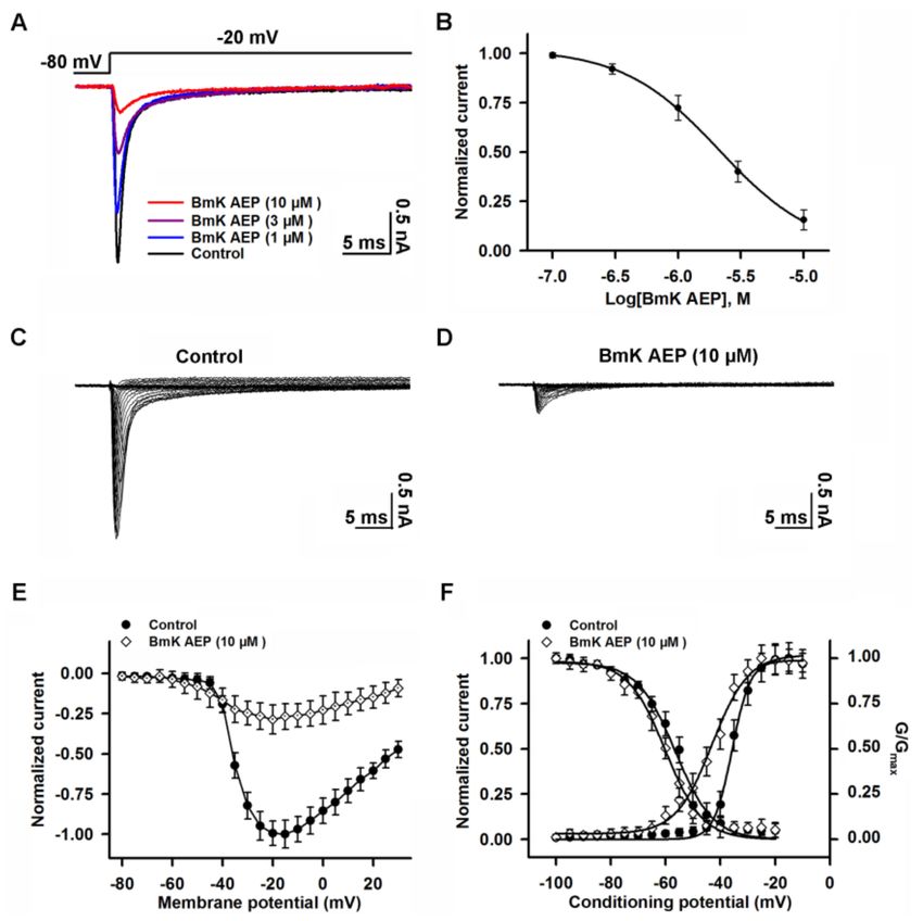
Int. J. Mol. Sci. 2019, 20, 729                                                                   2 of 13

channels [5,10,11]. The long-chain scorpion toxins can be categorized into excitatory mammalian
toxins and depressant insect toxins based on their activity in mammals and insects [12].
     The scorpion Buthus martensii Karsch (BmK) has been effectively used in traditional Chinese
medicine for alleviating pain, epilepsy, and facial paralysis [13]. BmK AEP, a scorpion toxin
identified from B. martensii Karsch, has been reported to display anti-epileptic activity in a coriaria
lactone-induced epileptic model in rat without mammalian toxicity [14]. Structurally, BmK AEP shares
high homology with depressant insect scorpion toxins, such as BmK ITa, BmK ITb [15], BmK IM [16],
and LqqIT2 [17]. Similar to BmK AEP, the recombinant BmK IM also displays anti-epileptic activity
in the pentylenetetrazol-induced seizure rat model [18]. The depolarized shift of activation voltage
on Na+ currents in hippocampal neurons was proposed to be responsible for the anti-epileptic effect
of BmK IM [18]. In addition, scorpion stings may cause epilepsy, especially children with immature
blood-brain barrier, possibly because α-scorpion toxins induce increments in action potential firing
and sympathetic excitation [19–21].
     VGSCs play a crucial role in generating the rising phase of action potentials (APs) in neurons
and, therefore, in controlling neuronal excitability. According to the sequence homology of the
α-subunits, VGSCs can be categorized into nine subtypes (Nav 1.1–1.9) with relatively tissue-specific
expression [22]. Among the nine VGSC subtypes, Nav 1.1–1.3 and Nav 1.6 are predominantly expressed
in the brain [23]. Many studies have demonstrated that channelopathy in VGSCs is one of the crucial
reasons contributing to epilepsy [23,24], and clinic drugs targeting to VGSCs (such as carbamazepine)
display therapeutic efficacy against seizures [25,26].
     In this study, we therefore examined the influence of BmK AEP on neuronal excitability and how
BmK AEP influences brain VGSC subtypes (Nav 1.1–1.3 and Nav 1.6). We demonstrate that BmK AEP
suppresses AP firing in primary cultured cortical neurons and inhibits Na+ peak current in cortical
neurons. Similar to its action on Na+ currents elicited in cortical neurons, BmK AEP suppresses the
Na+ currents of Nav 1.1, Nav 1.3, and Nav 1.6 with IC50 values of 3.20, 1.46, and 0.39 µM, respectively,
suggesting that BmK AEP is a relatively selective Nav 1.6 gating modifier. In contrast, BmK AEP
minimally suppresses the peak currents of Nav 1.2 but inhibits the inactivation of Nav 1.2. Considered
together, our data demonstrate that BmK AEP represents a unique toxin that distinctly affects the
gating properties of brain VGSC subtypes.

2. Results

2.1. Influence of BmK AEP on Action Potential Firing in Cerebral Cortical Neurons
      BmK AEP has been shown to display anti-epileptic activity in a coriaria lactone-induced epileptic
model in rat [14]. We therefore evaluated the effect of BmK AEP on neuronal excitability in primary
cultured cortical neurons. The APs were elicited by injection of a 30-pA current. Bath application of
BmK AEP concentration-dependently suppressed the AP firing frequency (Figure 1). At concentrations
of 3 and 10 nM, BmK AEP displayed negligible effects on AP firing. However, at higher concentrations
(30 and 100 nM), BmK AEP significantly suppressed AP firing. The IC50 value for BmK AEP
suppression of AP firing is 33.1 nM (95% confidence interval (95% CI): 21.6–57.2 nM).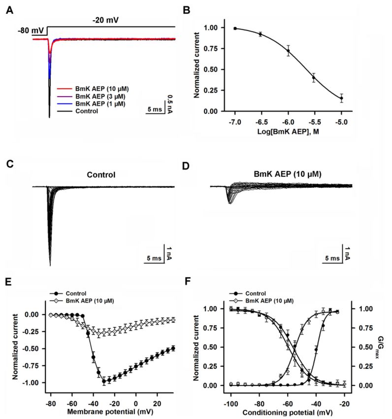
Int. J. Mol. Sci. 2019, 20, 729                                                                       3 of 13

      Figure 1. Influence of Buthus martensii Karsch (BmK) AEP on action potential firing in primary
      cultured mouse cortical neurons. (A) Representative traces of action potential firing evoked by
      a 30-pA current stimulation for 1000 ms in the presence of different concentrations of BmK AEP.
      (B) Concentration–response relationship of BmK AEP inhibition of action potential frequency. Each
      data point represents the mean ± SEM (n = 5–7).

2.2. Influence of BmK AEP on VGSCs in Primary Cultured Cortical Neurons
     VGSCs are responsible for initiating APs in brain neurons. To investigate the molecular targets
and mechanism of BmK AEP, we evaluated the influence of BmK AEP on VGSC currents in primary
cultured cortical neurons. VGSC currents were elicited by a 50-ms pulse depolarized from a holding
potential of −80 to −20 mV. Bath application of BmK AEP concentration-dependently inhibited VGSC
peak currents in primary cultured cortical neurons with an IC50 value of 2.12 µM (1.25–3.76 µM, 95%
CI) (Figure 2A,B). The maximal inhibition reached 85%. To test the influence of BmK AEP on VGSC
activation, the Na+ currents were triggered by depolarized pulses from −100 mV to +30 mV in a 5-mV
step in the absence and presence of BmK AEP (10 µM) (Figure 2C,D). At depolarization pulses below
−40 mV, BmK AEP slightly increased the VGSC currents, whereas at the stronger depolarization
potentials (< −40 mV), BmK AEP suppressed the Na+ peak currents (Figure 2E). BmK AEP shifted
the voltage of half-maximal activation of VGSCs towards the hyperpolarized direction by ~7.83 mV
(Figure 2F). BmK AEP minimally affected the steady-state inactivation of VGSCs (Figure 2F).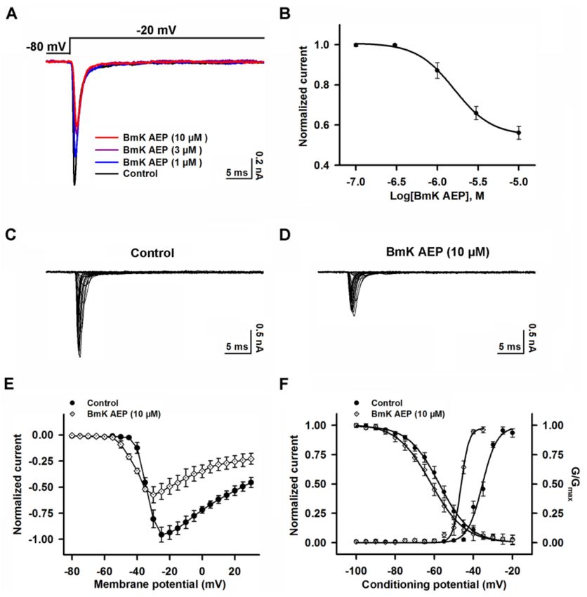
Int. J. Mol. Sci. 2019, 20, 729                                                                          4 of 13

      Figure 2. Influence of BmK AEP on voltage-gated sodium channel (VGSC) currents in primary cultured
      mouse cortical neurons. (A) BmK AEP inhibited the VGSC currents. Na+ currents were elicited by
      a 50-ms depolarization to −20 mV from the holding potential of −80 mV. (B) Concentration–response
      relationship of BmK AEP inhibition of Na+ currents. (C,D) Representative traces of Na+ currents
      in the absence and presence of 10 µM BmK AEP, respectively. Currents were triggered by 50-ms
      depolarizations from −100 to 30 mV in a 5-mV step. (E) Normalized peak current–membrane voltage
      (I–V) relationship of Na+ currents in the absence and presence of 10 µM BmK AEP. (F) Effect of BmK
      AEP (10 µM) on the steady-state activation and inactivation of VGSCs. Each data point represents the
      mean ± SEM (n = 5–8).

2.3. Influence of BmK AEP on hNav 1.1 Expressed in HEK-293 Cells
     Nav 1.1 is the primary subtype expressed in cortical neurons and plays a crucial role in action
potential firing in the CNS. We next evaluated the action of BmK AEP on brain VGSC subtypes. Bath
application of BmK AEP suppressed Nav 1.1 peak current evoked by a 50-ms depolarizing potential of
−20 mV from a holding potential of −80 mV with an IC50 value of 3.20 µM (1.73–6.32 µM, 95% CI),
and the maximal inhibition reached 82% (Figure 3A,B). Similar to the effect observed in cortical
neurons, BmK AEP increased the Nav 1.1 currents at the depolarization potentials below −45 mV,
whereas at the stronger depolarization potentials (> −45 mV), BmK AEP suppressed the Na+ peak
currents (Figure 3C–E). BmK AEP shifted the voltage of half-maximal activation of Nav 1.1 towards the
hyperpolarized direction by ~15.6 mV without affecting steady-state inactivation (Figure 3F).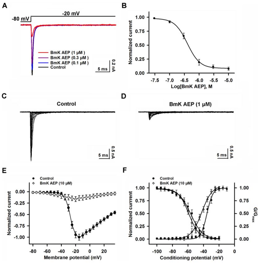
Int. J. Mol. Sci. 2019, 20, 729                                                                            5 of 13

      Figure 3. Influence of BmK AEP on hNav 1.1 expressed in HEK-293 cells. (A) BmK AEP suppressed
      the Nav 1.1 peak current. Nav 1.1 currents were elicited by a 50-ms depolarization to −20 mV from
      a holding potential of −80 mV. (B) Concentration–response relationships of BmK AEP inhibition on
      Nav 1.1 currents. (C,D) Representative traces of Nav 1.1 currents in the absence and presence of 10 µM
      BmK AEP, respectively. Currents were evoked by 50-ms depolarizations from −80 to 30 mV in a 5-mV
      step. (E) Normalized I–V relationship of Nav 1.1 in the absence and presence of 10 µM BmK AEP.
      (F) Influence of BmK AEP (10 µM) on the steady-state activation and inactivation of Nav 1.1. Each data
      point represents the mean ± SEM (n = 5–8).

2.4. Influence of BmK AEP on hNav 1.2 Expressed in HEK-293 Cells
     We next examined the effect of BmK AEP on Nav 1.2 heterologously expressed in HEK-293 cells.
In contrast to the significant effect on the suppression of Nav 1.1peak current, bath application of
10 µM BmK AEP slightly suppressed the Nav 1.2 peak current (~15%) (Figure 4A). However, BmK AEP
significantly delayed Nav 1.2 channel inactivation, and the EC50 value was calculated as 1.69 µM
(0.93–2.76 µM, 95%CI) (Figure 4B). BmK AEP (10 µM) shifted both the voltages of half-maximal
activation and steady-state inactivation towards the hyperpolarized direction by ~17.8 and ~7.7 mV,
respectively (Figure 4C–F).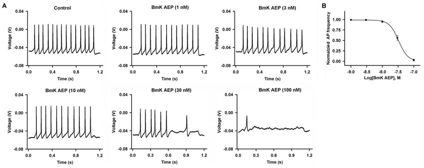
Int. J. Mol. Sci. 2019, 20, 729 6 of 13
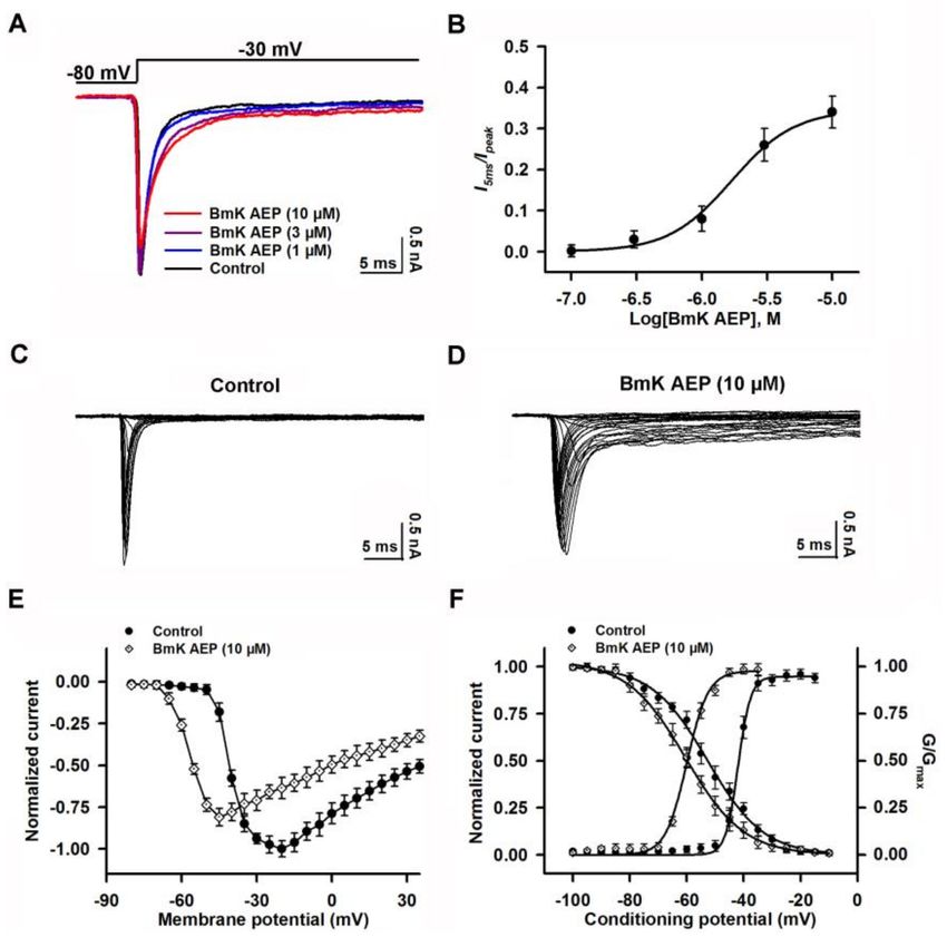
Figure 4. Influence of BmK AEP on hNav 1.2 expressed in HEK-293 cells. (A) BmK AEP delayed
Nav 1.2 channel inactivation. Nav 1.2 currents were triggered by a 50-ms depolarization to −30 mV
from a holding potential of −80 mV. (B) Concentration–response relationship of BmK AEP-delayed
inactivation of Nav 1.2 channels. (C,D) Representative traces of Nav1.2 currents in the absence and
presence of 10 µM BmK AEP, respectively. Currents were triggered by 50-ms depolarizations from
−100 to 30 mV in a 5-mV step. (E) Normalized I–V relationships of Nav 1.2 currents in the absence
and presence of 10 µM BmK AEP. (F) Effect of BmK AEP (10 µM) on the steady-state activation and
inactivation of Nav1.2. Each data point represents the mean ± SEM (n = 5–8).
2.5. Influence of BmK AEP on hNav 1.3 Stably Expressed in HEK-293 Cells
Similar to the effect on Nav 1.1, bath application of BmK AEP suppressed the Nav 1.3 peak current
evoked by a 50-ms depolarizing potential of −20 mV from a holding potential of −80 mV with an IC50
value of 1.46 µM (0.91–2.56 µM, 95% CI) (Figure 5A,B). However, the maximal inhibition on Nav 1.3
was only 56% (Figure 5B, Table 1). BmK AEP enhanced Nav 1.3 currents at the depolarization potentials
below −35 mV, whereas at the stronger depolarization potentials (> −35 mV), BmK AEP suppressed
the Na+ peak currents (Figure 5C–E). BmK AEP shifted the voltage for half-maximal activation towards
the hyperpolarized direction by ~10.0 mV while minimally affecting the steady-state inactivation of
Nav 1.3 (Figure 5F).Int. J. Mol. Sci. 2019, 20, 729 7 of 13
Figure 5. Influence of BmK AEP on hNav 1.3 expressed in HEK-293 cells. (A) Representative traces of
BmK AEP suppression of the Nav 1.3 peak current. Na+ currents were elicited by a 50-ms depolarization
to −20 mV from a holding potential of −80 mV. (B) Concentration–response relationship curve of
BmK AEP inhibition of Nav 1.3 currents. (C,D) Representative traces of Nav 1.3 currents in the absence
and presence of 10 µM BmK AEP, respectively. Currents were trigged by 50-ms depolarizations from
−100 to 30 mV in a 5-mV step. (E) Normalized I–V relationships of Nav 1.3 currents in the absence
and presence of 10 µM BmK AEP. (F) Effect of BmK AEP (10 µM) on the steady-state activation and
inactivation of Nav 1.3. Each data point represents the mean ± SEM (n = 5–8).
2.6. Influence of BmK AEP on hNav 1.6 Expressed in HEK-293 Cells
Nav 1.6 was reported to be expressed in the brain [27]. The influence of BmK AEP on Nav 1.6 was
evaluated. Similar to the effect on Nav 1.1, bath application of BmK AEP suppressed the Nav 1.6 peak
current evoked by a 50-ms depolarizing potential from a holding potential of −80 mV to −20 mV
(Figure 6A,B). At concentrations greater than 3 µM, BmK AEP nearly completely suppressed Nav 1.6
currents (Figure 6B). Non-linear regression revealed that the IC50 value for BmK AEP suppression
of Nav 1.6 current was 0.39 µM (0.26–0.57 µM, 95% CI) (Figure 6B, Table 1). BmK AEP slightly
enhanced the Nav 1.6 currents at the depolarization potentials below −35 mV, whereas at the stronger
depolarization potentials (> −35 mV), BmK AEP suppressed the Nav 1.6 peak currents (Figure 6C–E).
BmK AEP shifted the voltage for half-maximal activation of Nav 1.6 towards the hyperpolarized
direction by ~6.7 mV without affecting the steady-state inactivation of Nav 1.6 (Figure 6F).Int. J. Mol. Sci. 2019, 20, 729 8 of 13
Figure 6. Influence of BmK AEP on hNav 1.6 expressed in HEK-293 cells. (A) BmK AEP suppressed the
Nav 1.6 peak current. Na+ currents were elicited by a 50-ms depolarization to −20 mV from a holding
potential of −80 mV. (B) Concentration–response relationship curve of BmK AEP inhibition of Nav 1.6
currents. (C,D) Representative traces of Nav 1.6 currents in the absence and presence of 10 µM BmK
AEP, respectively. Currents were trigged by 50-ms depolarizations from −80 to 30 mV in a 5-mV step.
(E) Normalized I–V relationships of Nav 1.6 currents in the absence and presence of 10 µM BmK AEP.
(F) Effect of BmK AEP (10 µM) on the steady-state activation and inactivation of Nav 1.6. Each data
point represents the mean ± SEM (n = 5–7).
Table 1. The potency and maximum inhibition of BmK AEP in brain subtypes of VGSCs.
VGSC Maximum Selectivity for
Influence IC50 (µM)a 95% CI
Subtypes Inhibition/Delay (%) Nav 1.6
Inhibition of
Nav 1.1 82% 3.20 (1.73–6.32) 9.30
peak current
Delay -
Nav 1.2 34% 1.69 (0.93–2.76)
of inactivation
Inhibition of
Nav 1.3 56% 1.46 (0.91–2.56) 6.21
peak current
Inhibition of
Nav 1.6 93% 0.39 (0.26–0.57) 1
peak current
Selectivity = (MaxNav 1.6/MaxNav 1.x) × (IC50 Nav 1.x/IC50 Nav 1.6); a , IC50 values were measured at 10 Hz;
“–” represents no comparison with Nav 1.6; data are shown as means ± SEM; n = 5–8 for each subtype.Int. J. Mol. Sci. 2019, 20, 729 9 of 13
3. Discussion
Scorpion toxins provide valuable tools to study the channel gating and function of a variety
of ion channels/receptors [28]. BmK AEP has been reported to display anti-epileptic activity in
a coriaria lactone-induced epileptic model in the rat with comparable efficacy to diazepam [14]. In this
study, we demonstrated that BmK AEP suppressed AP firing and VGSC currents in cortical neurons
consistent with its inhibitory effect on neuronal excitability. The IC50 value for BmK AEP suppression
of AP firing is 33.1 nM, which is around 64-fold more potent than that on VGSC currents (2.12 µM).
Whether the action on VGSC was responsible for its inhibitory effect on AP firing needs further
examination. It should be noted that tetrodotoxin (TTX), a pore blocker of TTX-sensitive VGSCs,
also suppressed the AP firing at ~90 times more potent than that on VGSC currents recorded in spinal
cord neurons (Figure S1).
Long-chain scorpion toxins are generally recognized to be VGSC gating modifiers [29]. In general,
these toxins can be categorized into α- and β-toxins based on their binding sites and actions on
VGSCs [30,31]. α-Toxins bind to neurotoxin site 3 which is composed of the amino acid resides
located in the S3-S4 loop and S1-S2 loop in domain IV of VGSCs; it delays the inactivation kinetics
of the VGSCs by preventing the outward movement of the S4 segment of domain IV in response
to depolarization [30,32]. β-Toxins bind to neurotoxin site 4 (S3–S4 of domain II) and shift the
activation to negative potentials through a voltage sensor trapping mechanism [32]. We demonstrate
that BmK AEP shifts the activation voltage to a hyperpolarized direction and represses the peak
Na+ currents in primary cultured cortical neurons, two characteristics of β-toxin action on VGSCs.
Structurally, BmK AEP is highly homologous (>83%) the scorpion insect β-toxins BmK ITa, BmK ITb,
and LqqIT2 [15,17]. It has been demonstrated that BmK IM also displays anti-epileptic activity in the
pentylenetetrazol-induced seizure model in rats by shifting the voltage for half-maximal activation
towards the depolarized direction and suppressing Na+ currents [18]. These data imply that β-toxins
may display anti-epileptic activity, although they distinctly modify the activation of VGSCs.
While BmK AEP shifts the activation voltage to more negative potentials in Nav 1.1–1.3 and
Nav 1.6, which is consistent with β-toxin effects, it is also interesting that BmK AEP also delays
channel inactivation of Nav 1.2. These data suggest that in Nav 1.2, BmK AEP displays both α-like
and β-like properties. The IC50 value of BmK AEP on the suppression of Na+ currents of Nav 1.6 is
0.39 µM, while in Nav 1.1, and Nav 1.3, the IC50 values are 3.20 and 1.46 µM, respectively. In addition
to the potency difference, BmK AEP suppresses the Na+ currents of Nav 1.1, Nav 1.3, and Nav 1.6
to distinct degrees with maximum inhibition of 82%, 56%, and 93%, respectively. Therefore,
BmK AEP is a selective Nav 1.6 gating modifier with 9.30 and 6.21-fold selectivity over Nav 1.1,
and Nav 1.3, respectively. It is quite interesting that the degree of inhibition is inversely correlated
with hyperpolarized shift of voltage for half-maximal activation (or maximum augmentation of Na+
currents at relatively less depolarized potentials in Nav 1.1–1.3 and Nav 1.6).
Sodium channel blockers, including phenytoin, carbamazepine, lamotrigine, oxcarbazepine,
rufinamide, and lacosamide, represent the main anti-epileptic drugs [33]. Generally, these classical
sodium channel blocking drugs are nonselective for different subtypes of VGSCs (Nav 1.1–1.7).
These nonselective inhibitors are commonly used for controlling partial seizures and many different
causes of seizures. According to the drug-receptor hypothesis, the varying ability of these drugs
to treat different epilepsy diseases depends on their binding and action [34]. In the present study,
BmK AEP differentially suppressed the peak current of brain sodium channel subtypes (Nav 1.1, 1.3,
and Nav 1.6) with β-toxin effects (binding to the S3–S4 of domain II of VGSC), except for the Nav 1.2
channel. The inhibition of the peak current of sodium channels in the cortical neurons by BmK AEP
resulted in the suppression of APs and consequently blocked hyperexcitability in brain neurons,
therefore contributing to the anti-epileptic activity of BmK AEP. However, the nonselective sodium
channel anti-epileptic drugs affecting the interactions of the voltage-gated sodium channels are still
lacking, which could be due to the difficulty of the research. Further studies are needed to reveal how
BmK AEP affects the interactions of the brain sodium channel subtypes. It is noteworthy that BmKInt. J. Mol. Sci. 2019, 20, 729 10 of 13
AEP may have problems in crossing blood-brain barrier, which leads to its increase in concentration
in the blood, which will result in BmK AEP targeting VGSCs in the peripheral nervous system and
therefore causing potential adverse effects.
In summary, in the present study, we demonstrated that BmK AEP suppresses neuronal excitability
and displays β-toxin effects in in cortical neurons. BmK AEP selectively suppresses Nav 1.6 currents
when compared with other brain subtypes of VGSCs. An interesting feature of BmK AEP is that
although BmK displays β-toxin effects on Nav 1.1, Nav 1.3, and Nav 1.6, BmK AEP also delays the
inactivation kinetics of Nav 1.2. These data suggest that BmK AEP may present a useful tool to
investigate the gating mechanism of VGSCs.
4. Materials and Methods
4.1. Materials and Animals
Lyophilized crude venom of B. martensii Karsch was purchased from a domesticated scorpion
farm (Zhengzhou, Henan, China) and was the same source as described previously [35]. Sephadex
G-50 and CM-Sephadex C-50 resins were obtained from Pharmacia Fine Chemicals (Uppsala, Sweden).
Fetal bovine serum (FBS), trypsin, and L-glutamine were obtained from Thermo Fisher Scientific
(Grand Island, NY, USA). Trifluoroacetic acid and inorganic salts were obtained from Sigma-Aldrich
(St. Louis, MO, USA). HEK-293 cells stably expressing hNav 1.1, hNav 1.3, or hNav 1.6 were generous
gifts from Christoph Lossin (University of California, Davis, CA, USA) and are the same cells used in
the previous study [36]. HEK-293 cells stably expressing Nav 1.2 was gifted by Yuan Chen (Zhejiang
Agriculture & Forestry University, Deqing, China) as described previously [37]. C57BL/6J mice were
purchased from the Model Animal Research Center of Yangzhou University (Yangzhou, China).
4.2. BmK AEP Purification
The BmK crude venom was dissolved in ddH2 O and centrifuged to remove the sediment. After gel
filtration in a Sephadex G-50 column, 4 fractions (G1–G4) were collected. These fractions were subjected
to an LC-MS analysis to identify the BmK AEP. In the G-2 fraction, a peptide with molecular weight of
6730.3 Da close to that of BmK AEP (6730.4 Da) was found. The fraction was then subjected to HPLC
purification, and the targeted toxin was collected and lyophilized. To further confirm the identity,
the purified toxin was subjected to the Edman degradation and the first 15 amino acid residues
in N-terminal was determined to be DGYIRGSNGCKVSCL, which was identical to that of BmK
AEP. In addition, after reduction and S-carboxymethylation, the toxin was cleaved by Staphylococcus
aureaus V8 protease or Lysyl Endopeptidase and the enzymatic fragments were subjected to LC-MS
analysis. After S. aureaus V8 protease cleavage, three fragments with molecular weights of 607.4,
1275.5, and 537.2 Da corresponding to the molecular weights of fragments containing residues of 20–24,
46–56, and 47–61 of BmK AEP, respectively, were observed. Similarly, four fragments with molecular
weights of 1226.5, 2056.5, 1460.6, and 434.2 Da corresponding to the molecular weights of fragments
containing residues of 1–11, 24–39, 40–51, and 52–54 of BmK AEP, respectively, were observed. Together,
the purified toxin has the same sequence of BmK AEP.
4.3. Primary Cultures of Cortical Neurons
Animal experimentation protocols (81473539) were approved (12-05-2017) by the Animal
Experimentation Ethics Committee of China Pharmaceutical University. All experiments conformed to
the rule of minimizing animal suffering and numbers. The cortical neurons were dissociated from the
cortex of C57BL/6 mice (embryonic day 16 of either gender) as described previously and maintained
in Neurobasal complete medium (Neurobasal medium + 2% NS21, 1 mM L-glutamine, and 1% HEPES)
containing 5% FBS [35]. The dissociated cortical neurons were plated onto poly-L-lysine-coated
(50 µg/mL) 35-mm diameter dishes (Corning Incorporation, Corning, NY, USA) at a density of
1.5 × 10⁴ cells/dish. The culture medium was half-replaced by Neurobasal complete medium (serumInt. J. Mol. Sci. 2019, 20, 729 11 of 13
free) at days in vitro (DIV) 5 and 7. The neurons were maintained at standard conditions (5% CO2 ,
95% humidity at 37 ◦ C) and were used at DIV 8.
4.4. Culture of HEK-293 Cells Heterologously Expressing VGSC Subtypes
HEK-293 cells stably expressing individual VGSC subtypes were maintained in a culture medium
(DMEM supplemented with 10% FBS, 500 µg/mL G-418, 100 units/mL penicillin, and 0.1 mg/mL
streptomycin) in poly-D-lysine (10 µg/mL) pre-coated T75 flasks (Corning Incorporation, Corning,
NY, USA). At around 70% confluency, cells were digested with 0.05% trypsin and plated into
a poly-D-lysine pre-coated 35-mm diameter dish (Corning Life Sciences, Acton, MA, USA) at a density
of 1.5 × 10⁴ cells/dish. Cells were maintained at standard conditions (5% CO2 , 95% humidity at 37 ◦ C)
for 24 h before the experiment.
4.5. Voltage-Clamp and Current-Clamp Electrophysiology
Whole cell patch clamp was used to record the voltage-gated sodium currents in mouse cortical
neurons or HEK-293 cells expressing VGSC subtypes using an EPC-10 amplifier (HEKA Electronics,
Lambrecht, Germany) as described previously [5]. Fire-polished electrodes were fabricated from
1.5-mm glass capillaries using a horizontal micropipette puller (P-1000, Sutter Instrument Company,
Novato, CA, USA) with tip resistances of 2–3 MΩ. For recording Na+ currents in cortical neurons,
the bathing solution was (in mM) 30 NaCl, 5 CsCl, 1.8 CaCl2 , 5 KCl, 1 MgCl2 , 25 D-glucose, 5 HEPES,
and 90 tetraethylammonium-Cl (pH 7.4, adjusted with NaOH); and the pipette solution contained
(in mM) 135 CsF, 10 NaCl, and 5 HEPES (pH 7.2, adjusted with CsOH). For recording Na+ currents
in HEK-293 cells with VGSC subtype expression, the bathing solution contained (in mM) 130 NaCl,
1.5 CaCl2 , 1.5 MgCl2 , 4 KCl, 5 glucose, 5 HEPES, and 20 sucrose (pH 7.4, adjusted with NaOH);
and the pipette solution was (in mM) 90 CsF, 60 CsCl, 10 NaCl, and 5 HEPES (pH 7.2, adjusted with
CsOH). For current-clamp recording in the cortical neurons, fire-polished electrodes (3.0–5.0 MΩ)
were filled with a pipette solution that was (in mM) 140 KCl, 5 MgCl2 , 2.5 CaCl2 , 5 EGTA, 4 ATP,
0.3 GTP, and 10 HEPES (pH 7.3, adjusted with KOH); and neurons were bathed in a bathing solution
containing (in mM) 140 NaCl, 1 MgCl2 , 5 KCl, 2 CaCl2 , 10HEPES, and 10 glucose (pH 7.3, adjusted with
NaOH). To study the effect of BmK AEP (10 µM) on peak current-membrane voltage (I–V) relationships,
the Na+ currents were triggered by depolarized pulses from −100 mV to +30 mV in a 5-mV step.
The steady-state inactivation was tested by a standard double pulse protocol in which a series of
pre-pulses with potentials ranging from −130 mV to +20 mV with a 5-mV increment were applied
for 500-ms in the absence and presence of 10 µM BmK AEP before the Na+ current was triggered
by a 50-ms depolarizing pulse to −20 or −30 mV. Concentration–response curves of peptides on the
channels were fitted by the Hill equation as follows: Inor = C + A/[1 + ([BmK AEP]/IC50 )p ], where
Inor , V1/2 , and p are the normalized peak current, the half-maximal inhibitory concentration, and the
slop factor, respectively. The steady-state inactivation curve was fitted by the Boltzmann equation
as follows: [I/Imax = 1/(1 + exp(V1/2 -V)/k)], where V, V1/2 , and k are the membrane potential of the
conditioning step, the membrane potential at half-maximal inactivation, and the slope factor for the
inactivation curve, respectively. All data points represent the means ± SEM.
Supplementary Materials: Supplementary materials can be found at http://www.mdpi.com/1422-0067/20/3/729/
s1.
Author Contributions: F.Z. (Fan Zhang), Y.W., X.Z., Q.T, and F.Z (Fang Zhao) performed the experiments; F.Z.
(Fan Zhang) and Z.C. designed the experiments and wrote the manuscript.
Funding: This work was supported by National Natural Science Foundation of China (No. 81473539,
21777192), National Science and Technology Major Projects for “Major New Drugs Innovation and Development”
(2018ZX09101003-004-002), Jiangsu Provincial Natural Science Foundation (No. BK20160764, BK20160754), the State Key
Laboratory of Environmental Chemistry and Ecotoxicology (No. KF2015-13), the National Key Laboratory of Natural
Medicines, China Pharmaceutical University (No. SKLNMZZCX201825), and the Fundamental Research Funds for the
Central Universities (NO. 2016ZPY020).Int. J. Mol. Sci. 2019, 20, 729 12 of 13
Conflicts of Interest: The authors declare no conflict of interest.
Abbreviations
APs Action potentials
BmK Buthus martensii Karsch
CI Confidence Interval
DIV Days in vitro
FBS Fetal bovine serum
I-V Current-membrane voltage
VGSCs Voltage-gated sodium channels
References
1. Norton, R.S.; Chandy, K.G. Venom-derived peptide inhibitors of voltage-gated potassium channels.
Neuropharmacology 2017, 127, 124–138. [CrossRef] [PubMed]
2. Deuis, J.R.; Mueller, A.; Israel, M.R.; Vetter, I. The pharmacology of voltage-gated sodium channel activators.
Neuropharmacology 2017, 127, 87–108. [CrossRef] [PubMed]
3. Housley, D.M.; Housley, G.D.; Liddell, M.J.; Jennings, E.A. Scorpion toxin peptide action at the ion channel
subunit level. Neuropharmacology 2017, 127, 46–78. [CrossRef] [PubMed]
4. Bourinet, E.; Zamponi, G.W. Block of voltage-gated calcium channels by peptide toxins. Neuropharmacology
2017, 127, 109–115. [CrossRef] [PubMed]
5. He, Y.; Zou, X.; Li, X.; Chen, J.; Jin, L.; Zhang, F.; Yu, B.; Cao, Z. Activation of sodium channels by α-scorpion
toxin, BmK NT1, produced neurotoxicity in cerebellar granule cells: An association with intracellular Ca2+
overloading. Arch. Toxicol. 2017, 91, 935–948. [CrossRef] [PubMed]
6. Sarfo-Poku, C.; Eshun, O.; Lee, K.H. Medical application of scorpion venom to breast cancer: A mini-review.
Toxicon 2016, 122, 109–112. [CrossRef] [PubMed]
7. De Souza, J.M.; Goncalves, B.D.C.; Gomez, M.V.; Vieira, L.B.; Ribeiro, F.M. Animal toxins as therapeutic tools
to treat neurodegenerative diseases. Front. Pharmacol. 2018, 9, 145. [CrossRef]
8. Shen, B.; Cao, Z.; Li, W.; Sabatier, J.M.; Wu, Y. Treating autoimmune disorders with venom-derived peptides.
Expert Opin. Biol. Ther. 2017, 17, 1065–1075. [CrossRef]
9. Chan, Y.S.; Cheung, R.C.; Xia, L.; Wong, J.H.; Ng, T.B.; Chan, W.Y. Snake venom toxins: Toxicity and
medicinal applications. Appl. Microbiol. Biotechnol. 2016, 100, 6165–6181. [CrossRef]
10. De la Vega, R.C.; Possani, L.D. Overview of scorpion toxins specific for Na+ channels and related peptides:
Biodiversity, structure-function relationships and evolution. Toxicon 2005, 46, 831–844. [CrossRef]
11. Bergeron, Z.L.; Bingham, J.P. Scorpion toxins specific for potassium (K+ ) channels: A historical overview of
peptide bioengineering. Toxins 2012, 4, 1082–1119. [CrossRef] [PubMed]
12. Daly, N.L.; Wilson, D. Structural diversity of arthropod venom toxins. Toxicon 2018, 152, 46–56. [CrossRef]
[PubMed]
13. Guan, R.J.; Wang, C.G.; Wang, M.; Wang, D.C. A depressant insect toxin with a novel analgesic effect from
scorpion Buthus martensii Karsch. Biochimica. Biophys. Acta 2001, 1549, 9–18. [CrossRef]
14. Wang, C.G.; He, X.L.; Shao, F.; Liu, W.; Ling, M.H.; Wang, D.C.; Chi, C.W. Molecular characterization of
an anti-epilepsy peptide from the scorpion Buthus martensi Karsch. Eur. J. Biochem. 2001, 268, 2480–2485.
[CrossRef] [PubMed]
15. Wang, C.G.; Ling, M.H.; Chi, C.W.; Wang, D.C.; Pelhate, M. Purification of two depressant insect neurotoxins
and their gene cloning from the scorpion Buthus martensi Karsch. J. Pept. Res. 2003, 61, 7–16. [CrossRef]
[PubMed]
16. Peng, F.; Zeng, X.C.; He, X.H.; Pu, J.; Li, W.X.; Zhu, Z.H.; Liu, H. Molecular cloning and functional expression
of a gene encoding an antiarrhythmia peptide derived from the scorpion toxin. Eur. J. Biochem. 2002, 269,
4468–4475. [CrossRef] [PubMed]
17. Bosmans, F.; Martin-Eauclaire, M.F.; Tytgat, J. The depressant scorpion neurotoxin LqqIT2 selectively
modulates the insect voltage-gated sodium channel. Toxicon 2005, 45, 501–507. [CrossRef]
18. He, X.; Peng, F.; Zhang, J.; Li, W.; Zeng, X.; Liu, H. Inhibitory effects of recombinant neurotoxin BmK IM on
seizures induced by pentylenetetrazol in Rats. Chin. Med. J. 2003, 116, 1898–1903.Int. J. Mol. Sci. 2019, 20, 729 13 of 13
19. Bonilha, L.; Cendes, F.; Ghizoni, E.; Vieira, R.J.; Li, L.M. Epilepsy due to a destructive brain lesion caused by
a scorpion sting. Arch. Neurol. 2004, 61, 1294–1296. [CrossRef]
20. Bahloul, M.; Souissi, B.; Turki, O.; Dlela, M.; Ben Mahfoudh, K.; Bouaziz, M. Evidence of direct toxicological
effects of scorpion venom on central nervous system in Tunisian children. Case Rep. Crit. Care 2018, 2018,
8304375. [CrossRef]
21. Isbister, G.K.; Bawaskar, H.S. Scorpion envenomation. N. Engl. J. Med. 2014, 371, 457–463. [CrossRef]
[PubMed]
22. Wu, Y.; Ma, H.; Zhang, F.; Zhang, C.; Zou, X.; Cao, Z. Selective voltage-gated sodium channel peptide toxins
from animal venom: Pharmacological probes and analgesic drug development. ACS Chem. Neurosci. 2018, 9,
187–197. [CrossRef] [PubMed]
23. Kaplan, D.I.; Isom, L.L.; Petrou, S. Role of Sodium Channels in Epilepsy. Cold Spring Harb. Perspect. Med.
2016, 6, a022814. [CrossRef] [PubMed]
24. Fulton, S.P.; Van Poppel, K.; McGregor, A.L.; Mudigoudar, B.; Wheless, J.W. Vagus nerve stimulation in
intractable epilepsy associated with SCN1A gene abnormalities. J. Child Neurol. 2017, 32, 494–498. [CrossRef]
[PubMed]
25. Willow, M.; Gonoi, T.; Catterall, W.A. Voltage clamp analysis of the inhibitory actions of diphenylhydantoin
and carbamazepine on voltage-sensitive sodium channels in neuroblastoma cells. Mol. Pharmacol. 1985, 27,
549–558. [PubMed]
26. Mattson, R.H.; Cramer, J.A.; Collins, J.F. A comparison of valproate with carbamazepine for the treatment
of complex partial seizures and secondarily generalized tonic-clonic seizures in adults. The department of
veterans affairs epilepsy cooperative study No. 264 group. N. Engl. J. Med. 1992, 327, 765–771. [CrossRef]
[PubMed]
27. Estacion, M.; O’Brien, J.E.; Conravey, A.; Hammer, M.F.; Waxman, S.G.; Dib-Hajj, S.D.; Meisler, M.H.
A novel de novo mutation of SCN8A (Nav 1.6) with enhanced channel activation in a child with epileptic
encephalopathy. Neurobiol. Dis. 2014, 69, 117–123. [CrossRef] [PubMed]
28. Ortiz, E.; Possani, L.D. Scorpion toxins to unravel the conundrum of ion channel structure and functioning.
Toxicon 2018, 150, 17–27. [CrossRef]
29. Dutertre, S.; Lewis, R.J. Use of venom peptides to probe ion channel structure and function. J. Biol. Chem.
2010, 285, 13315–13320. [CrossRef]
30. Wang, J.; Yarov-Yarovoy, V.; Kahn, R.; Gordon, D.; Gurevitz, M.; Scheuer, T.; Catterall, W.A. Mapping the
receptor site for α-scorpion toxins on a Na+ channel voltage sensor. Proc. Natl. Acad. Sci. USA 2011, 108,
15426–15431. [CrossRef]
31. Cestèle, S.; Qu, Y.; Rogers, J.C.; Rochat, H.; Scheuer, T.; Catterall, W.A. Voltage sensor-trapping: Enhanced
activation of sodium channels by β-scorpion toxin bound to the S3-S4 loop in domain II. Neuron 1998, 21,
919–931. [CrossRef]
32. Rogers, J.C.; Qu, Y.; Tanada, T.N.; Scheuer, T.; Catterall, W.A. Molecular determinants of high affinity binding
of α-scorpion toxin and sea anemone toxin in the S3-S4 extracellular loop in domain IV of the Na+ channel α
subunit. J. Biol. Chem. 1996, 271, 15950–15962. [CrossRef]
33. Löscher, W.; Klitgaard, H.; Twyman, R.E.; Schmidt, D. New avenues for anti-epileptic drug discovery and
development. Nat. Rev. Drug Discov. 2013, 12, 757–776. [CrossRef] [PubMed]
34. Hille, B. Local anesthetics: Hydrophilic and hydrophobic pathways for the drug-receptor reaction.
J. Gen. Physiol. 1977, 69, 497–515. [CrossRef]
35. Zou, X.; Wu, Y.; Chen, J.; Zhao, F.; Zhang, F.; Yu, B.; Cao, Z. Activation of sodium channel by a novel
α-scorpion toxin, BmK NT2, stimulates ERK1/2 and CERB phosphorylation through a Ca2+ dependent
pathway in neocortical neurons. Int. J. Biol. Macromol. 2017, 104, 70–77. [CrossRef]
36. Coleman, N.; Nguyen, H.M.; Cao, Z.; Brown, B.M.; Jenkins, D.P.; Zolkowska, D.; Chen, Y.J.; Tanaka, B.S.;
Goldin, A.L.; Rogawski, M.A.; et al. The riluzole derivative 2-amino-6-trifluoromethylthio-benzothiazole
(SKA-19), a mixed KCa 2 activator and Nav blocker, is a potent novel anticonvulsant. Neurotherapeutics 2015,
12, 234–249. [CrossRef] [PubMed]
37. Chen, Y.; Yu, F.H.; Sharp, E.M.; Beacham, D.; Scheuer, T.; Catterall, W.A. Functional properties and differential
neuromodulation of Nav 1.6 channels. Mol. Cell Neurosci. 2008, 38, 607–615. [CrossRef] [PubMed]Int. J. Mol. Sci. 2019, 20, 729 14 of 13
© 2019 by the authors. Licensee MDPI, Basel, Switzerland. This article is an open access
article distributed under the terms and conditions of the Creative Commons Attribution
(CC BY) license (http://creativecommons.org/licenses/by/4.0/).You can also read