CA2Ⴙ SCRAPS LOCAL DEPLETIONS OF FREE CA2Ⴙ IN CARDIAC SARCOPLASMIC RETICULUM DURING CONTRACTIONS LEAVE SUBSTANTIAL CA2Ⴙ RESERVE
←
→
Page content transcription
If your browser does not render page correctly, please read the page content below
Ca2ⴙ Scraps
       Local Depletions of Free [Ca2ⴙ] in Cardiac Sarcoplasmic Reticulum
              During Contractions Leave Substantial Ca2ⴙ Reserve
                                       Thomas R. Shannon, Tao Guo, Donald M. Bers

Abstract—Free [Ca2⫹] inside the sarcoplasmic reticulum ([Ca2⫹]SR) is difficult to measure yet critically important in
  controlling many cellular systems. In cardiac myocytes, [Ca2⫹]SR regulates cardiac contractility. We directly measure
  [Ca2⫹]SR in intact cardiac myocytes dynamically and quantitatively during beats, with high spatial resolution. Diastolic
  [Ca2⫹]SR (1 to 1.5 mmol/L) is only partially depleted (24% to 63%) during contraction. There is little temporal delay in
  the decline in [Ca2⫹]SR at release junctions and between junctions, indicating rapid internal diffusion. The incomplete
  local Ca2⫹ release shows that the inherently positive feedback of Ca2⫹-induced Ca2⫹ release terminates, despite a large
  residual driving force. These findings place stringent novel constraints on how excitation-contraction coupling works in
  heart and also reveal a Ca2⫹ store reserve that could in principle be a therapeutic target to enhance cardiac function in
  heart failure. (Circ Res. 2003;93:40-45.)
          Key Words: calcium homeostasis 䡲 sarcoplasmic reticulum 䡲 ryanodine receptors 䡲 confocal imaging
                                              䡲 membrane transport

       easurement of cytosolic free [Ca2⫹] ([Ca2⫹]i) is routine         This gives quantitative data about [Ca2⫹]SRT but not [Ca2⫹]SR.
M      in most cell types and central to understanding the
critical and ubiquitous roles of [Ca2⫹]i in cellular signaling. In
                                                                        Intra-SR–trapped fluorescent Ca2⫹ indicators and Ca2⫹-
                                                                        sensitive proteins targeted to organelles8 –16 can assess
most cells, Ca2⫹ is stored in intracellular compartments, the           [Ca2⫹]SR, but truly quantitative data have been challenging to
endoplasmic or sarcoplasmic reticulum (ER or SR). The rapid             obtain, especially in cardiac muscle.
release of this stored Ca2⫹ via inositol trisphosphate or                  In the present study, we measure [Ca2⫹]SR directly in a
ryanodine receptor (RyR) channels is the triggering event for           spatially resolved, dynamic manner in intact ventricular
many cellular signaling cascades, including muscle contrac-             myocytes. Because the data are spatial as well as quantitative,
tion. The total amount of Ca2⫹ stored in the SR ([Ca2⫹]SRT) is          we also assess whether appreciable diffusional delays exist
critical to this Ca2⫹ signaling, by directly varying the amount         between [Ca2⫹]SR near release sites and sites far away.
available for release. In addition, in cardiac muscle, increas-            Cardiac ECC works by local Ca2⫹-induced SR Ca2⫹ re-
ing [Ca2⫹]SRT also increases fractional SR Ca2⫹ release for a           lease, where Ca2⫹ current is the trigger.3 This inherently
given release trigger.1– 4 This is probably due to an effect of         positive feedback would be expected to empty the SR,
luminal Ca2⫹ on RyR gating.5,6 The [Ca2⫹]SRT dependence of              although indirect evidence suggests that SR Ca2⫹ release is
release is nonlinear and extremely steep in the normal range            incomplete during a normal heartbeat.1– 4 However, this is
of SR Ca2⫹ loads. Thus, variation in [Ca2⫹]SRT may play an              controversial and our understanding of cardiac ECC is limited
important role in regulating SR Ca2⫹ release.1– 4                       by lack of knowledge of spatially resolved [Ca2⫹]SR. Data
   Although [Ca2⫹]SRT is important, SR Ca2⫹ is heavily buff-            presented here are critical to understanding how [Ca2⫹]SR is
ered, and it is free intra-SR [Ca2⫹] ([Ca2⫹]SR) that centrally          involved in regulating the release process.
determines (1) [Ca2⫹]SRT, (2) the effect of intra-SR Ca2⫹ on the
RyR, (3) the driving force for SR Ca2⫹ release, and (4) the                              Materials and Methods
maximal thermodynamic [Ca2⫹] gradient that the SR Ca2⫹-
ATPase can establish. Knowledge about [Ca2⫹]SR is increas-              Myocyte Isolation and Indicator Loading
ingly important in understanding cardiac excitation-                    Animal protocols were approved by the Loyola University Animal
                                                                        Studies Committee. Ventricular myocytes were isolated from New
contraction coupling (ECC) and numerous processes in                    Zealand White rabbits (Myrtle’s Rabbitry, Thompson Station, Tenn)
virtually all cells. [Ca2⫹]SRT can be measured by releasing SR          as previously described2 and were loaded with Fluo-5N AM (Mo-
Ca2⫹ by activation of RyR in intact cells (eg, by caffeine).7           lecular Probes) for 2 hours, and then 1.5 hours was allowed for

  Original received April 23, 2003; revision received May 22, 2003; accepted May 22, 2003.
  From the Department of Physiology (T.R.S., T.G., D.M.B.), Loyola University Chicago, Maywood, Ill; Department of Molecular Biophysics and
Physiology (T.R.S.), Rush University, Chicago, Ill.
  Correspondence to Thomas R. Shannon, DVM, PhD, Department of Molecular Biophysics and Physiology, Rush University, 1750 W Harrison,
Chicago, IL 60612. E-mail tshannon@rush.edu
  © 2003 American Heart Association, Inc.
  Circulation Research is available at http://www.circresaha.org                                  DOI: 10.1161/01.RES.0000079967.11815.19

                                                                   40
                             Downloaded from http://circres.ahajournals.org/ by guest on September 28, 2015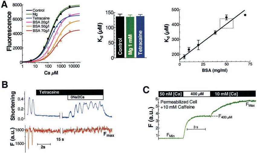
Shannon et al Local SR Depletions 41
Figure 1. Fluorescence signals from
intra-SR Fluo-5N. A, Fluorescence is
abolished by 10 mmol/L caffeine applica-
tion at striations but not in non-SR
regions. B, Enlargement of part of A
(with intensity profile). Striations are peri-
odic every 1.9 m, consistent with
intrasarcomeric spacing and are abol-
ished by caffeine.
deesterification and outward leak of cytosolic indicator, all at 37°C.17 cence is primarily from the SR (where expected [Ca2⫹]SR is
All experiments were performed at 23°C. Fluorescence was mea- ⬇1 mmol/L). This SR localization is supported by 3 obser-
sured both on confocal and epifluorescence microscopes at excita-
vations. First, the fluorescence pattern is localized to Z lines
tion⫽488 nm, emission ⬎500 nm for Fluo-5N. Cells exposed to
Di-8-ANNEPS to identify transverse tubules were imaged with and transverse tubules (stained by the lipophilic fluorophore
excitation⫽488 nm and fluorescence emission at ⬎600 nm. The Di-8-ANNEPS), exactly as expected for cardiac junctional
image in Figure 2C was deconvolved as in Gonzalez et al.18 SR (JSR, Figures 2A and 2B). That is, there is higher
All reagents and chemicals were purchased from Sigma Chemical fluorescence near transverse tubules (site of capacious JSR)
Company except as indicated. Cell superfusate contained
and weaker fluorescence strands through the sarcomere (site
(in mmol/L) CaCl2 2, NaCl 140, KCl 4, MgCl2 1, HEPES 5, and
glucose 10 (pH 7.4). Statistical significance was tested with two-way of more wispy, less dense longitudinal or free SR, FSR).
ANOVA. A value of P⬍0.05 was considered significant. Moreover, [Ca2⫹]SR is expected to be the same throughout the
resting SR, so brightness may reflect the expected ultrastruc-
Fluo-5N Calibration tural SR organization. The periodic bright fluorescence re-
In vitro calibration was performed in intracellular solutions (control, gions (1.9-m spacing; Figure 1B) correspond to sarcomeric
in mmol/L, KCl 140, HEPES 40 [pH 7.2]) with the [Ca2⫹] indicated.
spacing. Second, this distinct pattern is abolished by rapid
Solutions were suspended within a fluorometer, and Fluo-5N fluo-
rescence was measured under the indicated conditions. In vivo, FMax application of 10 mmol/L caffeine, which causes SR Ca2⫹
was determined in an intact myocyte by adding 1 mol/L isoproter- release to the cytosol (Figure 1B). The remaining sporadic
enol (ISO), then subsequently adding 0.5 mmol/L tetracaine to block bright spots and perinuclear rings that are little affected by
SR Ca2⫹ leak, and finally [Na⫹]o was removed to raise both [Ca2⫹]i caffeine (and avoided in Figure 1B) probably reflect Fluo-5N
and [Ca2⫹]SR. In vivo, Kd was estimated in permeabilized cells (50
g/mL saponin) with 10 mmol/L caffeine to allow [Ca2⫹] equilibra-
compartmentalization in non-SR regions with high [Ca2⫹].
tion across the SR. Intracellular solution, as previously described,19 Third, permeabilization of the sarcolemma with saponin does
included (in mmol/L) Cs-glutamate 200, HEPES 10 (pH 7.2), not alter the fluorescence pattern appreciably (not shown).
Mg-ATP 5, phosphocreatine ditris 5, MgCl2 0.5, glutathione 10 Thus, the image at high zoom in Figure 2C illustrates the
(reduced form), 5 U/mL creatine phosphokinase, 8% dextran (MW anatomy of the SR as it wraps around the myofilaments (dark
40 000), with variable free [Ca2⫹], 1 mol/L FCCP, 1 mol/L
ruthenium red, 2 mol/L oligomycin, and 8 mol/L cyclosporine to
regions within the sarcomere between wispy areas of FSR)
limit mitochondrial Ca2⫹ uptake. with the junctional SR located at the Z lines.
Figure 3A shows whole-cell fluorescence changes that reflect
Results transient [Ca2⫹]SR depletions during twitches and also caffeine-
The rabbit ventricular myocyte shown in Figure 1A is loaded induced [Ca2⫹]SR depletions. After caffeine removal, [Ca2⫹]SR
with Fluo-5N, a low-affinity Ca2⫹ indicator that has ex- only partially recovers unless pacing is resumed. The caffeine-
tremely low fluorescence when Ca2⫹-free.17 While there is sensitive fluorescence (attributed to the SR) at 0.5-Hz stimula-
surely some indicator in cytosol and mitochondria, [Ca2⫹] is tion is 53.2⫾1.8% (n⫽14, Figure 3A) of the total. All subse-
submicromolar in these compartments, such that the fluores- quent data refer only to this caffeine-sensitive component.
Downloaded from http://circres.ahajournals.org/ by guest on September 28, 2015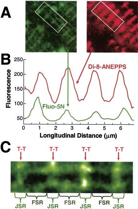
42 Circulation Research July 11, 2003
Figure 3. [Ca2⫹]SR measurements. A, Whole-cell [Ca2⫹]SR during
twitches evoked at 1 Hz (black) or 0.5 Hz (red), interrupted dur-
ing application of 10 mmol/L caffeine (used to deplete SR Ca2⫹).
Pacing was resumed only for 0.5 Hz. B, Single-twitch [Ca2⫹]SR
signal (1 Hz, average of 4) as fraction of caffeine-sensitive dia-
stolic fluorescence at 1 Hz (Fd1). Exponential curve fit (⫽100
ms) of [Ca2⫹]SR recovery in red. C, Measurement at a single
junction (as identified in Figure 1D), showing similar characteris-
tics (with 20 mmol/L butanedione monoxime, 4 ms/line). D, Sep-
arate signals analyzed from JSR and FSR regions. Traces are
average from 93 JSR and 29 FSR regions, with amplitude
normalized.
This is consistent with a [Ca2⫹]SR-dependent increase in frac-
Figure 2. Cellular anatomy of the SR. A and B, Images of Fluo-5N– tional release and also clearly demonstrates that [Ca2⫹]SR deple-
loaded myocytes before and after Di-8-ANNEPS addition to visual-
ize transverse tubules. Di-8-ANNEPS colocalizes with Fluo-5N sig-
tion is incomplete during a twitch.1,2 The of refilling (Figure
nal. C, Zoom of region in B, illustrating transverse tubules (T-T) 4E) speeds up with frequency, consistent with frequency-
with junctional and free SR regions in between (JSR and FSR). dependent acceleration of relaxation and [Ca2⫹]i decline.3
Because F may not be linearly related to [Ca2⫹]SR, calibrations
During the twitch, fluorescence declines to a minimum of ⬇75% are needed for greater quantitative evaluation. In vitro, Fluo-5N
at ⬇100 ms after [Ca2⫹]SR starts to decline and recovers with a Ca2⫹ affinity (Kd⫽135 mol/L) in solution measured in a
time constant () of ⬇100 ms (Figure 3B). Figure 3C shows that fluorometer is not altered by Mg or tetracaine (used below),
the [Ca2⫹]SR depletion measured at a single SR junction in although tetracaine partially quenches fluorescence (Figure 5A).
confocal microscopy is quite similar to the global [Ca2⫹]SR The presence of protein decreases both maximal fluorescence as
signal. We refer to these local [Ca2⫹]SR depletions as “Ca2⫹ well as apparent affinity of Fluo-5N for Ca2⫹ (Figure 5A). At
scraps” (since they are the intra-SR correlate of evoked Ca2⫹ cellular protein concentrations (50 to 100 mg/mL), Fluo-5N
sparks).20 Moreover, when Ca2⫹ scraps at junctional and free SR affinity is reduced ⬇3-fold (typical for fluorescent Ca2⫹ indica-
regions (as in Figure 2C) are compared, there is little kinetic tors in cells).22,23
difference (Figure 3D). This indicates that longitudinal diffusion Maximal fluorescence (FMax) in myocytes was defined by
within the SR is faster than we could readily detect (assuming stimulating SR Ca2⫹-ATPase by ISO and blocking SR Ca2⫹
SR Ca2⫹ release occurs at JSR).21 Thus, Ca2⫹ diffusion from free release by tetracaine (which dramatically increases SR Ca2⫹
to junctional SR does not appreciably limit SR Ca2⫹ availability content).24 Note that F did not rise much on tetracaine addition
for release, even at a single twitch. (even when we accounted for the modest quench by tetracaine).
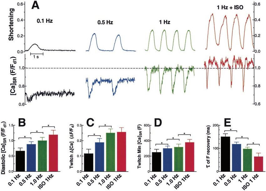
Figure 4A shows [Ca2⫹]SR and contractions at different pacing To further raise [Ca2⫹]SR, extracellular Na⫹ was abruptly re-
frequencies. As frequency increases, diastolic [Ca2⫹]SR increases, moved (causing Ca2⫹ entry via Na⫹-Ca2⫹ exchange), which
as does the extent of [Ca2⫹]SR depletion and the contraction caused spontaneous contractions and corresponding Ca2⫹ deple-
amplitude. With -adrenergic activation by ISO, there was a tions (Figure 5B). Under these conditions (with the RyR inhib-
further increase in diastolic [Ca2⫹]SR and extent of [Ca2⫹]SR ited), the SR Ca2⫹ pump should approach a limiting [Ca2⫹]SR/
depletion and faster [Ca2⫹]SR recovery ( decreased from 155 to [Ca2⫹]i gradient,25 and [Ca2⫹]SR should rise by the same factor as
70 ms), consistent with the expected acceleration of SR Ca2⫹- [Ca2⫹]i (and the elevation of average [Ca2⫹]i is indicated by the
ATPase by ISO. Figures 4B through 4D show mean diastolic F cellular contracture in Figure 5B). Since F still did not increase
(Fd versus that at 1 Hz, Fd1), peak fractional depletion (⌬F/ appreciably, despite the substantial rise in [Ca2⫹]SRT expected
Fd⫽25⫾2.6% for 1 Hz), and the minimum systolic value of with this protocol, FMax represents saturation of intra-SR Fluo-
[Ca2⫹]SR. All three parameters increase with frequency and ISO. 5N. FMin is taken as FCaf.
Downloaded from http://circres.ahajournals.org/ by guest on September 28, 2015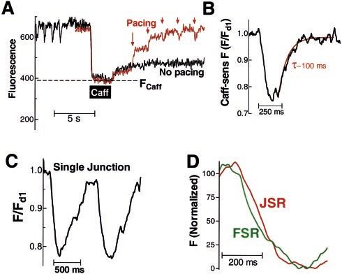
Shannon et al Local SR Depletions 43
Figure 4. Pacing frequency and ISO enhance [Ca2⫹]SR and depletion. A, Cell contraction (arbitrary units) and Fluo-5N fluorescence for
steady-state twitches. B, Mean diastolic fluorescence, where subscripts refer to frequency or ISO at 1 Hz (n⫽14 cells; normalized to
Fd1). C, Fractional decline in [Ca2⫹]SR during steady-state twitches (normalized to diastolic F, Fd; n⫽14). D, Minimum caffeine-sensitive F
during systole, where Fd-ISO⫽518⫾68. E, Time constant for recovery of [Ca2⫹]SR during twitches. *P⬍0.05.
To test whether Kd⫽400 mol/L is appropriate for intra-SR ected to be ⬇95% depleted early during the twitch, thereby
Fluo-5N, we used permeabilized myocytes (with RyRs opened limiting further SR Ca2⫹ release.26
by caffeine). When [Ca2⫹] was stepped from 50 nmol/L (FMin) to
400 mol/L (F400) to 10 mmol/L (FMax; Figure 5C), we found that Discussion
F400 was, on average, 47.5% of FMax⫺FMin. This confirms that We report the first direct measurements of [Ca2⫹]SR during
400 mol/L is an appropriate Kd in situ.
individual contractions and with subsarcomeric spatial reso-
Using these calibrations, we found that diastolic [Ca2⫹]SR
lution. These measurements and method provide valuable
increased from 0.48 to 0.92 to 1.65 mmol/L from 0.1 to 1 Hz
new quantitative information that is at the very heart of
(Figures 6A and 6B). These values are similar to time-averaged
cardiac ECC. It has also been argued that SR Ca2⫹ depletion
whole-heart NMR estimates of [Ca2⫹]SR (1.5 mmol/L)12 and our
does not participate in the shutoff of SR Ca2⫹ release,21 and
estimates (⬇1 mmol/L), based on [Ca2⫹]SR and [Ca2⫹]SRT in SR
vesicles plus cellular [Ca2⫹]SRT.25 However, both of those results indeed we show that SR Ca2⫹ release stops at local [Ca2⫹]SR
lacked kinetic or spatial information. ⬇0.4 mmol/L when there is still a large driving force for SR
The extent of [Ca2⫹]SR depletion increased from 24% to Ca2⫹ release. Cardiac ECC models have assumed that there is
63% over this range of frequencies. Diastolic ISO data were a major time delay (up to seconds) between recovery of
not calibrated because Fd was too near FMax. Minimum [Ca2⫹]SR near Ca2⫹ uptake sites (FSR) and at release sites
[Ca2⫹]SR attained during a twitch varied from 0.36 to (JSR).27 Such purported major time lags between JSR and
0.61 mmol/L (even with ISO). These [Ca2⫹]SR measurements FSR do not seem to occur during release (Figure 3C), and the
support previous, less direct [Ca2⫹]SRT measurements that [Ca2⫹]SR is restored rapidly during the twitch (even in the
suggested incomplete SR Ca2⫹ depletion.1,2,4 Importantly, JSR). The apparent delay or restitution of SR Ca2⫹ release
Figures 3B and 3C also indicate that JSR in individual (eg, at premature heartbeats) is probably due mainly to
junctions depletes only partially. These data are not consistent recovery of RyR (and/or L-type Ca2⫹ channel) availability,
with recent models of ECC where local [Ca2⫹]SR was proj- rather than the amount of releasable SR Ca2⫹.
Downloaded from http://circres.ahajournals.org/ by guest on September 28, 2015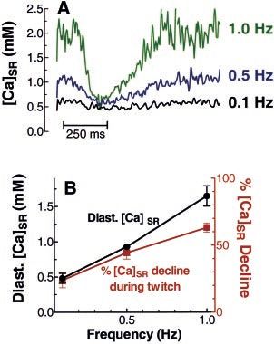
44 Circulation Research July 11, 2003
Figure 5. Fluo-5N calibration and [Ca2⫹]SR. A, In vitro calibration in intracellular solutions with the [Ca2⫹] indicated ⫾1 mmol/L MgCl2 or
0.5 mmol/L tetracaine and different bovine serum albumin (BSA) concentrations. B, In vivo FMax determination in an intact myocyte with
1 mol/L ISO, then 0.5 mmol/L tetracaine is added to block SR Ca2⫹ leak and [Na]o is removed to drive [Ca2⫹]i and [Ca2⫹]SR up (mean
FMax⫽1.37⫾0.09⫻Fd1). C, Permeabilized cell (50 g/mL saponin) with 10 mmol/L caffeine to allow [Ca2⫹] equilibration across the SR.
We propose that [Ca2⫹]SR depletion is dynamically involved in to release appreciable SR Ca2⫹.1– 4,20,28 This demonstrates that
terminating SR Ca2⫹ release, due to direct effects on RyR gating luminal [Ca2⫹]SR dynamically modulates SR Ca2⫹ release (and
(not by exhausting available SR Ca2⫹). Indeed, the SR retains a leak) during both diastole and ECC.
Ca2⫹ reserve, which is pharmacologically accessible, as indi- The fact that SR Ca2⫹ release does not go to completion even
cated by caffeine-induced Ca2⫹ transients, which completely locally (as expected for positive feedback) rules out substrate
deplete [Ca2⫹]SR (Figure 1B). When [Ca2⫹]SR is below 40% to limitation as the cause of release termination but leaves two
50% of its control value, resting SR Ca2⫹ leak (Ca2⫹ spark potential types of inactivation.29,30 One mechanism, stochastic
frequency) is very small and a normal Ca2⫹ current trigger fails attrition, would be when a sufficient number of Ca2⫹ channels in a
junction (L-type and RyR) close by chance to allow local [Ca2⫹]i to
fall and break the positive-feedback loop. This is unlikely to
produce reliable termination of SR Ca2⫹ release, given the high
number of channels at a junction (unless their gating is tightly
coupled).21,29–31 The second major class would be a time-dependent
RyR inactivation, which could depend explicitly on [Ca2⫹]i,
[Ca2⫹]SR, or both. There is evidence for [Ca2⫹]i-dependent inactiva-
tion (or adaptation).21 DelPrincipe et al32 found that after a global
cellular SR Ca2⫹ release, restitution required ⬎1 second and
suggested that SR Ca2⫹ depletion and slow functional repletion
were the likely explanation (because discrete local Ca2⫹ releases
showed much faster recovery). Our data indicate that the SR refills
rather rapidly and suggests that this restitution depends more on
recovery of RyR (or ICa) availability.
Interestingly, our own data demonstrate a trend upward in
[Ca2⫹]SR minimum during a twitch with increased release (Figure
4D). This would be consistent with a cytosolic Ca2⫹-dependent
inactivation site on the RyR, which binds more Ca2⫹, thus
Figure 6. Dynamic [Ca2⫹]SR profiles. A, Calibrated [Ca2⫹]SR sig-
nals. B, Mean diastolic [Ca2⫹]SR and fractional twitch depletion inactivating the channel faster and terminating release at a
at different frequencies. slightly higher [Ca2⫹]SR. However, our observation that release
Downloaded from http://circres.ahajournals.org/ by guest on September 28, 2015
Shannon et al Local SR Depletions 45
stops (and can only be very weakly activated) when [Ca2⫹]SR 9. Missiaen L, Taylor CW, Berridge MJ. Luminal Ca2⫹ promoting spontaneous
⬍0.5 mmol/L also suggests an equally important regulatory role Ca2⫹ release from inositol trisphosphate-sensitive stores in rat hepatocytes.
J Physiol. 1992;455:623–640.
for [Ca2⫹]SR (perhaps by influencing how RyR responds to 10. Tse FW, Tse A, Hille B. Cyclic Ca2⫹ changes in intracellular stores of gonado-
[Ca2⫹]i).5,6 Thus, both [Ca2⫹]SR and [Ca2⫹]i may both contribute tropes during gonadotropin-releasing hormone-stimulated Ca2⫹ oscillations. Proc
dynamically to the shutoff of SR Ca2⫹ release. A corollary is that Natl Acad Sci U S A. 1994;91:9750–9754.
11. Blatter LA. Depletion and filling of intracellular calcium stores in vascular
both RyR availability and [Ca2⫹]SR must recover to achieve smooth muscle. Am J Physiol. 1995;268:C503–C512.
restitution. So far, our results suggest that [Ca2⫹]SR recovers 12. Chen W, Steenbergen C, Levy LA, Vance J, London RE, Murphy E. Mea-
somewhat faster ( ⬇100 ms) than does the RyR ( ⬇300 surement of free Ca2⫹ in sarcoplasmic reticulum in perfused rabbit heart loaded
with 1,2-bis(2-amino-5,6-difluorophenoxy)ethane-N,N,N⬘,N⬘-tetraacetic acid by
ms).32,33 19F NMR. J Biol Chem. 1996;271:7398–7403.
[Ca2⫹]SR dictates the effect of SR Ca2⫹ on RyR gating, the 13. Golovina VA, Blaustein MP. Spatially and functionally distinct Ca2⫹ stores in
driving force for Ca2⫹ release, but it is also limited thermody- sarcoplasmic and endoplasmic reticulum. Science. 1997;275:1643–1648.
14. Miyawaki A, Llopis J, Heim R, McCaffery JM, Adams JA, Ikura M, Tsien RY.
namically. For diastolic [Ca2⫹]i⫽100 to 150 nmol/L at 0.5 to 1
Fluorescent indicators for Ca2⫹ based on green fluorescent proteins and calmod-
Hz (respectively), the [Ca2⫹]SR/[Ca2⫹]i gradient would be 9200 to ulin. Nature. 1997;388:882–887.
11 000. A gradient of 10 000 implies a high (78%) energetic 15. Robert V, De Giorgi F, Massimino ML, Cantini M, Pozzan T. Direct monitoring
efficiency for the SR Ca2⫹-ATPase (for 2 Ca2⫹/ATP) and a of the calcium concentration in the sarcoplasmic and endoplasmic reticulum of
skeletal muscle myotubes. J Biol Chem. 1998;273:30372–30378.
cytosolic ⌬GATP of 59 kJ/mol.25,34 16. Shmigol AV, Eisner DA, Wray S. Simultaneous measurements of changes in
Our data provide the first direct quantitative examination of sarcoplasmic reticulum and cytosolic [Ca2⫹] in rat uterine smooth muscle cells.
what is happening dynamically to local intra-SR free [Ca2⫹] J Physiol. 2001;531:707–713.
17. Kabbara AA, Allen DG. The use of the indicator fluo-5N to measure sarco-
during cardiac ECC. This is especially important because plasmic reticulum calcium in single muscle fibres of the cane toad. J Physiol.
[Ca2⫹]SR (rather than [Ca2⫹]SRT) is the central thermodynamic 2001;534:87–97.
parameter that governs buffering, allosteric regulation, driving 18. Gonzalez A, Kirsch WG, Shirokova N, Pizarro G, Stern MD, Rios E. The spark
and its ember: separately gated local components of Ca2⫹ release in skeletal
gradient, and transport limits. Thus, in cardiac myocytes, muscle. J Gen Physiol. 2000;115:139–158.
[Ca2⫹]SR is a major determinant of Ca2⫹ release and contraction. 19. Li Y, Kranias EG, Mignery GA, Bers DM. Protein kinase A phosphorylation of
Low [Ca2⫹]SR may also limit cardiac function in heart failure.35,36 the ryanodine receptor does not affect calcium sparks in mouse ventricular
Intra-SR Ca2⫹ diffusion is rapid, and local [Ca2⫹]SR never gets myocytes. Circ Res. 2002;90:309–316.
20. Cheng H, Lederer WJ, Cannell MB. Calcium sparks: elementary events
much less than ⬇50% of its diastolic value, even with strong underlying excitation-contraction coupling in heart muscle. Science. 1993;262:
activation of ECC. The less than complete depletion of local 740–744.
[Ca2⫹]SR during normal SR Ca2⫹ release implies a residual SR 21. Sham JSK, Song LS, Chen Y, Deng LH, Stern MD, Lakatta EG, Cheng HP.
Termination of Ca2⫹ release by a local inactivation of ryanodine receptors in
Ca2⫹ reserve that might be pharmacologically accessible for cardiac myocytes. Proc Natl Acad Sci U S A. 1998;95:15096–15101.
treatment of diseases such as heart failure. The experimental 22. Hove-Madsen L, Bers DM. Indo-1 binding to protein in permeabilized ventricular
approach described here should be very useful in further studies myocytes alters its spectral and Ca binding properties. Biophys J. 1992;63:89–97.
23. Harkins AB, Kurebayashi N, Baylor SM. Resting myoplasmic free calcium in
of SR Ca2⫹ in cardiac myocytes and other cell types. This novel frog skeletal muscle fibers estimated with fluo-3. Biophys J. 1993;65:865–881.
approach should allow new mechanistic and quantitative ques- 24. Overend CL, Eisner DA, O’Neill SC. The effect of tetracaine on spontaneous
tions to be addressed. Ca2⫹ release and sarcoplasmic reticulum calcium content in rat ventricular
myocytes. J Physiol. 1997;502:471–479.
25. Shannon TR, Bers DM. Assessment of intra-SR free [Ca] and buffering in rat
Acknowledgments heart. Biophys J. 1997;73:1524–1531.
This work was funded by NIH grant HL30077 and HL64098 26. Sobie EA, Dilly KW, Cruz J, Lederer WJ, Jafri MS. Termination of cardiac Ca2⫹
(D.M.B.) and AHA grant 0030381Z (T.R.S.). The authors thank Drs sparks: an investigative mathematical model of calcium-induced calcium release.
E. Ríos and L. Blatter for their help. Biophys J. 2002;83:59–78.
27. Luo CH, Rudy Y. A dynamic model of the cardiac ventricular action potential, I:
simulations of ionic currents and concentration changes. Circ Res. 1994;74:
References 1071–1096.
1. Bassani JWM, Yuan W, Bers DM. Fractional SR Ca release is regulated
28. Satoh H, Blatter LA, Bers DM. Effects of [Ca2⫹]i, SR Ca2⫹ load, and rest on Ca2⫹
by trigger Ca and SR Ca content in cardiac myocytes. Am J Physiol. spark frequency in ventricular myocytes. Am J Physiol. 1997;272:H657–H668.
1995;268:C1313–C1319. 29. Stern MD. Theory of excitation-contraction coupling in cardiac muscle. Biophys
2. Shannon TR, Ginsburg KS, Bers DM. Potentiation of fractional SR Ca release by J. 1992;63:497–517.
total and free intra-SR Ca concentration. Biophys J. 2000;78:334–343. 30. Bers DM. Cardiac excitation-contraction coupling. Nature. 2002;415:198–205.
3. Bers DM. Excitation-Contraction Coupling and Cardiac Contractile Force. 2nd 31. Marx SO, Gaburjakova J, Gaburjakova M, Henrikson C, Ondrias K, Marks AR.
ed. Dordrecht, Netherlands: Kluwer Academic Publishers; 2001. Coupled gating between cardiac calcium release channels (ryanodine receptors).
4. Trafford AW, Diaz ME, Eisner DA. Coordinated control of cell Ca2⫹ loading and Circ Res. 2001;88:1151–1158.
triggered release from the sarcoplasmic reticulum underlies the rapid inotropic 32. DelPrincipe F, Egger M, Niggli E. Calcium signalling in cardiac muscle: refrac-
response to increased L-type Ca2⫹ current. Circ Res. 2001;88:195–201. toriness revealed by coherent activation. Nat Cell Biol. 1999;6:323–329.
5. Lukyanenko V, Györke I, Györke S. Regulation of calcium release by calcium 33. Cheng H, Lederer MR, Lederer WJ, Cannell MB. Calcium sparks and [Ca2⫹]i
inside the sarcoplasmic reticulum in ventricular myocytes. Pflugers Arch. 1996; waves in cardiac myocytes. Am J Physiol. 1996;270:C148–C159.
432:1047–1054. 34. Allen DG, Morris PG, Orchard CH, Pirolo JS. A nuclear magnetic resonance
6. Györke I, Györke S. Regulation of the cardiac ryanodine receptor channel by study of metabolism in the ferret heart during hypoxia and inhibition of gly-
luminal Ca2⫹ involves luminal Ca2⫹ sensing sites. Biophys J. 1998;75: colysis. J Physiol. 1985;361:185–204.
2801–2810. 35. Pogwizd SM, Schlotthauer K, Li L, Yuan W, Bers DM. Arrhythmogenesis and
7. Varro A, Negretti N, Hester SB, Eisner DA. An estimate of the calcium content contractile dysfunction in heart failure: roles of sodium-calcium exchange, inward
of the sarcoplasmic reticulum in rat ventricular myocytes. Pflugers Arch. 1993; rectifier potassium current, and residual -adrenergic responsiveness. Circ Res.
423:158–160. 2001;88:1159–1167.
8. Somlyo AV, Gonzalez-Serratos HG, Shuman H, McClellan G, Somlyo AP. 36. Hobai IA, O’Rourke B. Decreased sarcoplasmic reticulum calcium content is
Calcium release and ionic changes in the sarcoplasmic reticulum of tetanized responsible for defective excitation-contraction coupling in canine heart failure.
muscle: an electron-probe study. J Cell Biol. 1981;90:577–594. Circulation. 2001;103:1577–1584.
Downloaded from http://circres.ahajournals.org/ by guest on September 28, 2015Ca2+ Scraps: Local Depletions of Free [Ca2+] in Cardiac Sarcoplasmic Reticulum During
Contractions Leave Substantial Ca2+ Reserve
Thomas R. Shannon, Tao Guo and Donald M. Bers
Circ Res. 2003;93:40-45; originally published online June 5, 2003;
doi: 10.1161/01.RES.0000079967.11815.19
Circulation Research is published by the American Heart Association, 7272 Greenville Avenue, Dallas, TX 75231
Copyright © 2003 American Heart Association, Inc. All rights reserved.
Print ISSN: 0009-7330. Online ISSN: 1524-4571
The online version of this article, along with updated information and services, is located on the
World Wide Web at:
http://circres.ahajournals.org/content/93/1/40
Permissions: Requests for permissions to reproduce figures, tables, or portions of articles originally published
in Circulation Research can be obtained via RightsLink, a service of the Copyright Clearance Center, not the
Editorial Office. Once the online version of the published article for which permission is being requested is
located, click Request Permissions in the middle column of the Web page under Services. Further information
about this process is available in the Permissions and Rights Question and Answer document.
Reprints: Information about reprints can be found online at:
http://www.lww.com/reprints
Subscriptions: Information about subscribing to Circulation Research is online at:
http://circres.ahajournals.org//subscriptions/
Downloaded from http://circres.ahajournals.org/ by guest on September 28, 2015You can also read