Chemoresistance Transmission via Exosome-Transferred MMP14 in Pancreatic Cancer
←
→
Page content transcription
If your browser does not render page correctly, please read the page content below
ORIGINAL RESEARCH
                                                                                                                                           published: 09 February 2022
                                                                                                                                        doi: 10.3389/fonc.2022.844648

                                              Chemoresistance Transmission via
                                              Exosome-Transferred MMP14 in
                                              Pancreatic Cancer
                                              Xinyuan Li , Kai Li , Mengmeng Li , Xiaoyu Lin , Yu Mei , Xuemei Huang and Huanjie Yang *

                                              School of Life Science and Technology, Harbin Institute of Technology, Harbin, China

                                              Pancreatic ductal adenocarcinoma (PDAC) is one of the deadliest malignancies.
                                              Gemcitabine is the most commonly used chemotherapy for the treatment of PDAC, but
                                              the development of drug resistance still remains challenging. Recently, exosomes have
                                              emerged as important mediators for intercellular communication. Exosomes affect
                                              recipient cells’ behavior through the engulfed cargos, however the specific cargos
                                              responsible for gemcitabine resistance in PDAC are poorly understood. Here, we
                                              reported that exosomes could transfer gemcitabine resistance via a metalloproteinase
                                              14 (MMP14)-dependent mechanism. MMP14 was identified as a major differentially
                                              secreted protein from the gemcitabine-resistant PDAC cells by comparative secretome.
                       Edited by:             It was packaged into the exosomes and transmitted from the chemoresistant cells to the
                           Jie Sun,           sensitive ones. The exosome-transferred MMP14 could enhance drug resistance and
  Wenzhou Medical University, China
                                              promotes the sphere-formation and migration abilities of the recipient sensitive PDAC
                     Reviewed by:
                          Hong Fan,
                                              cells. Mechanically, exosome-transferred MMP14 promotes the stability of CD44, the
         Southeast University, China          cancer stem cell marker in the recipient cells. Our results indicate that MMP14 is a key
                        Shan Wang,
                                              player for exosome-mediated transfer of gemcitabine resistance, thus targeting MMP14 in
   Guangxi Medical University Cancer
                     Hospital, China          exosomes may represent a novel strategy to limit gemcitabine resistance in PDAC.
                  *Correspondence:            Keywords: MMP14, exosome, gemcitabine resistance, CD44, pancreatic cancer
                        Huanjie Yang
                    yanghj@hit.edu.cn

                   Specialty section:         INTRODUCTION
         This article was submitted to
                      Cancer Genetics,        Pancreatic ductal adenocarcinoma (PDAC) is a devastating human malignancy with an average 5-
               a section of the journal       year survival rate less than 8% (1, 2). Due to early metastasis, most PDAC patients are diagnosed
                 Frontiers in Oncology        with advanced disease, which are not suitable for surgical resection (3). Gemcitabine represents the
      Received: 28 December 2021              first-line treatment of PDAC, but drug resistance is a major obstacle in improving the patient’s
        Accepted: 17 January 2022             response (4).
       Published: 09 February 2022                Recently, exosomes have emerged as important mediators for cell-to-cell communication (5).
                               Citation:      Exosomes engulf biologically active cargos including proteins, RNAs and lipids, which can be
          Li X, Li K, Li M, Lin X, Mei Y,     uptaken by adjacent cells and affect their behavior (6). For example, tumor-derived exosome-
          Huang X and Yang H (2022)
                                              transferred lncARSR has been reported to transmit sunitinib resistance from drug resistant cancer
       Chemoresistance Transmission
    via Exosome-Transferred MMP14
                                              cells to sensitive ones (7). Exosome-mediated EphA2 transmission transfers gemcitabine resistance
                   in Pancreatic Cancer.      in PDAC (8). In addition, exosomes shed from tumor microenvironment were found to promote
              Front. Oncol. 12:844648.        the stemness, epithelial-mesenchymal transition (EMT), metastasis and chemotherapy resistance of
      doi: 10.3389/fonc.2022.844648           cancer cells (9, 10).

Frontiers in Oncology | www.frontiersin.org                                          1                                      February 2022 | Volume 12 | Article 844648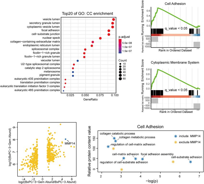
Li et al.                                                                                 Exosome-Transferred MMP14 Promotes Chemoresistance

    Matrix metalloproteinase 14 (MMP14), also known as                  70 minutes to remove any exosomes from the serum in advance,
membrane-type 1 MMP (MT1-MMP), is a transmembrane                       the medium was centrifuged at 500 g for 5 minutes to remove any
Zn2+-dependent MMP. MMP14 is localized in the leading edge              cell contamination. The resulting supernatants were centrifuged at
of migrating cancer cells where it proceeds extracellular matrix        12 000 g for 20 minutes to remove any possible apoptotic bodies
(ECM) remodeling by degrading protein components of the                 and large cell debris. The exosomes were collected as pellets after
ECM and promotes cancer cell migration, invasion and                    ultracentrifugation at 100 000 g for 70 minutes, then washed in
metastasis (11, 12). Studies also indicate that MMP14 regulates         PBS and pelleted again by ultracentrifugation.
cell motility, cancer stemness and other important biological               For PEG approach, appropriate amount of 5X PEG8000 was
processes non-proteolytically (13–17).                                  thoroughly mixed with the 48-hour cell culture medium to a final
    To investigate the role of extracellular components in              1x PEG8000 concentration. After incubation at 4°C for 12 hours,
modulating gemcitabine resistance, we performed LC/MS using             the samples were centrifuged at 12 000 g for 10 minutes. The
parental BxPC-3 and its subline BxPC-3-Gem which developed              supernatant was discarded as much as possible and exosome
gemcitabine resistance (18, 19) and found that MMP14 was a              pellets were collected, and then washed in PBS and pelleted again
major differential protein shed by resistant BxPC-3-Gem cells           by centrifugation.
versa parental BxPC-3 cells. As MMP14 is a membrane-bound                   Exosome preparations were verified by electron microscopy
protein, we hypothesized that MMP14 might be transferred via            (Quanta GEG250, FEI, Hillsboro, USA) and the size and particle
exosomes from chemoresistant PDAC cells, which might affect             concentration were analyzed using the ZetaPlus nanoparticle
the chemoresistance of the surrounding PDAC sensitive cells.            characterization system (Brookhaven, NY, USA).
Thus, we analyzed the influence of exosome-transferred MMP14
on recipient cells’ response to gemcitabine. Our results indicate       Exosome Staining and Quantification
that MMP14 is a key player for exosome-mediated transfer of             The isolated exosomes were stained with fluorescent dye PKH67
gemcitabine resistance.                                                 (Sigma, Saint Louis, USA) according to the manufacturer’s
                                                                        protocol. Briefly, exosomal protein concentration was
                                                                        determined by using Pierce BCA Protein Detection Kit
                                                                        (Thermo, Rockford, USA). Exosomes (20 mg) were suspended
MATERIALS AND METHODS                                                   in 1 mL a Diluent C and incubated with equal volume of Diluent
                                                                        C containing 5 mL of PK67 for 5 min. Serum (2 mL) was added to
Cell Lines and Cell Culture                                             terminate the staining. After washing with 1×PBS and
Human PDAC cell lines BxPC-3 and previously established                 centrifugation at 4°C, 120,000 g for 70 min, the stained
gemcitabine-resistant subline BxPC-3-Gem (18) were cultured             exosomes were re-suspended in 1×PBS.
in RPMI-1640 medium (Gibco, BRL Co. Ltd., USA) containing
10% fetal bovine serum (FBS) (Gibco, BRL Co. Ltd., USA)). Mia-          Constructs and Transfection
PaCa2, PANC-1 and HEK293T cells were cultured in Dulbecco’s             MMP14 overexpression vector (GFP-MMP14) was constructed
Modified Eagle’s Medium (DMEM) (Gibco, BRL Co. Ltd., USA)                by cloning the full-length cDNA of MMP14 into the Bgl II/EcoR
supplemented with 10% FBS. Cells were cultured in a humid               I sites of pEGFP-C1 vector. The nucleotides targeting MMP14
atmosphere containing 5% CO2 at 37°C. All cells were tested for         and negative control were synthesized by GenePharma
mycoplasma at regular intervals.                                        (Shanghai, China). The sequences of primers and siRNAs are
                                                                        listed in Table S1. Transient transfection was mediated by
Stable Cell Lines                                                       Lipofectamine 3000 (Invitrogen, Eugene, OR, USA) in
The plasmid pLVSIN-CMV-puro carrying GFP (pLVSIN-GFP)                   pLVSIN-RFP-BxPC-3-Gem, pLVSIN-GFP-BxPC-3, PANC-1
was constructed as described previously (19). pLVSIN-CMV-               and BxPC-3-Gem cells following the manufacturer’s protocol.
puro carrying RFP (pLVSIN-RFP) was constructed using the
same approach. Stable cell lines were established through               Sphere Formation and Colony
lentiviral transduction. Briefly, the constructed vectors and            Formation Assay
lentiviral packaging mix (VSV-G plasmid and Gag-Pol                     For the sphere formation assay, cells (500/well) were seeded into
plasmid) were co-transfected into HEK293T cells. The                    the ultra-low attachment 6-well plates (Corning, Inc., Corning,
supernatants containing lentiviruses were collected, filtered,           NY, USA) and cultured in DMEM-F12 medium (Gibco, Grand
and added into BxPC-3 and BxPC-3-Gem cells for 2 days. The              Island, NY, USA), containing 2% B27 (Gibco, MD, USA), 10 ng/
transduced cells (pLVSIN-RFP-BxPC-3-Gem, pLVSIN-GFP-                    mL of epidermal growth factor (EGF; Gibco, MD, USA), and 10
BxPC-3) were selected with puromycin (Santa Cruz,                       ng/mL of basic fibroblast growth factor (FGF; Gibco, MD, USA).
Texas, USA).                                                            After 14 days of culture, the spheres with diameter > 75 mM were
                                                                        counted. For colony formation assay, BxPC-3-GFP and BxPC-3-
Exosome Purification, Characterization,                                  Gem-RFP cells were seeded into 6-well plates (Corning, Inc.,
and Analysis                                                            Corning, NY, USA) individually or together, cultured for 14
Exosomes were i so late d f ro m PDAC c ell lin es by                   days, thereafter treated with gemcitabine for 72 hours. The
ultracentrifugation or PEG. After 48-hour cell culture in               fluorescence of the colonies was detected by the inverted
exosome-free medium, which was centrifuged at 100 000 g for             fluorescence microscope (IX71, Olympus Corporation, Japan).

Frontiers in Oncology | www.frontiersin.org                         2                                February 2022 | Volume 12 | Article 844648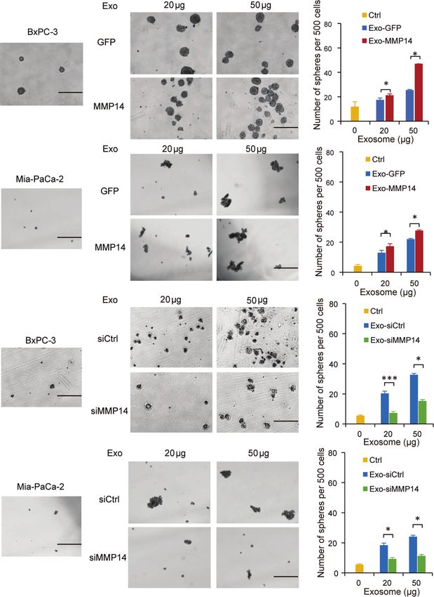
Li et al.                                                                                    Exosome-Transferred MMP14 Promotes Chemoresistance

Transwell Assay                                                            significant. All analyses were performed using SPSS v.17.0
The PDAC cells were seeded in the upper chamber of Transwell               software (SPSS Inc.).
(Costar Corp., Cambridge, MA, USA) and allowed to translocate
toward medium containing 20% FBS in the lower chamber for 48
hours. 4% formaldehyde and 0.5% crystal violet were used to fix
and stain the cells that migrated to the lower surface.                    RESULTS

RT-qPCR and Western Blot                                                   MMP14 Is a Predominant Protein Secreted
Total RNA was isolated using Trizol (Invitrogen, Eugene, OR,               From Chemoresistant PDAC Cells
USA) and reverse-transcribed (RT) into cDNA using ReverTra                 Our previous study indicates that the chemoresistant PDAC cells
Ace (TOYOBO, Japan). RT-qPCR was performed using SYBR                      secreted vital factors to enhance the resistance of the sensitive
Premix Ex Taq (Takara, Otsu, Shiga, Japan) on ViiA7 Real-time              PDAC cells (19). To understand the molecular mechanism, we
PCR System (Applied Biosystems Inc., Foster City, CA, USA).                re-visited and analyzed our comparative secretome data, and
GAPDH was used as the internal control for mRNA. Detailed                  obtained the network of proteins based on cell components and
information about the primers is shown in Table S1.                        molecular function (Supplementary Figures 1A, B). More
   Cell lysis and Western blot were conducted as previously                proteins of BxPC-3-Gem cells were associated with cell
described (18). Briefly, about 20-40 mg proteins per well were              adhesion, growth factor and receptor ligand activity, cytokine
resolved by SDS/PAGE and transferred on PVDF membranes                     receptor binding, clathrin binding and actin binding
(Millipore, Darmstadt, Germany). The membranes were                        (Supplementary Figures 1A, B). Gene Ontology (GO) analysis
incubated with antibodies against TSG101 (Proteintech,                     showed that the differential proteins of gemcitabine resistant
Wuhan, China), CD44 (Abcam, Cambridge, UK), MMP14                          cells were enriched in vesicle lumen, secretory granule lumen and
(Abcam, Cambridge, UK), and Activin A (ThermoFisher,                       cytoplasmic vesicle lumen relative to parental cells by cellular
Rockford, USA). b-actin (Santa Cruz, CA, USA) was used as                  component (Figure 1A). Because these multivesicular
loading control. Band intensity was quantified using ImageJ                 components are involved in the packaging of exosomes (21),
software (NIH).                                                            this result suggested that some differential proteins in the
                                                                           resistant cells might be secreted via exosomes. Among the
Comparative Secretome Analysis                                             differential proteins, MMP14 drew our attention because it was
The differentially secreted proteins in BxPC-3-gem vs BxPC-3               among the top 3 differentially expressed proteins in BxPC-3-
cells were expressed by fold change, and |log (FC)| > 1 was used           Gem in relative to parental cells (Figure 1B). To compare the
as the cut-off value. All statistical analyses were conducted with R       functions of the differential proteins, Gene Set Enrichment
software (Version 4.1) and Bioconductor version 4.0. KEGG                  Analysis (GSEA) analysis was performed. Results showed that
enrichment and GO function annotation analysis were                        BxPC-3-Gem cells secreted more proteins which were associated
performed by R package “clusterProfiler” (20). KEGG or GO                   with cell adhesion and cytoplasmic membrane system relative to
terms with BH-corrected p
Li et al.                                                                                                  Exosome-Transferred MMP14 Promotes Chemoresistance

                       A                                                                             C

                                                                                                     D

                       B                                              E

  FIGURE 1 | MMP14 was identified as the major differentially secreted protein by comparative secretome. (A) Gene Ontology (GO) functional enrichment (Cell
  Components, CC) for differential proteins in the conditioned medium of BxPC-3-Gem in relative to parental cells. Top 20 functions were exhibited in the plot. (B)
  Scatter plot of differentially secreted proteins in fold change of BxPC-3-GEM vs. BxPC-3 cells (x-axis) against BxPC-3-GEM cells (y-axis). The dot representing
  MMP14 was shown (blue dot). (C, D) GSEA analysis showed that differential proteins in the conditioned medium of BxPC-3-Gem vs BxPC-3 cells were enriched in
  cell adhesion system (C) and cytoplasmic membrane system (D) (q < 0.05, Bonferroni method; rank ordered by ratio of BxPC-3-Gem/BxPC-3). (E) Point plots of
  molecular functions involved in cell adhesion system. Function pathways included MMP14 (blue stars) or excluded MMP14 (orange star) in cell adhesion system were
  shown.

subjected to Western blot. Exosomes were confirmed by the                             gemcitabine-resistant BxPC-3-Gem cells. Using confocal
expression of exosome marker, the tumor susceptibility gene                          microscopy, we observed PKH67-dyed exosomes internalization
(TSG101), while MMP14 was detected in the portion of exosome                         into BxPC-3 and Mia-PaCa-2 cells within 48-hour co-incubation
pellets (Figure 2C), but not in the supernatant (Figure 2D).                         (Figure 3A). To determine whether exosome-transferred MMP14
Furthermore, more exosomes were secreted from chemoresistant                         could confer the resistant phenotype to recipient sensitive PDAC cells,
BxPC-3-Gem than parental cells through a quantified analysis of                       the exosome donor cells BxPC-3-Gem were overexpressed with
exosomes. Equal amounts of cells were indicated by equal                             MMP14 (Figure 3B) or knocked down of MMP14 (Figure 3C).
loading control in the two cell lines (Figures 2C, E). In                            The isolated exosomes carried more MMP14 proteins when it was
addition, higher level of MMP14 was detected in both the                             overexpressed, while less exosome-MMP14 proteins were detected
exosomes (Figure 2C) and whole cell lysates (Figure 2E) of                           when MMP14 was knocked down in BxPC-3-Gem cells compared
the resistant cells in comparison with parental cells.                               with the control (Figures 3D, E). MTT assay showed that the collected
                                                                                     exosomes had no effect on the proliferation of recipient Bx-PC-3 or
Exosome-Transferred MMP14 Confers to                                                 Mia-PaCa-2 cells upon gemcitabine treatment no matter whether
Gemcitabine Resistance of Recipient Cells                                            MMP14 was overexpressed or knocked down in the donor resistant
Neighboring cells can uptake exosomes (22). To evaluate whether the                  cells (Supplementary Figures 2A, B). To see whether a long period of
exosomes secreted from the resistant PDAC cells can be internalized                  incubation with exosomes could educate the recipient sensitive cells in
by the neighboring sensitive cells, BxPC-3 and Mia-PaCa-2cells were                  obtaining resistance to gemcitabine, co-culture colony formation
incubated with PKH67-dyed exosomes isolated from the                                 assay was performed using GFP-labeled sensitive BxPC-3 cells and

Frontiers in Oncology | www.frontiersin.org                                      4                                      February 2022 | Volume 12 | Article 844648
Li et al. Exosome-Transferred MMP14 Promotes Chemoresistance
A
B C
D E
FIGURE 2 | MMP14 was heavily secreted via exosome by BxPC-3-Gem cells. (A) NanoSight particle tracking analysis of the size distributions and concentration of
exosomes extracted from BxPC-3 (left) and BxPC-3-Gem cells (right) by ultracentrifugation. (B) Representative electron microscopy images of exosomes secreted by
BxPC-3 and BxPC-3-Gem cells by ultracentrifugation. Scale bar, 200 nm. (C) Immunoblotting assay of MMP14 expression in the exosomes extracted using PEG
and ultracentrifugation methods. TSG101 was used as exosome marker. (D) Immunoblotting assay of MMP14 and Activin A expression in the supernatant.
Secretory factor Activin A was used as the positive control. (E) Immunoblotting assay of MMP14 expression in whole cell lysis. Actin was used as the loading control.
Representative images were from three independent experiments.
RFP-labeled resistant BxPC-3-Gem cells. The BxPC-3 cells became performed in the recipient cells post exosome uptake from the
resistant to gemcitabine when co-cultured with gemcitabine-resistant resistant cells. The sphere-forming abilities were increased in
cells for two weeks and MMP14 overexpression in the resistant cells BxPC-3 and Mia-PaCa2 cells through uptaking exosomes from
further increased this effect (Figures 3F, G). To further address resistant PANC-1 cells, which were further enhanced when
whether exosome-transferred MMP14 was a key intercellular MMP14 was overexpressed in those cells (Figures 4A, B).
messenger for mediating chemoresistance, MMP14 was knocked Knockdown approach confirmed that exosome-transferred
down in RFP-labeled BxPC-3-Gem cells. The colony formation of MMP14 participated in the regulation of cancer stemness of
BxPC-3 cells post gemcitabine treatment was increased when co- the recipient cells. The sphere-forming abilities were greatly
cultured with RFP-labeled resistant BxPC-3-Gem cells, but was increased in BxPC-3 and Mia-PaCa2 cells by uptaking
significantly abrogated when MMP14 was knocked down in the exosomes from BxPC-3-Gem cells, which were dramatically
resistant cells (Figures 3H, I). We noticed that the abrogation effect decreased when MMP14 was knocked down in those cells
was comparable to MMP14 knockdown efficiency, indicating that (Figures 4C, D). GSEA enrichment analysis of comparative
MMP14 is a key player in exosome-mediating transmission secretome also showed that MMP14 was involved in the
of chemoresistance. regulation of cell growth and differentiation of stem cells
(Supplementary Figures 3A-C; Tables S4, S5).
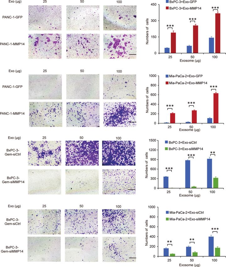
Exosome-Transferred MMP14 Promotes Transwell assays showed that the exosomes shed by MMP14-
Recipient Cells Sphere-Formation overexpressing PANC-1 cells could improve the migration abilities
And Migration of the sensitive BxPC-3 and Mia-PaCa-2 cells (Figures 5A, B).
Cancer cells that acquire cancer stem cell-like properties confer Knockdown approach confirmed that the exosomes from MMP14
to chemoresistance (23). To determine whether exosome- knockdown BxPC-3-Gem cells led to decreased migration abilities
transferred MMP14 could educate the recipient cells to obtain of BxPC-3 and Mia-PaCa-2 cells in comparison with that from
more aggressive properties, sphere formation assay was control BxPC-3-Gemcells (Figures 5C, D). Scratch assay confirmed
Frontiers in Oncology | www.frontiersin.org 5 February 2022 | Volume 12 | Article 844648
Li et al.                                                                                                  Exosome-Transferred MMP14 Promotes Chemoresistance

                    A

                    B                                                                            C

                    D                                                                            E

                    F                                                                            G

                    H                                                                            I

  FIGURE 3 | Exosome-transferred MMP14 promotes chemoresistance of the sensitive PDAC cells. (A) Fluorescent observation of BxPC-3 and Mia-PaCa-2 cells
  after 48-hour incubation with PKH67-labeled (green) exosomes derived from BxPC-3-Gem cells, the nucleus of BxPC-3 and Mia-PaCa-2 cells were stained by DAPI
  (blue). Scale bar, 25 mm. (B, C) Immunoblotting assays to test MMP14 overexpression (B) or knockdown in BxPC-3-Gem cells (C). (D, E) Immunoblotting assays to
  determine MMP14 levels in the exosomes obtained from the cells described in (B), and (C) (F, G) Colony formation in BxPC-3-GFP cells co-cultured with MMP14-
  overexpressing or control BxPC-3-Gem-RFP cells at a ratio of 1:1 for 2 weeks, followed by treatment with gemcitabine (40 nM) for 72 hours. Representative images
  from three independent experiments (F) and average numbers of colonies (G) were shown. (H, I) Colony formation of BxPC-3-GFP cells co-cultured with MMP14
  knockdown (si-MMP14) or control BxPC-3-Gem-RFP cells at a ratio of 1:1 for 2 weeks, followed by treatment with gemcitabine (40 nM) for 72 hours. Representative
  images from three independent experiments (H) and average numbers of colonies (I) were shown. Data in E, G are presented as mean ± SD, ***P < 0.001.

Frontiers in Oncology | www.frontiersin.org                                      6                                     February 2022 | Volume 12 | Article 844648
Li et al. Exosome-Transferred MMP14 Promotes Chemoresistance
A
B
C
D
FIGURE 4 | Exosome-transferred MMP14 promotes sphere-formation of the sensitive PDAC cells. (A, B) Sphere-forming assay in BxPC-3 (A) or Mia-PaCa-2 (B)
cells incubated with indicated exosomes from MMP14-overexpressing or control PANC-1 cells in a 6-well dish (500 cells per well) for 2 weeks. Representative
images (left) from three independent experiments and average number of spheres (right) were shown. (C, D) Sphere-forming assay of BxPC-3 (C) or Mia-PaCa-2 (D)
cells with indicated exosomes from MMP14 knockdown or control BxPC-3-Gem cells in a 6-well dish (500 cells per well) for 2 weeks. Representative images (left)
from three independent experiments and average number of spheres (right) were shown. Data in A-D are presented as mean ± SD, *P < 0.05, ***P < 0.001. Scale
bar, 200 mm.
that the sensitive cells could obtain increased motility upon plots on EMT suggested that both MMP14 and CD44 were
receiving exosome-transferred MMP14 (Supplementary involved this process (Supplementary Figures 4C, D; Table S6).
Figures 4A, B). Given that MMP14 can form a complex with CD44 on the cell
membrane (25), we speculated that exosome-transferred MMP14
Exosome-Transferred MMP14 Promotes might affect recipient cells’ stem-like properties via modulating the
CD44 Stability in the Recipient Cells activity of CD44. Western blot showed that the exosomes from
Epithelial to mesenchymal transition (EMT) is regarded as one of either MMP14 overexpressing PANC-1 cells or GFP control cells
the sources of cancer stem cells (24). Our GSEA analysis and point could increase CD44 protein levels in the recipient BxPC-3 cells
Frontiers in Oncology | www.frontiersin.org 7 February 2022 | Volume 12 | Article 844648Li et al. Exosome-Transferred MMP14 Promotes Chemoresistance
A
B
C
D
FIGURE 5 | Exosome-transferred MMP14 promotes migration of the sensitive PDAC cells. (A, B) Transwell assay in BxPC-3 (A) or Mia-PaCa-2 (B) cells pre-
incubated with indicated exosomes extracted from MMP14-overexpressing or control PANC-1 cells for 48 hours. Representative images from three independent
experiments were shown (left) and migrated cells were counted (right). (C, D) Transwell assay of BxPC-3 (C) or Mia-PaCa-2 (D) cells pre-incubated with indicated
exosomes extracted from MMP14 knockdown or control BxPC-3-Gem cells for 48 hours. Representative images from three independent experiments were shown
(left) and migrated cells were counted (right). Data in A-D are presented as mean ± SD, **P < 0.01, ***P < 0.001.
(Figure 6A). Furthermore, exosomes from MMP14- cells. To rule out the possibility that the increased CD44 might have
overexpressing PANC-1 cells led to greater CD44 level in resulted from exosomal transfer, CD44 levels in the exosomes were
comparison with that from the GFP control cells (Figure 6A). determined. Results showed that CD44 did exist in the exosomes,
We presumed that there are two possibilities associated with CD44 however the exosomes from MMP14-overexpressing PANC-1 cells
protein increase in the recipient cells. One possibility is the did not carry more CD44 protein in comparison with that from the
exosomes from MMP14-overexpressing PANC-1 cells might GFP control cells (Figure 6B). mRNA analysis indicated that CD44
carry more CD44 protein than the GFP control cells. The other mRNA level was not affected by the exosomes (Figures 6C, D). In
possibility is that the exosomes from MMP14-overexpressing addition, the exosomes from either MMP14-overexpressing
PANC-1 cells might lead to more CD44 expression or protein PANC-1 or MMP14 knockdown BxPC-3-Gem cells did not
accumulation in the recipient cells than that from GFP control change CD44 expression relative to the exosomes from the
Frontiers in Oncology | www.frontiersin.org 8 February 2022 | Volume 12 | Article 844648Li et al. Exosome-Transferred MMP14 Promotes Chemoresistance
A B
C D
E
F
FIGURE 6 | Exosome-transferred MMP14 promotes CD44 stability of the sensitive PDAC cells. (A) Immunoblotting assay of CD44 expression in BxPC-3 cells
incubated with indicated amounts of exosomes from MMP14-overexpressing or control PANC-1 cells for 48 hours. actin was used as loading control. (B)
Immunoblotting assay of CD44 expression in the exosomes extracted from MMP14-overexpressing or control PANC-1 cells. The amounts of loading exosomes (25
or 50 µg) were shown. TSG101 was used as the exosome marker. (C) RT-qPCR analysis of CD44 mRNA level in BxPC-3 cells incubated with indicated amounts of
exosomes from MMP14-overexpressing or control PANC-1 cells for 48 hours. CD44 expression in relative to GAPDH was normalized to control. (D) RT-qPCR
analysis of CD44 mRNA level in BxPC-3 cells incubated with indicated amounts of exosomes from MMP14-knockdown or control BxPC-3-Gem cells. CD44
expression in relative to GAPDH was normalized to control. (E) Immunoblotting assay of CD44 expression in BxPC-3 cells incubated with exosomes from MMP14-
overexpressing or control PANC-1 cells and treated with CHX (100 µg/mL) for up to 24 hours (left). Quantification of CD44 was normalized to the loading control and
expressed relative to 0 hour (right). (F) Immunoblotting assay of CD44 expression in BxPC-3 cells incubated with exosomes from MMP14-knockdown or control
BxPC-3-Gem cells and treated with CHX (100 µg/mL) for up to 24 hours (left). Quantification of CD44 was normalized to the loading control and expressed relative
to 0 hour (right). Representative images above were from three independent experiments. Data in (E, F) are presented as mean ± SE.
control cells (Figures 6C, D). Then, we used cycloheximide (CHX) (Figure 6E). Moreover, the exosomes from MMP14 knockdown
to block protein synthesis to see CD44 protein accumulation. The BxPC-3-Gem cells led to CD44 protein levels dramatically
exosomes from MMP14-overexpressing PANC-1 cells led to decreasing in BxPC-3 cells in comparison with the control cells
increase of CD44 protein in the recipient BxPC-3 cells in (Figure 6F). These results indicate that exosome-transferred
comparison with the exosomes from GFP control cells MMP14 promotes CD44 stability in recipient cells.
Frontiers in Oncology | www.frontiersin.org 9 February 2022 | Volume 12 | Article 844648Li et al. Exosome-Transferred MMP14 Promotes Chemoresistance
DISCUSSION cell invasion (38). MMP14 interacts with CD44 by cytoplasmic tails,
resulting in CD44 shedding (39). We did not detect CD44 cleavage
Chemoresistance is the major obstacle for effective interference by the exosome-transferred MMP14 in this study. Instead, we found
of PDAC (26). Intercellular communication via exosomes that exosome-transferred MMP14 increased the protein level of
represents a key feature of chemoresistance transmission (8). CD44 in the recipient cells. CD44 was detected in the exosomes of
The current study demonstrates that exosome-transferred PDAC cells, but the exosomes from MMP14-overexpressing cells
MMP14 is one of the reasons responsible for gemcitabine did not carry more CD44 than the exosomes from control cells.
resistance in PDAC cells. Thus, the possibility that CD44 increased along with MMP14 in
MMP14 is a membrane protein (27). Many studies were focused exosomal transfer was excluded. Protein stability assay indicated
on the proteolytic activity of MMP14 on the cell membrane where it that exosome-transferred MMP14 increased the stability of CD44
cleaves and activates multiple proteins to promote cancer cell protein in the recipient cells. Therefore, our results provide new
invasion (28, 29). However, few studies were focused on the mechanism that exosome-transferred MMP14 stabilizes CD44 in
function of MMP14 in cell-to-cell communication. Research the recipient cells.
indicated that MMP14 was more enriched in the exosomal MMP14 is over expressed in various cancers (40, 41). Recent
fraction of cultured corneal fibroblasts than the cell lysate (30), work revealed that MMP14 overexpression via knockdown of its
suggesting that MMP14 is an important molecule in mediating repressor potentiated tumor desmoplasia and chemoresistance in
intercellular communication. As a key player, it mediates corneal colon cancer (42). Collagen-rich fibrosis is also a pronounced
neovascularization by inducing migration in exosome recipient feature of PDAC, which confers to the chemodrug resistance
endothelial cells (31). MMP14 can be shed into extracellular space (43). These studies suggest the critical role of MMP14 in
from cancer cells. Both the full-length 60 kDa and the proteolytically mediating chemodrug resistance. Our data supports the
processed 43 kDa forms of MMP14 were detected in the exosomes chemoresistance transmission role of exosome-transferred
of fibrosarcoma and melanoma cells (32). In this study, we found MMP14 in PDAC, highlighting a promising therapeutic
that only the full-length MMP14 was engulfed in the exosomes of strategy. Targeting MMP14 with antibody demonstrates great
PDAC cells. The isolated exosomes were identified by their size as efficacy in limiting breast cancer growth and metastasis (44).
well as vesicle structure through electron microscopy and their Future studies are needed to explore whether MMP14 inhibition
exosomal marker protein TSG101. In addition, MMP14 was could overcome gemcitabine resistance in PDAC, thus providing
specifically detected in the exosome portion from PDAC cells. a targeted therapy for PDAC patients. Furthermore, it would be
Furthermore, PDAC cells resistant to gemcitabine shed more interesting to determine whether the current study could be
MMP14 via exosomes compared with the sensitive parental cells. translated into clinical application by using exosome-transferred
Exosomes from cancer cells can be internalized by adjacent cells MMP14 as a biomarker to predict PDAC patient’s response
and affect the recipient cells’ behavior depending on their cargos to gemcitabine.
(33). We found that the uptake of exosome-transferred MMP14 had
no influence on the proliferation of the recipient sensitive PDAC
cells, however it did educate the sensitive cells in obtaining
resistance to gemcitabine after a long period of incubation. CONCLUSION
Moreover, exosome-transferred MMP14 led to increased cancer
In summary, our data demonstrate that the exosome-transferred
stemness and invasion properties in the recipient cells, which are
MMP14 is a key mediator for the transmission of gemcitabine
also critical features of chemoresistance (34). Our proteomic
resistance in pancreatic cancer. The targeting of MMP14 in the
analysis indicated that MMP14 was the major protein which was
exosomes may represent a novel strategy to limit gemcitabine
heavily secreted by the resistant cells over the parental sensitive cells.
resistance in PDAC.
Exosomes from MMP14 knockdown cells lost their abilities to
promote stemness and invasion to some extent. Moreover, co-
culture with the chemoresistant cells resulted in increased colony
formation while knockdown of MMP14 led to dramatic decrease in DATA AVAILABILITY STATEMENT
the sensitive cells post-gemcitabine treatment. These results
demonstrate that exosome-transferred MMP14 is a key player for The original contributions presented in the study are included in
chemoresistance transmission in PDAC. Our results provide new the article/Supplementary Material. Further inquiries can be
evidence that MMP14 via exosome transmission promotes directed to the corresponding author.
gemcitabine resistance in the sensitive PDAC cells.
CD44 is a common surface marker of cancer stem cells and it
plays critical roles in the regulation of stemness and metastasis (35). AUTHOR CONTRIBUTIONS
CD44 functions as a receptor for ECM components such as
hyaluronan to activate the Nanog/Stat-3 signaling pathway, HY conceived the project and wrote the paper. XinL and KL
granting stem cells with increased abilities of self-renewal and designed and performed the experiments. XinL performed
maintenance (36, 37). Several studies revealed the interaction bioinformatics’ analysis. ML and YM helped some Western
between CD44 and MMP14. For example, CD44 regulates blot experiments. XH provided technical support. All the
MMP14 expression through Snail, leading to pancreatic cancer authors read and approved the final manuscript.
Frontiers in Oncology | www.frontiersin.org 10 February 2022 | Volume 12 | Article 844648Li et al. Exosome-Transferred MMP14 Promotes Chemoresistance
FUNDING SUPPLEMENTARY MATERIAL
This work was supported by National Natural Science The Supplementary Material for this article can be found online at:
Foundation of China (82172582, 8187249), Natural Science https://www.frontiersin.org/articles/10.3389/fonc.2022.844648/
Foundation of Heilongjiang Province (LH2020H072). full#supplementary-material
Pancreatic Cancer Cells. Cell Death Dis (2017) 8:e2924. doi: 10.1038/
REFERENCES cddis.2017.311
1. Siegel RL, Miller KD, Fuchs HE, Jemal A. Cancer Statistics, 2021. CA Cancer J 19. Li K, Zhang Z, Mei Y, Yang Q, Qiao S, Ni C, et al. Metallothionein-1G
Clin (2021) 71:7–33. doi: 10.3322/caac.21654 Suppresses Pancreatic Cancer Cell Stemness by Limiting Activin A Secretion
2. Rahib L, Smith BD, Aizenberg R, Rosenzweig AB, Fleshman JM, Matrisian via NF-kappaB Inhibition. Theranostics (2021) 11:3196–212. doi: 10.7150/
LM. Projecting Cancer Incidence and Deaths to 2030: The Unexpected thno.51976
Burden of Thyroid, Liver, and Pancreas Cancers in the United States. 20. Yu G, Wang LG, Han Y, He QY. Clusterprofiler: An R Package for Comparing
Cancer Res (2014) 74:2913–21. doi: 10.1158/0008-5472.CAN-14-0155 Biological Themes Among Gene Clusters. OMICS (2012) 16:284–7.
3. Neoptolemos JP, Kleeff J, Michl P, Costello E, Greenhalf W, Palmer DH. doi: 10.1089/omi.2011.0118
Therapeutic Developments in Pancreatic Cancer: Current and Future 21. Colombo M, Raposo G, Thery C. Biogenesis, Secretion, and Intercellular
Perspectives. Nat Rev Gastroenterol Hepatol (2018) 15:333–48. doi: 10.1038/ Interactions of Exosomes and Other Extracellular Vesicles. Annu Rev Cell Dev
s41575-018-0005-x Biol (2014) 30:255–89. doi: 10.1146/annurev-cellbio-101512-122326
4. Amrutkar M, Gladhaug IP. Pancreatic Cancer Chemoresistance to 22. Sun Z, Shi K, Yang S, Liu J, Zhou Q, Wang G, et al. Effect of Exosomal miRNA
Gemcitabine. Cancers (Basel) (2017) 9:157. doi: 10.3390/cancers9110157 on Cancer Biology and Clinical Applications. Mol Cancer (2018) 17(1):147.
5. Tkach M, Thery C. Communication by Extracellular Vesicles: Where We Are doi: 10.1186/s12943-018-0897-7
and Where We Need to Go. Cell (2016) 164:1226–32. doi: 10.1016/ 23. Steinbichler TB, Dudas J, Skvortsov S, Ganswindt U, Riechelmann H,
j.cell.2016.01.043 Skvortsova II. Therapy Resistance Mediated by Cancer Stem Cells. Semin
6. Thery C. Exosomes: Secreted Vesicles and Intercellular Communications. Cancer Biol (2018) 53:156–67. doi: 10.1016/j.semcancer.2018.11.006
F1000 Biol Rep (2011) 3:15. doi: 10.3410/B3-15 24. Morel AP, Lievre M, Thomas C, Hinkal G, Ansieau S, Puisieux A. Generation
7. Qu L, Ding J, Chen C, Wu ZJ, Liu B, Gao Y, et al. Exosome-Transmitted of Breast Cancer Stem Cells Through Epithelial-Mesenchymal Transition.
lncARSR Promotes Sunitinib Resistance in Renal Cancer by Acting as a PloS One (2008) 3:e2888. doi: 10.1371/journal.pone.0002888
Competing Endogenous RNA. Cancer Cell (2016) 29:653–68. doi: 10.1016/ 25. Anderegg U, Eichenberg T, Parthaune T, Haiduk C, Saalbach A, Milkova L,
j.ccell.2016.03.004 et al. ADAM10 Is the Constitutive Functional Sheddase of CD44 in Human
8. Fan J, Wei Q, Koay EJ, Liu Y, Ning B, Bernard PW, et al. Chemoresistance Melanoma Cells. J Invest Dermatol (2009) 129:1471–82. doi: 10.1038/
Transmission via Exosome-Mediated EphA2 Transfer in Pancreatic Cancer. jid.2008.323
Theranostics (2018) 8:5986–94. doi: 10.7150/thno.26650 26. Xie W, Chu M, Song G, Zuo Z, Han Z, Chen C, et al. Emerging Roles of Long
9. Hu JL, Wang W, Lan XL, Zeng ZC, Liang YS, Yan YR, et al. CAFs Secreted Noncoding RNAs in Chemoresistance of Pancreatic Cancer. Semin Cancer
Exosomes Promote Metastasis and Chemotherapy Resistance by Enhancing Biol (2020) S1044-579X(20):30222–4. doi: 10.1016/j.semcancer.2020.11.004
Cell Stemness and Epithelial-Mesenchymal Transition in Colorectal Cancer. 27. Bassiouni W, Ali MAM, Schulz R. Multifunctional Intracellular Matrix
Mol Cancer (2019) 18:91. doi: 10.1186/s12943-019-1019-x Metalloproteinases: Implications in Disease. FEBS J (2021) 288:7162–82.
10. Ren J, Ding L, Zhang D, Shi G, Xu Q, Shen S, et al. Carcinoma-Associated doi: 10.1111/febs.15701
Fibroblasts Promote the Stemness and Chemoresistance of Colorectal Cancer 28. Macpherson IR, Rainero E, Mitchell LE, van den Berghe PV, Speirs C,
by Transferring Exosomal lncRNA H19. Theranostics (2018) 8:3932–48. Dozynkiewicz MA, et al. CLIC3 Controls Recycling of Late Endosomal
doi: 10.7150/thno.25541 MT1-MMP and Dictates Invasion and Metastasis in Breast Cancer. J Cell
11. Turunen SP, Tatti-Bugaeva O, Lehti K. Membrane-Type Matrix Metalloproteases Sci (2014) 127:3893–901. doi: 10.1242/jcs.135947
as Diverse Effectors of Cancer Progression. Biochim Biophys Acta Mol Cell Res 29. Kajita M, Itoh Y, Chiba T, Mori H, Okada A, Kinoh H, et al. Membrane-Type
(2017) 1864:1974–88. doi: 10.1016/j.bbamcr.2017.04.002 1 Matrix Metalloproteinase Cleaves CD44 and Promotes Cell Migration. J Cell
12. Gifford V, Itoh Y. MT1-MMP-Dependent Cell Migration: Proteolytic and Biol (2001) 153:893–904. doi: 10.1083/jcb.153.5.893
Non-Proteolytic Mechanisms. Biochem Soc Trans (2019) 47:811–26. 30. Han KY, Dugas-Ford J, Seiki M, Chang JH, Azar DT. Evidence for the
doi: 10.1042/BST20180363 Involvement of MMP14 in MMP2 Processing and Recruitment in Exosomes
13. Sakamoto T, Seiki M. Cytoplasmic Tail of MT1-MMP Regulates Macrophage of Corneal Fibroblasts. Invest Ophthalmol Vis Sci (2015) 56:5323–9.
Motility Independently From its Protease Activity. Genes Cells (2009) 14:617– doi: 10.1167/iovs.14-14417
26. doi: 10.1111/j.1365-2443.2009.01293.x 31. Han KY, Chang JH, Azar DT. MMP14-Containing Exosomes Cleave VEGFR1
14. D’Alessio S, Ferrari G, Cinnante K, Scheerer W, Galloway AC, Roses DF, et al. and Promote VEGFA-Induced Migration and Proliferation of Vascular
Tissue Inhibitor of Metalloproteinases-2 Binding to Membrane-Type 1 Matrix Endothelial Cells. Invest Ophthalmol Vis Sci (2019) 60:2321–9. doi: 10.1167/
Metalloproteinase Induces MAPK Activation and Cell Growth by a Non- iovs.18-26277
Proteolytic Mechanism. J Biol Chem (2008) 283:87–99. doi: 10.1074/ 32. Hakulinen J, Sankkila L, Sugiyama N, Lehti K, Keski-Oja J. Secretion of Active
jbc.M705492200 Membrane Type 1 Matrix Metalloproteinase (MMP-14) Into Extracellular
15. Sakamoto T, Seiki M. A Membrane Protease Regulates Energy Production in Space in Microvesicular Exosomes. J Cell Biochem (2008) 105:1211–8.
Macrophages by Activating Hypoxia-Inducible Factor-1 via a Non-Proteolytic doi: 10.1002/jcb.21923
Mechanism. J Biol Chem (2010) 285:29951–64. doi: 10.1074/jbc.M110.132704 33. Morelli AE, Larregina AT, Shufesky WJ, Sullivan ML, Stolz DB, Papworth GD,
16. Nishida C, Kusubata K, Tashiro Y, Gritli I, Sato A, Ohki-Koizumi M, et al. et al. Endocytosis, Intracellular Sorting, and Processing of Exosomes by
MT1-MMP Plays a Critical Role in Hematopoiesis by Regulating HIF- Dendritic Cells. Blood (2004) 104:3257–66. doi: 10.1182/blood-2004-03-0824
Mediated Chemokine/Cytokine Gene Transcription Within Niche Cells. 34. Nio K, Yamashita T, Kaneko S. The Evolving Concept of Liver Cancer Stem
Blood (2012) 119:5405–16. doi: 10.1182/blood-2011-11-390849 Cells. Mol Cancer (2017) 16:4. doi: 10.1186/s12943-016-0572-9
17. Ulasov IV, Mijanovic O, Savchuk S, Gonzalez-Buendia E, Sonabend A, Xiao T, 35. Yan Y, Zuo X, Wei D. Concise Review: Emerging Role of CD44 in Cancer
et al. TMZ Regulates GBM Stemness via MMP14-DLL4-Notch3 Pathway. Int Stem Cells: A Promising Biomarker and Therapeutic Target. Stem Cells Transl
J Cancer (2020) 146:2218–28. doi: 10.1002/ijc.32636 Med (2015) 4:1033–43. doi: 10.5966/sctm.2015-0048
18. Gao Y, Zhang Z, Li K, Gong L, Yang Q, Huang X, et al. Linc-DYNC2H1-4 36. Bourguignon LY, Peyrollier K, Xia W, Gilad E. Hyaluronan-CD44 Interaction
Promotes EMT and CSC Phenotypes by Acting as a Sponge of miR-145 in Activates Stem Cell Marker Nanog, Stat-3-Mediated MDR1 Gene Expression,
Frontiers in Oncology | www.frontiersin.org 11 February 2022 | Volume 12 | Article 844648Li et al. Exosome-Transferred MMP14 Promotes Chemoresistance
and Ankyrin-Regulated Multidrug Efflux in Breast and Ovarian Tumor Cells. Mediated Membrane Type 1-Matrix Metalloproteinase Expression.
J Biol Chem (2008) 283:17635–51. doi: 10.1074/jbc.M800109200 Oncogene (2011) 30:1002–8. doi: 10.1038/onc.2010.485
37. Bourguignon LY, Wong G, Earle C, Chen L. Hyaluronan-CD44v3 Interaction 44. Ling B, Watt K, Banerjee S, Newsted D, Truesdell P, Adams J, et al. A Novel
With Oct4-Sox2-Nanog Promotes miR-302 Expression Leading to Self- Immunotherapy Targeting MMP-14 Limits Hypoxia, Immune Suppression
Renewal, Clonal Formation, and Cisplatin Resistance in Cancer Stem Cells and Metastasis in Triple-Negative Breast Cancer Models. Oncotarget (2017)
From Head and Neck Squamous Cell Carcinoma. J Biol Chem (2012) 8:58372–85. doi: 10.18632/oncotarget.17702
287:32800–24. doi: 10.1074/jbc.M111.308528
38. Jiang W, Zhang Y, Kane KT, Collins MA, Simeone DM, di Magliano MP, et al. Conflict of Interest: The authors declare that the research was conducted in the
CD44 Regulates Pancreatic Cancer Invasion Through MT1-MMP. Mol absence of any commercial or financial relationships that could be construed as a
Cancer Res (2015) 13:9–15. doi: 10.1158/1541-7786.MCR-14-0076 potential conflict of interest.
39. Terawaki S, Kitano K, Aoyama M, Mori T, Hakoshima T. MT1-MMP
Recognition by ERM Proteins and Its Implication in CD44 Shedding. Genes Publisher’s Note: All claims expressed in this article are solely those of the authors
Cells (2015) 20:847–59. doi: 10.1111/gtc.12276 and do not necessarily represent those of their affiliated organizations, or those of
40. Cui G, Cai F, Ding Z, Gao L. MMP14 Predicts a Poor Prognosis in Patients With the publisher, the editors and the reviewers. Any product that may be evaluated in
Colorectal Cancer. Hum Pathol (2019) 83:36–42. doi: 10.1016/j.humpath.2018.03.030 this article, or claim that may be made by its manufacturer, is not guaranteed or
41. Karamanou K, Franchi M, Vynios D, Brezillon S. Epithelial-To-Mesenchymal endorsed by the publisher.
Transition and Invadopodia Markers in Breast Cancer: Lumican a Key Regulator.
Semin Cancer Biol (2020) 62:125–33. doi: 10.1016/j.semcancer.2019.08.003 Copyright © 2022 Li, Li, Li, Lin, Mei, Huang and Yang. This is an open-access article
42. Ragusa S, Prat-Luri B, Gonzalez-Loyola A, Nassiri S, Squadrito ML, Guichard A, et al. distributed under the terms of the Creative Commons Attribution License (CC BY).
Antiangiogenic Immunotherapy Suppresses Desmoplastic and Chemoresistant The use, distribution or reproduction in other forums is permitted, provided the
IntestinalTumorsinMice.JClinInvest(2020)130:1199–216.doi:10.1172/JCI129558 original author(s) and the copyright owner(s) are credited and that the original
43. Dangi-Garimella S, Strouch MJ, Grippo PJ, Bentrem DJ, Munshi HG. publication in this journal is cited, in accordance with accepted academic practice. No
Collagen Regulation of Let-7 in Pancreatic Cancer Involves TGF-Beta1- use, distribution or reproduction is permitted which does not comply with these terms.
Frontiers in Oncology | www.frontiersin.org 12 February 2022 | Volume 12 | Article 844648You can also read