Specific immune status in Parkinson's disease at different ages of onset
←
→
Page content transcription
If your browser does not render page correctly, please read the page content below
www.nature.com/npjparkd

                  ARTICLE                 OPEN

                  Specific immune status in Parkinson’s disease at different ages
                  of onset
                  Jun Tian1,3, Shao-Bing Dai2,3, Si-Si Jiang1, Wen-Yi Yang1, Yi-Qun Yan1, Zhi-Hao Lin1, Jia-Xian Dong1, Yi Liu1, Ran Zheng1, Ying Chen1,
                  Bao-Rong Zhang1 ✉ and Jia-Li Pu 1 ✉

                      Recent evidence suggests that innate and adaptive immunity play a crucial role in Parkinson’s disease (PD). However, studies
                      regarding specific immune cell classification in the peripheral blood in PD remain lacking. Therefore, we aimed to explore the
                      different immune status in patients with PD at different ages of onset. We included 22 patients; among them were 10 who had
                      early-onset PD (EOPD) and 12 had late-onset PD (LOPD) and 10 young healthy controls (YHCs) and 8 elder HCs (EHCs). Mass
                      cytometry staining technology was used to perform accurate immunotyping of cell populations in the peripheral blood. Motor
                      symptoms and cognitive function were assessed using the Unified Parkinson’s Disease Rating Scale (UPDRS) III score and Mini-
                      mental State Examination (MMSE) score, respectively. T test and ANOVA statistical analysis were performed on the frequency of
                      annotated cell population. Linear regression model was used to analyze the correlation between clusters and clinical symptoms. We
                      characterized 60 cell clusters and discovered that the immune signature of PD consists of cluster changes, including decreased
                      effector CD8+ T cells, lower cytotoxicity natural killer (NK) cells and increased activated monocytes in PD patients. In summary, we
                      found that CD8+ T cells, NK cells, and monocytes were associated with PD. Furthermore, there may be some differences in the
1234567890():,;

                      immune status of patients with EOPD and LOPD, suggesting differences in the pathogenesis between these groups.
                      npj Parkinson’s Disease (2022)8:5 ; https://doi.org/10.1038/s41531-021-00271-x

                  INTRODUCTION                                                                                Aging is known to cause immunosenescence or the age-
                  Parkinson’s disease (PD) is the second most common neurode-                              induced peripheral immune system dysregulation; its main feature
                  generative disorder; it is characterized by progressive dopaminer-                       is the accumulation of memory cells and non-functional
                  gic neuron death and Lewy body, particularly α-synuclein,                                immunocytes16. A recent study showed that naive CD4+ and
                  accumulation1. Recent evidence shows the significant role of                              naive CD8+ T cells were significantly decreased, whereas central
                  neuroinflammation in PD2. Particularly, previous studies have                             memory CD4+ T cells were significantly increased in patients with
                  correlated PD with autoimmune diseases3, microglia activation4,5,                        early-stage PD17. They suggested that adaptive immune system
                  peripheral immune cell infiltration of brain histopathology6, and                         altered to a more pro-inflammatory state in the EOPD patients17.
                  immune cell and cytokine dysregulation in the peripheral blood7.                         Additionally, immune cell differentiation is diverse and complex,
                  An aberrant immune system increases the susceptibility of                                for example, CD8 T cells include naive CD8+ T cells, central
                  developing PD; therefore, identifying pathogenic immune targets                          memory T cells (TCM), effector memory T cells (TEM), and terminally
                  is critical to provide a new direction in exploring PD pathogenesis.                     differentiated effector memory re-expressing CD45RA T cells
                     Previous studies have shown that the peripheral immune cells                          (TEMRA). Different subpopulations of the same cell type have
                  and molecules contribute to the development of PD through                                different functions and changes, which can result in confounding
                  infiltration of the central nervous system (CNS) from an impaired                         results. Therefore, in this study, we aimed to explore specific
                  blood–brain barrier. Notably, α-synuclein reactive T cells could                         immune cell subtypes in the peripheral blood in patients with PD
                  even be detected even 10 years prior to PD diagnosis; this                               at different ages of onset.
                  supports the role of immune inflammation in the pathogenesis of
                  PD8. Dysregulation of both the innate and adaptive immune
                  systems in the peripheral blood of patients with PD has recently                         RESULTS
                  been documented9,10. Particularly, a study found high expression                         Characterization of peripheral blood immune populations
                  levels of interleukin-4 (IL-4), IL-6, and IL-10 in patients with PD;                     Mass cytometry profiling was performed in 40 peripheral blood
                  among them, IL-6 was associated with an increased risk of                                mononuclear cell (PBMC) samples of 40 subjects; among them, 10
                  mortality11. Similarly, the expression of cytokine chemokines is                         had EOPD and 12 had LOPD and 10 were age- and sex-matched
                  upregulated in monocytes such as monocyte chemotactic protein                            YHCs and 8 were EHCs. All the patients we recruited were
                  1 and IL-812. In addition to immune molecules, the role of                               medication naive and excluded those with hypertension, diabetes,
                  peripheral immune cells in PD has received increasing attention                          and other neurodegenerative disease. Clustering algorithm was
                  due to the increased understanding of the impairment of                                  applied to all cells, which were divided into distinct phenotypes
                  blood–brain barrier in PD13. In the peripheral blood of PD patients,                     based on marker expression18 (Fig. 1a). t-SNE was performed to
                  CD8+ T cells6, monocytes14, and NK cells10 are commonly                                  visualize the high-dimensional data in two dimensions19. Distribu-
                  increased; however, CD4+ T cells15 are decreased.                                        tion of each cluster (Fig. 1b) and the differences between two

                  1
                    Department of Neurology, the Second Affiliated Hospital, School of Medicine, Zhejiang University, Hangzhou, Zhejiang, China. 2Department of Anesthesiology, Women’s
                  Hospital, School of Medicine, Zhejiang University, Hangzhou 310009 Zhejiang, China. 3These authors contributed equally: Jun Tian, Shao-Bing Dai. ✉email: brzhang@zju.edu.cn;
                  jialipu@zju.edu.cn

                  Published in partnership with the Parkinson’s Foundation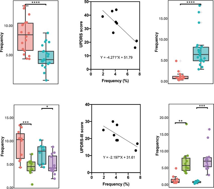
J. Tian et al.
2
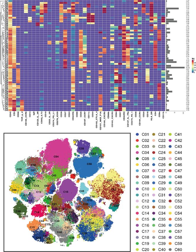
a
1234567890():,;
b
tSNE 2
tSNE 1
Fig. 1 Peripheral blood immune cell clusters sorting based on marker expression levels. a Heatmap of normalized immune cell marker
expression for 60 immune cell clusters. b Distribution of 60 clusters and difference in all groups. The clusters we focus on were marked with
red boxes. EHCs elder healthy controls, EOPD early-onset PD, HCs healthy controls; LOPD late-onset PD, PD Parkinson’s disease, t-SNE t-
distributed stochastic neighbor embedding, YHCs young healthy controls.
major groups of PD and HCs as well as among four subgroups of 13 subpopulations (Supplementary Tables 1 and 2). Cell numbers
EOPD, LOPD, YHCs, and EHCs are displayed in Fig. 1. Unsupervised were normalized to the number of PBMCs for each subject.
clustering was used to divide PBMCs into 60 cell clusters Normalized abundances were then compared between patients
(C01–C60) according to the pattern of marker expression. These with PD and HCs and among the four subgroups (EOPD, LOPD,
60 cell clusters belonged to nine lineages, including CD4+ T cells, YHCs, and EHCs). No significant difference was observed in the
CD8+ T cells, γδT cells, NKT cells, NK cells, dendritic cells, frequencies between total lineages except for monocytes. In
monocytes and B cells. These lineages were further divided into contrast, significant differences were observed between several
npj Parkinson’s Disease (2022) 5 Published in partnership with the Parkinson’s Foundation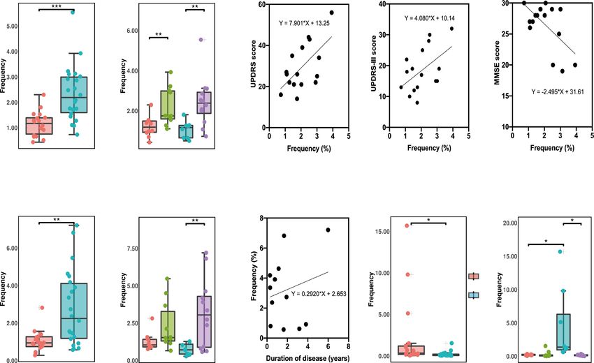
J. Tian et al.
                                                                                                                                                   3
                              C16                                     C37                                     C39
                    (CD57- Naïve CD8+ T)                      (CD57+ CD8+ TEMRA)                      (CD57+ CD8+ TEM)
           a                                             c                                        e

                                                  YHCs                                     YHCs                                   YHCs

                                                  EOPD                                     EOPD                                   EOPD

                                                  EHCs                                     EHCs                                   EHCs

                                                  LOPD                                     LOPD                                   LOPD

                              C16                                     C37                                     C18
                    (CD57- Naïve CD8+ T)                      (CD57+ CD8+ TEMRA)                           (CD8+ TEM)
           b                                             d                                        f

                                                                                                                                  HCs

                                                                                                                                  PD

Fig. 2 The frequency of CD8+ T-cell subsets between early- and late-onset PD patients and HCs and the changes of different clusters with
age and course of disease. a The C16 of EHCs was significantly lower compared to YHCs, and was significantly lower in patients with LOPD
compared to patients with EOPD. b The cluster of C16 cells decreased with age (p = 0.006, R2 = 0.323). c The C37 cluster of EHCs was
significantly higher compared to that of YHCs. The C37 of patients with LOPD was significantly lower compared to EHCs. The C37 cluster in
patients with EOPD was significantly higher compared to YHCs. d With the prolongation of the disease course, the C37 cells decreased (p =
0.037, R2 = 0.200). e The C39 of EHCs was significantly higher compared to YHCs. The C39 cluster of patients with EOPD was significantly
higher compared to YHCs. f The frequency of C18 of PD patients was significantly lower compared to HCs. Two-sided t tests were used to test
statistical significance between groups (*p,0.05, **p,0.01, ***p,0.001). Error bars show the mean ± SEM. EHCs elder healthy controls, EOPD early-
onset PD, HCs healthy controls; LOPD late-onset PD, PD Parkinson’s disease, YHCs young healthy controls.

subpopulations and clusters, particularly concentrating on CD8+           C37 decreased in patients with prolonged PD duration (Fig. 2d).
T cells, NK cells, and monocytes.                                         CD8+ TEMRA and TEM were effector CD8+ T cells and CD8+ TEM cells
                                                                          required repeated proliferation to transform into CD8+ TEMRA cells,
CD8+ T-cell clusters and immunosenescence in PD                           which was considered to be terminally differentiated with high
Among 14 clusters of CD8+ T cells, several clusters were                  cell killing capacity, increased perforin and granzyme B
significantly differed between groups or subgroups. The frequency          releasing20.
of C16 (CD57− naive CD8+ T, Fig. 2a) was lower in EOPD than in               Additionally, we speculated that there were several differences
LOPD; particularly, an inverse relationship was observed between          between patients with EOPD and LOPD. The frequency of C16 was
age and C16 frequency (Fig. 2b). Moreover, C16 was also                   lower in EOPD than in LOPD (Fig. 2a). C37 was higher in patients
decreased in EHCs compared with YHCs (Fig. 2a) although no                with EOPD than in YHCs and lower in patients with LOPD than in
statistical difference was noted using ANOVA with Bonferroni              EHCs (Fig. 2c). The trend was similar to C39 (Fig. 2e), though there
correction (Supplementary Fig. 2a). C18 (CD8+ TEM), a type of             were no significant difference was noted using ANOVA (Supple-
CD45RA− CCR7− effector memory T cells, was significantly                   mentary Fig. 2c, e).
decreased in the PD group than in the HC group (Fig. 2f). C37
belonged to CD57+ CD8+ TEMRA, a type of CD45RA+ CCR7−                     Toxicity of NK cells in PD
effector memory re-expressing CD45RA T cells, and increased with          Three clusters (C27, C29, and C32) of NK cells were significantly
age in HCs (Fig. 2c). Compared with the EHCs, C37 was decreased           different between groups and subgroups. Among them, C32
in patients with LOPD (Fig. 2c) although no statistical difference        (CD56+, CD16+, CD57−, CD28−) and C27 (CD56+, CD16+, CD57+,
was observed using ANOVA (Supplementary Fig. 2c). Additionally,           CD28−) increased in PD patients compared to those in HCs, and

Published in partnership with the Parkinson’s Foundation                                                    npj Parkinson’s Disease (2022) 5
J. Tian et al.
4

                      C32                             C32                                   C32                      C32                         C32
                (CD57- CD28- NK)                 (CD57- CD28- NK)                 (CD57- CD28- NK)              (CD57- CD28- NK)            (CD57- CD28- NK)
          a                                  b                         c                                  d                         e

                                       HCs                          YHCs

                                                                    EOPD

                                       PD                           EHCs

                                                                    LOPD

                      C27                             C27                                   C27                      C29                         C29
                (CD57+ CD28- NK)                 (CD57+ CD28- NK)                 (CD57+ CD28- NK)              (CD57+ CD28+ NK)            (CD57+ CD28+ NK)
          f                                  g                         h                                  i                             j

                                       HCs                          YHCs                                                           HCs                         YHCs

                                                                    EOPD                                                                                       EOPD

                                       PD                           EHCs                                                           PD                          EHCs

                                                                    LOPD                                                                                       LOPD

    Fig. 3 The frequency of NK cell clusters between PD patients and HCs and the changes of different clusters with disease course, UPDRS,
    UPDRS-III, and MMSE scores. a The frequency of C32 subgroup in patients with PD was significantly higher compared to HCs. b The C32
    cluster in patients with LOPD was significantly higher compared to EHCs and was significantly higher in patients with EOPD compared to
    YHCs. c The increase in C32 frequency was associated with an increase in the UPDRS score (p = 0.012, R2 = 0.375). d The UPDRS-III score
    increased (p = 0.046, R2 = 0.254). e The MMSE score decreased (p = 0.013, R2 = 0.367). f C27 in patients with PD was significantly higher
    compared to HCs. g C27 in patients with LOPD was significantly higher compared to EHCs. h Prolonged disease course was associated with
    increased C27 cell clusters in patients with LOPD (p = 0.041, R2 = 0.424). i C29 in patients with PD was significantly lower compared to HCs.
    j C29 in patients with LOPD was significantly lower compared to YHCs. (*p,0.05, **p,0.01, ***p,0.001). Error bars show the mean ± SEM. EHCs
    elder healthy controls, EOPD early-onset PD, HCs healthy controls, LOPD late-onset PD, PD Parkinson’s disease, YHCs young healthy controls.

    both increased in patients with LOPD compared to those in EHCs                                consistent immune trends, which may be attributed to the large
    (Fig. 3a, b, f, g). In contrast to C27 and C32, the C29 (CD56+,                               number of patients with LOPD.
    CD16+, CD57+, CD28+) cluster was lower in patients with PD (Fig.
    3i, j), although no significant difference was observed using                                  Activated monocytes in PD
    ANOVA between the four subgroups in C29 (Supplementary Fig.
                                                                                                  Finally, we found that all monocyte clusters (C53 and C52) were
    3j).
                                                                                                  different between groups and subgroups. C53 (CD14+, CD16−,
       Apparently, three clusters of NK cells differ in their surface
                                                                                                  CD45RA−) decreased in PD compared to HCs (Fig. 4a), which was
    expression of CD28 and CD57. The presence of CD28 has been
    considered a strong activator of NK cells, which induces                                      similarly found in the LOPD subgroup (Fig. 4b) and not in the
    degranulation, lysis of target cells and production of pro-                                   EOPD subgroups using ANOVA (Supplementary Fig. 4b). By
    inflammatory cytokines21. In addition, NK cell expression of                                   contrast, C52 (CD14+, CD16−, CD45RA+) increased in PD (Fig.
    CD57 could be considered a more mature phenotype, higher                                      4e), and a consistent trend was found in both the late- and early-
    cytotoxic capacity, and more sensitive to CD16 stimulation22. From                            onset patients (Fig. 4f).
    the expression of CD28 and CD57, we speculate that the                                           CD45RA was originally thought to be a marker for the naive
    cytotoxicity of C29 cells were the highest, followed by that of                               T cells. It was later found that CD45RA+ T cells accumulated in vivo
    C27 cells and C32 cells. We found that the increase in CD57−                                  due to persistent viral infection, inflammatory syndromes and
    CD28− NK cells (C32) were associated with increased UPDRS and                                 senescence23. Furthermore, several studies have also found that
    UPDRS-III scores (Fig. 3c, d) and decreased MMSE scores (Fig. 3e).                            CD45RA was expressed on activated monocytes, and was
    Additionally, an increase in the CD57+ CD28− NK cells (C27) of                                regarded as a marker of peripheral blood monocyte activation24.
    patients with LOPD were observed as the disease prolonged (Fig.                               We speculate that in patients with EOPD, the proportion of
    3h). In conclusion, the peripheral blood of patients with PD                                  inactivated monocytes (C53) decreased and the proportion of
    demonstrated a decrease in highly toxic NK cells and an increase                              activated monocytes (C52) increased. Regarding monocytes, the
    in less toxic NK cells. In addition to differences in immunological                           two cells (C52 and C53) presented similar tendencies in EOPD and
    background, the NK cell changes between EOPD and LOPD were                                    LOPD. Interestingly, the decrease in C53 was more pronounced in
    broadly similar. Patients with PD and LOPD had relatively                                     early-onset PD, whereas this change was not statistically different

    npj Parkinson’s Disease (2022) 5                                                                            Published in partnership with the Parkinson’s Foundation
J. Tian et al.
5
C53 C53 C52
(CD45RA- Monocytes) (CD45RA- Monocytes) (CD45RA+ Monocytes)
a c e
HCs HCs
PD PD
C53 C53 C52
(CD45RA- Monocytes) (CD45RA- Monocytes) (CD45RA+ Monocytes)
b d f
YHCs YHCs
EOPD EOPD
EHCs EHCs
LOPD LOPD
Fig. 4 The frequency of monocyte clusters between PD patients and HCs, and the changes in different clusters with UPDRS and UPDRS-III
scores. a The frequency of C53 in patients with PD was significantly lower compared to HCs. b The C53 cluster in patients with EOPD was
significantly lower compared to YHCs; additionally, C53 in patients with LOPD was significantly lower compared to EHCs. c Increased C53
frequency was associated with an increased UPDRS score (p = 0.018, R2 = 0.633) and d UPDRS-III scores increased in patients with LOPD (p =
0.042, R2 = 0.526). e The proportion of the C52 subgroup in patients with PD was significantly higher compared to HCs. f C52 in EOPD patients
was significantly higher compared to YHCs and was significantly higher in patients with LOPD compared to EHCs (*p,0.05, **p,0.01, ***p,0.001).
Error bars show the mean ± SEM. EHCs elder healthy controls, EOPD early-onset PD, HCs healthy controls, LOPD late-onset PD, PD Parkinson’s
disease, YHCs young healthy controls.
in late-onset PD (Supplementary Fig. 4b). In general, we found that effector T cells may be capable of rapid cytokinesis, cytotoxicity
the activation of monocytes was the common manifestation of the and IFN-γ production29. Interestingly, we found significantly lower
PBMCs in patients with PD. numbers of CD8+ effector T cells in patients with PD compared to
controls, despite the importance of senescence in the exacerba-
tion of neurodegenerative disorders such as PD. Moreover, recent
DISCUSSION studies have shown a decrease in markers of replicative
In this study, we identified that PD was closely associated with senescence in the CD8+ cells of PD patients, such as CD57 and
specific immune cell classification, such as clusters C16, C18 and TEMRA cells, which is consistent with our findings27. This may lead
C37 of CD8+ T cells; clusters C32, C27 and C29 of NK cells; and to insufficient antigen processing in T cells, which results in
clusters C53 and C52 of monocytes in the peripheral blood. These autoantigen accumulation, such as α-synuclein. In contrast, the
findings suggest the difference in the immune background in PD. decreased CD8+ effector T cells in peripheral blood may be due to
Aging is known to cause gradual weakening of body function, transfer to the brain. There is research evidence of tissue-resident
including the immune system25; this presents with abnormal memory CD8+ T cells in the postmortem brain tissue of patients
immune responses and senescence phenotype of immune cells. with PD, which were associated with synucleinopathy and
Thus, aging causes a decrease in CD57− naive CD8+ cells and an neuronal death6. In mouse models, CD8+ T infiltration was
increase in CD57+ CD8+ effector T cells26,27. CD57 expression is similarly found in the brain, which activated and converted
usually associated with senescent human CD8+ T cells and microglia to the M1 pro-inflammatory phenotype30.
represents “end-stage” effector T cells that are unable to NK cells are important in the field of oncology as a member of
proliferate28. However, it has also been shown that CD57+ CD8+ the innate immune system31. NK cells have recently been found to
Published in partnership with the Parkinson’s Foundation npj Parkinson’s Disease (2022) 5J. Tian et al.
6
was associated with a shorter disease duration and later onset40.
Table 1. Demographic characteristics of PD patients and healthy
Another study concluded that monocyte phagocytosis was higher
controls.
in early-moderate PD and the changes in monocytes were mainly
YHCs EOPD EHCs LOPD attributed to the autologous serum41. Similarly, it has been shown
that the inflammation susceptibility of the monocytes in PD may
Total participants enrolled, n 10 10 8 12 be attributed to the “second strike”, that was the lipopolysacchar-
Age (Mean) 40.30 40.30 68.25 64.23 ide (LPS)- and α-synuclein protein-induced inflammation42. LPS is
Gender (Female/Male) 1/9 3/7 4/4 4/5 a microbial metabolite that has been shown to promote the entry
of monocytes into the CNS causing different disorders42.
Age of onset (Mean) N/A 37.8 N/A 62.42***
The phenotype of immune cells from elderly individuals showed
Duration (Mean) N/A 2.44 N/A 1.8 significant changes. Particularly, thymic degeneration and
UPDRS score (Mean) N/A 26.5 N/A 32.38 repeated pathogen exposure caused a decrease in naive CD8+
UPDRS-III score (Mean) N/A 15.38 N/A 21.63 T cells and an accumulation in mutual memory cells, specifically
H-Y stage (Mean) N/A 1.31 N/A 1.44 terminally differentiated effector CD8+ T cells, which is consistent
MMSE score (Mean) N/A 26.63 N/A 26.38 with our findings. Additionally, CD8+ T cells and NK cells entering
a senescent state are characterized by the absence of CD28
Two-sided t-test was used to test statistical significance between groups. expression and increased expression of CD57. Aged immune cells
EHCs elder healthy controls, EOPD early-onset PD, LOPD late-onset PD, YHCs can produce several specific substances, such as chemokines and
young healthy controls. cytokines, leading to a pro-inflammatory environment in the
***p,0.001.
body43. However, immunosenescence has been reported to play a
protective role against autoimmune diseases such as PD due to
the decreased autoantigen response of non-functioning immune
play different roles in adaptive immune regulation. Chemokines cells44. By contrast, stronger autoimmune responses may be
produced by neurons, microglia, and astrocytes can recruit triggered in patients with EOPD. These conflicting findings show
peripheral NK cells to the CNS, which effectively clears α-synuclein the complex circumstance of immune aging in LOPD and EOPD.
aggregates through receptor-mediated endocytosis32. Addition- Indeed, we found the difference of peripheral blood immune cells
ally, NK cells were found in the substantia nigra of both humans33 between EOPD and LOPD such as CD8+ TEMRA and CD45RA−
and mice34; this is supported by the association found between monocytes.
NK cells and increased phosphorylated α-synuclein deposits in Peripheral blood immune status is relatively heterogeneous in
mouse models of synucleinopathies33. Previous studies have different individuals and could be influenced by various factors
found that the number of circulating NK cells in patients with PD is such as medicines, underlying diseases, mental45, diet46, exer-
increased compared to that in controls; additionally, their activity
cise46, etc. Studies have shown that a variety of PD medicines have
is associated with disease severity35. We found that CD57−, CD28−
the effect on immune cell phenotype and function. Dopamine is a
and CD57+, CD28− NK cells were increased and CD57+, CD28+ NK
significant modulator of immune function and can affect a wide
cells were decreased in PD patients compared with those in HCs.
range of immune cells, which express almost all dopamine
Both CD28 and CD57 are associated with the cytotoxicity of NK
receptors47. Among leukocytes, B cells and NK cells have the
cells. NK cells require CD28-mediated costimulatory signals for
optimal proliferation and are able to produce high levels of highest expression of dopamine receptors47. In the experimental
interferon (IFN)-γ and tumor necrosis factor (TNF) in response to autoimmune encephalomyelitis mice, pramipexole inhibited the
CD28 antibody stimulation36. Additionally, CD57 antigen can be production of inflammatory cytokines such as IL-17, IL-1β and
considered as a marker of terminal differentiation in NK cells and TNF-α in peripheral lymphoid tissues48. In addition, selegiline
is strongly associated with inflammation37. CD28− and CD57− NK changed the phagocytic activity of granulocytes in the mice,
cells may result in impaired antigen clearance and immunoin- which caused a decrease in B cells and an increase in T
flammatory disorders in PD. Further, recent studies have shown lymphocytes, especially CD8+ T cells in the spleen49. Overall, the
that NK cell depletion resulted in exacerbated synuclein pathology interference from PD drugs should be carefully considered when
in the mouse model, suggesting the protective role of NK cells in studying immune status in PD.
PD33. This was consistent with our findings that NK cells were Our study has some limitations. First, the relatively small
correlated with the severity of the disease because increase in C32 number of patients involved in the study may have caused bias in
NK cells were associated with increased UPDRS and UPDRS-III the results. Second, cytomegalovirus antibody levels were not
scores, but decreased MMSE scores of PD patients. Therefore, we evaluated, which are associated with CD8+ T-cell expression50.
speculated that impaired NK cell function was a sign of poor Third, our study has not further validated the function of the
prognosis. The exact mechanism of the protective role of NK cells immune cells through cellular or animal experiments. In the next
remains unknown; however, astrocytes were recently found to studies we will select the interested clusters to explore more
suppress CNS inflammation by triggering T-cell apoptosis. This deeply the mechanisms of changes in peripheral immune cells in
process was driven by the production of IFN-γ from meningeal NK PD. Furthermore, future studies with a larger sample size are
cells, the expression of which is regulated by gut microbiome38. warranted to consider these contributing factors and validate the
Studies have found that the chemokines released by microglia relevance of NK cells and monocytes in disease progression to
or the expression of full-length human α-synuclein allows the further explore the pathogenesis between immunity and PD.
peripheral pro-inflammatory monocyte infiltration of the sub- In summary, our data reflected the changes in peripheral blood
stantia nigra39. The relative number of monocytes was found to be immune cells in patients with PD. Particularly, we found that CD8+
significantly higher in the prefrontal cortex of patients with PD T cells, NK cells, and monocytes were changed and part of the
than in those of normal controls14. Activated monocytes can clusters were associated with age of onset, disease duration, or
infiltrate the brain parenchyma through similar mechanisms and scores of motor or psychiatric symptoms of PD, which provides an
kill neurons. Additionally, they may recognize and promote the evidence for the role of neuroinflammation in PD. Additionally, we
transmission of peripheral antigens, such as α-synuclein14. found differences regarding the changes of peripheral cells
Previous studies have similarly shown monocyte activation in between EOPD and LOPD. This finding suggests immunological
the periphery of PD. Patients with PD had significantly higher specificities and highlights the need for new explanations
proliferative capacity of monocytes compared to controls, which regarding the role of neuroinflammation in EOPD.
npj Parkinson’s Disease (2022) 5 Published in partnership with the Parkinson’s FoundationJ. Tian et al.
7
METHODS was used to visualize the high-dimensional data in two dimensions and to
Subjects show the distribution of each cluster and marker expression and
differences among each group or different sample types54.
Our study included 22 patients with PD. Among them, 10 had early-onset
PD (EOPD; 50 years old).
All patients were admitted to the neurology department at the Second Statistical analyses
Affiliated Hospital of Zhejiang University and diagnosed by senior The differences in the frequency of each cluster between patients with PD
movement disorder specialists based on the current diagnostic criteria51. and HCs were compared. Additionally, the differences in the frequency of
Additionally, 10 age- and sex-matched YHCs and 8 EHCs were recruited each cluster between YHCs and EHCs, EOPD patients and YHCs, LOPD
(Table 1 and Supplementary Table 4). Patients with hypertension, diabetes, patients and EHCs, and EOPD patients and LOPD patients were compared.
and other neurodegenerative diseases, including essential tremor, Two-sided t test statistical analysis was used to test the statistically
multiple-system atrophy, corticobasal degeneration, and Wilson’s disease, significant difference between groups and subgroups. Additionally, we
were excluded. All the patients we recruited were medication naive. Data also performed one-way ANOVA analysis among all four subgroups (YHCs,
regarding the age of onset, duration of disease, Hoehn and Yahr (H&Y) EHCs, EOPD and LOPD) and Bonferroni correction was used as multiple
stage, and UPDRS and MMSE scores were collected (Table 1), and the comparison adjustment. A linear regression model was used to analyze the
peripheral blood samples were obtained. correlation between clusters and clinical characteristics (age, duration of
disease, UPDRS score, UPDRS-III score, H&Y stage, and MMSE score) of
patients with PD. All statistical analyses were performed using SPSS version
Standard protocol approvals, registrations, and patient
25.0 (Armonk, NY: IBM Corp). Statistical significance was set at p < 0.05.
consents
Ethics approval was obtained through the Medical Ethics Committee of the
Second Affiliated Hospital of Zhejiang University School of Medicine Reporting summary
(2020–596). All patients and HCs provided their informed consent before Further information on research design is available in the Nature Research
blood withdrawal. Reporting Summary linked to this article.
Sample processing for mass cytometry
The blood samples were transferred to a 50-ml centrifuge tube with a 10- DATA AVAILABILITY
ml pipette. Ficoll separation solution (GE Healthcare) at 10 ml was added to Anonymized data not published within this article will be made available on request
the 50-ml centrifuge tube. The sample was diluted to 20 ml with PBS from any qualified investigator.
(GENOM) and was added slowly to the top layer of Ficoll separation
solution without breaking the upper liquid level of the Ficoll separation Received: 12 August 2021; Accepted: 20 December 2021;
solution. The 50-ml centrifuge tube containing Ficoll and sample was
placed in a centrifuge with plate rotor (Avanti J-15R, Beckman). Then, it was
centrifuged at 400 × g for 15 min (Acc/Dec Rate 1). The waste liquid above
the separated white film layer was absorbed with a suction pump, and
0.5 ml volume was reserved to prevent PBMC loss. The white layer was
REFERENCES
transferred to a new 50-ml centrifuge tube using a 1-ml manual pipette,
and repeated aspiration was performed until no obvious cells remained in 1. Samii, A., Nutt, J. G. & Ransom, B. R. Parkinson’s disease. Lancet 363, 1783–1793
the Ficoll layer. FACS buffer was added to PBMC initial extract to (2004).
supplement to 30 ml, followed by 400 × g centrifugation for 10 min to 2. Tan, E. K. et al. Parkinson disease and the immune system - associations,
adsorb and discard the supernatant. A 1-ml ACK (PLT) fine was added. Cell mechanisms and therapeutics. Nat. Rev. Neurol. 16, 303–318 (2020).
lysis was done; then, it was blown, mixed, and let stand for 2 min. FACS 3. Chang, C. C. et al. Autoimmune rheumatic diseases and the risk of Parkinson
buffer was added into PBMC suspension after cracking to complement to disease: a nationwide population-based cohort study in Taiwan. Ann. Med. 50,
10 ml. It was centrifuged at 400 × g for 5 min. The supernatant was 83–90 (2018).
4. Rispoli, V., Schreglmann, S. R. & Bhatia, K. P. Neuroimaging advances in Parkin-
discarded, and 4-ml FACS buffer was added and blown and resuspend. A
son’s disease. Curr. Opin. Neurol. 31, 415–424 (2018).
10-μl cell suspension was obtained, and Trypan blue dye (Solarbio) was
5. Subhramanyam, C. S., Wang, C., Hu, Q. & Dheen, S. T. Microglia-mediated neuroin-
added and diluted to count. The supernatant was discarded by
flammation in neurodegenerative diseases. Semin. Cell Dev. Biol. 94, 112–120 (2019).
centrifugation and cell precipitation was obtained.
6. Galiano-Landeira, J., Torra, A., Vila, M. & Bove, J. CD8 T cell nigral infiltration
precedes synucleinopathy in early stages of Parkinson’s disease. Brain 143,
Mass cytometry staining and data acquisition 3717–3733 (2020).
Cells were washed once with 1× PBS; to exclude dead cells, they were stained 7. Boyko, A. A., Troyanova, N. I., Kovalenko, E. I. & Sapozhnikov, A. M. Similarity and
with 100 μl of 250 nM cisplatin (Fluidigm) on ice for 5 min. Afterwards, they differences in inflammation-related characteristics of the peripheral immune
were incubated in Fc receptor blocking solution before staining with surface system of patients with Parkinson’s and Alzheimer’s diseases. Int. J. Mol. Sci. 18,
antibodies cocktail for 30 min on ice (Supplementary Table 3). Cells were https://doi.org/10.3390/ijms18122633 (2017).
washed twice with FACS buffer (1× PBS + 0.5% BSA) and fixed in 200 μl of 8. Lindestam Arlehamn, C. S. et al. alpha-Synuclein-specific T cell reactivity is
intercalation solution (Maxpar Fix and Perm Buffer containing 250 nM 191/193 associated with preclinical and early Parkinson’s disease. Nat. Commun. 11, 1875
Ir, Fluidigm) overnight. After fixation, cells were washed once with the FACS (2020).
buffer and then a perm buffer (eBioscience); then, cells were stained with 9. Lindestam Arlehamn, C. S., Garretti, F., Sulzer, D. & Sette, A. Roles for the adaptive
intracellular antibodies cocktail for 30 min on ice (Supplementary Table 3). Cells immune system in Parkinson’s and Alzheimer’s diseases. Curr. Opin. Immunol. 59,
were washed and resuspended with deionized water, adding to 20% EQ beads 115–120 (2019).
(Fluidigm), acquired on a mass cytometer (Helios, Fluidigm). 10. Cen, L. et al. Peripheral lymphocyte subsets as a marker of Parkinson’s disease in
a Chinese population. Neurosci. Bull. 33, 493–500 (2017).
11. Karpenko, M. N., Vasilishina, A. A., Gromova, E. A., Muruzheva, Z. M. & Bernadotte,
CyTOF data analysis A. Interleukin-1β, interleukin-1 receptor antagonist, interleukin-6, interleukin-10,
Data of each sample were de-barcoded from raw data with unique mass- and tumor necrosis factor-α levels in CSF and serum in relation to the clinical
tagged barcodes using a doublet-filtering scheme18. Bead normalization diversity of Parkinson’s disease. Cell. Immunol. 327, 77–82 (2018).
method was used to normalize the fcs file generated from different 12. Reale, M. et al. Peripheral cytokines profile in Parkinson’s disease. Brain Behav.
batches52. Data were manually gated using the FlowJo software to exclude Immun. 23, 55–63 (2009).
debris, dead cells, and doublets, leaving live, single immune cells. The 13. Sweeney, M. D., Sagare, A. P. & Zlokovic, B. V. Blood-brain barrier breakdown in
X-shift clustering algorithm was applied53 to all cells to partition the cells Alzheimer disease and other neurodegenerative disorders. Nat. Rev. Neurol. 14,
into distinct phenotypes based on their marker expression levels. Cell 133–150 (2018).
types of each cluster were annotated according to the marker expression 14. Harms, A. S. et al. Peripheral monocyte entry is required for alpha-Synuclein
pattern on a heatmap of cluster versus marker. The dimensionality induced inflammation and Neurodegeneration in a model of Parkinson disease.
reduction algorithm, t-distributed stochastic neighbor embedding (t-SNE), Exp. Neurol. 300, 179–187 (2018).
Published in partnership with the Parkinson’s Foundation npj Parkinson’s Disease (2022) 5J. Tian et al.
8
15. Baird, J. K., Bourdette, D., Meshul, C. K. & Quinn, J. F. The key role of T cells in 48. Lieberknecht, V. et al. Pramipexole, a dopamine D2/D3 receptor-preferring ago-
Parkinson’s disease pathogenesis and therapy. Parkinsonism Relat. Disord. 60, nist, prevents experimental autoimmune encephalomyelitis development in
25–31 (2019). mice. Mol. Neurobiol. 54, 1033–1045 (2017).
16. Costantini, E., D’Angelo, C. & Reale, M. The role of immunosenescence in neu- 49. Szczypka, M., Sobieszczanska, A., Suszko-Pawlowska, A. & Lis, M. Selegiline and
rodegenerative diseases. Mediators Inflamm. 2018, 6039171 (2018). clomipramine effects on lymphocyte subsets, regulatory T cells and sheep red
17. Yan, Z. et al. Dysregulation of the adaptive immune system in patients with early- blood cell (SRBC)-induced humoral immune response after in vivo administration
stage Parkinson disease. Neurol. Neuroimmunol. Neuroinflamm. 8, https://doi.org/ in mice. Eur. J. Pharmacol. 887, 173560 (2020).
10.1212/NXI.0000000000001036 (2021). 50. Smith, C. J., Quinn, M. & Snyder, C. M. CMV-specific CD8 T cell differentiation and
18. Zunder, E. R. et al. Palladium-based mass tag cell barcoding with a doublet-filtering localization: implications for adoptive therapies. Front Immunol. 7, 352 (2016).
scheme and single-cell deconvolution algorithm. Nat. Protoc. 10, 316–333 (2015). 51. Postuma, R. B. et al. MDS clinical diagnostic criteria for Parkinson’s disease. Mov.
19. van Unen, V. et al. Visual analysis of mass cytometry data by hierarchical stochastic Disord. 30, 1591–1601 (2015).
neighbour embedding reveals rare cell types. Nat. Commun. 8, 1740 (2017). 52. Finck, R. et al. Normalization of mass cytometry data with bead standards.
20. Koch, S. et al. Multiparameter flow cytometric analysis of CD4 and CD8 T cell Cytometry A 83, 483–494 (2013).
subsets in young and old people. Immun. Ageing 5, 6 (2008). 53. Samusik, N., Good, Z., Spitzer, M. H., Davis, K. L. & Nolan, G. P. Automated
21. Zhuang, X. & Long, E. O. CD28 homolog is a strong activator of natural killer cells mapping of phenotype space with single-cell data. Nat. Methods 13, 493–496
for lysis of B7H7(+) tumor cells. Cancer Immunol. Res. 7, 939–951 (2019). (2016).
22. Lopez-Verges, S. et al. CD57 defines a functionally distinct population of mature NK 54. Esaulova, E. et al. Single-cell RNA-seq analysis of human CSF microglia and
cells in the human CD56dimCD16+ NK-cell subset. Blood 116, 3865–3874 (2010). myeloid cells in neuroinflammation. Neurol. Neuroimmunol. Neuroinflamm. 7,
23. Carrasco, J., Godelaine, D., Van Pel, A., Boon, T. & van der Bruggen, P. CD45RA on https://doi.org/10.1212/NXI.0000000000000732 (2020).
human CD8 T cells is sensitive to the time elapsed since the last antigenic
stimulation. Blood 108, 2897–2905 (2006).
24. Kersten, B. et al. CD45RA, a specific marker for leukaemia stem cell sub- ACKNOWLEDGEMENTS
populations in acute myeloid leukaemia. Br. J. Haematol. 173, 219–235 (2016). This work was supported by grants from the National Natural Science Foundation of
25. Rawji, K. S. et al. Immunosenescence of microglia and macrophages: impact on China (grant number 81771216) and the Key Research and Development Program of
the ageing central nervous system. Brain 139, 653–661 (2016). Zhejiang Province (grant number 2020C03020). We thank PLT Tech Co., Ltd.
26. Fukushima, Y., Minato, N. & Hattori, M. The impact of senescence-associated T cells on (Hangzhou, China) for CyTOF experiments and analyses and all of the PD patients and
immunosenescence and age-related disorders. Inflamm. Regen. 38, 24 (2018). healthy volunteers who participated in this study.
27. Williams-Gray, C. H. et al. Abnormalities of age-related T cell senescence in Par-
kinson’s disease. J. Neuroinflammation 15, 166 (2018).
28. Brenchley, J. M. et al. Expression of CD57 defines replicative senescence and
AUTHOR CONTRIBUTIONS
antigen-induced apoptotic death of CD8+ T cells. Blood 101, 2711–2720 (2003).
29. Morris, S. R. et al. Inflammescent CX3CR1+CD57+CD8+ T cells are generated and J.T.: Conceptualization, methodology, writing - original draft, writing - review &
expanded by IL-15. JCI Insight 5, https://doi.org/10.1172/jci.insight.132963 (2020). editing, visualization. S.-B.D.: Conceptualization, methodology, writing - review &
30. Brochard, V. et al. Infiltration of CD4+ lymphocytes into the brain contributes to editing, visualization. J.T. and S.-B.D. are co-first authors. S-S.J.: Methodology, writing -
neurodegeneration in a mouse model of Parkinson disease. J. Clin. Investig 119, original draft, writing - review & editing, supervision. W.-Y.Y.: Methodology,
182–192 (2009). investigation, writing - original draft, writing - review & editing. Y.-Q.Y.: Writing -
31. Morvan, M. G. & Lanier, L. L. NK cells and cancer: you can teach innate cells new review & editing, supervision. Z.-H.L.: Writing - review & editing, supervision. J.-X.D.:
tricks. Nat. Rev. Cancer 16, 7–19 (2016). Writing - review & editing, supervision. R.Z.: writing - review & editing, supervision.
32. Earls, R. H. & Lee, J. K. The role of natural killer cells in Parkinson’s disease. Exp. Y.C.: Writing - review & editing, supervision. J.-L.P.: Writing - review & editing,
Mol. Med 52, 1517–1525 (2020). supervision. B.-R.Z.: Writing - review & editing, supervision.
33. Earls, R. H. et al. NK cells clear alpha-synuclein and the depletion of NK cells
exacerbates synuclein pathology in a mouse model of alpha-synucleinopathy.
Proc. Natl Acad. Sci. USA 117, 1762–1771 (2020). COMPETING INTERESTS
34. Earls, R. H. et al. Intrastriatal injection of preformed alpha-synuclein fibrils alters The authors declare no competing interests.
central and peripheral immune cell profiles in non-transgenic mice. J. Neuroin-
flammation 16, 250 (2019).
35. van de Wouw, M., Boehme, M., Dinan, T. G. & Cryan, J. F. Monocyte mobilisation, ADDITIONAL INFORMATION
microbiota & mental illness. Brain Behav. Immun. 81, 74–91 (2019). Supplementary information The online version contains supplementary material
36. Sanmamed, M. F. et al. Agonists of co-stimulation in cancer immunotherapy directed available at https://doi.org/10.1038/s41531-021-00271-x.
against CD137, OX40, GITR, CD27, CD28, and ICOS. Semin Oncol. 42, 640–655 (2015).
37. Kared, H., Martelli, S., Ng, T. P., Pender, S. L. & Larbi, A. CD57 in human natural Correspondence and requests for materials should be addressed to Bao-Rong Zhang
killer cells and T-lymphocytes. Cancer Immunol. Immunother. 65, 441–452 (2016). or Jia-Li Pu.
38. Sanmarco, L. M. et al. Gut-licensed IFNgamma(+) NK cells drive LAMP1(+)TRAIL
(+) anti-inflammatory astrocytes. Nature 590, 473–479 (2021). Reprints and permission information is available at http://www.nature.com/
39. Batchu, S. Prefrontal cortex transcriptomic deconvolution implicates monocyte reprints
infiltration in Parkinson’s disease. Neurodegener. Dis. 20, 110–112 (2020).
40. Nissen, S. K. et al. Alterations in blood monocyte functions in Parkinson’s disease. Publisher’s note Springer Nature remains neutral with regard to jurisdictional claims
Mov. Disord. 34, 1711–1721 (2019). in published maps and institutional affiliations.
41. Wijeyekoon, R. S. et al. Monocyte function in Parkinson’s disease and the impact
of autologous serum on phagocytosis. Front Neurol. 9, 870 (2018).
42. Grozdanov, V. et al. Inflammatory dysregulation of blood monocytes in Parkin-
son’s disease patients. Acta Neuropathol. 128, 651–663 (2014). Open Access This article is licensed under a Creative Commons
43. Ventura, M. T., Casciaro, M., Gangemi, S. & Buquicchio, R. Immunosenescence in Attribution 4.0 International License, which permits use, sharing,
aging: between immune cells depletion and cytokines up-regulation. Clin. Mol. adaptation, distribution and reproduction in any medium or format, as long as you give
Allergy 15, 21 (2017). appropriate credit to the original author(s) and the source, provide a link to the Creative
44. Mayne, K., White, J. A., McMurran, C. E., Rivera, F. J. & de la Fuente, A. G. Aging and Commons license, and indicate if changes were made. The images or other third party
neurodegenerative disease: is the adaptive immune system a friend or foe? Front. material in this article are included in the article’s Creative Commons license, unless
Aging Neurosci. 12, https://doi.org/10.3389/fnagi.2020.572090 (2020). indicated otherwise in a credit line to the material. If material is not included in the
45. Hang, S. & Huh, J. R. The immune-mind connection. Cell 179, 803–805 (2019). article’s Creative Commons license and your intended use is not permitted by statutory
46. Song, M. & Chan, A. T. The potential role of exercise and nutrition in harnessing regulation or exceeds the permitted use, you will need to obtain permission directly
the immune system to improve colorectal cancer survival. Gastroenterology 155, from the copyright holder. To view a copy of this license, visit http://creativecommons.
596–600 (2018). org/licenses/by/4.0/.
47. Kawano, M., Takagi, R., Saika, K., Matsui, M. & Matsushita, S. Dopamine regulates
cytokine secretion during innate and adaptive immune responses. Int Immunol.
30, 591–606 (2018). © The Author(s) 2022
npj Parkinson’s Disease (2022) 5 Published in partnership with the Parkinson’s FoundationYou can also read