Clinical Significance of Circulating Serum Levels of sCD95 and TNF-α in Cytoprotection of Cervical Cancer
←
→
Page content transcription
If your browser does not render page correctly, please read the page content below
Reports of Biochemistry & Molecular Biology
                                             Vol.10, No.4, Jan 2022

                                             Original article                                                                                            www.RBMB.net

                                                            Clinical Significance of Circulating
                                                           Serum Levels of sCD95 and TNF-α in
                                                            Cytoprotection of Cervical Cancer
                                               Saurabh Kumar Agnihotri1,2#, Balawant Kumar3#, Ankita Jain2, Anjali Anjali3, 4,
                                                  Mahendra Pal Singh Negi3, Rekha Sachan4, Madan Lal Brahma Bhatt1,
                                                              Raj Kamal Tripathi3, 4, Monika Sachdev*2,5
                                             Abstract
                                             Background: This study correlates the serum levels of sCD95 & TNF-α with a simple cell-based assay to
                                             evaluate the capacity of the serum sample to induce apoptosis in Jurkat cells. Interlinking of these parameters
                                             can be explored to design a minimum invasive diagnostic strategy for cervical cancer (CC).
                                             Methods: Sera samples were assessed to induce apoptosis in Jurkat cells through FACS. Serum levels of
                                             sCD95 and TNF-α were measured by ELISA. JNK phosphorylation was evaluated in sera incubated Jurkat
                                             cells. Data was scrutinized through statistical analysis.
                                             Results: Significantly higher serum levels of sCD95 and lower TNF-α levels were observed in CC patients;
                                             their sera samples inhibited induction of apoptosis in Jurkat cells through reduced JNK phosphorylation.
                                             Statistical analysis linked these three parameters for the early screening of CC.
                                             Conclusions: Distinct sera levels of sCD95 & TNF-α in CC patients showed an anti-apoptotic effect, which can be
                                             considered for early detection of CC.

                                             Keywords: Apoptosis, sCD95, Jurkat Cells, Tumor Necrosis Factor-alpha, Uterine Cervical Neoplasms.
[ Downloaded from rbmb.net on 2022-03-13 ]

                                             Introduction
                                             Apoptotic pathways are usually disrupted or                          recruiting initiator caspases into the death-
                                             inactivated in cancerous cells and engenders                         inducing signalling complex (DISC) which
                                             uncontrolled cell growth and tumor cell                              leads to their activation (4). CD95 is also
                                             resistance (1). Apart from internal cytotoxic                        known to activate non-apoptotic pathways
                                             stress that predominantly triggers apoptosis, it                     including NFκB, Erk1/2& JNK1/2, p38
                                             can also be induced by cell-membrane-                                mitogen-activated protein kinase pathways
                                             anchored signalling pathways of the TNF-                             and the 3-kinase/Akt phosphatidylinositol
                                             superfamily: the CD95-receptor/CD95-ligand-                          pathway (5,6).
                                             system (Fas/FasL or APO-1) and the tumor                                CD95 consists of two isoforms, one is a
                                             necrosis factor (TNF)-related apoptosis                              soluble form (sCD95) and other one is
                                             inducing ligand (TRAIL or APO-2L) with the                           anchored to the cellular membrane (mCD95)
                                             TRAIL receptors 1 and 2 (TRAIL-R1 and R2)                            (7,8). Binding of sCD95 to CD95L leads to
                                                                                                                  unavailability of CD95L to bind with mCD95
[ DOI: 10.52547/rbmb.10.4.711 ]

                                             (2,3). The best known death receptor is APO-
                                             1/ Fas, which induces apoptosis through                              and leads to the downregulation of cellular

                                             1: Department of Radiotherapy, King George’s Medical University, Lucknow 226 003, India.
                                             2: Division of Endocrinology, CSIR-Central Drug Research Institute, Lucknow 226 031, India.
                                             3: Division of Toxicology & Experimental Medicine, CSIR-Central Drug Research Institute, Lucknow 226 031, India.
                                             4: Department of Obstetrics & Gynaecology, King George’s Medical University, Lucknow 226 003, India.
                                             5: Academy of Scientific and Innovative Research (AcSIR), Ghaziabad 201 002, India.
                                             #The first and the second authors contributed equally to this work.
                                             *Corresponding author: Monika Sachdev; Tel: +91 522 2772450; E-mail: monika@cdri.res.in.
                                             Received: 16 Mar, 2021; Accepted: 1 May, 2021
Kumar Agnihotri S et al
apoptosis. Elevated levels of sCD95 have been parameters include the sera levels of sCD95
reported in sera samples of patients with and TNF-α as well as capacity to induce
various malignancies (9–12). Soluble form of apoptosis in Jurkat T cells through the patients’
CD95L is generated by a metalloproteinase- serum factors.
like protease, which binds to sCD95 and
induce apoptosis in cytotoxic T-cells thus Materials and Methods
preventing the recognition of tumor cells (13). Ethics statement
Therefore, detection of sCD95 in serum could The study was conducted according to the
be a new approach to detect the cancer. guidelines of the declaration of Helsinki and
Basically, CD95 is a membrane protein approved by the Institutional Ethics Committee
belonging to the TNF family (14,15). TNF-α is (Approval ref# 51 E.C.M. IIA/P1), Office of the
a pleiotropic inflammatory cytokine, critical for Research Cell, King George’s Medical
the various cellular events. TNF-α may exist as University, Lucknow. Written informed
a 26-kDa membrane tethered form (mTNF-α) consents were obtained from all enrolled
or a soluble 17-kDa cytokine (sTNF-α). Both participants to participate in this study.
these isoforms have contradictory effects on
tumor growth but the mechanism has not been Collection of blood samples and separation of sera
understood properly (16). Blood samples (~5 ml) were collected from the
Usually CC remains asymptomatic and CC patients (n=20) visiting King George’s
goes undiagnosed, but can be cured if detected Medical University Lucknow, along with age
early. Presently Pap test is widely used to matched (n=20) healthy controls (HC) by
detect precancerous changes in the cervix, venipuncture in serum separating vacutainers
which may lead to the progression of CC (17) (367956, BD). After blood clotting, serum was
but has low sensitivity (18,19). Numerus separated through centrifugation in 2000 g for
aspects limits the test sensitivity including 10 minutes at 4 °C and stored at -80 ºC until
lesion size, inaccessible location, lesser further analysis.
number of abnormal cells as well as their small
size; manifestation of blood due to Cell lines and cell culture
[ Downloaded from rbmb.net on 2022-03-13 ]
inflammation further complicates the cell Human T lymphocyte cell line Jurkat cells and
visualization. The persistent infection of human embryonic kidney cell line HEK-293
human papillomavirus (HPV) also leads to were obtained from institutional cell
high-grade cervical intraepithelial neoplasia repository, originally procured from ATCC
(CIN) (20,21) and ultimately progress to CC (American Type Cell Culture). Jurkat & HEK-
(22). Most of the clinically confirmed CC 293 cells were maintained in RPMI and
cases are associated with infection of high-risk DMEM high glucose media respectively
HPV (16,20,24) and can be transmitted supplemented with 10% FBS (10082147,
through sexual activity (25,26). Consequently, Gibco) along with 1X antibiotic solution
the combination of the Pap test with HPV- (15140122, Gibco) containing 1 mM sodium
DNA testing can increase noteworthy pyruvate (58636, Sigma Aldrich). Culture was
sensitivity, but methodological and economic maintained at 37 °C temperature and 5% CO2
constrains limits its feasibility. along with ~ 90% humidity and cells were
Therefore, it is warranted to look for novel allowed for growth and proliferation.
simple and cost-effective complementary
prognostic methods for accurate and early Analysis of induced apoptosis in sera incubated cells
[ DOI: 10.52547/rbmb.10.4.711 ]
prediction of the disease. In the present study, Jurkat cells and HEK-293 cells were seeded at
comparatively, a simpler and more authentic a density of 2.5 × 10⁵ and 3 × 10⁵ cells/ml media
approach has been established to predict the respectively in each well of 6-well plates.
risk of developing CC based on three different Further, 100 µl of serum from clinical subjects
parameters of patients’ sera samples. These was added in the wells containing 2 ml to total
712 Rep. Biochem. Mol. Biol, Vol.10, No.4, Jan 2022sCD95 and TNF-α in Cervical Cancer
media. After 48 hours of incubation, apoptosis in PBST (1X PBS + 0.1% Tween 20) at 25 °C
was assessed using Annexin V FITC and PI for 1 hr followed by probing with specific
apoptosis detection kit (V13242, Invitrogen) primary antibody (anti-phospho-p54/46JNK or
following manufacturer’s instructions. Total anti-p54/46JNK) for overnight (14 hrs) at 4 °C.
10,000 events were analyzed for each sample in After washing membrane was incubated with
BD FACS Calibur flow cytometer system the specific HRP conjugated secondary
(USA). Cells having positive scoring for antibody for 2 hrs at 25 °C. Protein bands were
Annexin-V but negative for PI were considered detected using Immobilon Western
as apoptotic and the subsequent data was Chemiluminescent HRP Substrate
analyzed using Cell QuesecPro software. (WBKLSO500, Millipore) and developed with
chemi-documentation imaging system (Image
Quantitation of sCD95 and sCD95L in sera samples Quant LAS 4000, GE Life Science, PA, USA).
by Enzyme Linked Immunosorbent Assay (ELISA)
sCD95 and sCD95L level were measured in Statistical analysis
each serum sample with the Human APO- Data were summarized as Mean ± SE (standard
1/FAS ELISA Kit (KHS9502, BioSource error of the mean). Two independent groups of
International) and Human sFAS Ligand ELISA HC and CC patients were compared by
Kit (KHS9521, BioSource International) Student’s T-test. Pearson correlation analysis
respectively, as described by the manufacturers. was done to assess the association between the
The range of the kit for sCD95 was 0.23-15 variables i.e. serum concentration of sCD95 and
ng/ml. TNF-α as well as apoptosis induction of Jurkat
cells through sera samples of HC Vs. CC
Detection of TNF-α level in sera by ELISA patients. Simple linear regression analysis was
Serum concentration of TNF-α was determined done to assess the strength of association
by ELISA using Human TNF-α Kit (KHC3011, between these variables. Diagnostic accuracy
Invitrogen) as described by the manufacturer. (sensitivity and specificity) of these parameters
Twenty CC patient and 20 healthy volunteer’s (sCD95, TNF-α and apoptosis induction) for
serum samples were thawed completely and the patients was done through receiver
[ Downloaded from rbmb.net on 2022-03-13 ]
mixed well prior to analysis. All standards, operative characteristics (ROC) curve analysis.
controls and samples were analyzed in duplicate. A two-tailed (α=2) with pKumar Agnihotri S et al
cells was 31.5%, 58.3 % and 84.1% at 100, 150 cells grown without serum exposure (Fig. 1D).
and 200 µl of serum respectively (Fig. 1C). These observations strongly depict an
While induced apoptosis in HEK-293 cells association of the CD95 pathway in the
hardly remained 9% after the exposure to apoptosis of Jurkat cells through healthy
healthy serum, which was similar to control women sera.
[ Downloaded from rbmb.net on 2022-03-13 ]
Fig. 1. Normal sera induce apoptosis in the Jurkat cells as compared to the HEK-293 cells. Representative FACS dot-plot
with 150 µl of healthy sera cocktail showing (A) Induction of apoptosis in Jurkat cells, whereas (B) HEK 293 cells did not
show considerable induction of the apoptosis. (C) Jurkat cells and (D) HEK-293 cells, were incubated for 48 hrs with 0-200
µl cocktail of 20 healthy women sera. Apoptosis rate in each cell line was measured by flow cytometry using Annexin-V/PI
markers. A total of 10000 events were captured by BD FACS Calibur flow cytometer system (USA) and analysis was
performed with the CellQuesecPro software. The data are represented as means ±SEM from three independent experiments.
Serum induced apoptosis is mediated by JNK1/p38
phosphorylation
It is quite evident from our results that the sera level of JNK phosphorylation through western
of CC subjects contain high concentration of blotting (Fig. 2). This analysis indicated that
sCD95 receptor which interact with CD95L Jurkat cells incubated with sera of CC subjects
(Fas ligand) and causes dilution of CD95L in (Fig. 2B) showed significantly lesser activation
the serum. Consequently, CD95L is unavailable of JNK (ratio between integrated density of
in high concentration to interact with cell phospho-p54/46JNK and Total JNK levels) as
[ DOI: 10.52547/rbmb.10.4.711 ]
membrane associated CD95 receptor which compared to the cells incubated with sera of HC
could lead to activation of death receptor (Fig. 2A). Further assessment for the p38
domain which further activates ASK1 and its phosphorylation (data not shown) in the similar
downstream JNK/p38 resulting in apoptosis. experimental set up demonstrated higher
Jurkat cells were incubated with sera from CC phosphorylation with the CC patients’ sera,
and HC subjects for 48 hrs and analyzed for the when compared with sera of healthy women.
714 Rep. Biochem. Mol. Biol, Vol.10, No.4, Jan 2022sCD95 and TNF-α in Cervical Cancer
Overall, these results strongly suggest that by CD95/CD95L interactions that activate the
apoptosis induction in Jurkat T cell is mediated JNK1 and p38 downstream.
Fig. 2. Reduced JNK phosphorylation was observed during induced Apoptosis of Jurkat cells through the sera samples of
CC patients. Jurkat cells were incubated with 100 µl serum of (A) Healthy women or (B) Cancerous women for 48 hrs. Cell
were harvested after 48 hrs. followed by western blot and densitometry analysis to determine relative extent of JNK
activation (ratio between integrated density of phospho-p54/46JNK and total JNK levels). Serum of healthy women induce
the p54/46JNK phosphorylation as compared to cancerous patients’ sera.
[ Downloaded from rbmb.net on 2022-03-13 ]
Cervical cancer patient’s serum prevents apoptosis potential of sera to induce cell death in Jurkat
induction in Jurkat cells cells.
Sera samples from HC and CC subjects were
added to Jurkat cells and incubated for 48 hrs. Correlation of sCD95 levels with inhibition of
Results clearly demonstrated that sera from apoptosis in Jurkat cells
healthy women were able to induce apoptosis As previous experiments showed the
ranging from 35.33-65.33% in Jurkat cells. involvement of CD95 pathway in the induction
Whereas highly reduced apoptosis range of of apoptosis in Jurkat cells. Quantification of
2.50-10.33% was observed in Jurkat cells after sCD95 protein levels in all sera samples was
induction with the sera of CC patients (Fig. done through ELISA. The sCD95 in HC ranged
3A). Overall the average percentage of from 1.00-3.00 ng/ml with mean (± SE) 1.64 ±
apoptosis in the HC with mean (± SE) 51.29 ± 0.09 ng/ml and median 1.66 ng/ml whereas in
2.45% and median 53.50% whereas CC patients it ranged from 1.45-5.50 ng/ml with
corresponding value in CC patients was mean (± SE) 3.13 ± 0.21 ng/ml and median 3.05
significantly lower (PKumar Agnihotri S et al
HC (Fig. 3B). Statistical analysis of the and low apoptosis induction in Jurkat cells,
experimental data clearly demonstrated a while using CC serum unlike healthy
correlation between elevated levels of sCD95 volunteers.
Fig. 3. Differential parameters i.e. apoptosis induction in Jurkat cells as well as sCD95 & TNF-α concentration in sera
samples of HC (n=20) Vs. CC patients (n=20). (A) Induction of apoptosis (%) in Jurkat cells after incubation with 100 µl
sera samples of CC patients. (B) Increased levels of sCD95 (Fas receptor) in CC patients’ sera as compared to healthy sera
samples. (C) Decreased levels of TNF-α in sera samples of CC patients Vs. HC. Concentrations of sCD95 and TNF-α were
measured by ELISA in sera obtained from HC and CC patients separately in triplicate. These results are represented as mean
± Standard Error of mean. **p < 0.01 or ***psCD95 and TNF-α in Cervical Cancer
[ Downloaded from rbmb.net on 2022-03-13 ]
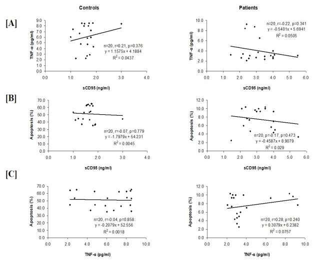
Fig. 4. Correlation and best fit regression analysis for the serum concentration of sCD95 and TNF-α as well as apoptosis
induction of Jurkat cells through sera samples of HC Vs. patients. Pearson correlation analysis was done to assess association
between the variables. (A) Positive (direct) correlation (r=0.21) was observed between sCD95 and TNF-α concentration of
sera in HC, whereas a negative (inverse) correlation (r=-0.22) was observed in CC patients. (B) Negative correlation was
observed between sCD95 concentration and induced apoptosis for both HC (r=-0.07) and CC patients’ groups (r=-0.17). (C)
For TNF-α concentration and induced apoptosis, HC showed a negative correlation (r=-0.04) in contrast to positive
correlation (r=0.28) of CC patients.
Sensitivity and specificity for sCD95, TNF-α levels and the best parameter in terms of diagnostic
apoptosis induction to discriminate patients from accuracy with AUC=1.000 (p1.9 ng/ml with
Receiver Operative Characteristics (ROC) AUC=0.942 (pKumar Agnihotri S et al
Fig. 5. Receiver operative characteristics (ROC) curve analysis to check diagnostic accuracy for all the three variables and
the cut off value to discriminate CC patients from HC. (A) Cut-off for Apoptosis induction (%) in Jurkat cells was found to
be ≤ 10.33%, while its sensitivity and specificity is of 100.0%. (B) Cut-off concentration of sCD95 was found to be >1.9
ng/ml, while its sensitivity and specificity is of 95.0%. (C) Cut-off concentration of TNF-α was found to be ≤ 4.01 pg/ml,
while its sensitivity and specificity is of 80.0%.
Discussion
It is known that CD95-ligand binds to the un-induced Jurkat T cells, which were
sCD95, which further protect the cancerous incubated with the CC patients’ sera samples.
cells from CD95-mediated apoptosis and Similarly, increased phosphorylation of p38
represents one of the method of evading was also detected (data not shown); hence it can
immune surveillance (2, 28). Based on these be depicted that serum of healthy volunteer
previous reports, the present study established a induces the apoptosis through JNK1 and p38
cell-based assay, where serum-induced phosphorylation (Fig. 2).
[ Downloaded from rbmb.net on 2022-03-13 ]
apoptosis in Jurkat cells was directly mediated Induced apoptosis of Jurkat cells reduced
by the CD95 pathway. At the same time, significantly, when cells were supplemented
transformed HEK-293 cells were used as with sera samples of CC patients, whereas
negative control, as these cells are known to be supplementation with healthy sera samples
resistant to CD95L and do not die from CD95 resulted in higher apoptotic indices (Fig. 3A).
apoptosis (Fig. 1). Based upon these observations, which
Interaction of CD95-ligand leads to the suggested a crucial role of CD95 in this
activation of death receptor and signal is mechanism; the next objective was to check the
transmitted to MAP kinase pathway that sCD95 levels in the serum of all the CC
culminated in apoptosis through JNK1 and patients. This experiment showed elevated
p38 phosphorylation. Therefore, in order to levels of sCD95 in the sera of CC patients as
check, whether this apoptosis pathway is compared to healthy control subjects (Fig. 3B).
mediated by JNK1 and p38 phosphorylation Reports about the other malignancies also
and can be induced by FasL-Fas interaction; authenticated this data as the elevated levels of
next objective was to analyze the JNK1 and sCD95 have been observed in patients’ sera
[ DOI: 10.52547/rbmb.10.4.711 ]
p38 phosphorylation in Jurkat T cells during samples of ovarian, gall bladder, lung,
induced apoptosis. Immuno-blotting result of melanoma, head and neck (29), renal
these Jurkat T cells demonstrated that induced carcinomas (30) as well as autoimmune
apoptosis was mediated by the elevated rheumatic diseases (3). Sera levels of sCD95
phosphorylation of JNK as lower has also been well correlated with the survival
phosphorylation of JNK was observed in the rate of the CC patients as the lower sCD95
718 Rep. Biochem. Mol. Biol, Vol.10, No.4, Jan 2022sCD95 and TNF-α in Cervical Cancer
levels (1.5ng/ml). Present data both CC patients and HC, but the correlation
is in absolute agreement with these reports, coefficient was slightly higher for the CC
equally present study also found very low levels patients as compare to the HC (Fig. 4).
of sCD95 in the sera samples of HC. Further, to assess the diagnostic accuracy of
The next segment of the study was TNF-α; these variables (sCD95, TNF-α and induced
in order to understand the role of TNF-α in CC, apoptosis) through their sensitivity and
its concentration was assessed in the sera specificity, ROC curve and AUC analysis was
samples. According to the previous studies, done. Highly significant accuracy was observed
TNF-α has been reported in dual roles; where for the apoptosis induction; hence this
it regulates the proliferation of cell through parameter can be widely recommended to
activation of NFκB and induce the apoptosis in assess the risk of the disease. Increased sera
cancer killing cell (31,32). TNF-α tends to levels of sCD95 in patients, was also found to
affect most of the body's organs, and cytokine be significant, whereas decreased sera levels of
performs a range of functions, many of which TNF-α in CC patients showed slightly lower
are still unexplored. In the present study, lower diagnostic accuracy (Fig. 5). But at the same
levels of TNF-α were observed in the sera time, it is important to consider all the three
samples of the CC patients as compared to the parameters together, as the apoptotic pathways
HC (Fig. 3C). Higher concentration of TNF-α are usually dependent on both the other
in healthy women may regulate discrete parameters.
signaling pathways associated with nuclear It is evident from the present study that the
factor κB (NF-κB) and c-Jun N-terminal distinct sera levels of sCD95 & TNF-α in CC
kinase (JNK). NF-κB is a foremost anti- patients showed an anti-apoptotic effect, which
apoptotic cell survival signal while sustained may create a micro-environment to evade the
JNK activation leads to cell death. In fact, the anti-tumor immune response. Hence, it has
crosstalk between NF-κB and JNK is involved been suggested that these parameters can be
in determining the cellular outcomes in explored for the risk assessment of CC.
[ Downloaded from rbmb.net on 2022-03-13 ]
response to TNF-α (6). However, more elaborative studies can be done
Correlation and best fit regression analysis with larger sample size for the further
between sCD95, TNF-α and apoptosis deployment in the clinical scenario.
induction (in Jurkat cells) for CC patients and
HC was done through Pearson correlation Acknowledgements
analysis to assess association between the Sophisticated Analytical Instrumental Facility
variables. In CC patients, it was observed that of the CSIR-Central Drug Research Institute
the lower levels of TNF-α were strongly Lucknow is thankfully acknowledged. This
associated with the reduction in induced work was supported by Council of Scientific
apoptosis as compare to the HC; whereas and Industrial Research (CSIR), New Delhi,
association between sCD95 & TNF-α for CC Government of India.
patients turned out to be negatively correlated, The authors have no conflicts of interest to
which showed positive correlation for the HC. declare.
References
[ DOI: 10.52547/rbmb.10.4.711 ]
1. Danial NN, Korsmeyer SJ. Cell Death: Critical 3. Jodo S, Kobayashi S, Kayagaki N, Ogura N,
Control Points. Cell. 2004;116(2):205–19. Feng Y, Amasaki Y, et al. Serum levels of soluble
2. Walczak H, Krammer PH. The CD95 (APO- Fas/APO-1 (CD95) and its molecular structure in
1/Fas) and the TRAIL (APO-2L) apoptosis patients with systemic lupus erythematosus (SLE)
systems. Exp Cell Res. 2000 Apr;256(1):58–66. and other autoimmune diseases. Clin Exp
Immunol. 1997;107(1):89–95.
Rep. Biochem. Mol. Biol, Vol.10, No. 4, Jan 2022 719Kumar Agnihotri S et al
4. Townson JL, Naumov GN, Chambers AF. The 16. Hatai T, Matsuzawa A, Inoshita S, Mochida
role of apoptosis in tumor progression and Y, Kuroda T, Sakamaki K, et al. Execution of
metastasis. Curr Mol Med. 2003 Nov;3(7):631–42. apoptosis signal-regulating kinase 1 (ASK1)-
5. Igney FH, Krammer PH. Immune escape of induced apoptosis by the mitochondria-dependent
tumors: apoptosis resistance and tumor caspase activation. J Biol Chem.
counterattack. J Leukoc Biol. 2002;71(6):907–20. 2000;275(34):26576–81.
6. Liu ZG, Hsu H, Goeddel D V., Karin M. 17. Wentzensen N, Von Knebel Doeberitz M.
Dissection of TNF receptor 1 effector functions: Biomarkers in cervical cancer screening. Dis
JNK activation is not linked to apoptosis while Markers. 2007;23(4):315–30.
NF-κB activation prevents cell death. Cell. 18. Lettini AA, Guidoboni M, Fonsatti E,
1996;87(3):565–76. Anzalone L, Cortini E, Maio M. Epigenetic
7. Nagata S. Fas ligand-induced apoptosis. Annu remodelling of DNA in cancer. Histol
Rev Genet. 1999;33:29–55. Histopathol. 2007;22(10–12):1413–24.
8. Chang HY, Yang X, Baltimore D. Dissecting 19. Syrjänen KJ. Spontaneous evolution of
Fas signaling with an altered-specificity death- intraepithelial lesions according to the grade and
domain mutant: Requirement of FADD binding type of the implicated human papillomavirus
for apoptosis but not Jun N-terminal kinase (HPV). Eur J Obstet Gynecol Reprod Biol.
activation. Proc Natl Acad Sci USA. 1996;65(1):45–53.
1999;96(4):1252–6. 20. Kanamoto T, Mota M, Takeda K, Rubin LL,
9. Kondera-Anasz Z, Mielczarek-Palacz A, Miyazono K, Ichijo H, et al. Role of apoptosis
Sikora J. Soluble Fas receptor and soluble Fas signal-regulating kinase in regulation of the c-Jun
ligand in the serum of women with uterine N-terminal kinase pathway and apoptosis in
tumors. Apoptosis. 2005;10(5):1143–9. sympathetic neurons. Mol Cell Biol.
10. Suda T, Nagata S. Purification and 2000;20(1):196–204.
characterization of the fas-ligand that induces 21. Zur Hausen H. Papillomaviruses and cancer:
apoptosis. J Exp Med. 1994;179(3):873–9. From basic studies to clinical application. Nat Rev
11. Oehm A, Behrmann I, Falk W, Pawlita M, Cancer. 2002;2(5):342–50.
Maier G, Klas C, et al. Purification and molecular 22. Bosch FX, De Sanjosé S. The epidemiology
[ Downloaded from rbmb.net on 2022-03-13 ]
cloning of the APO-1 cell surface antigen, a of human papillomavirus infection and cervical
member of the tumor necrosis factor/nerve cancer. Dis Markers. 2007;23(4):213–27.
growth factor receptor superfamily. Sequence 23. Walboomers J, Jacobs M, Manos M, Bosch
identity with the Fas antigen. J Biol Chem. F, Kummer J, Shah K, et al. Human
1992;267(15):10709–15. Papillomavirus Is a Necessary Cause. J Pathol.
12. Itoh N, Yonehara S, Ishii A, Yonehara 1999;189:12–9.
M, Mizushima S, Sameshima M, et al. The 24. Kischkel FC, Lawrence DA, Tinel A,
polypeptide encoded by the cDNA for human cell LeBlanc H, Virmani A, Schow P, et al. Death
surface antigen Fas can mediate apoptosis. Cell. receptor recruitment of endogenous caspase-10
1991;66(2):233–43. and apoptosis initiation in the absence of caspase-
13. Alderson MR, Tough TW, Davis-Smith T, 8. J Biol Chem. 2001;276(49):46639–46.
Braddy S, Falk B, Schooley KA, et al. Fas ligand 25. Lavrik I, Golks A, Krammer PH. Death
mediates activation-induced cell death in human receptor signaling. J Cell Sci. 2005;118(2):265–7.
T lymphocytes. J Exp Med. 1995;181(1):71–7. 26. Smith CA, Farrah T, Goodwin RG. The TNF
14. Mizutani Y, Yoshida O, Bonavida B. receptor superfamily of cellular and viral proteins:
Prognostic significance of soluble Fas in the activation, costimulation, and death. Cell.
[ DOI: 10.52547/rbmb.10.4.711 ]
serum of patients with bladder cancer. J Urol. 1994;76(6):959–62.
1998;160(2):571–6. 27. Aguilar-Lemarroy A, Romero-Ramos JE,
15. Ueno T, Toi M, Tominaga T. Circulating Olimon-Andalon V, Hernandez-Flores G, Lerma-
soluble Fas concentration in breast cancer Diaz JM, Ortiz-Lazareno PC, et al. Apoptosis
patients. Clin Cancer Res. 1999;5(11):3529–33. induction in Jurkat cells and sCD95 levels in
720 Rep. Biochem. Mol. Biol, Vol.10, No.4, Jan 2022sCD95 and TNF-α in Cervical Cancer
women’s sera are related with the risk of 30. Kimura M, Tomita Y, Imai T, Saito T,
developing cervical cancer. BMC Cancer. Katagiri A, Tanikawa T, et al. Significance of
2008;8:1–12. serum-soluble CD95 (Fas/APO-1) on prognosis
28. Vejda S, Posovszky C, Zelzer S, Peter B, in renal cell cancer patients. Br J Cancer.
Bayer E, Gelbmann D, et al. Plasma from cancer 1999;80(10):1648–51.
patients featuring a characteristic protein 31. Yang J, Lin Y, Guo Z, Cheng J, Huang J,
composition mediates protection against Deng L, et al. The essential role of MEKK3 in
apoptosis. Mol Cell Proteomics. 2002;1(5):387– TNF-induced NF-κB activation. Nat Immunol.
93. 2001;2(7):620–4. A
29. Midis GP, Shen Y, Owen-Schaub LB. 32. Devin A, Cook A, Lin Y, Rodriguez Y,
Elevated soluble Fas (sFas) levels in Kelliher M, Liu Z. The distinct roles of TRAF2
nonhematopoietic human malignancy. Cancer and RIP in IKK activation by TNF-R1: TRAF2
Res. 1996;56(17):3870–4. recruits IKK to TNF-R1 while RIP mediates IKK
activation. Immunity. 2000;12(4):419–29.
[ Downloaded from rbmb.net on 2022-03-13 ]
[ DOI: 10.52547/rbmb.10.4.711 ]
Rep. Biochem. Mol. Biol, Vol.10, No. 4, Jan 2022 721
Powered by TCPDF (www.tcpdf.org)You can also read