Coexistence of anterior cranial fossa dural arteriovenous fistula and arteriovenous malformation with the same drainage system: illustrative case
←
→
Page content transcription
If your browser does not render page correctly, please read the page content below
J Neurosurg Case Lessons 3(13): CASE2222, 2022
DOI: 10.3171/CASE2222

Coexistence of anterior cranial fossa dural arteriovenous fistula and arteriovenous
malformation with the same drainage system: illustrative case
Daisuke Yamamoto, MD, PhD,1 Ichiyo Shibahara, MD, PhD,1 Madoka Inukai, MD, PhD,1 Hiroyuki Koizumi, MD, PhD,1
Yuri Hyakutake, MD,1 Jun Niki, MD,1 Daisuke Ishima, MD,2 Ryo Usui, MD,2 Ayato Kimura, MD,2 Takuichiro Hide, MD, PhD,1 and
Toshihiro Kumabe, MD, PhD1

Departments of 1Neurosurgery and 2Neurology, Kitasato University School of Medicine, Sagamihara, Kanagawa, Japan

  BACKGROUND The authors report a rare case of coexistence of dural arteriovenous fistula (DAVF) and arteriovenous malformation (AVM), with a
  common trunk drainer from both DAVF and AVM in the left anterior cranial fossa (ACF) with simple DAVF in the right ACF.
  OBSERVATIONS A 63-year-old female presented with seizure. Cerebral angiography showed bilateral DAVFs in the ACF and AVM in the left frontal lobe.
  A dilated frontal vein acted as a simple drainer of the right DAVF. In contrast, a dilated vein with large varix was the common drainer of both the left DAVF
  and the AVM. During surgery, indocyanine green videoangiography was performed with direct observation. In the left ACF, the drainer occlusion of the
  DAVF resulted in partial shrinkage of the varix and decreased distal blood flow. Additional main feeder occlusion of the AVM could decrease the blood flow
  further, but not completely because of the residual pial supplies for the AVM. Finally, the nidus of the AVM with varix was removed by en bloc resection.
  LESSONS Neurosurgeons should be aware of the coexistence of DAVF and AVM with a common trunk drainer. Only simple occlusion of the drainer
  from DAVF is not sufficient, so removal of the AVM is essential.

  https://thejns.org/doi/abs/10.3171/CASE2222
  KEYWORDS anterior cranial fossa; arteriovenous malformation; coexistence; dural arteriovenous fistula; common drainage system; indocyanine
  green videoangiography

    Intracranial dural arteriovenous fistulas (DAVFs) and arteriovenous                occurrence because simple occlusion of the DAVF drainer as a result of
malformations (AVMs) are relatively rare lesions, with detection rates of             misidentification as only DAVF will not achieve complete cure and allow
0.15–0.29 per 100,000 per year and 1.12 per 100,000 per year, respec-                 the possibility of hemorrhage from the coexisting AVM.
tively.1–3 Coexistence of intracranial DAVF and AVM is extremely rare,
with only three reported cases of intracranial DAVF and AVM in different              Illustrative Case
regions. We experienced a case of DAVF and AVM coexisting in the                          A 63-year-old female was referred to our department without
same anterior cranial fossa (ACF), with a common trunk drainer from                   any appreciable past medical history. Computed tomography dem-
the shunt of the DAVF and AVM. During the surgery, we could visualize                 onstrated old left frontal intracranial hemorrhage. Magnetic reso-
the hemodynamic changes during occlusion of the DAVF drainer and                      nance imaging revealed apparent cortical venous dilation beside
AVM feeder using stepwise indocyanine green (ICG) videoangiography.                   the hemorrhage and suspected the nidus of the AVM (Fig. 1).
We also obtained complete histological examination of the AVM, and the                    Right and left external carotid angiography showed the arteriove-
common drainer with varix from both the DAVF and AVM.                                 nous shunt supplied by the bilateral infraorbital arteries, ethmoidal
    Here, we describe the coexistence of DAVF and AVM in the ACF with                 artery, and middle meningeal artery, which revealed bilateral DAVFs
the same drainage system. Neurosurgeons should not overlook this rare                 in the ACF (Fig. 2). The arteriovenous shunts were both located at

ABBREVIATIONS AVM = arteriovenous malformations; ACF = anterior cranial fossa; DAVF = dural arteriovenous fistula; ICG = indocyanine green; SSS = superior sagittal
sinus.
INCLUDE WHEN CITING Published March 28, 2022; DOI: 10.3171/CASE2222.
SUBMITTED January 13, 2022. ACCEPTED February 2, 2022.
© 2022 The authors, CC BY-NC-ND 4.0 (http://creativecommons.org/licenses/by-nc-nd/4.0/).

                                                                                                           J Neurosurg Case Lessons | Vol 3 | Issue 13 | March 28, 2022 | 1

                                                                                                                   Unauthenticated | Downloaded 06/12/22 09:19 PM UTC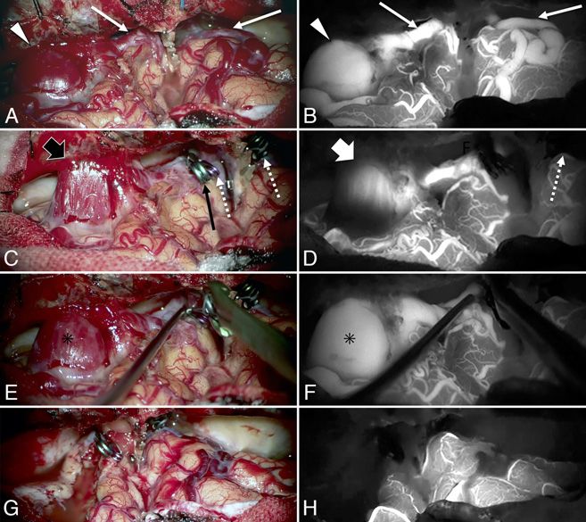
FIG. 1. Initial axial computed tomography scan (A) and T2-weighted magnetic resonance image (B and C)
                            demonstrating the cavity caused by old intracranial hemorrhage (white arrow), dilated drainer (white broken
                            arrow), varix (white arrowhead), and nidus (large white arrow).

the cribriform plate and drained into the bilateral cortical veins to the             especially dilation in the left drainer (Fig. 3). We diagnosed bilateral
superior sagittal sinus (SSS). Left internal carotid angiography sho-                 ACF DAVFs, with coexistence of left ACF DAVF and AVM that had a
wed the arteriovenous shunt with nidus and intranidal aneurysm sup-                   common trunk drainer. We planned surgical intervention.
plied by the left fronto-orbital artery, which revealed the AVM. The left                 A bifrontal osteoplastic craniotomy with both side dural incisions
draining cortical vein was a common trunk from the shunt of the left                  was performed. The dilated cortical vein, venous varix, nidus, and
DAVF and AVM. Venous varix was observed at the bilateral drainers,                    shunting points of the bilateral DAVFs could be observed. The nidus
                                                                                      and a proper feeder of the AVM in the left frontal lobe was identified
                                                                                      between the shunting point of the left DAVF and dilated varix
                                                                                      (Fig. 4A). We performed initial ICG videoangiography before inducing
                                                                                      hemodynamic changes (Fig. 4B). The DAVF in the right ACF had sim-
                                                                                      ple architecture with a single drainer, so drainer occlusion could result
                                                                                      in shrinkage of the dilated distal drainers (Fig. 4C and D). In the left
                                                                                      ACF, drainer occlusion of DAVF at the cribriform plate was performed
                                                                                      to reduce the blood flow to the drainer both from the left ACF DAVF
                                                                                      and AVM. This simple drainer occlusion could result in shrinkage of
                                                                                      the varix with residual shunting flow. Then, occlusion of the left
                                                                                      fronto-orbital artery, which was a proper feeder of the nidus, shrank
                                                                                      the dilated left varix immediately but still incompletely because of the
                                                                                      remaining pial arterial supply from the AVM (Fig. 4C and D). Releas-
                                                                                      ing the temporary occlusion of the left fronto-orbital artery caused the
                                                                                      re-expansion of the varix (Fig. 4E and F). Therefore, we removed the

  FIG. 2. Three-dimensional reconstruction images using rotational digi-
  tal subtraction angiography of both right (A, anteroposterior view; B,             FIG. 3. Three-dimensional reconstruction images using rotational digi-
  lateral view) and left (C, anteroposterior view; D, lateral view) external         tal subtraction angiography of the left internal carotid artery (A, antero-
  carotid arteries depicting the bilateral dural arteriovenous fistulae               posterior view; B, lateral view) depicting the nidus of the arteriovenous
  (DAVFs) in the anterior cranial fossa (ACF), shunting point (white bro-            malformation (AVM) (white broken arrow) in the left frontal lobe and its
  ken arrows), bilateral frontal draining veins (white arrows), and varix in         draining vein (white arrows) with varix (white arrowheads) that was
  the frontal lobe (white arrowheads).                                               the common drainer of the left DAVF.

2 | J Neurosurg Case Lessons | Vol 3 | Issue 13 | March 28, 2022

                                                                                                               Unauthenticated | Downloaded 06/12/22 09:19 PM UTC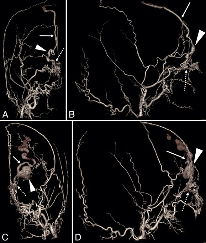
FIG. 4. Intraoperative initial microscopic view (A) and indocyanine
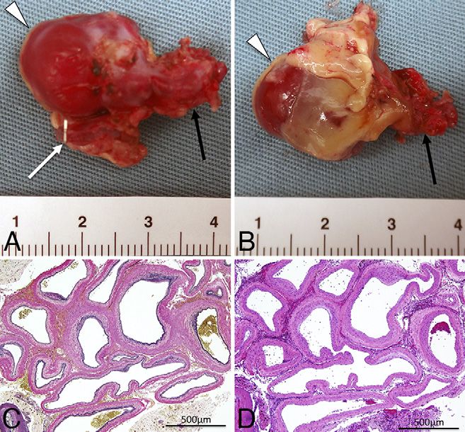
green (ICG) videoangiography (B) showing the bilateral frontal dilated FIG. 5. Photographs of the surgical specimen (A and B) showing the
drainers (white arrows) and expanded varix in the left frontal lobe varix (white arrowheads), drainer (white arrow), and nidus (black
(white arrowheads). Microscopic view (C) and ICG videoangiography arrow). Photomicrograph (C, elastic van Gieson staining, original mag-
(D) after occlusion of the drainers of the bilateral DAVFs in the ACF nification 40) demonstrating the arterial and venous structures in the
(white broken arrows) and main feeder of the AVM (black arrow) cerebral parenchyma. These findings corresponded to AVM. Photomi-
revealing the shrinking varix (large black arrow) with residual shunting crograph (D, hematoxylin and eosin staining, original magnification
flow (large white arrow). Microscopic view (E) and ICG videoangiogra- 40) showing hemosiderin infiltrating around the vessels correspond-
phy (F) after occlusion of the bilateral drainers and release of the main ing to old intracranial hemorrhage.
feeder of the AVM demonstrating reexpansion of the varix (asterisk).
Intraoperative final microscopic view (G) and ICG videoangiography
(H) showing complete removal of the AVM and normalization of the and intraoperative visualization using ICG videoangiography during
bilateral frontal lobe hemodynamics. stepwise clipping of the drainer of the DAVF and feeder of the
AVM. The concept of surgery for DAVF in the ACF is disconnection
AVM and varix completely by coagulated the proper feeder with pial of the fistula which drains into a dilated vein.9 In the present case,
supply of AVM and coagulated drainer at the just distal to varix the right DAVF was quite simple, so drainer occlusion could cure
(Fig. 4G). Finally, we confirmed that no residual AVM and venous dilata- the right DAVF. However, the same procedure could not be effective
tion on the frontal lobe remained using ICG videoangiography (Fig. 4H). on the left side, because the arterial supply for the AVM drained
Histological examination revealed vessels of various sizes in the into the same dilated cortical vein as the left DAVF. These very
brain parenchyma with hemosiderin, and the diagnosis was AVM interesting hemodynamics were confirmed by intraoperative ICG
(Fig. 5). In addition, intimal thickening with elastic lamina was par- videoangiography using stepwise temporary clipping of the drainer
tially observed, and the final diagnosis was varix. Postoperative of the DAVF and feeder of the AVM. This ICG videoangiography
angiography detected no residual DAVF and AVM. The patient’s also demonstrated the pial supply for the AVM. Consequently, we
postoperative course was uneventful. could remove the AVM completely. If we had remained unaware of
the complicated hemodynamic system in the left ACF and selected
Discussion the surgical strategy of drainer occlusion only as a treatment for the
The present case of DAVF coexisting with AVM using the same DAVF, the patient might have suffered a miserable outcome.
draining system in the ACF is unique. Only two cases of DAVF in Histological examination revealed that the AVM contained ves-
the ACF with bilateral frontal drainers have been treated surgi- sels of various sizes in the brain parenchyma with hemosiderin.
cally.4,5 Therefore, the present bilateral case was certainly rare. In The AVM is a lesion that is usually present at birth and grows pro-
addition, our case featured coexistence of the intracranial DAVF portionately with age.10 Consequently, venous hypertension follow-
and AVM in the same region. The three previous cases of coexis- ing sinus thrombosis is the primary mechanism of the formation of
tence of intracranial DAVF and AVM had these lesions located in DAVFs.6,11,12 Venous hypertension may promote the growth of
different regions as follows: SSS DAVF and left occipital AVM; SSS microscopic arteriovenous shunts, which are found within the vasa
DAVF and right temporal AVM; and tentorial DAVF and left cerebel- vasorum of normal pachymeninges, and may stimulate the release
lopontine fissure AVM.6–8 of angiogenic factors in experimental models.12–14 In addition, head
trauma is reported to be one of the etiologies of DAVF.2,15
Observations Our case had a history of head trauma 20 years previously. The
In our case, the left DAVF in the ACF shared a common drain- present case can be summarized as bilateral DAVFs in the ACF
ing system with the AVM, as confirmed by cerebral angiography coexisting with unilateral frontal AVM. The assumed cause of this
J Neurosurg Case Lessons | Vol 3 | Issue 13 | March 28, 2022 | 3
Unauthenticated | Downloaded 06/12/22 09:19 PM UTC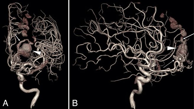
interesting clinical condition can be explained as follows: (1) left frontal    8. Ahmed R, Lopez C, Philip K, Gould G. Dural arteriovenous fistula
AVM caused long-term venous hypertension of SSS; (2) subsequent                    and arteriovenous malformation presenting as trigeminal neuralgia.
increasing cortical venous pressure in the bilateral anterior frontal              BMJ Case Rep. 2021;14(1):e240483.
                                                                                9. Lawton MT, Chun J, Wilson CB, Halbach VV. Ethmoidal dural arte-
lobes; (3) acquired bilateral DAVFs developed in the ACF with expres-
                                                                                   riovenous fistulae: an assessment of surgical and endovascular
sion of multiple angiogenic factors; (4) head trauma might contribute to           management. Neurosurgery. 1999;45(4):805–811.
the development of DAVF; and (5) asymptomatic left frontal intracranial        10. Phillips J, Tang C, Armstrong D, De Chalain T, Zuker R. Congenital
hemorrhage occurred from the AVM or DAVF. Further clinical investiga-              arteriovenous malformations: a follow-up of treatment. Can J Plast
tion may reveal more facts about this clinical condition.                          Surg. 2005;13(1):23–26.
                                                                               11. Phatouros CC, Halbach VV, Dowd CF, et al. Acquired pial arteriove-
                                                                                   nous fistula following cerebral vein thrombosis. Stroke. 1999;30(11):
Lessons                                                                            2487–2490.
   Neurosurgeons should be aware of the possibility of the coexistence         12. Kojima T, Miyachi S, Sahara Y, et al. The relationship between
of DAVF and AVM. Preoperative angiography to establish the angio-                  venous hypertension and expression of vascular endothelial growth
architecture in detail and plan the approximate operation strategies is            factor: hemodynamic and immunohistochemical examinations in a
important. ICG videoangiography during the stepwise occlusion of                   rat venous hypertension model. Surg Neurol. 2007;68(3):277–284.
drainer of the DAVF and feeder of the AVM was quite useful to under-           13. Terada T, Tsuura M, Komai N, et al. The role of angiogenic factor
                                                                                   bFGF in the development of dural AVFs. Acta Neurochir (Wien).
stand the complicated hemodynamics of DAVF coexisting with AVM.                    1996;138(7):877–883.
                                                                               14. Lawton MT, Jacobowitz R, Spetzler RF. Redefined role of angiogen-
References                                                                         esis in the pathogenesis of dural arteriovenous malformations.
 1. Al-Shahi R, Bhattacharya JJ, Currie DG, et al. Prospective, population-        J Neurosurg. 1997;87(2):267–274.
    based detection of intracranial vascular malformations in adults: the      15. Ishikawa T, Houkin K, Tokuda K, Kawaguchi S, Kashiwaba T.
    Scottish Intracranial Vascular Malformation Study (SIVMS). Stroke.             Development of anterior cranial fossa dural arteriovenous malformation
    2003;34(5):1163–1169.                                                          following head trauma. Case report. J Neurosurg. 1997;86(2):291–293.
 2. Satomi J, Satoh K. Epidemiology and etiology of dural arteriovenous
    fistula. Article in Japanese. Brain Nerve. 2008;60(8):883–886.              Disclosures
 3. Elhammady MS, Ambekar S, Heros RC. Epidemiology, clinical pre-             The authors report no conflict of interest concerning the materials or
    sentation, diagnostic evaluation, and prognosis of cerebral dural          methods used in this study or the findings specified in this paper.
    arteriovenous fistulas. Handb Clin Neurol. 2017;143:99–105.
 4. Deshmukh VR, Chang S, Albuquerque FC, McDougall CG, Spetzler               Author Contributions
    RF. Bilateral ethmoidal dural arteriovenous fistulae: a previously          Conception and design: Yamamoto, Shibahara, Kumabe. Acquisition of
    unreported entity: case report. Neurosurgery. 2005;57(4):E809.             data: Yamamoto, Shibahara, Inukai, Niki, Usui, Kimura, Kumabe.
 5. Kohama M, Nishimura S, Mino M, et al. Anterior cranial fossa dural         Analysis and interpretation of data: Yamamoto, Inukai, Kumabe.
    arteriovenous fistula with bilateral cortical drainers–case report.         Drafting the article: Yamamoto, Shibahara, Hide, Kumabe. Critically
    Neurol Med Chir (Tokyo). 2010;50(3):217–220.                               revising the article: Yamamoto, Kumabe. Reviewed submitted version
 6. Bai Y, He C, Zhang H, Ling F. De novo multiple dural arteriovenous         of manuscript: Yamamoto, Hide, Kumabe. Approved the final version of
    fistulas and arteriovenous malformation after embolization of cere-         the manuscript on behalf of all authors: Yamamoto. Administrative/
    bral arteriovenous fistula: case report. Childs Nerv Syst. 2012;            technical/material support: Yamamoto, Koizumi, Hyakutake, Ishima.
    28(11):1981–1983.                                                          Study supervision: Shibahara, Kumabe.
 7. Sattur MG, Abi-Aad KR, Tian F, Welz ME, Anderies B, Bendok BR.
    Treatment strategy of a patient with a brain arteriovenous malforma-       Correspondence
    tion and cranial dural fistula: 2-dimensional operative video. Oper         Daisuke Yamamoto: Kitasato University School of Medicine, Kanagawa,
    Neurosurg (Hagerstown). 2019;16(5):636.                                    Japan. daiyama@med.kitasato-u.ac.jp; baystars_saiko@hotmail.com.

4 | J Neurosurg Case Lessons | Vol 3 | Issue 13 | March 28, 2022

                                                                                                      Unauthenticated | Downloaded 06/12/22 09:19 PM UTC
You can also read