A systematic review of the Neotropical social wasp genus Angiopolybia Araujo, 1946 (Hymenoptera: Vespidae): species delimitation, morphological ...
←
→
Page content transcription
If your browser does not render page correctly, please read the page content below

Arthropod Systematics & Phylogeny 80, 2022, 75–97 | DOI 10.3897/asp.80.e71492                                                                                                         75

A systematic review of the Neotropical social wasp genus
Angiopolybia Araujo, 1946 (Hymenoptera: Vespidae):
species delimitation, morphological diagnosis, and
geographical distribution
Paulo Cézar Salgado Barroso1, Rodolpho Santos Telles Menezes2,3, Marcio Luiz de Oliveira1,
Alexandre Somavilla1
1 Programa de Pós-Gradução em Ciências Biológicas (Entomologia), Coordenação de Biodiversidade, Instituto Nacional de Pesquisas da
     Amazônia (INPA), Av. André Araújo, 2.936, Petrópolis, 69067-375, Manaus, Brazil
2 Programa de Pós-Graduação em Biodiversidade Animal, Centro de Ciências Naturais e Exatas, Universidade Federal de Santa Maria
     (UFSM), Av. Roraima, 1000, Camobi, 97105-900, Santa Maria, Brazil
3 Laboratório de Biologia Comparada e Abelhas, Departamento de Biologia, Faculdade de Filosofia, Ciências e Letras (FFCLRP),
    Universidade de São Paulo (USP), Av. Bandeirantes, 3900, Monte Alegre, 14040-901, Ribeirão Preto, SP, Brazil

http://zoobank.org/F85F11D5-313D-4A75-B390-7FDCCCA9376F

Corresponding authors: Paulo Cézar Salgado Barroso (pc.salgadobarroso@gmail.com), Rodolpho Santos Telles Menezes (rstmenezes@gmail.com)

Received 12 July 2021
Accepted 03 February 2022                                                                     Academic Editors               Beny Wipfler, Mónica M. Solórzano-Kraemer
Published 02 March 2022

Citation: Barroso PCS, Menezes RST, de Oliveira ML, Somavilla A (2022) A systematic review of the Neotropical social wasp genus Angiopolybia
Araujo, 1946 (Hymenoptera: Vespidae): species delimitation, morphological diagnosis, and geographical distribution. Arthropod Systematics &
Phylogeny 80: 75–97. https://doi.org/10.3897/asp.80.e71492

Abstract
For the Neotropical genus Angiopolybia Araujo 1946, several phenotypic forms were previously described, however, they have not
been studied within an integrative taxonomic framework. Here, we used molecular data (variation of two mitochondrial genetic
markers with molecular species delimitation methods) and morphology (adult morphology, male genitalia, and scanning electron
microscopy images) to test the number of species within Angiopolybia. Specifically, we investigated the taxonomic validity of the
morphological variants A. pallens dark morph, A. paraensis morph paraensis, A. paraensis morph ruficornis, and A. paraensis morph
obscurior. Moreover, we reviewed the taxonomy and geographic distribution of the genus. Our results of morphological and molecu-
lar analyses are compatible with the current classification of Angiopolybia, and we did not find reasons to propose the morphological
variants of A. pallens and A. paraensis as valid species. Additionally, we reassess the spatial range of the four Angiopolybia species
and provide refined maps of their geographical distributions.

Keywords
Integrative taxonomy, morphological variation, mtDNA, paper wasps, phylogenetic systematics, swarm-founding social wasps.

Copyright Barroso et al.: This is an open access article distributed under the terms of the Creative Commons Attribution License (CC BY 4.0), which permits unrestricted use, distribution,
and reproduction in any medium, provided the original author and source are credited.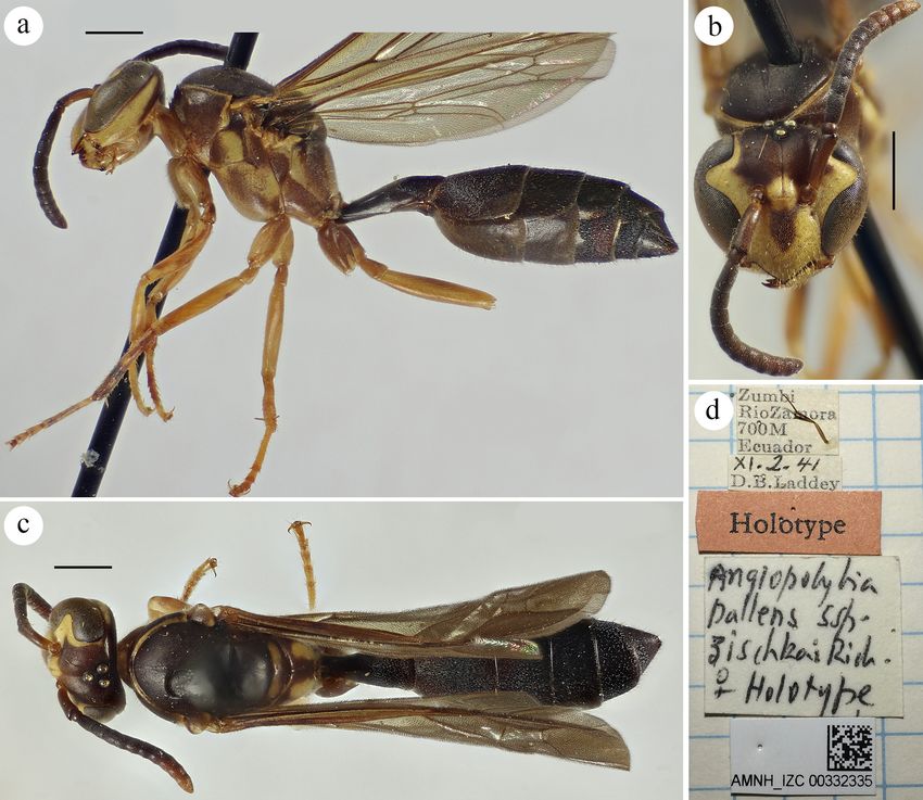
76 Barroso et al.: Systematic review of Angiopolybia 1. Introduction Species boundaries are frequently hard to delimit due A. paraensis morph ruficornis, and A. paraensis morph to intraspecific morphological variation. Hence, the use obscurior. However, Andena et al. (2007) made the fol- of different sources of biological information such as lowing comment about the “morphs” of A. paraensis: “is biogeography, behavior, ecology, molecular data, and a category without nomenclatural standing, hence these morphology is generally regarded as a good practice for morphs are synonyms of A. paraensis”. species delimitation (Bickford et al. 2007; Padial et al. Here, we analyzed the variation of mitochondrial genet- 2010). Intraspecific polymorphism has been designated ic markers with molecular species delimitation methods, as “morph” for swarm-founding social wasps (Vespidae: adult morphology, male genitalia, and scanning electron Polistinae: Epiponini) (Richards 1978), but taxonomic microscopy images to characterize in detail the species investigations using a variety of characters from inde- limits within Angiopolybia. Thus, we tested whether the pendent datasets are scarce for the group (Menezes et al. “morphs” of A. pallens and A. paraensis can be recog- 2015; Lopes and Menezes 2017; Somavilla et al. 2021). nized based on morphological and molecular data, as well Angiopolybia Araujo, 1946 is a neotropical swarming- as providing diagnostic features for Angiopolybia species founding social wasp genus composed of four species: A. and a reassessment of their geographical distribution. pallens (Lepeletier, 1836), A. paraensis (Spinola, 1851), A. obidensis (Ducke, 1904), and A. zischkai Richards, 1978. Angiopolybia is recovered as the sister lineage of all remaining Epiponini genera (Menezes et al. 2020; 2. Material and Methods Noll et al. 2021). From a morphological perspective, An- giopolybia is characterized by the pronotum with later- al fovea, scutum with posterolateral lamella absent an- 2.1. Taxonomic sampling teriorly and not adjacent to the tegula, mesoepisternum with dorsal groove, clypeus with square lateral lobes, and We obtained type material through loans from the Natural sharply pointed apex (Carpenter 2004). According to An- History Museum (NHM, London, England) and Museu dena et al. (2007), the monophyly of the genus is sup- de Zoologia da Universidade de São Paulo (MZUSP, São ported by six synapomorphies: prestigma about as long as Paulo, Brazil). Additionally, we obtained images of type wide, proepisternum with reduced carina, scutal lamella specimens deposited at the following institutions: Amer- reduced, scutellum without an impressed line, practically ican Museum of Natural History (AMNH, New York, straight dorsal groove, and flattened metapleural basal- USA), Harvard Museum of Comparative Zoology (MCZ, ar area. Phylogenetic studies recovered two clades with Cambridge, MA, USA), and Muséum National d’Histoire the following relationship for Angiopolybia species: ((A. Naturelle (MNHN, Paris, France). We also obtained su- paraensis + A. obidensis) + (A. pallens + A. zischkai)) pplementary material from the following scientific insti- (Andena et al. 2007; Noll et al. 2021). tutions: AMNH, Coleção de Hymenoptera do Museu de The genus occurs from Costa Rica to the south-cen- Zoologia da Universidade Federal da Bahia (MZUFBA, tral region of Brazil (Richards 1978), and A. pallens is Salvador, Brazil), Coleção Entomológica da Universidade the only species that occurs in the Amazon region and Federal do Espírito Santo (UFES, Vitória, Brazil), Institu- the Brazilian Atlantic Forest (Carvalho et al. 2014, 2015, to Nacional de Biodiversidad (INABIO, Quito, Ecuador), 2021). The nests of Angiopolybia are characterized as Instituto Nacional de Pesquisas da Amazônia (INPA, Ma- exposed on leaves, branches, and stone slabs and con- naus, Brazil), Museo de Zoología - Pontificia Universidad sisting of several stacked combs (Ducke 1914), fused or Católica del Ecuador (QCAZ, Quito, Ecuador), MZUSP, suspended from each other by a central pedicel, and the Museu Paraense Emílio Goeldi (MPEG, Belém, Brazil), combs gradually grow along the margins (Wenzel 1998). and NHM. All specimens were identified using the identi- The nests of Angiopolybia species have a single envelope, fication keys proposed by Richards (1978) and Andena et which can be ovoid shape or bottle-shaped, with a single al. (2007), and also by comparison with the type material entry at the lower part with a tubular form and horizontal- and original descriptions of Lepeletier (1836), Spinola ly curved (Richards 1978; Wenzel 1998). Morphological (1851), Ducke (1904), and Richards (1978). difference between queens and workers is subtle (Rich- ards 1978), and only the male of A. pallens was described by Richards (1978). 2.2. Morphological analysis Morphological differences between A. paraensis and A. obidensis and between A. pallens and A. zischkai are We used adult specimens for a redescription of the female subtle, such as the height of the anterior pronotal lamella and description of the male. We examined 469 females and the development of the pronotal lobe, respectively and 32 males of A. pallens (eight males for genitalia anal- (Richards 1978; Andena et al. 2007). Moreover, Rich- ysis), four females of A. zischkai, 104 females and three ards (1978) characterized multiple morphological vari- males of A. obidensis (two males for genitalia analysis), ants of A. pallens and A. paraensis as “morph”, such as and 80 females and six males of A. paraensis (four males A. pallens dark morph, A. paraensis morph paraensis, for genitalia analysis). We also analyzed images of five
Arthropod Systematics & Phylogeny 80, 2022, 75–97                                                                    77

type specimens of the species. They were analyzed un-        software MESQUITE v.3.61 (Maddison and Maddison
der a Nikon SMZ645 stereo microscope with an acces-          2019). The most appropriate model of nucleotide evolu-
sory magnifying lens of Nikon G-AL 2x. We obtained           tion and the best-fitting partitioning scheme were selected
the proportions and morphological measurements with          using PARTITIONFINDER2 (Lanfear et al. 2016) under
the ocular lens AmScope reticulated WF10X/22 and the         the Bayesian information criterion (see Table S3).
measurements in the images using the IMAGEJ 1.52a
(Rasband 2019). The explanation of how each measure-
ment was performed is described in Supplementary Ma-         2.4. Phylogenetic and molecular
terial (Table S1). For the morphological terminology, we          species delimitation analyses
followed Richards (1978), and for male terminalia, we
used Richards (1978), Buck et al. (2012), and Somavil-       Phylogenetic inference was conducted by Maximum
la et al. (2018). We obtained the images of specimens        Likelihood (ML) using the IQ-TREE v.1.6.12 (Nguy-
and morphological characters with the aid of a Leica         en et al. 2015) with a concatenated morphological and
DMC4500 digital camera coupled to a Leica M205A              molecular dataset. We calculated branch supports with
stereomicroscope with a self-assembly system, using the      1000 replicates of SH-like approximate likelihood ra-
Leica Application Suite v.4.10.0-Montage® software. We       tio test (SH-aLRT) (Guindon et al. 2010) and Ultrafast
edited the images and assembled the plates using the soft-   Bootstrap approximation (UFBoot) (Minh et al. 2013).
ware ADOBE PHOTOSHOP® CS6 v.6.1. We employed                 We used Apoica thoracica du Buysson, 1906 and Agelaia
scales of 1 mm and 0.5 mm for images of adults and male      fulvofasciata (DeGeer, 1773) as outgroups based on pre-
genitalia, respectively. We also generated Scanning Elec-    vious phylogenetic studies (Andena et al., 2007; Menezes
tron Microscopy images (equipment Oxford Instruments         et al. 2020; Noll et al. 2021). Visualization and edition
INCAx-act, model 51-ADD0007) without the use of              of the phylogenetic trees were performed using the pro-
preparation technique of coating. Finally, we built a ma-    gram FIGTREE v.1.4.2 (http://tree.bio.ed.ac.uk/software/
trix of morphological characters based on the studies of     figtree). We also performed analysis under Maximum
Ducke (1914), Richards (1943, 1978), Carpenter (1991),       Parsimony (MP) with our concatenated matrix using
and Andena et al. (2007), as well as proposing new mor-      the program TNT – Tree analysis using New Technolo-
phological characters for Angiopolybia species.              gy v.1.5 (Goloboff and Catalano 2016). We performed a
                                                             Traditional search by Wagner trees with 1 random seed,
                                                             1000 replicates, saving 30 by replication, and collapsing
2.3. Molecular dataset                                       the tree after the search. The polarization of states was
                                                             carried out by comparison with outgroups (Nixon and
We extracted total DNA from a mid and a hind leg of          Carpenter 1993). The visualization of the MP phyloge-
specimens preserved in ethanol and pinned museum spec-       netic tree was performed using the program WINCLADA
imens. We extracted DNA from a single specimen of A.         v.1.00. 08 (Nixon 2002).
pallens, A. zischkai, A. obidensis, A. paraensis yellow,        We used four species delimitation methods for Cox1
A. paraensis brown and yellow and A. paraensis dark          and 16S data separately and also concatenated as follows:
brown, the complete list of specimens used in our anal-      Automatic Barcode Gap Discovery (ABGD) (Puillandre
yses is presented in Supplementary Material (Table S2).      et al. 2012), Assemble Species by Automatic Partition-
For extraction, we used the DNeasy® Blood & Tissue ex-       ing (ASAP) (Puillandre et al. 2021), bayesian Poisson
traction kit (QIAGEN®, Valencia, California, USA) fol-       Tree Processes (bPTP) (Zhang et al. 2013) and multi-rate
lowing the manufacturer’s protocol. We amplified two mi-     Poisson Tree Processes (mPTP) (Kapli et al. 2017). For
tochondrial gene fragments, Cytochrome Oxidase subunit       ABGD and ASAP, we used the online platforms https://
I (Cox1) and 16S ribosomal DNA (16S). Specific prim-         bioinfo.mnhn.fr/abi/public/abgd and https://bioinfo.
ers and conditions for PCR amplification are described       mnhn.fr/abi/public/asap respectively, using the Kimura
by Menezes et al. (2015, 2017). The PCR products were        Two-Parameter (K2P) model (Kimura 1980). We used the
purified using exonuclease I and shrimp alkaline phos-       phylogenetic trees generated by IQ-Tree ver.1.6.12 for
phatase. Sanger sequencing was conducted by the Centro       the bPTP and mPTP methods. For the bPTP and mPTP,
de Recursos Biológicos e Biologia Genômica (CREBIO),         we used the online platforms https://species.h-its.org and
Universidade Estadual Paulista ‘Júlio de Mesquita Filho’     https://mptp.h-its.org/#/tree respectively, removing the
(UNESP, Jaboticabal, São Paulo, Brazil). The sequencing      outgroups and with default parameters, except for the
was carried out in both directions, and the consensus se-    number of generations of Monte Carlo Markov Chains
quences were assembled on GENEIOUS R7 (Kearse et             (MCMC) of 300,000 in bPTP. Additionally, we analyzed
al. 2012). All sequences were deposited at GenBank (ac-      the genetic distance among the specimens for both mito-
cess numbers are MZ496308–MZ496314 and MZ513916              chondrial markers in MEGA X using the K2P model.
for Cox1, and MZ496381–MZ496387 and MZ513917 for
16S). We performed the alignment using the MUSCLE
algorithm (Edgar 2004), implemented in the program           2.5. Geographic distribution maps
MEGA X (Kumar et al. 2018) and with default parame-
ters. We visually inspected and corrected all alignments.    We used the recorded localities for Angiopolybia speci-
We concatenated the Cox1 and 16S sequences using the         mens to build a geographical database of species occur-
78                                                                                        Barroso et al.: Systematic review of Angiopolybia

rence. The recorded localities were based on the label                    (currently Angiopolybia) by Ducke (1914), these genera
data of all analyzed specimens and information obtained                   are not well-defined by the proposed characters. Based
in the literature with the precise locality. We avoided the               on this, some morphological characters that had been
use of non-geo-located information, such as presence                      proposed to separate Angiopolybia from Agelaia such as
data in states or provinces. The Google Maps platform                     stelocyttarous nest (with envelope) (Ducke 1914), very
was used to determine each specimen’s geographical                        weak or absent occipital carina (Richards 1943), and
coordinates and convert them to decimal degrees. We                       flat metapleural basalar area (Richards 1978; Andena et
used the resulting database to characterize in detail the                 al. 2007) were not considered here because they are not
geographic range of each Angiopolybia species with the                    exclusive to Angiopolybia, since they are present in few
software QGIS v.3.10.11 (QGIS.org, 2020, Geographic                       species of Agelaia. Moreover, the scutellum with line or
Information System, http://www.qgis.org 2020) and a bi-                   depression (Richards 1943; Andena et al. 2007) was not
ome map reported by Dinerstein et al. (2017).                             considered because they are not exclusive to Agelaia,
                                                                          since some Angiopolybia species present the line, despite
                                                                          it is inconspicuous. Richards (1943) commented that the
                                                                          characters determined by him, isolated, had no effect on
3.       Results                                                          separating the taxa.
                                                                              Based on previous and this study, A. pallens and A.
                                                                          zischkai are closer, as are A. paraensis and A. obidensis.
To characterize in detail the species limits within An-                   Angiopolybia pallens and A. zischkai are distinguished
giopolybia, we considered several morphological charac-                   by rounded gena and propodeum with posterior subme-
ters, including females, males, male genitalia, and nest                  dian translucent mark inserted in a depression, whereas
architecture (Figs 1–8), and two mitochondrial markers,                   the other species have angled gena and submedian trans-
Cox1 and 16S, under a phylogenetic and molecular spe-                     lucent mark of the propodeum not inserted in a depres-
cies delimitation approach (Fig. 9). Additionally, we per-                sion. We documented other differences in the step 1 of the
formed a taxonomic revision for Angiopolybia and char-                    identification key for the genus (see below).
acterized the geographical range of each species (Fig.
10). Moreover, we proposed an identification key with                     Geographic distribution. Bolivia, Brazil, Colombia,
new morphological characters and images.                                  Costa Rica, Ecuador, French Guiana, Guyana, Panama,
                                                                          Peru, Suriname, Trinidad and Tobago, Venezuela.

3.1.     Revision of Angiopolybia Araujo,
         1946                                                             Angiopolybia pallens (Lepeletier, 1836)

Rhopalidia Lepeletier, 1836: 538 (a genus with two species); ICZN,        Figs 1a–f, 2a–c, 6a–e, 7a–g
    1976: 240, 241 (Opinion 1051 - suppressed under the powers of the
    plenary, nº. 2072 in the Official Index of Rejected and Invalid Ge-   Rhopalidia pallens Lepeletier, 1836: 539; Spinola, 1851: 63 (nest); de
    neric Names in Zoology). Type species, Rhopalidia pallens Lepele-         Saussure, 1854: 189 (synonym of Polybia pallipes (Olivier, 1792));
    tier, 1836, subsequent designation by Schulz, 1912: 60.                   du Buysson, 1906: 342 (synonym of Apoica pallida var. pallens
Angiopolybia Araujo, 1946: 166, 169 (designation of new name for Ste-         (Fabricius, 1804)); Ducke, 1910: 542 (specimen of the collection of
    lopolybia Ducke, 1914). Specie-type, Rhopalidia pallens Lepeletier,       Spinola = S. infernalis (de Saussure,1854)); Schulz, 1912: 60 (syn-
    1836 (original designation).                                              onym: S. infernalis (de Saussure, 1854)).
Stelopolybia (Angiopolybia) Richards and Richards, 1951: 69; Rich-        Polistes rufina Erichson, 1848: 590; Spinola, 1851: 79 (synonym of
    ards, 1973: 49 (suppression of Rhopalidia Lepeletier, 1836 by             Rhopalidia pallens Lepeletier, 1836).
    ICZN, 1976).                                                          Polybia infernalis de Saussure, 1854: 195, plate XXV: fig. 3 (in division
                                                                              My); Ducke, 1905a: 662 (synonym: P. ampullaria Möbius, 1856);
Diagnosis. Lateral ocellus separated from the eye by two                      Richards, 1943: 45 (invalid designation of the type species of Ste-
times its diameter. Compound eyes with bristles. Clypeus                      lopolybia); Richards, 1978: 232 (lectotype designation). Type local-
with acute apex, lateral margins parallel in part upper and                   ity: “Le Para”, female (MNHN, Paris) [examined by images].
rectangular lateral lobes. Short malar space. Occiput with                Polybia ampullaria Möbius, 1856: 133, 155 (key identification of
carina. Labial palpus with four segments. Proepisternum                       nests), 165 (plate VII), VII (figs. 1–8 - female, nest).
with reduced lateral carina. Pronotal carina limited to a                 Eumenes flavopectus Provancher, 1888: 422.
small length in the center of the pronotum and acute. Pro-                Stelopolybia infernalis; Ducke, 1910: 519 (key), 524 (synonym: Poly-
notum with lateral fovea. Mesoscutum with reduced pos-                        bia ampullaria Möbius, 1856), 525 (fig. 12 - nest); Ducke, 1913:
terolateral lamella. Dorsal groove of the mesoepisternum                      331 (synonym: R. pallens Lepeletier, 1836).
present and transverse to the sclerite. Metasomal tergum                  Stelopolybia pallens; Bequaert, 1944: 293 (key), 294 (synonym: Poly-
I, in dorsal view, with lateral margins diverging gradually                   bia infernalis Saussure, 1854, Polybia ampullaria Möbius, 1856,
from the base to apex.                                                        Polistes rufina Erichson, 1884, Eumenes flavopectus Provancher
                                                                              1888).
Additional comments. Considering the Gymnopolybia                         Angiopolybia pallens; Araujo, 1946: 169; Richards, 1978: 231 (key),
description (currently Agelaia) from Stelopolybia species                     232 (description of male, and diagnose of female and male); Andena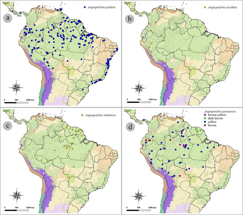
Arthropod Systematics & Phylogeny 80, 2022, 75–97                                                                                     79

Figure 1. Lectotype, female of Polybia infernalis de Saussure, 1856 [currently Angiopolybia pallens (Lepeletier, 1836)]: a. lateral
view, b. dorsal view, c. head in frontal view. Male: d. head in frontal view, e. lateral view, f. dorsal view. Scale: 1 mm. Source:
Agnièle Touret-Alby MNHN for images a, b, and c.

    et al., 2007: 59 (phylogenetic discussion), 60 (table 2 – characters     directed to the posterior region; posterior submedian
    matrix), 61 (figs. 1A, 2A), 62 (figs. 3B, 5 – cladogram), 63 (key), 64   translucent mark of the propodeum inserted in a round
    (locality of examined material); Carvalho et al., 2014, 2021 (evolu-     depression; basal metapleural area with parallel upper
    tionary hypothesis of species distribution).                             and lower margins.
Stelopolybia (Angiopolybia) pallens; Richards and Richards, 1951: 77
    (list and notes about the nests).                                        Redescription of female (Fig. 1a, b, c). Size. (1) Head
Angiopolybia infernalis; Overal, 1978: 9 (list of species).                  1.27 mm long, 2 mm high, and 2.27 mm wide; (2) me-
                                                                             sosoma 3.50 mm long, anterior wing 8.11 mm long, and
Type locality. Cayenne, French Guiana.                                       posterior wing 5.27 mm long; (3) metasoma 5.53 mm
                                                                             long. Head. (1) Lateral ocelli with 0.15 mm and median
Diagnosis. Anterior wing of 7–8.5 mm; eyes with me-                          ocellus with 0.17 mm of diameter, not inserted in a de-
dium-sized and sparse bristles; rounded gena; pronotal                       clivity of the vertex, and the lateral ocelli separated from
lamella low on the anterior margin, one fifth of the height                  the eyes for twice its diameter. (2) Compound eyes with
of antennal socket; pronotal lobe developed in the ante-                     medium-sized and sparse bristles. (3) Frons with inter-
rior lateral region, below the pronotal fovea; defined and                   antennal space about twice the height of antennal sock-
deep pronotal fovea; axillary fossa with anterior margin                     et. Anterior tentorial fovea closer to the antennal socket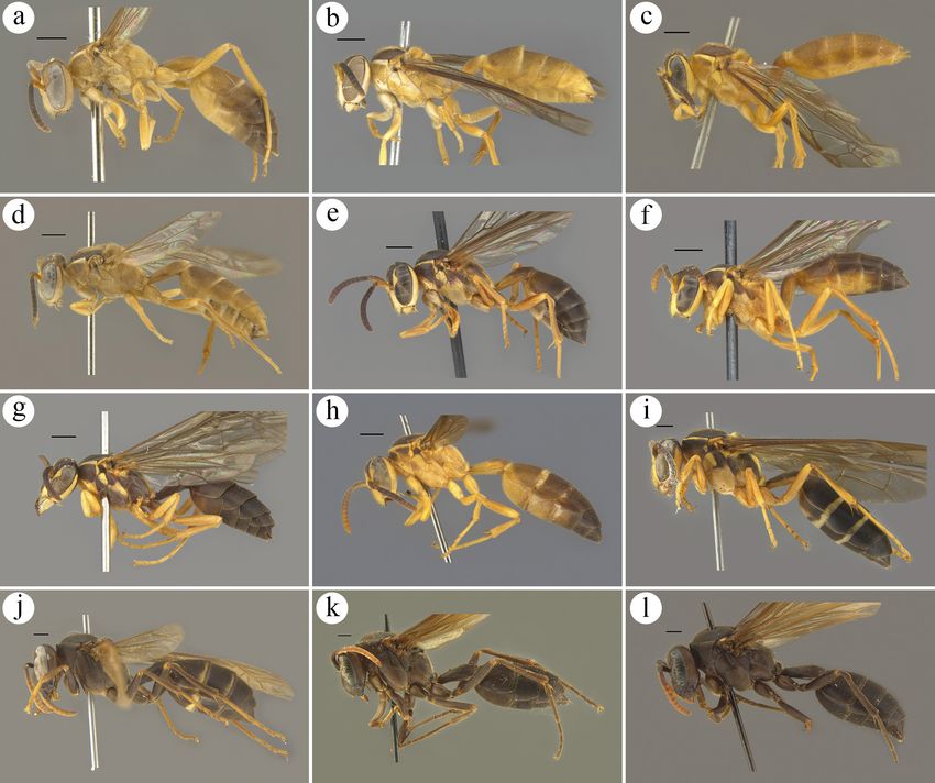
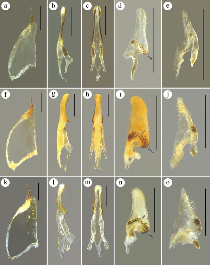
80                                                                                Barroso et al.: Systematic review of Angiopolybia

Figure 2. Structure variation of the nest of Angiopolybia pallens: a. typical form in external view, b. and c. variation found in ex-
ternal and internal view, respectively.

than to the eye. The central region of the frons with long          Redescription of male (Fig. 1d, e, f). Size. (1) Head
bristles. (4) Antennal socket 0.22 mm high. (5) Clypeus             1.01 mm long, 1.83 mm high, and 2.09 mm wide; (2)
as high as wide, contact with eyes for a distance greater           mesosoma 2.87 mm long, anterior wing 7.1 mm long,
than twice the height of antennal socket, and lateral lobe          and posterior wing 4.5 mm long; (3) metasoma 6.08 mm
touching the eye. Medium bristles in the basal half and             long. Head. (1) Lateral ocelli with 0.17 mm and median
long bristles in the apical half. (6) Gena with half of the         ocellus with 0.18 mm of diameter. (2) Compound eyes
width of the eye at the level of the ocular sinus. Meso-            with small-sized and sparse bristles. (3) Frons with in-
soma. (1) Anterior lamella of pronotum with a height of             terantennal space with 1.85 times the height of antennal
one fifth of the height of antennal socket. Pronotal fo-            socket. Anterior tentorial fovea closer to the eye that to
vea with a circular shape, deep and with slight anterior            the antennal socket. (4) Antennal socket with 0.21 mm
prominence. (2) Mesoscutum convex and as long as wide.              high. (5) Clypeus as high as wide and apex less acute
(3) Tegula 1.7 times longer than wide. (4) Axillary fossa           than in the female. Pubescence stronger than in the fe-
with anterior margin directing to the posterior region. (5)         male. (6) Gena with half of the width of the eye at the
Propodeum with translucent posterior submedian mark,                level of the ocular sinus. Mesosoma. (1) Mesoscutum
anterior to the propodeal valve, inserted in a round de-            0.9 times longer than wide. (2) Propodeum with trans-
pression. Propodeal valve complete and expanded, me-                lucent posterior submedian mark, anterior to the prop-
dian region with half of the height of antennal socket.             odeal valve, inserted in a not round depression. (3) An-
(6) Anterior wing with prestigma 1.4 times longer than              terior wing with prestigma 1.6 times longer than wide.
wide. (7) Posterior wing with eight hamuli. Metasoma.               Metasoma. (1) Metasomal tergum I 1.9 times longer
(1) Metasomal tergum I two times longer than broad.                 than broad. (2) Metasomal tergum II 0.8 times longer
Tergum with angulation in the posterior third, in later-            than wide. Genitalia (Fig. 6a–e). Paramere 1.6 mm
al view. (2) Metasomal tergum II 0.82 times longer than             long and 0.6 mm wide; parameral spine with one fifth
broad. Color. Brown in general. Yellow: lateral of the              of the paramere, curved upwards and with small-sized
apex, lateral of the front, interantennal region, disc of           and sparse bristles; lobe with a rounded apex and slight-
the clypeus, mandibles, gena, antennal segments: scape,             ly curved downwards. Aedeagus 1.2 mm long; enlarged
dorsal of FL6 (flagellomere 6) and 7, FL8–10, band con-             valve with a small emargination in the tip; apical portion
touring the posterior margin of the pronotum, tegula, thin          0.42 mm long and straight, ventral margin with denticles
bands in the posterior margins of the metasomal terga               directed for the anterior region; denticulation decreasing
I, II and III, and metasomal sterna II and III. Yellowish           in size from the apex to the base and more sclerotized
brown: central longitudinal and lateral region of the me-           than the rest of the apical portion; small-sized bristles
soscutum, tibiae and tarsi, femora median and posterior,            with alveolar base, closer in the lower half and sparse in
metasomal tergum I, anterior half of metasomal tergum               the upper half; median expansion without denticles and
II, metasomal sterna I–IV. Black: FL1–5, ventral of FL 6            with acute apex; lateral apodeme not flattened dorsoven-
and 7, metasomal terga 4–6 and metasomal sterna 5 and               trally at the apex; basal apodeme arched to the venter.
6. Wings with hyaline cells, pterostigma and venation in            Digitus 2.5 times longer than wide; apical process not
general yellowish-brown, except brown in the veins C,               curved in the region of the upper half and with bristles
Sc+R, M+Cu and M.                                                   of alveolar base small and sparse; rounded anteroventral
Arthropod Systematics & Phylogeny 80, 2022, 75–97                                                                                   81

lobe with a strip of black scale-like bristles crossing it     this study not reported by Richards (1978) and Andena et
obliquely at the base of the digitus; bristles absent in the   al. (2007). They treated the occipital carina absent for the
basal articulation. Cuspis approximately 0.48 mm long,         taxon. The prestigma longer than wide, also, was not cited
with five black scale-like bristles on the lateral lobe, and   by Richards (1978) and Andena et al. (2007). However,
small bristles with alveolar base and close throughout the     this character was found by Silveira and Carpenter (1995)
area of the cuspis, except sparse in the central region and    for some specimens, and here we also found the two
on the ventral margin.                                         forms, prestigma about as long as wide and longer than
                                                               wide. The redescription of A. pallens was made based on
Morphological variation (Fig. 7a–g). Anterior wing be-         the lectotype of Polybia infernalis de Saussure, 1854 (ju-
tween 7–8.5 mm in length. Anterior margin of the prono-        nior synonym of A. pallens). The information about the
tum (below the fovea) more curved. Pronotum, in lateral        male specimen described is: BRA, Roraima, Amajarí,
view, with frontal region more projected forward. Col-         Serra do Tepequém, SESC Tepequém. 1–16.i.2016 / Ma-
oration varied between populations from black with yel-        laise grande, J.A. Rafael, F.F. Xavier Filho col.
low marks [like A. pallens dark morph (sensu Richards,
1978)] to yellowish brown.                                     Lectotype. ♀, TYPE / MUSEUM PARIS, Amérique,
                                                               Leprieur 1834 / Polybia infernalis Sauss., Type. / 289694
Nest (Fig. 2a–c). The nest of A. pallens was initially de-     / Polybia infernalis Sauss., Lectotype ♀, Richards, 1971
scribed by Möbius (1856) for Polybia ampullaria (junior        (MNHN, Paris), record MNHN, Paris EY25588 (Fig. 1a,
synonym of the taxon), and later described by Wenzel           1b, 1c). Polybia infernalis is a junior synonym of Rhopa-
(1998) as a nest with a flask-shaped envelope and with a       lidia pallens Lepeletier, 1836, but here we used it to the
long downward entrance with its hole horizontally; ped-        redescription of the species. Type specimen analyzed by
icel initially single and later being able to be multiple;     images.
flexible card with long fibers, usually yellow or amber;
and adjacent combs, suspended or fused from each other         Additional material examined. We examined 469 fe-
and without contact with the envelope. Additionally, the       males and 32 males for A. pallens; see supplementary
following nest variation was found: built on an irregular      material S1.
substrate (thin branch with several leaves), connected to
the substrate by a central pedicel (thick) and several sup-    Geographic distribution. Bolivia: Beni, Cochabamba;
port pedicels (fines); 11 combs overlapping and also con-      Brazil: Acre, Alagoas (new record), Amapá, Amazonas,
nected by a central pedicel and multiple support, with the     Bahia, Ceará, Espírito Santo, Maranhão, Mato Gros-
third and fourth combs with the largest circumferences         so, Pará, Pernambuco, Piauí, Rio de Janeiro, Rondônia,
and combs decreasing in circumference towards the ends,        Roraima, Santa Catarina, São Paulo Sergipe; Colombia:
and without contact with the envelope; hexagonal cells         Meta, Caquetá, Nariño, Vaupés, Putumayo, Amazonas;
of the combs with diameter of 2.5 mm; single envelope          Ecuador: Esmereldas, Napo, Pichincha; French Guiana;
with long fibers arranged longitudinally, circumference        Guyana; Panama; Peru: Loreto, San Martin, Huánuco,
gradually decreasing towards the entrance and without          Pasco, Junín, Cuzco; Suriname; Trinidad and Tobago;
entrance of tubular shape.                                     Venezuela: Monagas (Fig. 10a).

Comparative comments. Angiopolybia pallens is dis-
tinguished by the presence of a lobe in the lateroanterior     Angiopolybia zischkai Richards, 1978
region of the pronotum, absent in A. zischkai, and meta-
pleural basalar area with parallel upper and lower mar-        Figs 3a–d, 7h
gins, diverging in A. zischkai. Some A. pallens specimens
can be confused with A. zischkai because it can resemble       Angiopolybia zischkai Richards, 1978: 30 (list of mimicry), 231 (key,
the typical form of A. zischkai, of color darker.                 fig. 94), 234 (description); Andena et al., 2007: 59 (phylogenetic
                                                                  discussion), 60 (table 2 - character matrix), 62 (fig. 3A, 5 - clado-
Additional comments. Despite the geographic disjunc-              gram), 63 (key), 64 (locality of examined specimens) [examined by
tion by thousand kilometers in the distribution of A. pal-        images].
lens between the Amazon and Atlantic Forest (Carvalho et
al. 2014, 2021), considering the morphological analysis of     Type locality. Zumbi, Ecuador.
specimens collected in both biomes, we verified that the
differences, when found, are very subtle and follow the        Diagnosis. Anterior wing of 8–9.5 mm; eyes with me-
variation found between different populations. Consider-       dium-sized and sparse bristles; rounded gena; pronotal
ing the populations from the Atlantic Forest, the color var-   lamella low on the anterior margin, one fifth of the height
ied from yellow with brown marks to completely brown,          of antennal socket; pronotal lobe not developed in the
while among the populations from the Amazon Forest it          lateral anterior region, below of the pronotal fovea; de-
varied from black with yellow marks to yellowish brown,        fined pronotal fovea, but little deep; axillary fossa with
with some forms existing in both biomes. The occipital         anterior margin directed to the posterior region; posterior
carina present in A. pallens, despite weak or not, found in    submedian translucent mark of the propodeum inserted in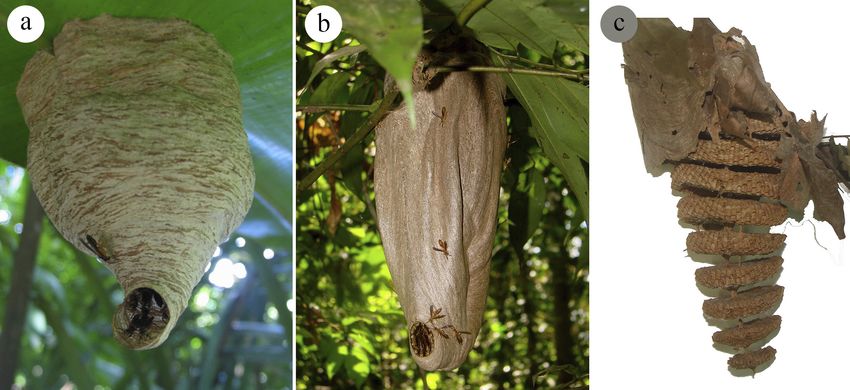
82                                                                                  Barroso et al.: Systematic review of Angiopolybia

Figure 3. Holotype, female of Angiopolybia zischkai Richards, 1978: a. lateral view, b. head in frontal view, c. dorsal view, d. labels
of type specimen. Scale: 1 mm. Source: Steve Thurston, AMNH.

a round depression; metapleural basalar area with diver-             height of one fifth of the height of antennal socket. Prono-
gent upper and lower margins.                                        tal fovea with ellipsoid shape, little deep and with slight
                                                                     anterior prominence. (2) Mesoscutum subconvex and as
Redescription of female (Fig. 3a–c). Size. (1) Head 1.03             long as wide. (3) Tegula 1.5 times longer than wide. (4)
mm long, 2.09 mm high, and 2.24 mm wide; (2) meso-                   Axillary fossa with anterior margin directing to the pos-
soma 3.54 mm long, anterior wing 9.42 mm long, and                   terior region. (5) Propodeum with translucent posterior
posterior wing 6.10 mm long; (3) metasoma 5.9 mm long.               submedian mark, anterior to the propodeal valve, insert-
Head. (1) Lateral ocelli with 0.16 mm and median ocel-               ed in a round depression. Propodeal valve complete and
lus with 0.18 mm of diameter, not inserted in a declivity            expanded, median region with two thirds of the height of
of the vertex and the lateral ocelli separated from the eyes         antennal socket. (6) Anterior wing with prestigma as long
for twice its diameter. (2) Compound eyes with medi-                 as wide. (7) Posterior wing with eight hamuli. Metaso-
um-sized and sparse bristles. (3) Frons with interanten-             ma: (1) Metasomal tergum I 2.1 times longer than wide.
nal space with 1.75 times the height of antennal socket.             Tergum with angulation in the posterior third, in later-
Anterior tentorial fovea closer to the antennal socket than          al view. (2) Metasomal tergum II 0,82 times longer than
to the eye. Central region of the frons with long bristles.          broad. Color. Dark brown in general. Yellow: lateral of
(4) Antennal socket 0.24 mm high. (5) Clypeus 0.9 times              the vertex, lateral of the frons, clypeus (but dark brown
higher than wide, contact with eyes for a distance great-            disc), interantennal region, mandibles, malar space, gena,
er than twice the height of antennal socket, and lateral             slender band contouring the posterior margin of the pro-
lobe touching the eye. Long bristles all over the clypeus            notum, outer margins of the tegula, wide spot at along
and very long bristles on the apical margin. (6) Gena with           the anterior margin of the scrobal furrow and along the
half of the width of the eye at the level of the ocular si-          dorsal groove of the mesoepisternum, anterior half of the
nus. Mesosoma. (1) Anterior lamella of pronotum with                 metanotum, submedian longitudinal band on the propo-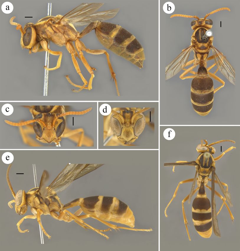
Arthropod Systematics & Phylogeny 80, 2022, 75–97                                                                                         83

deum, lateral margin of the propodeum, upper region of               mayo, Valle del Cauca (new record); Costa Rica: He-
the metapleural basalar area. Yellowish brown: FL7–10                redia; Ecuador: Orellhana, Zamora-Chinchipe; Panama:
of the antenna, anterior and median coxae, trochanters,              Colón; Peru: Cuzco, Huánuco, Junín, Loreto, Pasco,
femora, tibiae and tarsi (but with dorsal brown spots).              Ucayali (Fig. 10b).
Black: ocellar area, FL1–6 of the antenna, mesoscutum
and metasoma. Wings with hyaline cells, except yellow
in the costal, medial, submarginal I and marginal; and               Angiopolybia obidensis (Ducke, 1904)
yellowish-brown venation, except brown in the veins C,
Sc+R, M+Cu, M and in the beginning of the Cu.                        Figs 4a–f, 6f–j, 7i

Male. Unknown.                                                       Polybia obidensis Ducke, 1904: 348 (key), 354; Ducke, 1907: 140 (syn-
                                                                         onym: P. paraensis var. luctuosa Schulz, 1905); Richards, 1978: 234
Morphological variation (Fig. 7h). We found a speci-                     (lectotype designation) [examined by images].
men with yellow color and black marks, and the abdo-                 Polybia paraensis luctuosa Schulz, 1905: 132; Ducke, 1907: 140 (syn-
men, apparently, wider in dorsal view.                                   onym of P. obidensis Ducke, 1904); Richards, 1978: 234 (lectotype
                                                                         designation) [examined].
Nest. Not described, but Valverde et al. (2019) in the               Stelopolybia obidensis; Ducke, 1910: 519 (key), 526.
identification key of social wasps from Costa Rica com-              Angiopolybia obidensis; Araujo, 1946: 169; Richards, 1978: 231 (key),
mented that the nest envelope resembles an inverted flask.               234; Andena et al., 2007: 59 (phylogenetic discussion), 60: (table
                                                                         2 - characters matrix), 61 (fig. 2B), 62 (fig. 4A, 5 - cladogram), 63
Comparative comments. Angiopolybia zischkai resem-                       (key) e 64 (locality of examined material).
bles A. pallens, but it is distinguished by the pronotum             Stelopolybia (Angiopolybia) obidensis Richards and Richards, 1951: 81
without a developed lobe in the lateroanterior region,                   (list of species).
which is present in A. pallens; metapleural basalar area
with divergent upper and lower margins, which are par-               Type locality. Óbidos, Pará, Brazil.
allel in A. pallens; and pronotal fovea with translucent
mark of elliptical shape, which is circular in A. pallens.           Diagnosis. Anterior wing of 12–14 mm; eyes with very
                                                                     small-sized and sparse bristles; angulate gena with en-
Additional comments. One specimen designated as                      larged lower and upper region; pronotal lamella very el-
holotype of Angiopolybia brunnescens (Fig. 7h) but                   evated along the anterior margin, one third of the height
not described by Richards (specimen deposited in the                 of antennal socket; axillary fossa with anterior margin
NHM) is an A. zischkai specimen with more yellowish                  directed to the anterior region; posterior submedian trans-
coloration, similar to the color of some A. pallens. The             lucent mark not inserted in a depression.
two specimens with the labels: Paratype / PERU: 1.609,
Maracapata [Marcapata] (Perú) / Stelopolybia infernalis              Redescription of female (Fig. 4a, b, c). Size. (1) Head
Ducke rev.11. [1911] (1 ♂, MZUSP), and 1.609, Maraca-                1.77 mm long, 3.13 mm high, and 3.56 mm wide; (2)
pata [Marcapata] (Perú) / Stelopolybia infernalis Ducke              mesosoma 5.69 mm long, anterior wing 13.38 mm long,
rev.11. [1911] / A. pallens (Lep.) f. zischkai, Rich. 4 ♀ (1         and posterior wing 8.55 mm long; (3) metasoma 8.57 mm
♀, MZUSP), that are two of the paratypes of A. zischkai,             long. Head. (1) Lateral ocelli with 0.26 mm and median
are specimens of A. pallens with coloration resemble to              ocellus with 0.28 mm of diameter, inserted in a declivi-
the holotype of A. zischkai. Although Gomes et al. (2018,            ty of the vertex, and the lateral ocelli separated from the
2020) reported A. zischkai samples from the Brazilian                eyes for 1.7 times its diameter. (2) Compound eyes with
states Rondônia and Acre, we did not find A. zischkai for            very small-sized and sparse bristles. (3) Frons with inter-
these regions.                                                       antennal space with 1.7 times the height of antennal sock-
                                                                     et. Anterior tentorial fovea closer to the antennal socket
Holotype. ♀, Holotype / Zumbi, Rio Zamora, 700M,                     than to the eye. Central region of the frons with very long
Ecuador / XI.2.41, D.B.Laddey / Angiopolybia pallens                 bristles. (4) Antennal socket 0.34 mm high. (5) Clypeus
ssp., zischkai Rich., ♀ Holotype / AMNH_IZC 00332335                 as high as wide, contact with eyes for a distance of ap-
(AMNH, New York) (Fig. 3). Type specimen analyzed                    proximately the height of antennal socket and lateral lobe
by images.                                                           not touching the eye. Clypeus with long bristles, but with
                                                                     very long bristles on the apical margin. (6) Gena wider
Type material examined. Paratype: Paratype / PERU: Dept. Huanuco,    than half of the width of the eye at the level of the ocu-
Divisoria, 7.viii.1949, J.M.Schuncke., B.M.1952-645 / Angiopolybia   lar sinus. Mesosoma. (1) Anterior lamella of pronotum
zischkai Rich., ♀, paratype (1 ♀, NHM).                              with height of one third of the height of antennal socket.
                                                                     Pronotal fovea with ellipsoid shape, shallow and with-
Additional material examined. We examined three fe-                  out anterior prominence. (2) Mesoscutum convex and
males of A. zischkai; see supplementary material S1.                 as long as wide. (3) Tegula 1.5 times longer than wide.
                                                                     (4) Axillary fossa with anterior margin directing to the
Geographic distribution. Bolivia: Cochabamba; Co-                    anterior region. (5) Propodeum with translucent posteri-
lombia: Amazonas, Cundinamarca, Magdalena, Putu-                     or submedian mark, anterior to the propodeal valve, not
84                                                                                Barroso et al.: Systematic review of Angiopolybia

Figure 4. Lectotype, female of Angiopolybia obidensis (Ducke, 1904): a. lateral view, b. dorsal view, c. head in frontal view. Male:
d. head in frontal view, e. lateral view, f. dorsal view. Scale: 1 mm. Source: Agnièle Touret-Alby MNHN for images a, b, and c.

inserted in a depression. Propodeal valve complete and             the posterior margins of the metasomal terga I–III and
expanded, median region with two third of the height of            sternum II. Yellowish-brown: vertex and gena. Black:
antennal socket. (6) Anterior wing with prestigma as long          metasomal terga II–VI and metasomal sterna II–VI. Red-
as wide. (7) Posterior wing with 11 hamuli. Metasoma.              dish-brown: inferior margin of the clypeus and the man-
(1) Metasomal tergum I 1.7 times longer than broad. Ter-           dibular teeth. Wings with yellowish-brown in the cells
gum with angulation in the posterior third, in lateral view.       and venation, except reddish-brown in the veins C, Sc+R,
(2) Metasomal tergum II 0.8 times longer than broad and            M+Cu, M and in the beginning of the Cu.
without a row of very long bristles on the posterior mar-
gin. Color. Brown in general. Yellow: median longitudi-            Description of male (Fig. 4d, e, f). Size. (1) Head 1.6
nal band and lateral of the frons, interantennal elevation,        mm long, 2.7 mm high, and 3.2 mm wide; (2) mesosoma
band surrounding the disc of the clypeus, mandibles, low-          5.2 mm long, anterior wing 12.4 mm long, and posterior
er quarter of the gena, malar space, band contouring the           wing 8.10 mm long; (3) metasoma 8.3 mm long. Head.
posterior margin of the pronotum, tegula, spot anterior            (1) Lateral ocelli with 0.24 mm and median ocellus with
to the scrobal furrow of the mesepisternum, longitudinal           0.27 mm of diameter. (2) Frons with interantennal space
submedian band and thin lateral band in the mesoscutum,            with 1.5 times the height of antennal socket. Anterior ten-
axilla, anterior half of the scutellum, metanotum, subme-          torial fovea closer to the eye than to the antennal socket.
dian band in the propodeum, margin anterior to the prop-           (3) Antennal socket 0.31 mm high. (4) Clypeus 1.2 times
odeal valve, upper region of the metapleural basalar area,         higher than wide and apex less acute than in the female.
apex of the coxae, femora, tibiae and tarsi, bands along           Pubescence stronger than in the female. (5) Gena with
Arthropod Systematics & Phylogeny 80, 2022, 75–97 85
half of the width of the eye at the level of the ocular si- 1904 (MNHN, Paris), record MNHN, Paris EY25586
nus. Mesosoma. (1) Mesoscutum 1.1 times longer than (Fig. 4a, b, c). Type specimen analyzed by images.
wide. Metasoma. (1) Metasomal tergum I 1.7 times lon-
ger than broad. (2) Metasomal tergum II 0.8 times longer Type material examined. PARALECTOTYPE / Surinam, ex.coll.
than wide. Posterior margin with slight emargination in Fruhstorfer / spec. Typ. / Polybia paraensis luctuosa Schlz. ♀ an ♀, W.
the center. Genitalia (Fig. 6f–j). Paramere 2 mm long A. Schulz det. / Schulz Coll., 1908-157. / B.M. TYPE, HYM., 18.767b /
and 0.81 mm wide; parameral spine with one fifth of (1 ♀, NHM). Polybia paraensis luctuosa is a junior synonym of Polybia
the paramere, straight and with long bristles; lobe with obidensis Ducke, 1904, redescribed species here.
rounded apex and slightly curved downwards. Aedeagus
1.72 mm long; enlarged valve with a small emargination Additional material examined. We examined 102 fe-
in the tip; apical portion 0.66 mm long and curved to the males and three males for A. obidensis; see supplemen-
venter, ventral margin with denticles directed for the an- tary material S1.
terior region; denticulation with large and conical denti-
cles in the basal and apical thirds and small denticles with Geographic distribution. Brazil: Acre, Amapá, Amazo-
widened bases in the median third, more sclerotized than nas, Mato Grosso, Pará, Roraima (new record); French
the rest of the apical portion; small-sized bristles with Guiana; Guyana; Suriname (Fig. 10c).
alveolar base, closer in the lower half and sparse in the
upper half; median expansion with one denticle and with
acute apex; lateral apodeme not flattened dorsoventrally Angiopolybia paraensis (Spinola, 1851)
at the apex; basal apodeme arched to the venter. Digitus
3.4 times longer than wide; apical process little curved in Figs 5a–f, 6k–o, 7j–l
the region of the upper half and with bristles of alveolar
base small and sparse; rounded anteroventral lobe with a Polistes paraensis Spinola, 1851: 60.
strip of black scale-like bristles crossing it obliquely at Polybia paraensis; de Saussure, 1854: 185, pl. XXIII fig. 2 (in division
the base of the digitus; bristles absent in the lower margin Phi).
and basal articulation. Cuspis approximately 0.44 mm Polybia ruficornis Ducke, 1905b: 20; Ducke 1910: 526 (synonym of
long, with 16 black scale-like bristles on the lateral lobe, paraensis (Spinola, 1953)); Richards, 1978: 235 (lectotype designa-
and long bristles with alveolar base and close throughout tion) [examined by images].
the area of the cuspis, except sparse in the central region Stelopolybia paraensis; Ducke, 1910: 519 (key), 526 (nest, synonym: P.
and on the ventral margin. ruficornis Ducke, 1905b); Bequaert, 1944: 293 (key), 294 (typical
paraensis).
Morphological variation (Fig. 7i). Some A. obidensis Stelopolybia paraensis var. ruficornis; Ducke, 1910: 526.
specimens found in São Gabriel da Cachoeira (Amazo- Stelopolybia paraensis var. (or subspecies) obscurior Bequaert, 1944:
nas) and Parque Nacional Serra da Mocidade (Roraima) 295 [examined by images].
are darker, like A. paraensis of coloration transitional be- Angiopolybia paraensis; Araujo, 1946: 169; Andena et al., 2007: 60
tween the yellow form and the black and yellow forms, (characteres matrix), 61 (fig. 1B), 62 (fig. 4B, 5 - cladogram), 63
which also occurs in these regions. (key), 64 (locality of examined material).
Stelopolybia (Angiopolybia) paraensis Richards and Richards, 1951: 80
Nest. Unknown. (list).
Angiopolybia paraensis obscurior; van der Vecht, 1972: 737.
Comparative comments. A. obidensis resembles A. par-
aensis, but it is distinguished by the lamella along the Type locality. Pará, Brazil.
anterior margin of the pronotum which is very elevated
(one third of the height of antennal socket) in A. obiden- Diagnose. Anterior wing of 13–15 mm; eyes with very
sis, and low (one fifth of the height of antennal socket) in small-sized and sparse bristles; angulate gena with en-
A. paraensis; pronotum with prominence absent in front larged lower region; pronotal lamella low on the anterior
of the fovea, but slight prominence in front of the fovea margin, one fifth of the height of antennal socket; axillary
in A. paraensis; angulate gena with enlarged lower and fossa with anterior margin directed to the anterior region;
upper region, but gena with only enlarged lower region in posterior submedian translucent mark not inserted in a
A. paraensis; parameral spine straight, but spine curved depression.
upwards in A. paraensis; aedeagus with lateral apodeme
not flattened dorsoventrally at the apex, but apex of the Redescription of female (Fig. 5a, b, c). Size. (1) Head
lateral apodeme flattened in A. paraensis. 2.01 mm long, 3.35 mm high, and 3.71 mm wide; (2) me-
sosoma 6 mm long, anterior wing 14.78 mm long, and
Additional comments. The label information of the male posterior wing 10.37 mm long; (3) metasoma 10.8 mm
specimen described is Brazil, AM, Itacoatiara, Mil Ma- long. Head. (1) Lateral ocelli with 0.27 mm and median
deireira. 16.xii.1999, Malaise, J. Vidal Leg. ocellus with 0.29 mm of diameter, inserted in a declivi-
ty of the vertex and the lateral ocelli separated from the
Lectotype. ♀, TYPE / Obidos / Polybia obidensis Ducke, eyes for two times its diameter. (2) Compound eyes with
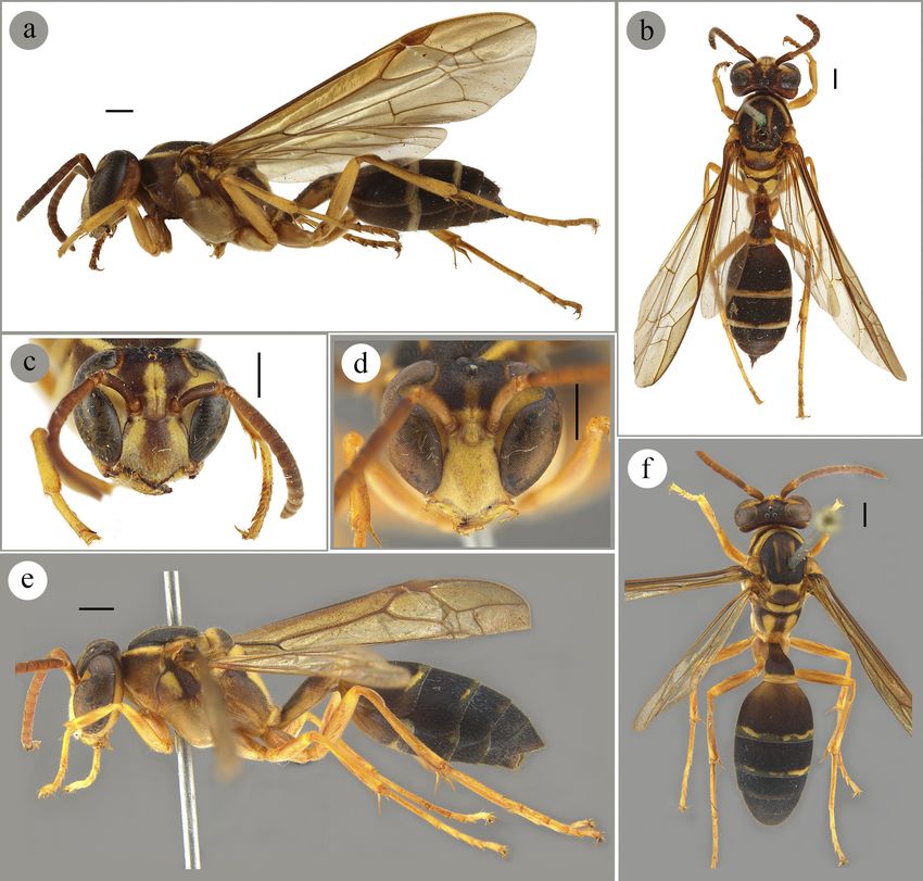
♀ typ. / MUSEUM PARIS, Brésil, Obidos, A. Ducke very small-sized and sparse bristles. (3) Frons with inter-86 Barroso et al.: Systematic review of Angiopolybia Figure 5. Female of Angiopolybia paraensis (Spinola, 1951) used in the redescription of the species: a. lateral view, b. dorsal view, c. head in frontal view. Male: d. head in frontal view, e. lateral view, f. dorsal view. Scale: 1 mm. antennal space with 1.7 times the height of antennal sock- the anterior region. (5) Propodeum with translucent pos- et. Anterior tentorial fovea closer to the antennal socket terior submedian mark, anterior to the propodeal valve, than to the eye. Central region of the frons with very long not inserted in a depression. Propodeal valve complete bristles. (4) Antennal socket 0.37 mm high. (5) Clypeus and expanded, median region with half of the height of as high as wide, contact with eyes for a distance of ap- antennal socket. (6) Anterior wing with prestigma as long proximately the height of antennal socket and lateral lobe as wide. (7) Posterior wing with 13 hamuli. Metasoma. not touching the eye. Clypeus with long bristles, but with (1) Metasomal tergum I 1.5 times longer than broad. Ter- very long bristles on the apical margin. (6) Gena wider gum with angulation in the posterior third, in lateral view. than half of the width of the eye at the level of the ocular (2) Metasomal tergum II 0.7 times longer than broad and sinus. Mesosoma. (1) Anterior lamella of pronotum with with a row of very long bristles on the posterior margin. height of one fifth of the height of antennal socket. Pro- Color. Yellowish-brown in general. Yellow: lateral of the notal fovea with ellipsoid shape, shallow, and with slight vertex, lateral of the frons, interantennal elevation, lateral anterior prominence. (2) Mesoscutum convex and 1.2 and the lower margin of the clypeus, mandibles, gena, an- times longer than wide. (3) Tegula 1.6 times longer than terior half of the lower quarter of the gena, malar space, wide. (4) Axillary fossa with anterior margin directing to band contouring the posterior margin of the pronotum,
Arthropod Systematics & Phylogeny 80, 2022, 75–97                                                                                 87

Figure 6. Male genitalia of three Angiopolybia species. Angiopolybia pallens: a. paramere, b. aedeagus in left lateral view, c. ae-
deagus in ventral view, d. digitus, e. cuspis (image mirrored). Angiopolybia obidensis: f. paramere, g. aedeagus in left lateral view,
h. aedeagus in ventral view, i. digitus, j. cuspis. Angiopolybia paraensis: k. paramere, l. aedeagus in left lateral view, m. aedeagus
in ventral view, n. digitus, o. cuspis. Scale: 0.5 mm.

tegula, spot anterior to the scrobal furrow of the mese-             valve, upper region of the metapleural basalar area, apex
pisternum, longitudinal submedian band and thin lateral              of the femora, tibiae and tarsi, bands on the posterior mar-
band in the mesoscutum, axilla, metanotum, submedian                 gins of the metasomal terga I–V, in the anterior margin of
band in the propodeum, margin anterior to the propodeal              the sternum II, and in the posterior margins of the sterna
88                                                                                  Barroso et al.: Systematic review of Angiopolybia

Figure 7. Morphological variation of Angiopolybia species. Angiopolybia pallens: a. Cayenne, French Guiana; b. Amazonas, Bra-
zil; c. Bahia, Brazil; d. São Paulo, Brazil; e. Loreto, Peru; f. Napo, Ecuador; g. Chaparé, Bolivia. Angiopolybia zischkai: h. Pipeline,
Panama. Angiopolybia obidensis: i. Roraima, Brazil. Angiopolybia paraensis: j. Roraima (brown and yellow specimen), Brazil; k.
Napo, Ecuador (brown specimen); l. Crique Alama, French Guiana (dark brown specimen). Scale: 1 mm.

II–IV. Brown: a spot that extends from the vertex to the              interantennal space with 1.5 times the height of antennal
middle of the frons, transversal band in the pronotum,                socket. Anterior tentorial fovea closer to the eye than to
scrobal furrow, region posterior to the scrobal furrow,               the antennal socket. (3) Antennal socket with 0.34 mm
metapleural basalar area, lateral bands of the propode-               high. (4) Clypeus 1.2 times higher than wide and apex
um, and metasomal terga I and II. Dark brown: ocellar                 less acute than in the female. Pubescence stronger than
area, mesoscutum, posterior half of the scutellum, pos-               in the female. (5) Gena with half of the width of the eye
terior margin of the metanotum, bands in furrow and in                at the level of the ocular sinus. Mesosoma. (1) Mesoscu-
anterior marginal of the propodeum, and metasomal terga               tum 1.1 times longer than wide. (2) Propodeum without
III and IV. Reddish-brown: lower margin of the clypeus                translucent posterior submedian mark. Propodeal valve
and mandibular teeth. Reddish yellow antenna. Wings                   with a median region with two third of the height of an-
with yellowish-brown in cells and in venation, except                 tennal socket. (3) Anterior wing with prestigma as long
reddish-brown in the veins C, Sc+R, M+Cu, M, at the                   as wide. (4) Posterior wing with 14 hamuli. Metasoma.
beginning of the Cu and pterostigma.                                  (1) Metasomal tergum I 1.7 times longer than broad. (2)
                                                                      Metasomal tergum II 0.8 times longer than wide. Gen-
Description of male (Fig. 5d, e, f). Size. (1) Head 1.40              italia (Fig. 6k–o). Paramere 2.3 mm long and 0.9 mm
mm long, 3.08 mm high, and 3.47 mm wide; (2) meso-                    wide; parameral spine with one fifth of the paramere,
soma 5.28 long, anterior wing 14.2 mm long, and pos-                  curved upwards and with long bristles; lobe with rounded
terior wing 9.9 mm long; (3) metasoma 10.5 mm long.                   apex and not curved. Aedeagus 1.68 mm long; enlarged
Head. (1) Lateral ocelli with 0.24 mm and median ocel-                valve with a small emargination in the tip; apical portion
lus with 0.28 mm of diameter, and lateral ocelli separated            0.72 mm long and curved to the venter, ventral margin
from the eyes for 1.7 times its diameter. (2) Frons with              with denticles directed for the anterior region; denticula-
Arthropod Systematics & Phylogeny 80, 2022, 75–97 89
tion with large and conical denticles in the basal and api- notum with low lamella in the anterior margin (one fifth
cal thirds and denticles reduced in the middle third, more of the height of antennal socket), being high lamella (one
sclerotized than the rest of the apical portion; small-sized third of the height of antennal socket) in A. obidensis;
bristles with alveolar base, closer in the lower half and pronotum with a slight prominence in front of the fovea,
sparse in the upper half; median expansion without denti- but prominence absent in A. obidensis; gena not enlarged
cles and with acute apex; lateral apodeme flattened dorso- in the upper region, but enlarged in the upper region in A.
ventrally at the apex; sinuous basal apodeme. Digitus 2.7 obidensis; parameral spine curved upwards, but straight
times longer than wide; apical process little curved in the parameral spine in A. obidensis; aedeagus with the apex
region of the upper half and with bristles of alveolar base of the lateral apodeme dorsoventrally flattened, but apex
small and sparse; rounded anteroventral lobe with a strip not flattened in A. obidensis.
of black scale-like bristles crossing it obliquely at the
base of the digitus; bristles absent in the lower margin and Additional comments. Angiopolybia paraensis was
in the basal articulation. Cuspis approximately 0.46 mm described by Spinola (1851) and his type specimen (or
long, with 26 black scale-like bristles on the lateral lobe, specimens) was deposited in the Museo Regionale di
and long bristles with alveolar base and close throughout Scienze Naturali di Torino (MRSN, Torino, Italy). Rich-
the area of the cuspis, except sparse in the central region. ards (1978) did not find any A. paraensis type specimen
during his study about the social wasps of the Americas,
Morphological variation (Fig. 7j–l). Anterior wing of and we did not receive any answer from the Museum
13–15 mm; posterior wing of 13–19 hamuli; Angiopoly- about the type specimen. Additionally, on the online page
bia paraensis occurs in three color variants, identified by Checklist of Epiponini wasps (http://iunh2.sci.ibaraki.
Richards (1978) as A. paraensis morph paraensis (yel- ac.jp/wasp/Epiponini/epiponini.htm; consulted in 2021)
low specimens), A. paraensis morph ruficornis (brown produced by Dr. James M. Carpenter, the presence of the
and yellow specimens) and A. paraensis morph obscu- type specimen in the Museum’s collection is also uncer-
rior (brown specimens). Angiopolybia paraensis show tain. Based on this, the redescription of the species was
small changes between yellow populations and between carried out using a specimen identified by Ducke in 1909
brown and yellow populations, such as a slightly darker and from the same locality of the type specimen (Bra-
color or some yellow marks, respectively, but they are zil, Pará, 26.9.1901, Ducke / Polybia paraensis Spin. ♀,
well-defined. Despite the color variation, the morpholog- det. Ducke 1909 / Brazil., Mus.Goeldi., 1910-90. (1 ♀,
ical characteristics of female and male adults, and male NHM)). The information about the male specimen de-
genitalia used in the description are preserved in the three scribed is: BRA, Roraima, Amajarí, Serra do Tepequém,
forms, so they should not be treated as subspecies or SESC Tepequém. 1–15.iii.2016 / Malaise grande, J.A.
differentiated as morphs. Only a few A. paraensis pop- Rafael, F.F. Xavier Filho col.
ulations, with dark brown color from French Guiana and Although some checklists treat A. paraensis as regis-
Suriname, showed morphological variations such as the tered for Bahia state [for example, the Checklist of Epi-
absence of very long bristles in the posterior margin of ponini wasps produced by Dr. James M. Carpenter (http://
the metasomal tergum II and propodeum with a region iunh2.sci.ibaraki.ac.jp/wasp/Epiponini/epiponini.htm;
anterior to the spiracle less projected in the metapleural consulted in 2021) and Barbosa et al. (2016)], we did not
basalar area. However, we believe that this evidence is confirm this information.
still insufficient to justify a new species. Moreover, the
analysis of species delimitation with molecular data (see Type specimen. Without information.
below) showed that these variants are within the intraspe-
cific limits of A. paraensis. Additional material examined. We examined 79 fe-
males and six males for A. paraensis; see supplementary
Nest. Described by Schulz (1903) as a spherical nest material S1.
about 25 mm in diameter (possibly incorrect unit of
measurement was used, with centimeter (cm) being the Geographic distribution. Bolivia: Cochabamba, La
correct). Ducke (1910) complements the description of Paz; Brazil: Acre, Amapá, Amazonas, Maranhão, Mato
Schulz (1903), stating that the nest is composed of four Grosso, Pará, Rondônia, Roraima; Colombia: Amazonas,
overlapping combs, joined by a central pedicel, with a Caquetá; Ecuador; French Guiana; Guyana; Peru: Cuzco,
simple and very resistant envelope, and with transverse Huánuco, Junín, Loreto, Madre de Dios, Pasco, San Mar-
streaked with light and dark colors. tin; Panama; Suriname; Venezuela: Amazonas, Bolívar
(Fig. 10d).
Comparative comments. Angiopolybia paraensis re-
sembles A. obidensis, but it is distinguished by the pro-
3.2. Key to Angiopolybia species
The following key is a revised and adapted version with few modifications of the keys provided by Richards (1978)
and Andena et al. (2007).You can also read