Current facts constituting an understanding of the nature of adenomyosis
←
→
Page content transcription
If your browser does not render page correctly, please read the page content below

Reviews

Current facts constituting an understanding
of the nature of adenomyosis
Veronika Aleksandrovych1,A–F, Paweł Basta2,B,C,E,F, Krzysztof Gil1,A,D–F
1
    Department of Pathophysiology, Jagiellonian University Medical College, Kraków, Poland
2
    Department of Gynecology and Oncology, Jagiellonian University Medical College, Kraków, Poland

A – research concept and design; B – collection and/or assembly of data; C – data analysis and interpretation;
D – writing the article; E – critical revision of the article; F – final approval of the article

    Advances in Clinical and Experimental Medicine, ISSN 1899–5276 (print), ISSN 2451–2680 (online)                                       Adv Clin Exp Med. 2019;28(6):00–00

Address for correspondence
Veronika Aleksandrovych
                                                                 Abstract
E-mail: v.aleksandrovych@doctoral.uj.edu.pl                      Adenomyosis seems to be the most widespread coexistent pathology included under the umbrella of common
                                                                 benign disorders of the human uterus. The incidence of adenomyosis is under discussion since different imag-
Funding sources
None declared
                                                                 ing criteria are used. In the majority of cases, prevalence is determined among women with uterine fibroids
                                                                 and endometriosis or severe gynecological symptoms. This common benign pathology is asymptomatic in 1/3
Conflict of interest                                             of cases. Up to 50% of women with infertility are affected by adenomyosis. It seems to be an important risk
None declared                                                    factor for spontaneous pre-term delivery and pre-term premature rupture of the membranes. Nowadays,
                                                                 the etiology of adenomyosis is still unclear and requires deeper investigation. This review summarizes the
                                                                 aspects of prevalence, co-existence, risk factors, classification, mechanisms of pathogenesis, genes and im-
Received on September 4, 2017                                    munological features, main histological features, animal models, and clinical manifestation of adenomyosis.
Reviewed on October 16, 2017
Accepted on October 25, 2017                                     It might facilitate understanding of the independent nature of such a dual enigma as adenomyosis.
Published online on August 7, 2018                               Key words: infertility, adenomyosis, myometrium, metaplasia, endometrial junctional zone

DOI
10.17219/acem/79176

Copyright
© 2018 by Wroclaw Medical University
This is an article distributed under the terms of the
Creative Commons Attribution Non-Commercial License
(http://creativecommons.org/licenses/by-nc-nd/4.0/)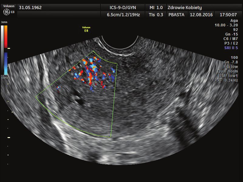
2                                                                 V. Aleksandrovych, P. Basta, K. Gil. Current facts about the nature of adenomyosis

Introduction                                                    myometrium as “adenomyosis uteri”, thereby singling out
                                                                adenomyosis among adenomyomas.10–12
  Adenomyosis is a benign condition of the uterus char-            Finally, in 1972, Bird proposed the modern explanation
acterized by the presence of ectopic endometrial glands         of adenomyosis. He described that adenomyosis is a non-
and stroma below the endometrial-myometrial junction            malignant endometrial invasion into the myometrium and
(at a depth of at least 2.5 mm below the basal layer of the     leads to macroscopic changes of uterus size. Under a mi-
endometrium).1,2 The focuses of endometrial glands and          croscope, hypertrophy and hyperplasia of the myometrium
stroma in the myometrium are typically surrounded by its        are observed with penetration of the endometrium inside
hyperplastic tissue.3 Lymphatic and vascular channels car-      (glands and stroma). Nowadays, this gynecological disease
ry out penetration of normal myometrium.4,5 The level           is characterized by the invasion of endometrial compo-
of endometrial invasion into a myometrium has been the          nents more than 2.5 mm below the endo-myometrial junc-
issue of heated debate.6 The majority of cases are observed     tion in the microscope’s low-power field.2,9,12
in multiparous premenopausal women. Likelihood estima-
tion of diagnosis demands obligatory histological analyses,     Prevalence and co-existence
which are commonly provided after hysterectomy. By far,
the majority of adenomyosis cases are detected in autopsy
                                                                of adenomyosis
and coexist with endometriosis and uterine fibroids, which         The incidence of adenomyosis is unclear and debatable
explain a wide range of distribution (from 5% to 70%) ac-       due to the use of a variety of criteria.12 Numerous publi-
cording to literature data.4,7                                  cations have shown that the prevalence of adenomyosis
                                                                widely varies (from 5–8% to 40–70% of all uterine speci-
                                                                mens), with a mean of 20–25%.4,7,8 The same statistical
Historical notes                                                data was present after histological analysis of postoperative
                                                                material. Based on a literature review, prevalence of adeno-
  A Bohemian physician and pathologist, Carl von Roki-          myosis ranges from a high of 61.5% to a low of 8.8%, with
tansky, first described adenomyoma as a focal core of en-       a peak among women between 35 and 50 years of age.7,14
dometrial glands and stroma surrounded by smooth                   The diagnosis of adenomyosis can be time-consuming
muscle cells in 1860. He referred to it as “cystosarcoma        and challenging, based on lucky histological observation
adenoids uterinum” with a neoplastic nature.8–12 Surpris-       of the material. Interestingly, the rate of detection depends
ingly, Knapp, an expert in the history of medicine, in 1999     on the quantity and quality of samples observed and can
claimed that the 1st who described a similar condition (lat-    vary from 31% to 62 % in the same uterus.9
er named adeno- or endometriosis) was Daniel Schrön                Current studies demonstrate that about 4/5 of cases
from Germany in 1690 in the work “Disputatio Inauguralis        with adenomyosis coexist with uterine fibroids and endo-
Medica de Ulceribus Uteri”. Moreover, some of pathologi-        metriosis.5,15 According to statistical data, approx. every
cal descriptions made between the 17th and 18th centuries       5th removed uterus with leiomyomata has a focus of adeno-
are similar to the modern classification of endometriosis       myosis, while up to 55% of adenomyosis samples reveal devel-
or adenomyosis. Knapp challenged the primacy in describ-        oping uterine fibroids. Kitawaki proved these conclusions
ing the pathology in his Letter to Editor of Fertility and      with his own analysis of data presented of the coexistence
Sterility, which was published. Unfortunately, his request      of both pathologies: adenomyosis with leiomyomata in
remained unanswered due to the author's sudden death            35–55% of cases, while leiomyomata with adenomyosis
within a few months after publication.12                        in 64%. At the same time, Kitawaki noted that adenomyosis
  The name adenomyoma was coined around the end                 is associated with endometriosis too: adenomyosis with
of the 19th century. In 1896, both Cullen and Von Reck-         endometriosis in 70% of cases, while “vice versa” only in
linghausen described the condition, followed by Pick and        6–20% of all cases.7
Rolly in 1897.11 In addition, Cullen provided the 1st ever         It is important to note that diseases such as polyps of the
classification of adenomyomas, while its pathogenetic           endometrium, typical and atypical endometrial hyper-
nature was depicted by Meyer in 1903. He emphasized             plasia and endometrial carcinoma often coexist with ad-
that it is a variant of “epithelial invasion of inflammatory    enomyosis, compared to their separate detection.7 On the
infiltrated tissue” and affects 2 kinds of epithelium: dys-     other hand, no study has demonstrated the natural trans-
topic and orthotopic (embryonic and mature). The origin         formation of adenomyosis to adenocarcinoma.16
of dystopic nature was also supported by Orloff, who de-
scribed it in 1895 as “glandular spaces under the serosa
covering uterine myomata”, and focused on developing            Risk factors
from “embryonic cells”.10–12
  Until the 1920s, adenomyosis and endometriosis were             It is important to consider that adenomyosis often co-
considered to be a part of the same entity.13 In 1925, Frankl   exists with other gynecological disorders and is revealed
depicted and named the invasion of mucosal tissue in the        by histological examination. As it appears from these
Adv Clin Exp Med. 2019;28(6):00–00                                                                                            3

features, suggested risk factors could not be clearly evalu-     through its receptors. This may suggest a link between
ated and most of them were received after retrospective          adenomyosis and depression with a common pathogenic
studies.                                                         factor.9
   Adenomyosis is common for women in the 5th decade
of life but it has also been revealed in patients in the post-
menopausal period after chemotherapy of breast cancer            Classification
with tamoxifen.9,17 Increasing age up to menopause has
long been considered a risk factor.18                              Adenomyosis has 2 types: diffuse and focal, which is also
   Multiparity is associated with adenomyosis. This might        named “adenomyoma” in the literature. In comparison
be explained by a mechanism of trophoblast invasion into         with the anterior and lateral sides of the uterus, the pos-
the myometrium during pregnancy.9,19 Romanek et al.              terior wall is affected more often.6,12
proved by analysis that the relative risk of adenomyosis           Bird et al. in 1972 divided the depth of invasion into
was almost 2 times lower among nulliparous women                 3 grades. The 1st grade is characterized by the existence
in comparison with multiparous.19,20 Different studies           of adenomyosis within 1 low-power field below the basal
have presented controversial data regarding abdominal            endometrium. The 2nd grade is represented by deeper
delivery and further risk of pathology development; the          penetration, to the mid-myometrium, while the 3rd grade
role of cesarean sections also remains unclear.19                penetrates beyond the mid-myometrium. His team as-
   Undoubtedly, sex hormones have a great impact on adeno-       sessed the extent (density) of glandular involvement of the
myosis.21 It has been demonstrated that estrogen receptors       myometrium as follows: “slight” – a few (1–3) adenomyotic
are always present in adenomyosis tissue, however, their         glands per low-power field; “moderate” – several (4–9) and
quantity was reduced compared to a corresponding normal          “marked” – many (10 or more).6
myometrium.7 Progesterone and androgen receptors were
also found in adenomyotic tissue. Some authors have noted
a balance between the amount of both steroids in healthy         Definition of endomyometrial
and adenomyotic tissue, while others have demonstrated
a slightly higher density of progesterone receptors when
                                                                 junctional zone
compared to estrogen. It is important to note that ectopic          The endomyometrial junctional zone (JZ) is a distinct,
as well as eutopic endometrium reflect all cyclic changes.22     hormonally sensitive part of the endomyometrial interface
   Several studies have revealed that smoking can also be        that was first visualized more than 20 years ago by magnetic
a risk factor of adenomyosis. Shrestha showed that the risk      resonance imaging (MRI).3,10 However, the realization that
of adenomyosis is higher in smokers, especially in those         the inner portion of the human myometrium constitutes
who smoked for more than 10 years.20 On the other hand,          a separate entity within the uterine musculature is more
Taran et al. noted that smokers appear less likely to have       than a century old. Brosens et al. referred to Werth and
adenomyosis and this might be explained by declining es-         Grusdew, who called it the “archimyometrium” in 1898.25
trogen levels in the blood, which is common for smokers.7,9         The JZ is not the outer myometrium.12 They have different
   Panganamamula et al. focused attention on previous            origins: the JZ originates from the Müllerian ductus and the
surgery on endometrium, proved the first hypothesis              outer myometrium from the mesenchyme. As endometrium,
that endometrial resection’s involvement in pathogenesis         the JZ expresses steroid hormone receptors, has a cycle-de-
of adenomysosis, made by Coltate and Smith in 1991.23            pendent type of growth and is involved in implantation and
The data rendering a strict direct link between adenomyo-        deep placentation. Also, normal uterine peristaltic activity
sis and uterine trauma is still inconsistent. As often as not,   originates from the JZ. This zone has been cited in literature
in the literature, such traumatic conditions as spontaneous      as the archimyometrium, stratum basale, inner myometrium,
abortion, dilation, curettage, and endometrial ablation are      junctional zone myometrium, endometrio–myometrial inter-
discussed.23 Levgur et al. noticed that the instrumental         face, transitional zone, and sub-endometrial myometrium.
curettage of the uterus could be a risk factor only in the       Ultrasound and MRI detect this zone in human uteri. Com-
case of abortion, whereas in the nonpregnant uterus it did       monly, the thickness of the JZ is 5 mm or less, whereas it has
not impact subsequent development of adenomyosis.4,23,24         a tendency to grow with age. Physiologically, JZ could be up
If it is not expelled, a pregnancy could be a crucial point      to 0.8 cm among women without adenomyosis and 1.1 cm
in the expansion of adenomyosis. One argument in favor           among those affected, respectively.26,27
of this is the fact that, despite about 20% of nulliparous          The changing of JZ size and characteristics (>12 mm,
women presenting with adenomyosis, a higher percent-             hemorrhagic high-signal myometrial spots) is a poor prog-
age of women after any term of pregnancy are affected            nostic for adenomyosis and has a strong correlation with
by the disease.7                                                 prognosis.25,28 It has been called the gold standard in re-
   Some studies have shown that production of prolactin          vealing adenomyosis. Bergeron et al. hypothesized that
by the endometrium, myometrium and even in leiomyomas            a disturbance of JZ may be involved by triggering an “in-
could stimulate a mitogenic activity in smooth muscle cells      vasive” factor.5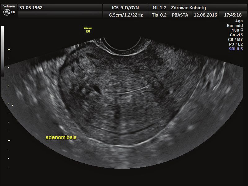
4                                                                 V. Aleksandrovych, P. Basta, K. Gil. Current facts about the nature of adenomyosis

Pathogenesis                                                    thrombospondin 1 (TSP-1), and platelet-derived growth
                                                                factor (PDGF) are potent angiogenic factors.28
   The etiology of adenomyosis is still uncertain. Some            In general, adenomyosis is characterized by overex-
theories equally explain the possible pathogenesis of the       pression of VEGF and hypoxia-inducible factor 1-alpha
disorder while at the same time complementing each other.       (HIF1-alpha), increasing the number of small vessels.
They focus on local invasion, cellular proliferation and        Kang et al. observed 2 alleles of VEGF genes (22578A and
angiogenesis.                                                   21154A) and concluded that they might be protective fac-
   This pathological condition originates from the deep         tors of adenomyosis.13,34 Tokyol et al. emphasized that the
basal layer of the endometrium as a result of its invagi-       quantity and intensity of cyclo-oxygenase (COX-2) expres-
nation in the myometrium, possibly due to loss of tissue        sion in the endometrium was growing during both phases
cohesion. Tissue affected by adenomyosis is character-          of the cycle in patients with adenomyosis in comparison
ized by higher expression of estradiol receptors (ER) com-      to healthy controls.35 Another characteristic feature of the
pared to typical endometrium, combined with expression          pathology is the prevalence of defective myometrial spiral
of the apoptosis-suppressing gene product – B cell lym-         artery remodeling associated with alteration.36
phoma/leukemia-2 (Bcl-2) protein. Enhancement of Bcl-2             In adenomyosis-induced animal models, the insulin
expression is involved in the pathogenesis of uterine           growth factor-II (IGF-II) mRNA was found to be mark-
adenomyosis.5,29–31                                             edly downregulated compared to normal endometrium, as
   From another point of view, the basalis invaginates into     observed by a global gene transcription profiling analysis.
the myometrium along the lymphatic vessels. This was            Levy et al. proved this with the same tendency of IGF-II
proved by pathological examination of postsurgical sam-         expression.21 Despite that, its expression in uterine fibroid
ples – adenomyotic focuses were found within intra-myo-         has the opposite character and this likely indicates dif-
metrial lymphatics. As the endometrial and myometrial           ferent molecular and pathogenic backgrounds of uterine
tissues originate from the Müllerian ducts, adenomyosis         fibroid and adenomyosis. However, the expression of IGF-II
can be the result of metaplasia from de novo ectopic intra-     can stimulate abnormal myometrial growth in patients
myometrial endometrial tissue. This was defined as the 3rd      with adenomyosis, explaining the frequent coexistence
theory of the pathogenesis of adenomyosis.5                     of both pathologies.
   It should be noted that myofibroblasts express some ex-
tracellular matrix proteins and definitely take part in the     Histological features
development of pathological focuses. The invasion of endo-
metrial tissue inside the myometrium leads to its hypertro-       Adenomyosis is identified in 20–30% of all uteri removed
phy as a consequent response.28 Mast cells in the endome-       at surgery, where it causes globular and cystic enlargement
trial tissue are located in close vicinity to smooth muscle     of the myometrium, with some cysts filled with extrava-
cells and impact its differentiation and development by se-     sated, often hemolyzed red blood cells and siderophages.5
creting nerve growth factor (NGF), preadipocyte factor-1        However, in specimens larger than 280 g it is much less
(Pref-1) and insulin-like growth factor-2. It is important      common.4 Microscopically, populations of spindle cells in-
to note that NGF is involved in the mechanism of pain           clude smooth muscle cells and components of adenomyotic
and might reflect the severity of the process, which can be     lesions, and make contact with the adenomyotic stroma.28
useful from the clinical point of view. Preadipocyte factor-1   On gross examination, the myometrium contains small,
could be a possible protective factor for further differen-     soft, red areas, some of which are cystic. Microscopic ex-
tiation of cells. Koike et al. concluded that the mast cell     amination of these lesions reveals glands lined by mildly
might preserve the balance in an affected uterus.28 Zhao        proliferative to inactive endometrium and surrounded
et al. observed the expression levels of caveolin 1 (CAV1)      by endometrial stroma. Secretory changes are rare, except
in adenomyosis-affected tissue and showed that focuses          during pregnancy and in patients treated with progestins.
of adenomyosis have a low level of CAV1 expression, which       Myometrial changing (hyperplasia and hypertrophy) could
could be associated with adenomyosis-related dysmenor-          lead to enlarging of the uterus.19 Over time, the uterus
rhea.32 The latest data provided by An et al. depicted that     may also be enlarged from cyclic bleeding into adenomy-
macrophages have an impact on epithelial–mesenchymal            otic foci. Varying degrees of glandular hyperplasia may be
transition (EMT)-like processes, and thereby could be ef-       seen, and occasionally hyperplastic surface endometrium
fector cells in that disease.33                                 extends into the foci of adenomyosis.37
   Currently, the interest of researchers is focused on ab-       Pathological foci are usually seen at a depth of at least
normal angiogenesis in JZ, which might be involved in the       2 mm in the myometrium on more than 1 microscopic field
pathogenesis of adenomyosis. Moreover, abnormal vas-            at ×10 magnification of the JZ. This is useful and impor-
cularization is common for half of the patients: vascular       tant for the uterus during pregnancy and postmenopause.
distribution is generally irregular and vessels are thick,      Glands and stroma in adenomyosis are usually in the
dilated, and/or reticular.13 Vascular endothelial growth        proliferative phase, whereas they may contain secretory
factor (VEGF), fibroblast growth factor (FGF)-1, FGF-2,         to menstrual transformations. The stromal fibroblasts are
Adv Clin Exp Med. 2019;28(6):00–00 5
histologically different from myocytes.5 During differential adenomyosis have elevated expression of IL-10 in eutopic
histological analysis, it is important to pay attention to the and ectopic endometrium (in the secretory phase), which
features of adenomyoma (different from adenomyosis): could play a significant role in the pathogenesis of this dis-
usually circumscribed, nodular aggregate of smooth mus- ease by altering the immune system.41 Later, Qin et al. ob-
cle, endometrial glands and (usually) endometrial stroma.5 served the expression of interleukin-10 receptor 1 (IL-10R1)
and interleukin-10 receptor 2 (IL-10R2) in adenomyosis.
They showed that expression of IL-10R1 and IL-10R2 was
Genes and immunity higher in adenomyotic samples in comparison with eutopic
endometrium of women with adenomyosis or in normal
of adenomyosis endometrium. They suggested that IL-10 receptors have
Genetic factors can explain the nature of a variety a possible connection to the immunotolerance and/or anti-
of diseases, and adenomyosis is no exception. Wang et al. inflammatory process during adenomyosis.42
has not revealed any chromosomal aberrations during Leukemia inhibitory factor (LIF) is a pleiotropic cytokine
examination of 25 cases of adenomyosis by comparative of the interleukin-6 family, produced by the endometrium
genomic hybridization.7 Mutations in k-ras and p53 are during the implantation window. Xiao et al. found signifi-
common in adenomyosis.5 Kitawaki has mentioned that cantly lower LIF immunolabeling intensity in women with
Goumenou et al. observed 31 cases of adenomyosis and adenomyosis in comparison to fertile women. The dys-
revealed the incidence of loss of heterozygosity on 2p22.3- regulation of both LIF mRNA and protein in the endo-
p16.1, 3p24.2-p22 and 9p21 chromosomal regions as 19.4%, metrium during the implantation window suggests that
9.7% and 6.5%, respectively.7 Human leukocyte antigen-DR adenomyosis may be associated with impaired implanta-
(HLA-DR) expression is significantly higher in stromal tion.43,44 Garavaglia et al. noted an overxpression of the hu-
cells compared to glandular cells in adenomyosis tissue. man leukocyte antigens (HLA) of class II and an increase
This might be involved in the immune reactions which of complement components C3 or C4 in adenomyosis.13,31
occur in adenomyosis.38
Goumenou et al. observed the expression of p16, retino-
blastoma (pRb) and cyclin D1 oncoproteins in patients with Model systems
adenomyosis. Loss of expression of either pRb or p16, or the
overexpression of cyclin D1, could lead to tissue growth. Nowadays, different animal models are widely used for
His team showed that for endometrial tissue in patients the observation and re-creation of adenomyosis, while for
with adenomyosis during the 1st phase of the cycle, high endometriosis only humans and non-human primates are
expression of p16 was and low of pRb is common, whereas used.45 Usually, as the object of experimental models for
for endometrioma this balance was reversed. He concluded adenomyosis, non-human primates, horses, dogs, cats, rab-
that the prevalence of p16 expression was common for bits, and laboratory rodents are used.28 This pathology
adenomyosis in comparison with endometriomas. This occurs naturally in non-human primates, notably Macaca
clarified the differences in pathogenic mechanisms during mulata. Several mouse strains also develop adenomyosis
both widespread gynecological pathologies.39 spontaneously and it can be found in a small percentage
Fibroblast growth factor 2 (FBG2) is located on chromo- of adult animals.17,46 Moreover, spontaneous development
some 4q26–27. It can upregulate the expression of VEGF of adenomyosis with a prevalence of ~80% by 12 months
and synergistically act with VEGF in stimulating new vessel of age was revealed in CD-1 mice.42 Adenomyosis has also
formation. Kang et al. demonstrated that the FGF2 754C/G been induced in mice by hormonal manipulation such as
polymorphism is correlated with increased risk of adeno- by the implantation of a single anterior pituitary gland
myosis among Chinese women.28,34,40 into the uterine lumen.17 Other experiments were focused
For identification of endometrial stromal cells among the on the administration of tamoxifen and further develop-
myometrium in adenomyotic foci, immunohistochemical ment of adenomyosis in mice. Far fewer studies on ad-
staining for CD10 is used, while it cannot distinguish such enomyosis have been performed in rats than in mice.
pathological conditions as myometrial invasion and carcinoma Rabbit is a convenient animal model because pathology
of endometrium from those involving adenomyosis (in situ). can spontaneously manifest and its prevalence can be
The cells close to the neoplasm also express this marker.5 enhanced by estrogen administration during a period
Vimentin expression is lower in adenomyosis in com- of 1–2 years.45
parison with eutopic endometrium, while expression
of α-smooth muscle actin and desmin is consistent.28
Interleukin-10 (IL-10) is an important immunomodula- Symptoms
tory cytokine produced by many cell populations. It is one
of the major anti-inflammatory cytokines and plays im- Adenomyosis does not present pathognomonic clinical
portant role in several chronic inflammatory diseases symptoms.6,13 About 1/3 of women with adenomyosis do
and cancers. Wang et al. demonstrated that patients with not have any symptoms.4,5,12 The most frequent symptoms6 V. Aleksandrovych, P. Basta, K. Gil. Current facts about the nature of adenomyosis
are common with other gynecological disorders: men-
orrhagia, dysmenorrhea, metrorrhagia, dyspareunia,
and chronic, erratic or constant pelvic pain. Symptoms
of heavy/abnormal bleeding are thought to be positively
associated with the depth of penetration of adenomyosis
into the myometrium.25,47–49 Dysmenorrhea is correlated
with the prevalence of adenomyotic glands.6
Diagnosis
In general, symptomatology does not allow detection
of adenomyosis in time. The diagnosis can include vaginal
examination, transvaginal sonography (TVS) and MRI.18
Unfortunately, diagnostic hysteroscopy does not make
it possible to discover pathognomonic signs in such cas- Fig. 2. Transvaginal ultrasound color Doppler scan. Sagittal view.
Increased vascularity within myometrium is demonstrated, which is one
es.12 On pelvic examination, the uterus may be enlarged, of the ultrasound adenomyosis features
soft and mildly tender, especially if a patient is examined
premenstrually. Ultrasonography is helpful to observe the
size and shape of the uterus (a globular or asymmetrical
uterus, myometrial cysts), localization of heterogeneous
myometrium and of focal abnormal echotexture. Three-
dimensional reconstruction of the uterine anatomy in the
coronal plane provides new and unrivaled views of the JZ.3
Sometimes, the adenomyosis may be confined to a part
of the myometrium in the form of a well-circumscribed
lump, which is called adenomyoma. This is in contrast
to the uterine myoma, which does not present a well-de-
fined capsule.15
Although pelvic ultrasound is a widely accepted imaging
modality, the reported sensitivity for the trans-abdominal
approach is only 32–63% with a corresponding specificity
of 95–97%.50 Improvement may be achieved with a trans-
vaginal approach. Hysterosalpingography may be of help,
Fig. 3. Transvaginal ultrasound B-mode scan (amber filter). Sagittal view.
while diverticula extending into the myometrium are Typical for adenomyosis changes within the uterus: sub-endometrial
present. Magnetic resonance imaging allows observation echogenic linear striations and acoustic shadowing as a result
of the inner myometrium and detection of its thickness of ultrasound beam diffraction by irregular structure of myometrium
– endometrial tissue causes a hyperplastic reaction (“Venetian blind”
appearance)
and nature of changes, which is considered the hallmark
of adenomyosis.12
Undoubtedly, the histological examination of a few
postsurgical uterine transmural sections (from body and
fundus) provide the final diagnosis. 5 Only histological
examination can reveal the existence of ectopic glands
in the endometrium (the “adeno” component), different
from those received by different imaging techniques,
which in the majority observe the organization of smooth
muscle (the “myosis” component).10
Treatment
Fig. 1. Transvaginal ultrasound B-mode scan. Sagittal view. Typical The primary indication for treatment is the pres-
changes within the uterus for adenomyosis: enlarged vascular spaces
within the inner layer of myometrium and small myometrial cysts (sub- ence of symptoms that negatively affect woman’s daily
endometrial cysts – specific sign) life. Conservative treatment of pain with nonsteroidalAdv Clin Exp Med. 2019;28(6):00–00 7
Fig. 4. Contrast-enhanced pelvic magnetic
resonance imaging (MRI) of the same patient.
T2-weighted image in sagittal section. Magnetic
resonance imaging view corresponds with diffuse
adenomyosis – high T2 signal regions on the
anterior wall of the uterus, representing cystic
changes
anti-inflammatory drugs (NSAIDs) and hormonal con- Several studies have proved that women with adenomyosis,
trol of excessive cyclic bleeding maintain the first line revealed before pregnancy, had significantly increased risk
of management. Oral progesterone, contraceptive pills, of preterm delivery, preterm premature rupture of mem-
hormonal patches or rings, or levonorgestrel intrauterine branes, fetal malpresentation and cesarean delivery. Hypo-
device (IUD) can effectively control symptoms. When con- thetically, this could be connected with the enlargement
servative management is not efficient or contraindicated, of adenomyotic focuses during pregnancy and further its
surgical manipulations (endometrial ablation/resection, association with inflammatory consequences.52–54 Adeno-
myometrial excision/reduction, myometrial electrocoagu- myosis is connected with impaired implantation, destroy-
lation, uterine artery ligation) are used for the treatment ing the uterine cavity’s receptivity, accompanied by lower
of symptoms. However, these methods are not very effec- expression of the adhesion molecules necessary for embryo
tive.9 For superficial adenomyosis, using endoscopic endo- implantation (integrin β-3 and osteopontin). These factors
metrial ablation is preferred.5,51 The coexistence of uterine make it possible to evaluate patients with adenomyosis as
fibroids and adenomyosis in 1 patient could complicate a target group for in vitro fertilization (IVF) treatment.39,55
the diagnosis as the have a similarity in symptoms. Thus, Neoplastic transformation of adenomyosis is rare: only
in both pathologies uterine artery embolization (UAE) a few case reports of adenocarcinomas have been pub-
is performed.9 Less than a half of patients after such surgi- lished. Koike et al. analyzed all the available data and pre-
cal treatment have regression in 2–3 years.19 Unfortunately, sented in their review, to date, 44 cases that have been
variants with leiomyomata are still complicated to identify documented.28
among patients with adenomyosis. The former detailed
diagnosis of both pathologies might be helpful before UAE, References
by reason of poor post-surgical prognosis among women
1. Kido A, Fujimoto K, Matsubara N, Kataoka M, Konishi I, Togashi K.
with adenomyosis.14 If a woman is not a candidate for any
A layer of decreased apparent diffusion coefficient at the endome-
medical or surgical conservative management or if that trial-myometrial junction in uterine adenomyosis. Magn Reson Med
treatment cannot sufficiently control the symptoms, hys- Sci. 2015;15(2):220–226.
terectomy is the final line of treatment. 2. Zhou C, Zhang T, Liu F, et al. The differential expression of mRNAs
and long noncoding RNAs between ectopic and eutopic endome-
tria provides new insights into adenomyosis. Mol Biosyst. 2016;12(2):
Outcome 362–370.
3. Exacoustos C, Brienza L, Di Giovanni A, et al. Adenomyosis: Three-
dimensional sonographic findings of the junctional zone and corre-
The junctional zone is quite important for the process lation with histology. Ultrasound Obstet Gynecol. 2011;37(4):471–479.
of human placentation and is characterized by unique 4. Levgur M, Abadi MA, Tucker A. Adenomyosis: Symptoms, histology,
vascular plasticity in terms of physiological remodeling and pregnancy terminations. Obstet Gynecol. 2000;95(5):688–691.
5. Bergeron C, Amant F, Ferenczy A. Pathology and physiopatholo-
of the myometrial spiral arteries in pregnancy. Increasing
gy of adenomyosis. Best Pract Res Clin Obstet Gynaecol. 2006;20(4):
thickness of JZ, changes in neo-angiogenesis and altera- 511–521.
tions in this zone, which are common for adenomyosis 6. Peric H, Fraser IS. The symptomatology of adenomyosis. Best Pract
and endometriosis, may be associated with a shallow type Res Clin Obstet Gynaecol. 2006;20(4):547–555.
7. Kitawaki J. Adenomyosis: The pathophysiology of an oestrogen-depen-
of defective deep placentation.10–12 As it appears from this dent disease. Best Pract Res Clin Obstet Gynaecol. 2006;20(4):493–502.
statement, both pathologies may form the pathogenic back- 8. Graziano A, Lo Monte G, Piva I. Diagnostic findings in adenomyosis:
ground for negative obstetric outcome (preterm birth, fe- A pictorial review on the major concerns. Eur Rev Med Pharmacol Sci.
2015;19(7):1146–1154.
tal growth retardation and postpartum hemorrhage). 368 V. Aleksandrovych, P. Basta, K. Gil. Current facts about the nature of adenomyosis
9. Taran FA, Stewart EA, Brucker S. Adenomyosis: Epidemiology, risk 34. Kang S, Li SZ, Wang N, et al. Association between genetic poly-
factors, clinical phenotype and surgical and interventional alterna- morphisms in fibroblast growth factor (FGF)1 and FGF2 and risk
tives to hysterectomy. Geburtshilfe Frauenheilkd. 2013;73(9):924–931. of endometriosis and adenomyosis in Chinese women. Hum Reprod.
10. Benagiano G, Brosens I. History of adenomyosis. Best Pract Res Clin 2010;25(7):1806–1811.
Obstet Gynaecol. 2006;20(4):449–463. 35. Tokyol C, Aktepe F, Dilek FH, Sahin O, Arioz DT. Expression of cyclo-
11. Benagiano G, Brosens I, Lippi D. The history of endometriosis. Gyne- oxygenase-2 and matrix metalloproteinase-2 in adenomyosis and
col Obstet Invest. 2014;78(1):1–9. endometrial polyps and its correlation with angiogenesis. Int J Gyne-
12. Benagiano G, Brosens I, et al. Glob. libr. women's med., (ISSN: 1756- col Pathol. 2009;28:148–156.
2228) 2010. doi: 10.3843/GLOWM.10460 36. Brosens I, Pijnenborg R, Benagiano G. Defective myometrial spi-
13. Di Donato N, Montanari G, Benfenati A, et al. Prevalence of adeno- ral artery remodelling as a cause of major obstetrical syndromes
myosis in women undergoing surgery for endometriosis. Eur J Obstet in endometriosis and adenomyosis. Placenta. 2013;34(2):100–105.
Gynecol Reprod Biol. 2014;181:289–293. 37. Rubin E, Farber JL. Pathology. 2nd ed. Philadelphia, PA: Lippincott;
14. Woźniakowska E, Milart P, Paszkowski T. Embolizacja tętnic macicz- 1988.
nych – zagadnienia kliniczne. Ginekol Pol. 2013;84:1051–1054. 38. Koumantakis EE, Panayiotides JG, Goumenou AG, et al. Different
15. Aleksandrovych V, Bereza T, Sajewicz M, Walocha JA, Gil K. Uterine HLA-DR expression in endometriotic and adenomyotic lesions: Cor-
fibroid: Common features of widespread tumor (Review article). Folia relation with transvaginal ultrasonography findings. Arch Gynecol
Med Cracov. 2015;55(1):61–75. Obstet. 2010;281:851–856.
16. Verit FF, Yucel O. Endometriosis, leiomyoma and adenomyosis: The 39. Goumenou AG, Matalliotakis IM, Tzardi M, Fragouli IG, Mahutte NG,
risk of gynecologic malignancy. Asian Pac J Cancer Prev. 2013;14(10): Arici A. p16, retinoblastoma (pRb), and cyclin D1 protein expres-
5589–5597. sion in human endometriotic and adenomyotic lesions. Fertil Steril.
17. Parrott E, Butterworth M, Green A, White IN, Greaves P. Adenomyosis: 2006;85(Suppl 1):1204–1207.
A result of disordered stromal differentiation. Am J Pathol. 2001;159(2): 40. Kang S, Zhao J, Liu Q, Zhou R, Wang N, Li Y. Vascular endothelial
623–630. growth factor gene polymorphisms are associated with the risk
18. Naftalin J, Hoo W, Pateman K, Mavrelos D, Holland T, Jurkovic D. How of developing adenomyosis. Environ Mol Mutagen. 2009;50:361–366.
common is adenomyosis? A prospective study of prevalence using 41. Wang F, Li H, Yang Z, Du X, Cui M, Wen Z. Expression of interleukin-10
transvaginal ultrasound in a gynaecology clinic. Hum Reprod. 2012;27 in patients with adenomyosis. Fertil Steril. 2009;91(5):1681–1685.
(12):3432–3439. 42. Qin X, Zhang H, Wang F, Xue J, Wen Z. Expression and possible role
19. Romanek K, Bartuzi A, Bogusiewicz M, Rechberger T. Risk factors for of interleukin-10 receptors in patients with adenomyosis. Eur J Obstet
adenomyosis in patients with symptomatic uterine leiomyomas. Gynecol Reprod Biol. 2010;161(2):194–198.
Ginekol Pol. 2010;8:678–680. 43. Xiao Y, Sun X, Yang X, et al. Leukemia inhibitory factor is dysregulat-
20. Shrestha A. Risk factors for adenomyosis. J Nepal Health Res Counc. ed in the endometrium and uterine flushing fluid of patients with
2012;10(22):229–233. adenomyosis during implantation window. Fertil Steril. 2010;94(1):
21. Levy M, Mittal K, Chiriboga L, Zhang X, Yee H, Wei JJ. Differential 85–89.
expression of selected gene products in uterine leiomyomata and 44. Tremellen KP, Russell P. The distribution of immune cells and mac-
adenomyosis. Fertil Steril. 2007;88(1):220–223. rophages in the endometrium of women with recurrent reproduc-
22. Shen M, Liu X, Zhang H. Transforming growth factor β1 signaling tive failure. II: Adenomyosis and macrophages. J Reprod Immunol.
coincides with epithelial–mesenchymal transition and fibroblast- 2012;93(1):58–63.
to–myofibroblast transdifferentiation in the development of ade- 45. Greaves P, White IN. Experimental adenomyosis. Best Pract Res Clin
nomyosis in mice. Hum Reprod. 2016;31(2):355–369. Obstet Gynaecol. 2006;20(4):503–510.
23. Panganamamula UR, Harmanli OH, Isik-Akbay EF, Grotegut CA, Dan- 46. Chen Y, Zhu B, Zhang H, et al. Epigallocatechin-3-gallate reduces
dolu V, Gaughan JP. Is prior uterine surgery a risk factor for adeno- myometrial infiltration, uterine hyperactivity, and stress levels and
myosis? Obstet Gynecol. 2004;104(5 Pt 1):1034–1038. alleviates generalized hyperalgesia in mice induced with adenomy-
24. Curtis KM, Hillis SD, Marchbanks PA, Peterson HB. Disruption of the osis. Reprod Sci. 2013;20(12):1478–1491.
endometrial–myometrial border during pregnancy as a risk factor 47. Ates S, Ozcan P, Aydin S. Differences in clinical characteristics for the
for adenomyosis. Am J Obstet Gynecol. 2002;187(3):543–544. determination of adenomyosis coexisting with leiomyomas. J Obstet
25. Brosens I, Derwig I, Brosens J, Fusi L, Benagiano G, Pijnenborg R. Gynaecol Res. 2016;42(3):307–312.
The enigmatic uterine junctional zone: The missing link between 48. Weiss G, Maseelall P, Schott LL. Adenomyosis a variant, not a disease?
reproductive disorders and major obstetrical disorders? Hum Reprod. Evidence from hysterectomized menopausal women in the Study
2010;25(3):569–574. of Women’s Health across the Nation (SWAN). Fertil Steril. 2009;91(1):
26. Hauth EA, Jaeger HJ, Libera H, Lange S, Forsting M. MR imaging of the 201–206.
uterus and cervix in healthy women: determination of normal val- 49. Goluda M, Ujec M, Gabryś M, Kmieciak K. Adenomioza. Wrocław,
ues. Eur Radiol. 2007;17(3):734–742. Poland: Wydawnictwo Cornetis; 2003.
27. Tian T, Zhang GF, Zhang H, Liu H. Intravoxel incoherent motion diffu- 50. Weerakkody Y, Gaillard F, et al. Adenomyosis of the uterus. https://
sion-weighted imaging in differentiating uterine fibroid from focal radiopaedia.org/articles/adenomyosis-of-the-uterus. Accessed on
adenomyosis: Initial results. Springerplus. 2016;5:9. January 12, 2017.
28. Koike N, Tsunemi T, Uekuri C, et al. Pathogenesis and malignant trans- 51. Scarperi S, Pontrelli G, Campana C. Laparoscopic radiofrequen-
formation of adenomyosis (Review). Oncol Rep. 2013;29(3):861–867. cy thermal ablation for uterine adenomyosis. JSLS. 2015;19(4):
29. Russell P, Brennan B. Aberrant bcl-2 modulated apoptosis of the pii:e2015.00071. doi: 10.4293/JSLS.2015.00071
endometrium. Pathology. 2005;37:387–389. 52. Kim SC, Lee NK, Yun KY, Joo JK, Suh DS, Kim KH. A rapidly grow-
30. Zhang L, Li J, Li M. Expression of bcl-2 protein in adenomyosis [in ing adenomyosis associated with preterm delivery and postpartum
Chinese]. Zhonghua Fu Chan Ke Za Zhi. 2000;35(9):533–535. abscess formation. Taiwan J Obstet Gynecol. 2016;55(4):620–622.
31. Garavaglia E, Audrey S, Annalisa I. Adenomyosis and its impact on 53. Mochimaru A, Aoki S, Oba MS, Kurasawa K, Takahashi T, Hirahara F.
women fertility. Iran J Reprod Med. 2015;13(6):327–336. Adverse pregnancy outcomes associated with adenomyosis with
32. Zhao L, Zhou S, Zou L, Zhao X. The expression and functionality of uterine enlargement. J Obstet Gynaecol Res. 2015;41(4):529–533.
stromal caveolin 1 in human adenomyosis. Hum Reprod. 2013;28(5): 54. Juang CM, Chou P, Yen MS, Twu NF, Horng HC, Hsu WL. Adenomyo-
1324–1338. sis and risk of preterm delivery. BJOG. 2007;114(2):165–169.
33. An M, Li D, Yuan M, Li Q, Zhang L, Wang G. Interaction of macro- 55. Harada T, Khine YM, Kaponis A, Nikellis T, Decalvas G, Taniguchi F.
phages and endometrial cells induces epithelial–mesenchymal tran- The impact of adenomyosis on women’s fertility. Obstet Gynecol Surv.
sition-like processes in adenomyosis. Biol Reprod. 2017;96(1):46–57. 2016;71(9):557–568.You can also read