Deep Neural Networks for COVID-19 Detection and Diagnosis using Images and Acoustic-based Techniques: A Recent Review
←
→
Page content transcription
If your browser does not render page correctly, please read the page content below
Noname manuscript No.
                                         (will be inserted by the editor)

                                         Deep Neural Networks for COVID-19 Detection and Diagnosis
                                         using Images and Acoustic-based Techniques: A Recent Review
                                         Walid Hariri · Ali Narin
arXiv:2012.07655v2 [cs.CV] 23 Jan 2021

                                         Received: date / Accepted: date

                                         Abstract The new coronavirus disease (COVID-19)               is infected by coronavirus or it is another lung disease.
                                         has been declared a pandemic since March 2020 by the          In this study, we also give a brief review of the lat-
                                         World Health Organization. It consists of an emerg-           est applications of cough analysis to early screen the
                                         ing viral infection with respiratory tropism that could       COVID-19, and human mobility estimation to limit its
                                         develop atypical pneumonia. Experts emphasize the im-         spread.
                                         portance of early detection of those who have the COVID-
                                         19 virus. In this way, patients will be isolated from other   Keywords Viral Pneumonia · COVID-19 · Deep
                                         people and the spread of the virus can be prevented.          learning · Chest CT scan · X-Ray Image · Cough
                                         For this reason, it has become an area of interest to         analysis.
                                         develop early diagnosis and detection methods to en-
                                         sure a rapid treatment process and prevent the virus
                                         from spreading. Since the standard testing system is          1 Introduction
                                         time-consuming and not available for everyone, alter-
                                         native early-screening techniques have become an ur-          The novel severe acute respiratory syndrome-related
                                         gent need. In this study, the approaches used in the          coronavirus (SARS-CoV-2) started from Wuhan, China
                                         detection of COVID-19 based on deep learning (DL)             in December 2019 and spread to all the countries world-
                                         algorithms, which have been popular in recent years,          wide. This virus caused pneumonia of unknown cytol-
                                         have been comprehensively discussed. The advantages           ogy and is named COVID-19. This infectious disease
                                         and disadvantages of different approaches used in liter-      has been classified as a public health crisis of the in-
                                         ature are examined in detail. The Computed Tomogra-           ternational community concern on January 30, 2020,
                                         phy of the chest and X-ray images give a rich represen-       because of its high infectivity and mortality. The lack
                                         tation of the patient’s lung that is less time-consuming      of successful diagnosis or preventive measures has led
                                         and allows an efficient viral pneumonia detection using       to a rise in the number of cases, an increase in the cost
                                         the DL algorithms. The first step is the pre-processing       of hospitalizations and palliative treatments. Therefore,
                                         of these images to remove noise. Next, deep features          scientists and medical industries around the world in-
                                         are extracted using multiple types of deep models (pre-       cited to find a prompt and accurate detection of COVID-
                                         trained models, generative models, generic neural net-        19 for early prevention, screening, forecasting, drug de-
                                         works, etc.). Finally, the classification is performed us-    velopment, and contact tracing to save more time for
                                         ing the obtained features to decide whether the patient       the scientific community and healthcare expert to pass
                                                                                                       to the next diagnosis stage to reduce the death rate
                                         Corresponding author ∗ Walid Hariri
                                         Labged Laboratory, Badji Mokhtar Annaba University
                                                                                                       reverse transcription polymerase chain reaction (RT-
                                         E-mail: hariri@labged.net                                     PCR) is recommended to diagnose COVID-19. Addi-
                                         Ali Narin
                                                                                                       tionally, there are studies in the literature using various
                                         Department of Electrical and Electronics Engineering,         imaging methods (computed tomography (CT) and X-
                                         Zonguldak Bulent Ecevit University                            ray). [93, 24, 85, 77, 97]. It may occur in situations that
                                         E-mail: alinarin@beun.edu.tr                                  negatively affect these methods. The changes of viruses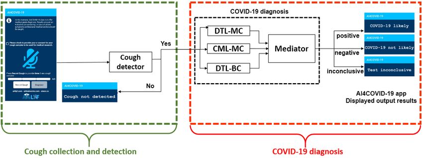
ii Walid Hariri, Ali Narin
by the appearance of new mutations make the classi-
fications a more challenging task [25]. Moreover, one
of the biggest problems with COVID-19 patients is vi-
ral pneumonia (VP). Differentiating between viral and
non-viral pneumonia (nVP) is not easy. Coexistence of
COVID-19 and viral pneumonia can have dire conse-
quences.
Oxford COVID-19 Evidence Service Team Center
follows some tips in identifying these problems. Muscle
pain, loss of sense of smell and shortness of breath with-
out pleuritic pain are the most common symptoms, es-
pecially in the case of COVID-19 infection. On the other
hand, symptoms such as bilateral positive lung findings,
tachycardia or tachypnea disproportionate to temper-
ature, and low temperature indicate VP (not COVID-
19) symptoms [34]. nVP, however, is most susceptible
if it becomes rapidly unwell after a short period from
the appearance of symptoms and does not have simi-
lar symptoms of COVID-19, pleuritic pain, or purulent
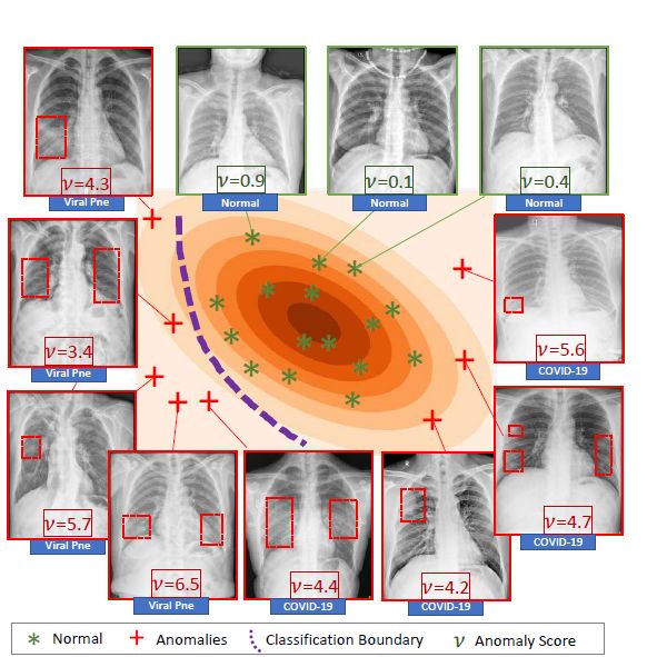
sputum. Fig. 1: Distinguishing between VP cases (anomalies)
and nVP cases and normal controls [98].
Many studies have been introduced to solve this
problem, for instance, Zhang et al. [98] proposed to
lessen the process of anomaly detection into a one-class- 2 Medical imaging technologies versus RT-PCR
classification problem using a confidence aware module. test
Deep learning (DL) is then used for the classification
task as shown in Figure 1. Recent reviews show that the The medical imaging field has considerably emerged
use of novel technology with artificial intelligence (AI) in the last years offering reliable automated methods
and machine learning (ML) techniques considerably im- for clinical decision making. It has received wide ac-
proves the screening, contact tracing, forecasting, and ceptance by the scientists and the medical community.
drug and vaccine development with high reliability. In the case of COVID-19, CT scans and X-ray images
can play a vital role in the early diagnosis of the dis-
Since the COVID-19 pandemic started, it has been
ease. Infected patients have clinical symptoms including
clear that deep learning algorithms from ML technolo-
cough and fever, however, an important proportion of
gies seem to be used extensively to detect COVID-
infected patients can be asymptomatic. In Germany, it
19, VP, bacterial pneumonia (BP) and other similar
has been confirmed in the study of Rothe et al. [75]
cases. The advantages and disadvantages of these stud-
that an asymptomatic patient was able to transmit the
ies should be evaluated. In this study, it is aimed to
virus to another patient. According to the study of Al-
present a detailed review on studies using DL approaches
Tawfiq et al. [6] from 9 countries, 18 from 144 cases were
using various images in the literature to detect COVID-
asymptomatic, the equivalent of 12.5%. The study has
19. In addition, studies that detection of COVID-19 us-
been done using the RT-PCR test.
ing acoustic sound data are included.
Due to the high risk of transmission of COVID-19,
The rest of this paper is organized as follows: Section accurate diagnostic methods are urgently needed to pre-
2 presents the medical imaging technologies. In Sec- vent the spread of the virus and for humanity to breathe
tion 3 we review the most important DL methods pro- comfortably. Besides being the gold standard of the RT-
posed to diagnose the COVID-19, as well as the recent PCR test, the results are time consuming (requires 5 to
advanced applications. Section 4 presents some addi- 6 hours) to obtain.In addition, the high rate of false
tional DL applications to fight against COVID-19 such detection of RT-PCR test is questioned whether it is a
as acoustic analysis and human mobility estimation. In good diagnostic method. [91, 55]. In this case, it is rec-
Section 5 an overall discussion and proposed solutions ommended that patients with typical imaging findings
have been presented to accurately diagnose and to re- should be separated ones from one another and more
duce the spread of the COVID-19. Conclusions, future than one RT-PCR test should performed to avoid mis-
trends and challenges end the paper. diagnosis.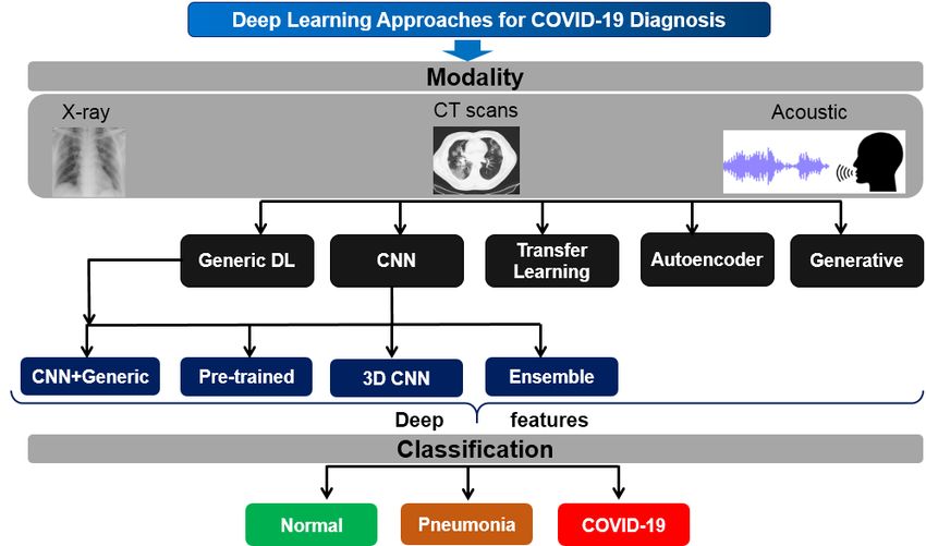
Title Suppressed Due to Excessive Length                                                                            iii

     The X-ray, however, is an efficient screening method,   diagnosis, and control. X-ray scans are used worldwide
it is fast at capturing, cheaper than the RT-PCR test,       to diagnose the injured part and for the detection or
and largely available worldwide. CT-scans, on the other      other diseases in order to treat patients [21]. The X-ray
hand, can be obtained much faster and more accurately        facility is available even in the remotest parts and thus
in the presence of an efficient algorithm (notably DL al-    X-ray images can be easily acquired for patients even
gorithms) to accurately identify the infected patients.      in their home or in their quarantine location. These
In [51], it has been proven that DL offers highly promis-    images have been extensively used for COVID-19 di-
ing results for medical diagnostics compared to health-      agnosis [63]. The most common reported abnormal in
care professionals. Figure 2 presents the change that oc-    Chest X-ray (CXR) findings are ground-glass opacities
curs in the COVID-19 pneumonia cases on some days.           (GGOs) [95]. Figure 3 presents an example of an X-ray
In the following section, detailed information about CT      scan for COVID-19 patients. CXR is the most widely
and X-ray images is presented.                               used imaging technology by researchers because it is
                                                             easily available and inexpensive. However, GGOs are
                                                             often the first sign of a diagnosis of COVID-19 pneu-
2.1 Chest Computed Tomography                                monia.

CT is an imaging method that uses a special x-ray beam
to create detailed scans of areas inside the body (e.g.
lungs, heart, blood vessels, airways, and lymph nodes).
These images are taken from different angles to gen-
erate tomographic images which give the possibility to
the radiographers to directly see inside the body instead
of surgery. CT images are considered to be an effective
way of making clinical decisions. They showed high effi-
ciency in diagnosing COVID-19 especially patients with
false-negative RT-PCR results, assuming a role for the
CT as a reliable tool for COVID-19 diagnosis during
this epidemic period [49, 5, 91, 35]. Therefore, the Na-
tional Health Commission of the People’s Republic of
China suggested CT examination in monitoring disease
progression and controlling treatments of COVID-19 in
its 6th version of the diagnosis and treatment program        Fig. 3: An example of X-ray image for a COVID-19
[99].                                                                              patient.

                                                             3 Deep learning approaches in the COVID-19
                                                             pandemic
 Fig. 2: CT scans in the early fast gradually stage of
 COVID-19 pneumonia cases. a: GGO plus reticular             DL is a subset of ML that offers considerable power
 pattern on the forth day. b: GGO plus consolidation         for improving the accuracy and speed of diagnosis by
  on the third day. c: GGO on the second day. [100]          automating the screening through medical imaging in
                                                             collaboration with radiologists and/or physicians. Sub-
                                                             sequently, it has received wide acceptance and interest
                                                             by the medical community leads to emphasizing the de-
                                                             velopment of such diagnostic technologies [51]. In the
2.2 X-Ray Image                                              following, we review the most important DL approaches
                                                             adopted to diagnose the pneumonia of COVID-19 since
Wilhelm Conrad Röntgen has discovered the first X-ray       its spread in December 2019 until today. Figure 4 presents
in 1895 during experimenting with Lenard tubes and           the taxonomy of these approaches using different im-
Crookes tubes. X-ray has a very important role in the        ages and acoustic features. In the following, we detail
medical field, it can help in the prevention of infection,   each of the distinguished nine groups: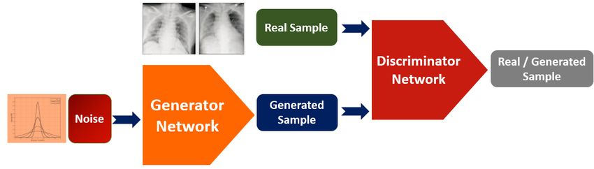
iv                                                                                                Walid Hariri, Ali Narin

                  Fig. 4: Taxonomy of deep learning based-approaches for COVID-19 diagnosis.

3.1 Generic deep learning                                      technique. Then, the infected regions were processed
                                                               and quantized using specific metrics in the CT scan.
Generic DL methods without any specific modification               We can also find generic convolutional neural net-
have been proposed to detect COVID-19. For example,            works in which the authors use the generic CNN trained
Wang el al. [89] have used CT images of 5,372 patients         with their datasets without any combination with other
from 7 different cities in China to train a deep neural        ML algorithms or pre-trained models. For example, in
network (DNN).                                                 [20], the authors trained the CNN model with the data
    Pneumonia Detection Challenge dataset (RSNA) is            collected from Wuhan Jin Yin-Tan hospital in order
used in [56] to train a DL model in order to locate lung       to classify the CT images into one of the five follow-
opacities on chest radiographs. RSNA dataset contains          ing classes: healthy lung, COVID-19, pneumonia, non-
two classes: Normal and Pneumonia (non-normal). The            COVID-19 VP, BP and pulmonary tuberculosis.
total of 16,680 images have been used from this data set
where 8,066 are from healthy class (normal), whereas
8,614 as classified as pneumonia.                              3.2 Transfer learning
    The authors in [83] collected from two hospitals of in
China the CT images of 88 infected patients (COVID-            Transfer learning (TL) is a ML technique in which a
19), 101 patients diagnosed with bacteria pneumonia,           trained model for one task is redesigned in a related
where the rest are healthy (86 persons). Using this dataset,   second task (see Figure 5). This approach is explicitly
they applied a DL-based CT diagnosis system namely:            useful when there are not sufficient datasets like in the
DeepPneumonia to localize the principal lesion features,       case of COVID-19 in order to either reduce the neces-
especially GGO and thus to identify the infected pa-           sary fine-tuning data size or improve performance. TL
tients. The first step is the segmentation of the lung         can be used in two scenarios: supervised (with labeled
region. Next, they introduced the DRE-Net (Details             data from the target domain) or unsupervised (with-
Relation Extraction neural network) to draw the top-K          out any labeled data from the target domain: the pre-
features in the CT images and to receive the image-level       training process is supervised, but unsupervised dur-
predictions. Finally, the image-level predictions is used      ing fine-tuning). A DNN is proposed in [40] to detect
to diagnose the patient.                                       COVID-19 using X-ray images. To do so, the authors
    Another generic DL framework is proposed in [96]           applied a TL approach on the deep Pruned Efficient-
to automatically extract and analyze regions with high         Net model. Then, it has been interpolated by post-hoc
possibility to be infected with COVID-19. To do so, the        analysis to be able to explain the obtained predictions.
authors applied a segmentation stage using a DL-based          TL based-framework for the detection of pneumonia is
Title Suppressed Due to Excessive Length v
proposed in [15]. The features have been extracted from 3.3 Augmentation and Generation Techniques
X-ray images using five different pre-trained models:
DenseNet121, ResNet18, GoogLeNet, AlexNet and In-
Recently, generative adversarial networks (GANs) are
ceptionV3. Next, an ensemble model has been added
considered the most powerful and successful method for
to combine outputs from all pre-trained models. The
data augmentation. Since the outbreak of COVID-19 is
obtained results are as follows: accuracy of 96.4%; re-
recent, it is difficult to gather a significant amount of
call of 99.62% on non-trained data from the Guangzhou
radiographic images and datasets in such a short time.
Women and Children’s Medical Center database.
Therefore, DL networks especially (CNNs) need addi-
Fine-tuned deep TL with generative adversarial net- tional training data to overcome this problem and to
work (GAN) is presented in [42] to learn a limited dataset enhance the efficiency of CNN in detecting COVID-
and to avoid the overfitting problem. To do so, the au- 19 (See Figure 6). Various methods have been applied
thors applied the pre-trained models: Squeeznet, AlexNet, the GANs for this reason. For instance, in [87], authors
GoogLeNet, and Resnet18 as deep TL models to de- generate more X-ray images using Auxiliary Classifier
tect pneumonia from chest x-rays. Applying a com- Generative Adversarial Network (ACGAN) based on
bination of GAN and deep transfer models enhanced the CovidGAN model. Accordingly, the classification
the accuracy of the proposed system and realized 99%. accuracy has been significantly enhanced from 85% us-
After applying image preprocessing algorithms to the ing the CNN alone, to 95% using the ACGAN with
chest X-ray images to identify and remove diaphragm CovidGAN model.
regions, the pre-trained VGG-16 model [81] has been
fine-tuned in [32] using the obtained images to predict Also, to handle the problem of the lack of datasets
COVID-19 infected pneumonia. Another work proposed for COVID-19, Loey et al. [52] proposed classical data
in [8] to detect the COVID-19 in small medical image augmentation techniques along with Conditional GAN
datasets. To do so, they worked with two different data (CGAN) on the basis of a deep transfer learning model
sets from public databases. In the first dataset, there for COVID-19 detection using CT images. Similar rep-
are 224 COVID-19, 700 BP and 504 Normal X-ray im- resentation has been used in [54] to classify the CT
ages. The second dataset includes 224 COVID-19, 714 images into the following four classes : the COVID-19,
BP and VP, and 504 Normal X-ray images. They ob- normal, pneumonia bacterial, and pneumonia virus. To
tained 96.78% accuracy, 98.66% sensitivity and 96.46% do so, the authors have used a dataset of 307 images.
specificity performance values. Three deep transfer models are then carried out in this
Multi-Channel TL-based method with X-ray images work for investigation. The models are the Googlenet,
have been proposed in [61]. Multi-channel pre-trained Alexnet and Restnet18. three strategies have been con-
ResNet model is then used to perform the diagnosis ducted, in each strategy the authors applied a differ-
of COVID-19. To classify the X-ray images on a one- ent deep TL using the three pre-trained models men-
against-all strategy, three ResNet models have been re- tioned above. The testing accuracies achieved by the
trained. The three allowed classifications are: 1) normal Googlenet, Alexnet and Restnet18 are 80.6%, 85.2%
or diseased, 2) pneumonia or non-pneumonia, and 3) and 100%, respectively.
COVID-19 or non-COVID19 individuals. The method Another method proposed in [41] aims to generate
achieved a precision of 94% and a recall of 100%. Other synthetic medical images using DL CGAN to overcome
TL-based methods can be found in [60, 57, 12, 27, 1, 73, the dataset limitation that leads to over-fitting. The
68,53, 69]. proposed model has been implemented in a form to sup-
port a lightweight architecture without transfer learn-
ing without performance degradation. It can deal with
any non-uniformity in the data distribution and the
limited accessibility of training images in the classes.
It consists of a single convolutional layer with filter size
32 and kernel 4 × 4, followed by ReLU activation func-
tion and Max Pooling layer for down-sampling the im-
age (input representation) and enabling feature extrac-
tion. After a flatten layer there exists a dense layer of
size 128, followed by dropout and a final dense layer
with softmax activation function for a binary output.
Fig. 5: An example of transfer learning process for Other methods based on data augmentation to detect
COVID-19 detection. the COVID-19 can be found in [4].vi Walid Hariri, Ali Narin
Fig. 6: Generative adversarial network representation
for COVID-19 detection.
Fig. 7: An example of Autoencoder model for
COVID-19 detection.
3.5 Pre-trained Deep Neural Networks
3.4 Autoencoder-based models
Pre-trained models were originally trained on existing
large-scale labeled dataset (e.g. ImageNet) and later
Another ML technique to handle the problem of in- fine-tuned over the chest CT and X-ray images to ac-
sufficient data for the affected COVID-19 cases is the complish the diagnosis process. The last layer in these
Autoencoder (AE) [10]. It is a neural network method models has been removed and a new fully connected
with competent data encoding and decoding strategies (FC) layer is added with an output size of two that rep-
used for unsupervised feature learning. The AE models resents two separate classes (COVID-19 or normal). In
are comprised of two main steps: encoder and decoder. the obtained models, only the final FC layer is trained,
The input samples are mapped typically to a lower- while other layers are initialized with pre-trained weights
dimensional space with beneficial feature representation [64]. These models can be a very useful solution to the
in the encoding step. In the second step, the decoding lack of large datasets for COVID-19. However, some
consists of reverting data to its original space, trying challenges exist. One of the risen problems here is that
to create data from lower space representation. Figure the transfer across datasets from a domain to another
7 shows the conceptual diagram of AE with its two can lead to deterioration of performance due to the
main steps. The advantage of adopting such unsuper- gap existing between the domains. This is often the
vised classification to handle the problem of COVID- case with medical images taken from different centers.
19 detection compared to its counterpart (supervised Moreover, there is an over-fitting problem with small
classification) is to avoid the long time spent in assem- amounts of COVID-19 datasets. Therefore, pre-trained
bling large amounts of data which could increase the models are generally used with some particular modifi-
risk of mortality and postpones medical care. For ex- cations in order to avoid the over-fitting problem.
ample, in [44], the authors introduced the COVIDomaly In [64], eight pre-trained CNN models have been
which aims to diagnose new COVID-19 cases using a compared including GoogleNet, AlexNet, MobileNet-
convolutional autoencoder framework. They tested two V2, VGG-16, SqueezeNet, ResNet-50; ResNet-34 and
strategies on the COVIDX dataset acquired from the Inception-V3. The obtained results for the classification
chest radiographs by training the model on chest X- of COVID-19 from normal cases show that ResNet-34
rays: the first strategy used only healthy adults, the outperformed the other pre-trained models and achieved
second tested healthy and BP, and infected adults with an accuracy of 98.33%. This evaluation has been con-
COVID-19. Using 3-fold cross-validation, they obtained ducted on a total of 286 scans of COVID-19 and nor-
a pooled Receiver Characteristic Operator-Area Under mal classes as a training set, and 120 scans for the test
the Curve (ROC-AUC) of 76.52% and 69.02% with the (60 scans for each class). When dealing with the aug-
two strategies respectively. mented dataset, the total of the training scans is 1002,
where 428 scans are used for the validation and 120
In [22], the authors extracted discriminative features scans for the test. ResNet-18 has been applied in [66]
from the autoencoder and Gray Level Co-occurence using limited training datasets and achieved a sensitiv-
Matrix using CT images. The obtained features are ity of 100% and 76.90% of precision for the COVID-19
then combined with random forest classifier for COVID- class. Figures 8 and 9 show an example of AlexNet and
19 detection. They achieved the following results: ac- VGG-16 architectures respectively. DenseNet201 pre-
curacy of 97.78%, specificity of 98.77% and recall of trained model is used in [39] on chest CT images. To
96.78%. Other autoencoder-based methods can be found classify the patients into positive or negative COVID-
in [13, 80, 43]. 19, deep transfer learning is carried out, and obtainedTitle Suppressed Due to Excessive Length vii
a training accuracy of 99.82%, and validation accu-
racy of 97.48%. The extreme version of the Inception
(Xception) model is applied in [17] and achieved an
accuracy of 97.40%, f measure of 96.96%, sensitivity
of 97.09% and specificity of 97.29% for three classes
COVID-19, pneumonia, and other diseases. Hemdan et Fig. 9: VGG-16 architecture proposed in [81].
al. [33] proposed COVIDX-Net framework to diagnose
the COVID-19 cases using X-ray images. It includes
three main steps to accomplish the diagnostic process
of the COVID-19 as follows: pre-processing, Training
Model and Validation, Classification. In consequence
of the absence of public COVID-19 datasets, the ex-
periments are carried out on 50 Chest X-ray images
where only 25 have been diagnosed with COVID-19, for
the validation. The COVIDX-Net combined the follow-
ing seven different architectures of deep CNN models:
VGG-19, DenseNet121, InceptionV3, ResNetV2, Inception-
ResNet-V2, Xception, and the second version of Google
MobileNet. Figure 10 presents an overview of the COVIDX-
Net framework. Another model called InstaCovNet-19 Fig. 10: COVIDX-Net framework [33].
makes use of five pre-trained models including Xcep-
tion, ResNet101, InceptionV3, MobileNet, and NASN
is proposed in [26]. Two classification strategies have model. The obtained volumes were fed into the pro-
been conducted : (COVID-19, Pneumonia, Normal) and posed DeCoVNet (3D deep convolutional neural Net-
(COVID, NON-COVID). Very high precision and clas- work to Detect COVID-19). A weakly-supervised clas-
sification accuracy have been achieved using the two sification is then applied and achieved high COVID-19
strategies (See Table 1). Similar method has been pro- classification performance and good lesion localization
posed by Narin et al. [63] using five pre-trained CNNs results. Muller et al. [62] have also used UNet model
and three three different binary datasets including COVID- instead of computational complex CNNs to reduce the
19, normal (healthy), bacterial and viral pneumonia pa- over-fitting problem during the segmentation of the in-
tients. Gour et al. [23] proposed a new CNN model fected lung region. In [88], 3D-ResNet is applied for
based on the VGG-19. They used a 30-layered CNN end-to-end training to classify the acquired lung images
model for the training with X-ray images, and obtained into pneumonia or healthy.
sub-models using logistic regression. Other methods us-
In order to predict the risk of COVID-19, Yang et
ing pre-trained CNNs can be found in [59, 2, 46].
al. [94] applied end-to-end training from CT images
using the 3D Inception V1 model pre-trained on the
ImageNet dataset. The obtained accuracy was 95.78%
overall, and 99.4% on a a part of the dataset contain-
ing 1,684 COVID-19 patients. Li et al. [48] introduced a
3D DL system that aims to early detect the COVID-19,
called COVNet. The COVNet model is composed essen-
tially of RestNet50, which have a range of CT scans as
entry and produces features for the equivalent scans.
Fig. 8: AlexNet architecture proposed in [45].
The obtained features from all scans are then involved
by a max-pooling process. The final feature map is used
as an input to a fully connected layer and softmax ac-
tivation function to produce an output of a likelihood
3.6 3D Convolutional Neural Networks result for the three classes: COVID-19, non-pneumonia
and Community-acquired pneumonia (CAP). Han et al.
3D CNN models have also been used in the literature. [29] introduced a deep 3D multiple instance learning to
They mainly extract 3D features from the segmented detect the COVID-19 using CT images. High accuracy
3D lung region using CT images. For example, Wang et has been achieved (97.9%) and AUC of 99.0%. Other
al. [90] segmented the lung region a pre-trained UNet 3D CNN-based methods can be found in [86, 50].viii Walid Hariri, Ali Narin
3.7 Combination of Generic CNNs with traditional 3.8 Ensemble models
ML algorithms
Handling the problem of COVID-19 detection using a
single DL model without any specific addition might
Another strategy is to use CNN models differently by not achieve a high accuracy classification using CXR
combining them with traditional ML algorithms. images or CT scans. For this reason, the use of many
DL models combined with each other can be a good
In [84], the authors presented a CNN model trained solution, namely: ensemble model and the learning ap-
on X-ray images to recognize pneumonia. The proposed proach is called ensemble learning. For example, the au-
architecture consists of a combination of the convolu- thors in [82] proposed a DL model namely Attention-
tion, max-pooling, and rating layers. The obtained fea- based VGG-16. This model used VGG-16 to capture
tures comprise four convolutional layers, a max-pooling the spatial relationship between the ROIs in CXR im-
layer, and a RELU activator between them. The tradi- ages. By using an appropriate convolution layer (4th
tional ML algorithm ANN (Artificial neural network) pooling layer) of the VGG-16 model in addition to the
is finally applied for classification. ANN and AlexNet attention module, they added a novel DL model to per-
architecture have been combined in [9] to systemati- form fine-tuning in the classification process. In [28] en-
cally find out COVID-19 pneumonia subjects using CT semble of three pre-trained models including Resnet50
scans. Firstly, a segmentation using ANN algorithm and VGG16 and an own small CNN is applied for a
is performed to localize the lungs. Next, COVID-19 test set of 33 new COVID-19 and 218 pneumonia cases.
classes are augmented to produce more images. Finally, The overall accuracy realized is of 91.24%. Shalaf et al.
pre-trained Alexnet architecture is used in one time [78] an ensemble deep transfer learning system with 15
with only a transfer learning process, the obtained accu- pre-trained CNN architectures on CT images. They ob-
racy is 98.14%. And with additional layer namely called tained the following results: accuracy (85%), precision
(Bidirectional Long Short-Term Memories) in the sec- (85.7%) and recall (85.2%).
ond time, with an accuracy 98.70%. Nour et al. [65]
proposed a scratch CNN model including five convolu- 3.9 Smart phone applications
tion layered serial network. Three ML algorithms have
been trained on the obtained deep features involving To further automate the screening of COVID-19 and to
k-NN, SVM, and DT. The highest accuracy is obtained make it faster, mobile phones can be a very interesting
by the SVM with 98.97%. framework for that due to their facility and numerous
sensors with important computing proficiencies. Specif-
Instead of using pre-trained deep CNNs only as fea- ically, a smartphone has is able to scan CT images of
ture extractor, in [38], two other strategies have been COVID-19 patients to use them for analysis screening.
conducted to accurately classify Chest X-ray images Moreover, multiple CT images of the same COVID-19
into positive of negative COVID-19 including fine-tuning patient can be gathered into one smartphone for simi-
strategy and end to end training. The following mod- larity examination of how disfigurement have been de-
els have been used as a feature extractor : ResNet101, veloped [71]. However, the computing capability of a
VGG19, ResNet50, ResNet18, and VGG16 where SVM mobile to treat a large amount of data is lower than a
is used for ML-based classification. Whereas, a new grand machine or a computer. Therefore, a lightweight
CNN model is used for the fine-tuning strategy. Finally, representation is needed to accomplish this task. Conse-
end-to-end training with a dataset of 180 COVID-19 quently, various recent methods have been proposed to
and 200 normal is carried out as a third strategy. 94.7% detect COVID-19 in mobile devices using a slight rep-
of accuracy is achieved using ResNet50 model and SVM resentation. In [101], a lightweight DL model namely
classifier, where fine-tuned strategy with ResNet50 model LightCovidNet has been offered to detect COVID-19
achieved 92.6%. Finally, the end-to-end training strat- using a mobile platform. To enhance the performance
egy of the developed CNN model realized a 91.6% re- of the proposed model, supplementary data have been
sult. Deep CNN and long short-term memory (LSTM) generated and added to the training dataset using the
have been combined in [37] to diagnose COVID-19 au- conditional deep convolutional GAN. In order to reduce
tomatically from X-ray images. The obtained accuracy the memory usage of the proposed model, five units of
of the classification of three classes (COVID-19, nor- feed-forward CNN are built using separable convolu-
mal, and other pneumonia) is 99.4%. Similar methods tion operators. Multi-scale features are then learned to
that combine deep features and classical ML techniques be suitable for the X-ray images which have been ac-
can be found in [77]. quired from all over the world separately. Instead ofTitle Suppressed Due to Excessive Length ix
COVID-19 diagnosis and detection, various lightweight
applications have been introduced to delay the spread
of the virus. These applications could be designed to
be compatible with the capabilities of a smartphone to
further speed up their operation. Among these applica-
tions we can find: masked face recognition [30], facial
mask detection [14, 16], social distance monitoring [3,
74] and human mobility estimation [92]. Other mobile-
based technique using to fight against COVID-19 has Fig. 11: AI4COVID-19 system architecture [36].
been proposed in [58]. Using DL algorithms, the au-
thors arrived to efficiently evaluate the level of pneu-
monia and thus to determine whether it is a COVID-19 cloud, and give a result during two minutes. To over-
case or not. come the lack of COVID-19 cough training data, the
authors applied transfer learning using ESC-50 dataset
[70] that contains 50 classes of cough and non-cough
sounds acquired using a smartphone. Figure11 presents
4 Deep learning for other applications
the offered system architecture and a drawing of AI4COVID-
Other applications have been proposed to fight against 19. The obtained results show high overall accuracy of
COVID-19 induced pneumonia, the most important are 95.60%, a sensitivity of 96.01%, a specificity of 95.19%,
cough detection and human mobility estimation. and precision of 95.22%. Schuller et al. [76] studied what
computer audition could possibly contribute to the on-
going battle against the COVID-19. Other recent acous-
tic analysis for the detection of COVID-19 can be found
4.1 Cough Detection in [19, 7, 67, 72, 47, 67, 18].
In addition to the DL approaches using X-ray and chest
CT scans for COVID-19 detection, scientists affirm that 4.2 Estimating Human Mobility
audio sounds generated by the respiratory system can
be diagnosed and analyzed to decide whether the pa- Human mobility (movement) is one of the main fac-
tient is infected or not. Therefore, cough analysis has tors that promote the transmission of the virus. Policy-
been used to screen and diagnose COVID-19. ML tech- makers find huge difficulties to find an optimal proto-
niques can supply useful cues enabling the develop- col to insure the social distancing and barrier measures.
ment of a diagnostic instrument. To do so, cough data To solve this problem, Bao et al. [11] proposed a sys-
of COVID and non-COVID is required. Accordingly, tem that aims to evaluate and estimate maps of people
Sharma et al. [79] proposed a database called Coswara, movement responses by learning from existing ground
of respiratory sounds, namely, cough, breath, and voice. truth data. The proposed system is based on a DL
Some experiments have been recently carried out to based-data generation called COVID-GAN. It merges
screen COVID-19 from acoustic features, for example, a diversity of features involving contextual features,
in [31], Recurrent Neural Network (RNN) has been used COVID-19 details and data history, as well as policies
in its new architecture, namely the Long Short-Term from various origins such as news, reports and Safe-
Memory (LSTM) to extract six speech features from a Graph. Experiment results showed that COVID-GAN
collected dataset (i.e. Spectral centroid, Spectral Roll- can well imitate real-world human movement reactions
off, Zero-Crossing Rate, Zero-Crossing Rate, MFCC, and the area-constraint-based correction can consider-
and MFCC). In this work, 70% of the data was used ably upgrade the solution value. To further explain the
for training, and 30% for testing. The obtained results relation between people mobility and COVID-19 con-
show that the best accuracy is achieved for breathing tamination, Xiong et al. [92] presented a study using
sound, reaching up to 98.2% followed by cough sounds, mobile device data to give more insights to decision
an accuracy of 97% is attained. Whereas, the accuracy makers about the national mobility tendencies before
of voice analysis is of 88.2%. and during the pandemic.
A smartphone application using cough-based diag-
nosis for COVID-19 detection is proposed in [36]. This
application is based on an AI-powered screening solu-
tion called AI4COVID-19. Its principle is to send three
3 second cough sounds to an AI engine running in thex
Table 1: Summary of DL-based methods for COVID-19 pneumonia classification.
C.V: refers to Cross validation. Tr: refers to training, Val: refers to the validation, AE refers to Autoencoder. Sens refers to the Sensitivity. Spec refers
to the Specificity. Acc refers to the Accuracy.
Author Modality Dataset 2D/3D All data All COVID-19 Network and technique C.V Sens(%) Spec(%) Acc (%)
COVID-19/normal 3141 96.1
[63] CXR COVID-19/pneumonia 2D 1834 341 5 pre-trained CNNs 5 / / 99.5
COVID-19/bacterial 3113 99.7
Tr: 78.93 Tr: 89.93
[89] CT COVID-19/pneumonia/normal 3D 1,266 924 DNN / /
Val: 80.39 Val: 81.16
[83] CT COVID-19/normal/bacterial 2D 275 88 DRE-Net / 93 96 99
[42] CXR pneumonia dataset 2D 624 50 GAN + TL / / / 99
[8] CXR COVID-19/normal/bacterial 2D 1,427 224 MobileNet v2 10 98.66 96.46 94.72
[61] CXR COVID-19/pneumonia/normal 2D 6,008 184 Three ResNet models 5 / / 93.9
[44] CXR COVID-19/pneumonia/normal 2D 8,850 498 AE : COVIDomaly 3 / / 76.52
[65] CXR COVID-19/pneumonia/normal 2D 2,905 219 CNN+k-NN+SVM / / / 98.70
[9] CXR COVID-19/pneumonia/normal 2D 2,905 219 ANN+AlexNet / / / 98.97
[66] CXR COVID-19/pneumonia/normal 2D 502 180 ResNet-18 / 76.90 100 /
Ensemble:Resnet50
[28] CXR COVID-19/pneumonia/normal 2D 2,905 219 10 / / 91.24
and VGG16
[39] CXR COVID-19/normal 2D 2,492 1,262 TL and DenseNet201 / / / 99.82
[17] CXR COVID-19/pneumonia/normal 2D / / Xception / 97.09 97.29 97.40
5 pre-trained models
[38] CXR COVID-19/normal 2D 380 180 / / / 94.7
+ SVM
COVID-19/pneumonia/normal 99.08
[26] CXR 2D 2905 219 pre-trained models / / /
COVID/non-COVID 99.53
[59] CXR COVID-19/pneumonia/normal 2D / / 5 pre-trained CNNs / / / 95
Bacterial, Non-COVID Viral,
[2] CXR 2D / / 5 COVID-CAPS / 90 95.8 95.7
COVID-19
[60] CXR COVID-19/normal 2D 5,000 184 5 TL+pre-trained models / 100 98.3 /
[57] CXR+CT COVID-19/normal 2D 526 238 TL+AlexNet model / 72 100 94.1
TL+InceptionV3
[12] CXR+CT COVID-19/normal 2D 320 160 / 72 100 99.01
and ResNet50
[37] CXR COVID-19/pneumonia/normal 2D 4,575 1,525 LSTM+CNN / 99.2 99.9 99.4
4,448 2,479 / / 95.78
[94] CXR COVID-19/pneumonia/normal 2D 3D Inception V1 10
101 52 98.08 91.30 93.3
Conditional GAN :
[101] CXR COVID-19/pneumonia/normal 2D 1343 446 5 / / 97.28
LightCovidNet
Total: 8,504 Total: 445
[32] CXR COVID-19/normal 2D TL VGG-16 model / 98.0 100 94.5
Training: 6,899 Training: 366
TL+ Ensemble of
[78] CT COVID-19/normal 2D 746 349 / / / 85
15 pre-trained models
[22] CT COVID-19/normal 2D 2,482 1,252 AE+random forest / / 98.77 97.87
[33] CXR COVID-19/normal 3D 50 25 COVIDX-Net / 100 80 /
Walid Hariri, Ali NarinTitle Suppressed Due to Excessive Length xi
5 Discussion guished. A very high performance test dataset can be
created. This overestimates the results obtained. It can
Table 1 includes some studies conducted with DL mod- be noted that a CV method should be used in which
els. It can be seen in Table 1 that studies have fo- the whole data set can be tested in studies. Although
cused on two popular images, namely CT and X-ray there are many studies using DL-based methods, it is
images. The most used of these is X-ray images. This very difficult to produce sufficiently transparent, stable
is because it is easily available everywhere. In addition, and reliable models. It was clearly stated above that
both its low memory space and high results in its per- there are many parameters affecting the results.
formance encouraged researchers to use X-ray images. As an alternative to the studies performed on X-ray
In addition, the greater availability of X-ray data from and CT images, researches on the detection of COVID-
COVID-19 patients in public databases has led to the 19 are carried out with sound and cough-based acoustic
large number of these studies. In articles in the field sound analysis. Alternative approaches are very impor-
of medicine, it is often stated that CT images show tant in the detection of COVID-19. It may be possible
higher performance. However, these high accuracies are to save more lives with real-time detection and diag-
not seen in DL based CAD systems. This may be be- nosis systems with online scanning systems installed
cause radiologists can easily distinguish patients from with mobile or computer. As a result of all these ap-
CT images. CT images have too many cross-sections of proaches, namely RT-PCR test, DL-based X-ray and
the same person. They are much more complex than CT images, detection and acoustic examinations, it is
X-ray images. This complex situation is a disadvantage predicted that patients with COVID-19 can be detected
in distinguishing them in DL methods. The combina- more stable and with higher accuracy. Because, consid-
tion of X-ray and CT images, performed in few studies, ering that the epidemic started with a person, it is very
also shows that it gives good results. It can be said important to correctly diagnose even a person.
that studies are carried out with multi-class solutions
rather than the binary classification problem. It is seen 6 Conclusion
that studies carried out with 3D data have lower per-
formance. It has mostly been studied with 2D data. Although the RT-PCR test is considered the gold stan-
The results obtained from these data are very high. dard for COVID-19 diagnosis, it is time-consuming to
Considering the number of data, it is known that DL make a decision because of high false-negative levels in
models work stably with a lot of data. According to the results. Therefore, medical imaging modalities such
this fact, the number of data used is not sufficient. It as chest X-ray and chest CT scans are the best alter-
can be stated that this is one of the biggest problems native according to scientists. Chest X-ray radiography
in the detection of COVID-19. It is very important to is of low cost and low radiation dose, it is available and
compare the studies conducted with the data obtained easy to use in general or community hospitals. This re-
from many different centers. Otherwise, the accuracy of view presents a detailed study of the existing solutions
the studies can be fooled. For this, we recommend that that are mainly based on DL techniques to early diag-
data collected from many different centers be offered nose the COVID-19. This study gives more of an insight
to researchers from a single center. However, in studies into the scientists’ and decision-makers’ thought pro-
conducted with a small number of data, it is seen that cesses - not only during the wave periods but also dur-
pre-trained models are used to ensure high model train- ing that of the vaccination that could require real-time
ing. Especially Keras, Tensorflow, PyTorch and MAT- mass testing. The lack of data, however, is the manda-
LAB also include these pre-trained models. The biggest tory problem to achieve efficient and real-time results.
problem here is that these models are trained with the Many solutions have been presented and discussed in
ImageNet dataset. Having a lot of data belonging to this review study to give more ideas to future trends and
very different classes in the ImageNet dataset can re- also for eventual future diseases that might suffer from
duce the trust in these models. However, it is seen that the missing-data problem. We believe that with more
the performances obtained are also very high. public databases, better DL based-approaches can be
Traditional ML algorithms are also used by using developed to detect and diagnose the COVID19 accu-
the feature extraction part of DL models. This approach rately. Also, when policy-makers and citizens are mak-
appears to improve performance. It is generally seen ing their best to submit to the difficult constraints of
that the SVM algorithm is used. Most of the stud- lockdown and social distancing, AI can be used to cre-
ies do not use any cross-validation (CV) method. We ate more intelligent robots and autonomous machines
think that this decreases the reliability of the results. to help health workforce and to reduce their workload
Because it is not known how the test data are distin- by disinfection, working in hospitals, food distributingxii Walid Hariri, Ali Narin
and helping the patients. The challenge of this solution 6. Al-Tawfiq, J.A.: Asymptomatic coronavirus infection:
is that people lack confidence in autonomous machines Mers-cov and sars-cov-2 (covid-19). Travel medicine and
infectious disease (2020)
and prefer to be served by a human even if there is 7. Alsabek, M.B., Shahin, I., Hassan, A.: Studying the sim-
a risk of virus transmission. Moreover, entrusting chat- ilarity of covid-19 sounds based on correlation analysis
bots to diagnose patients needs a large amount of medi- of mfcc. In: 2020 International Conference on Commu-
cal data from experts. Also, the difference in languages nications, Computing, Cybersecurity, and Informatics
(CCCI), pp. 1–5. IEEE (2020)
from a country to another makes an already difficult 8. Apostolopoulos, I.D., Mpesiana, T.A.: Covid-19: au-
task still more arduous. On the other hand, when deal- tomatic detection from x-ray images utilizing transfer
ing with voice analysis, there are still many challenges learning with convolutional neural networks. Physical
and Engineering Sciences in Medicine p. 1 (2020)
to be taken up. For example, until now, annotated data 9. Aslan, M.F., Unlersen, M.F., Sabanci, K., Durdu, A.:
of patients’ voices are not publicly available for research Cnn-based transfer learning-bilstm network: A novel ap-
purposes of COVID-19 detection and diagnosis. Collect- proach for covid-19 infection detection. Applied Soft
ing these data is mostly made in unconstrained envi- Computing p. 106912 (2020)
10. Baldi, P.: Autoencoders, unsupervised learning, and
ronments (i.e. in-the-wild) using smartphones or other deep architectures. In: Proceedings of ICML workshop
voice recorders. These environments are generally noisy on unsupervised and transfer learning, pp. 37–49 (2012)
and contain reverberation, which leads to bad quality of 11. Bao, H., Zhou, X., Zhang, Y., Li, Y., Xie, Y.: Covid-
gan: Estimating human mobility responses to covid-19
data and makes the diagnosis and detection of COVID-
pandemic through spatio-temporal conditional genera-
19 more challenging. Finally, one of the most important tive adversarial networks. In: Proceedings of the 28th
future trends is to concentrate on further decreasing the International Conference on Advances in Geographic In-
false negative rate and, as far as practicable, reducing formation Systems, pp. 273–282 (2020)
12. Benbrahim, H., Hachimi, H., Amine, A.: Deep transfer
the false positive rate by the same token to accurately learning with apache spark to detect covid-19 in chest
differentiate viral from BP. x-ray images. Romanian Journal of Information Science
and Technology 23, S117–S129 (2020)
13. Berenguer, A.D., Sahli, H., Joukovsky, B., Kvasnyt-
Acknowledgements The authors would like to thank the sia, M., Dirks, I., Alioscha-Perez, M., Deligiannis, N.,
’Agence Nationale de Valorisation des Résultats de la Recherche Gonidakis, P., Sánchez, S.A., Brahimetaj, R., et al.:
et du Développement Technologique (DGRSDT), Algérie’. Explainable-by-design semi-supervised representation
learning for covid-19 diagnosis from ct imaging. arXiv
preprint arXiv:2011.11719 (2020)
Conflict of interest 14. Chen, Y., Hu, M., Hua, C., Zhai, G., Zhang, J., Li, Q.,
Yang, S.X.: Face mask assistant: Detection of face mask
service stage based on mobile phone. arXiv preprint
The authors declare that they have no conflict of inter- arXiv:2010.06421 (2020)
est. 15. Chouhan, V., Singh, S.K., Khamparia, A., Gupta, D.,
Tiwari, P., Moreira, C., Damaševičius, R., De Albu-
querque, V.H.C.: A novel transfer learning based ap-
References proach for pneumonia detection in chest x-ray images.
Applied Sciences 10(2), 559 (2020)
16. Chua, M.H., Cheng, W., Goh, S.S., Kong, J., Li, B.,
1. Abbas, A., Abdelsamea, M.M., Gaber, M.M.: Classi-
Lim, J.Y., Mao, L., Wang, S., Xue, K., Yang, L., et al.:
fication of covid-19 in chest x-ray images using de-
Face masks in the new covid-19 normal: Materials, test-
trac deep convolutional neural network. arXiv preprint
ing, and perspectives. Research 2020 (2020)
arXiv:2003.13815 (2020) 17. Das, N.N., Kumar, N., Kaur, M., Kumar, V., Singh,
2. Afshar, P., Heidarian, S., Naderkhani, F., Oikonomou, D.: Automated deep transfer learning-based approach
A., Plataniotis, K.N., Mohammadi, A.: Covid-caps: for detection of covid-19 infection in chest x-rays. Irbm
A capsule network-based framework for identification (2020)
of covid-19 cases from x-ray images. arXiv preprint 18. Deshmukh, S., Ismail, M.A., Singh, R.: Interpreting
arXiv:2004.02696 (2020) glottal flow dynamics for detecting covid-19 from voice.
3. Ahmed, I., Ahmad, M., Rodrigues, J.J., Jeon, G., Din, arXiv preprint arXiv:2010.16318 (2020)
S.: A deep learning-based social distance monitoring 19. Deshpande, G., Schuller, B.W.: Audio, speech, lan-
framework for covid-19. Sustainable Cities and Society guage, & signal processing for covid-19: A comprehen-
p. 102571 (2020) sive overview. arXiv preprint arXiv:2011.14445 (2020)
4. Ahmed, S., Hossain, M.F., Noor, M.B.T.: Convid-net: 20. Fu, M., Yi, S.L., Zeng, Y., Ye, F., Li, Y., Dong, X.,
An enhanced convolutional neural network framework Ren, Y.D., Luo, L., Pan, J.S., Zhang, Q.: Deep learning-
for covid-19 detection from x-ray images. In: Proceed- based recognizing covid-19 and other common infectious
ings of International Conference on Trends in Computa- diseases of the lung by chest ct scan images. medRxiv
tional and Cognitive Engineering, pp. 671–681. Springer (2020)
(2021) 21. Ghosh, A., Saha, S.: Automatic identification of frac-
5. Ai, T., Yang, Z., Hou, H., Zhan, C., Chen, C., Lv, W., ture region within bone in x-ray image. In: 2018 2nd
Tao, Q., Sun, Z., Xia, L.: Correlation of chest ct and International Conference on Electronics, Materials En-
rt-pcr testing in coronavirus disease 2019 (covid-19) in gineering & Nano-Technology (IEMENTech), pp. 1–7.
china: a report of 1014 cases. Radiology p. 200642 (2020) IEEE (2018)Title Suppressed Due to Excessive Length xiii
22. Goel, C., Kumar, A., Dubey, S.K., Srivastava, V.: Ef- (covid-19) using x-ray images. Informatics in Medicine
ficient deep network architecture for covid-19 detection Unlocked 20, 100412 (2020)
using computed tomography images. medRxiv (2020) 38. Ismael, A.M., Şengür, A.: Deep learning approaches for
23. Gour, M., Jain, S.: Stacked convolutional neural net- covid-19 detection based on chest x-ray images. Expert
work for diagnosis of covid-19 disease from x-ray images. Systems with Applications 164, 114054 (2020)
arXiv preprint arXiv:2006.13817 (2020) 39. Jaiswal, A., Gianchandani, N., Singh, D., Kumar, V.,
24. Gozes, O., Frid-Adar, M., Greenspan, H., Browning, Kaur, M.: Classification of the covid-19 infected patients
P.D., Zhang, H., Ji, W., Bernheim, A., Siegel, E.: Rapid using densenet201 based deep transfer learning. Journal
ai development cycle for the coronavirus (covid-19) pan- of Biomolecular Structure and Dynamics pp. 1–8 (2020)
demic: Initial results for automated detection & patient 40. Jaiswal, A.K., Tiwari, P., Rathi, V.K., Qian, J., Pandey,
monitoring using deep learning ct image analysis. arXiv H.M., Albuquerque, V.H.C.: Covidpen: A novel covid-
preprint arXiv:2003.05037 (2020) 19 detection model using chest x-rays and ct scans.
25. Grubaugh, N.D., Hanage, W.P., Rasmussen, A.L.: Mak- medRxiv (2020)
ing sense of mutation: what d614g means for the covid- 41. Karakanis, S., Leontidis, G.: Lightweight deep learning
19 pandemic remains unclear. Cell 182(4), 794–795 models for detecting covid-19 from chest x-ray images.
(2020) Tech. rep., EasyChair (2020)
26. Gupta, A., Gupta, S., Katarya, R., et al.: Instacovnet- 42. Khalifa, N.E.M., Taha, M.H.N., Hassanien, A.E., El-
19: A deep learning classification model for the detec- ghamrawy, S.: Detection of coronavirus (covid-19) asso-
tion of covid-19 patients using chest x-ray. Applied Soft ciated pneumonia based on generative adversarial net-
Computing p. 106859 (2020) works and a fine-tuned deep transfer learning model us-
27. Haghanifar, A., Majdabadi, M.M., Ko, S.: Covid-cxnet: ing chest x-ray dataset. arXiv preprint arXiv:2004.01184
Detecting covid-19 in frontal chest x-ray images using (2020)
deep learning. arXiv preprint arXiv:2006.13807 (2020)
43. Khobahi, S., Agarwal, C., Soltanalian, M.: Coronet:
28. Hall, L.O., Paul, R., Goldgof, D.B., Goldgof, G.M.: A deep network architecture for semi-supervised task-
Finding covid-19 from chest x-rays using deep learn- based identification of covid-19 from chest x-ray images.
ing on a small dataset. arXiv preprint arXiv:2004.02060 medRxiv (2020)
(2020)
44. Khoshbakhtian, F., Ashraf, A.B., Khan, S.S.: Covido-
29. Han, Z., Wei, B., Hong, Y., Li, T., Cong, J., Zhu, X.,
maly: A deep convolutional autoencoder approach for
Wei, H., Zhang, W.: Accurate screening of covid-19 us-
detecting early cases of covid-19. arXiv preprint
ing attention based deep 3d multiple instance learning.
arXiv:2010.02814 (2020)
IEEE Transactions on Medical Imaging (2020)
45. Krizhevsky, A., Sutskever, I., Hinton, G.E.: Imagenet
30. Hariri, W.: Efficient masked face recogni-
classification with deep convolutional neural networks.
tion method during the covid-19 pandemic.
Communications of the ACM 60(6), 84–90 (2017)
PREPRINT (Version 3) available at Research Square
[https://doi.org/10.21203/rs.3.rs-39289/v3] (2020) 46. Kumar, R., Khan, A.A., Zhang, S., Wang, W., Abuidris,
31. Hassan, A., Shahin, I., Alsabek, M.B.: Covid-19 detec- Y., Amin, W., Kumar, J.: Blockchain-federated-learning
tion system using recurrent neural networks. In: 2020 and deep learning models for covid-19 detection using
International Conference on Communications, Comput- ct imaging. arXiv preprint arXiv:2007.06537 (2020)
ing, Cybersecurity, and Informatics (CCCI), pp. 1–5. 47. Laguarta, J., Hueto, F., Subirana, B.: Covid-19 artificial
IEEE (2020) intelligence diagnosis using only cough recordings. IEEE
32. Heidari, M., Mirniaharikandehei, S., Khuzani, A.Z., Open Journal of Engineering in Medicine and Biology
Danala, G., Qiu, Y., Zheng, B.: Improving the perfor- (2020)
mance of cnn to predict the likelihood of covid-19 using 48. Li, L., Qin, L., Xu, Z., Yin, Y., Wang, X., Kong, B.,
chest x-ray images with preprocessing algorithms. In- Bai, J., Lu, Y., Fang, Z., Song, Q., et al.: Artificial intel-
ternational journal of medical informatics 144, 104284 ligence distinguishes covid-19 from community acquired
(2020) pneumonia on chest ct. Radiology (2020)
33. Hemdan, E.E.D., Shouman, M.A., Karar, M.E.: Covidx- 49. Li, Y., Xia, L.: Coronavirus disease 2019 (covid-19): role
net: A framework of deep learning classifiers to di- of chest ct in diagnosis and management. American
agnose covid-19 in x-ray images. arXiv preprint Journal of Roentgenology 214(6), 1280–1286 (2020)
arXiv:2003.11055 (2020) 50. Liu, S., Georgescu, B., Xu, Z., Yoo, Y., Chabin, G.,
34. Heneghan, C.: Differentiating viral from bacte- Chaganti, S., Grbic, S., Piat, S., Teixeira, B., Balachan-
rial pneumonia (2020 (accessed December 07, dran, A., et al.: 3d tomographic pattern synthesis for
2020)). URL https://www.cebm.net/covid-19/ enhancing the quantification of covid-19. arXiv preprint
differentiating-viral-from-bacterial-pneumonia arXiv:2005.01903 (2020)
35. Huang, P., Liu, T., Huang, L., Liu, H., Lei, M., Xu, W., 51. Liu, X., Faes, L., Kale, A.U., Wagner, S.K., Fu, D.J.,
Hu, X., Chen, J., Liu, B.: Use of chest ct in combination Bruynseels, A., Mahendiran, T., Moraes, G., Shamdas,
with negative rt-pcr assay for the 2019 novel coronavirus M., Kern, C., et al.: A comparison of deep learning per-
but high clinical suspicion. Radiology 295(1), 22–23 formance against health-care professionals in detecting
(2020) diseases from medical imaging: a systematic review and
36. Imran, A., Posokhova, I., Qureshi, H.N., Masood, U., meta-analysis. The lancet digital health 1(6), e271–e297
Riaz, S., Ali, K., John, C.N., Nabeel, M.: Ai4covid- (2019)
19: Ai enabled preliminary diagnosis for covid-19 52. Loey, M., Manogaran, G., Khalifa, N.E.M.: A deep
from cough samples via an app. arXiv preprint transfer learning model with classical data augmenta-
arXiv:2004.01275 (2020) tion and cgan to detect covid-19 from chest ct radiogra-
37. Islam, M.Z., Islam, M.M., Asraf, A.: A combined deep phy digital images. Neural Computing and Applications
cnn-lstm network for the detection of novel coronavirus pp. 1–13 (2020)xiv Walid Hariri, Ali Narin
53. Loey, M., Manogaran, G., Taha, M.H.N., Khalifa, 70. Piczak, K.J.: Esc: Dataset for environmental sound clas-
N.E.M.: A hybrid deep transfer learning model with ma- sification. In: Proceedings of the 23rd ACM interna-
chine learning methods for face mask detection in the tional conference on Multimedia, pp. 1015–1018 (2015)
era of the covid-19 pandemic. Measurement 167, 108288 71. Purswani, J.M., Dicker, A.P., Champ, C.E., Cantor, M.,
(2021) Ohri, N.: Big data from small devices: the future of
54. Loey, M., Smarandache, F., M Khalifa, N.E.: Within the smartphones in oncology. In: Seminars in radiation on-
lack of chest covid-19 x-ray dataset: A novel detection cology, vol. 29, pp. 338–347. Elsevier (2019)
model based on gan and deep transfer learning. Sym- 72. Quatieri, T.F., Talkar, T., Palmer, J.S.: A framework for
metry 12(4), 651 (2020) biomarkers of covid-19 based on coordination of speech-
55. Long, C., Xu, H., Shen, Q., Zhang, X., Fan, B., Wang, production subsystems. IEEE Open Journal of Engi-
C., Zeng, B., Li, Z., Li, X., Li, H.: Diagnosis of the coro- neering in Medicine and Biology 1, 203–206 (2020)
navirus disease (covid-19): rrt-pcr or ct? European jour- 73. Rahaman, M.M., Li, C., Yao, Y., Kulwa, F., Rahman,
nal of radiology p. 108961 (2020) M.A., Wang, Q., Qi, S., Kong, F., Zhu, X., Zhao, X.:
56. Luz, E., Silva, P.L., Silva, R., Moreira, G.: Towards an Identification of covid-19 samples from chest x-ray im-
efficient deep learning model for covid-19 patterns detec- ages using deep learning: A comparison of transfer learn-
tion in x-ray images. arXiv preprint arXiv:2004.05717 ing approaches. Journal of X-ray Science and Technol-
(2020) ogy (Preprint), 1–19 (2020)
57. Maghdid, H.S., Asaad, A.T., Ghafoor, K.Z., Sadiq, A.S., 74. Rezaei, M., Azarmi, M.: Deepsocial: Social distancing
Khan, M.K.: Diagnosing covid-19 pneumonia from x-ray monitoring and infection risk assessment in covid-19
and ct images using deep learning and transfer learning pandemic. Applied Sciences 10(21), 7514 (2020)
algorithms. arXiv preprint arXiv:2004.00038 (2020) 75. Rothe, C., Schunk, M., Sothmann, P., Bretzel, G.,
58. Maghdid, H.S., Ghafoor, K.Z., Sadiq, A.S., Curran, K., Froeschl, G., Wallrauch, C., Zimmer, T., Thiel, V.,
Rabie, K.: A novel ai-enabled framework to diagnose Janke, C., Guggemos, W., et al.: Transmission of 2019-
coronavirus covid 19 using smartphone embedded sen- ncov infection from an asymptomatic contact in ger-
sors: Design study. arXiv preprint arXiv:2003.07434 many. New England Journal of Medicine 382(10), 970–
(2020) 971 (2020)
59. Makris, A., Kontopoulos, I., Tserpes, K.: Covid-19 de- 76. Schuller, B.W., Schuller, D.M., Qian, K., Liu, J.,
tection from chest x-ray images using deep learning and Zheng, H., Li, X.: Covid-19 and computer audition: An
convolutional neural networks. In: 11th Hellenic Con- overview on what speech & sound analysis could con-
ference on Artificial Intelligence, pp. 60–66 (2020) tribute in the sars-cov-2 corona crisis. arXiv preprint
60. Minaee, S., Kafieh, R., Sonka, M., Yazdani, S., Soufi,
arXiv:2003.11117 (2020)
G.J.: Deep-covid: Predicting covid-19 from chest x-ray
77. Sethy, P.K., Behera, S.K.: Detection of coronavirus
images using deep transfer learning. arXiv preprint
disease (covid-19) based on deep features. Preprints
arXiv:2004.09363 (2020)
2020030300, 2020 (2020)
61. Misra, S., Jeon, S., Lee, S., Managuli, R., Jang, I.S.,
78. Shalbaf, A., Vafaeezadeh, M., et al.: Automated detec-
Kim, C.: Multi-channel transfer learning of chest x-ray
tion of covid-19 using ensemble of transfer learning with
images for screening of covid-19. Electronics 9(9), 1388
deep convolutional neural network based on ct scans. In-
(2020)
ternational journal of computer assisted radiology and
62. Müller, D., Rey, I.S., Kramer, F.: Automated chest ct
surgery pp. 1–9 (2020)
image segmentation of covid-19 lung infection based on
79. Sharma, N., Krishnan, P., Kumar, R., Ramoji,
3d u-net. arXiv preprint arXiv:2007.04774 (2020)
63. Narin, A., Kaya, C., Pamuk, Z.: Automatic detection S., Chetupalli, S.R., Ghosh, P.K., Ganapathy, S.,
of coronavirus disease (covid-19) using x-ray images et al.: Coswara–a database of breathing, cough, and
and deep convolutional neural networks. arXiv preprint voice sounds for covid-19 diagnosis. arXiv preprint
arXiv:2003.10849 (2020) arXiv:2005.10548 (2020)
64. Nayak, S.R., Nayak, D.R., Sinha, U., Arora, V., Pa- 80. Shoeibi, A., Khodatars, M., Alizadehsani, R., Ghassemi,
chori, R.B.: Application of deep learning techniques for N., Jafari, M., Moridian, P., Khadem, A., Sadeghi, D.,
detection of covid-19 cases using chest x-ray images: A Hussain, S., Zare, A., et al.: Automated detection and
comprehensive study. Biomedical Signal Processing and forecasting of covid-19 using deep learning techniques:
Control p. 102365 (2020) A review. arXiv preprint arXiv:2007.10785 (2020)
65. Nour, M., Cömert, Z., Polat, K.: A novel medical di- 81. Simonyan, K., Zisserman, A.: Very deep convolutional
agnosis model for covid-19 infection detection based on networks for large-scale image recognition. arXiv
deep features and bayesian optimization. Applied Soft preprint arXiv:1409.1556 (2014)
Computing p. 106580 (2020) 82. Sitaula, C., Hossain, M.B.: Attention-based vgg-16
66. Oh, Y., Park, S., Ye, J.C.: Deep learning covid-19 fea- model for covid-19 chest x-ray image classification. Ap-
tures on cxr using limited training data sets. IEEE plied Intelligence pp. 1–14 (2020)
Transactions on Medical Imaging (2020) 83. Song, Y., Zheng, S., Li, L., Zhang, X., Zhang, X., Huang,
67. Pal, A., Sankarasubbu, M.: Pay attention to the cough: Z., Chen, J., Zhao, H., Jie, Y., Wang, R., et al.: Deep
Early diagnosis of covid-19 using interpretable symp- learning enables accurate diagnosis of novel coronavirus
toms embeddings with cough sound signal processing. (covid-19) with ct images. medRxiv (2020)
arXiv preprint arXiv:2010.02417 (2020) 84. Stephen, O., Sain, M., Maduh, U.J., Jeong, D.U.: An
68. Perumal, V., Narayanan, V., Rajasekar, S.J.S.: Detec- efficient deep learning approach to pneumonia classifi-
tion of covid-19 using cxr and ct images using transfer cation in healthcare. Journal of healthcare engineering
learning and haralick features. Applied Intelligence pp. 2019 (2019)
1–18 (2020) 85. Ucar, F., Korkmaz, D.: Covidiagnosis-net: Deep bayes-
69. Pham, T.D.: Classification of covid-19 chest x-rays with squeezenet based diagnostic of the coronavirus disease
deep learning: new models or fine tuning? Health Infor- 2019 (covid-19) from x-ray images. Medical Hypotheses
mation Science and Systems 9(1), 1–11 (2021) p. 109761 (2020)You can also read