Determinants of resistance to VEGF-TKI and immune checkpoint inhibitors in metastatic renal cell carcinoma
←
→
Page content transcription
If your browser does not render page correctly, please read the page content below
Sharma et al. Journal of Experimental & Clinical Cancer Research                 (2021) 40:186
https://doi.org/10.1186/s13046-021-01961-3

 REVIEW                                                                                                                                              Open Access

Determinants of resistance to VEGF-TKI and
immune checkpoint inhibitors in metastatic
renal cell carcinoma
Revati Sharma1,2, Elif Kadife1, Mark Myers2, George Kannourakis1,2, Prashanth Prithviraj1† and Nuzhat Ahmed1,2,3,4*†

  Abstract
  Vascular endothelial growth factor tyrosine kinase inhibitors (VEGF-TKIs) have been the mainstay of treatment for
  patients with advanced renal cell carcinoma (RCC). Despite its early promising results in decreasing or delaying the
  progression of RCC in patients, VEGF-TKIs have provided modest benefits in terms of disease-free progression, as 70%
  of the patients who initially respond to the treatment later develop drug resistance, with 30% of the patients innately
  resistant to VEGF-TKIs. In the past decade, several molecular and genetic mechanisms of VEGF-TKI resistance have been
  reported. One of the mechanisms of VEGF-TKIs is inhibition of the classical angiogenesis pathway. However, recent
  studies have shown the restoration of an alternative angiogenesis pathway in modulating resistance. Further, in the last
  5 years, immune checkpoint inhibitors (ICIs) have revolutionized RCC treatment. Although some patients exhibit potent
  responses, a non-negligible number of patients are innately resistant or develop resistance within a few months to ICI
  therapy. Hence, an understanding of the mechanisms of VEGF-TKI and ICI resistance will help in formulating useful
  knowledge about developing effective treatment strategies for patients with advanced RCC. In this article, we review
  recent findings on the emerging understanding of RCC pathology, VEGF-TKI and ICI resistance mechanisms, and
  potential avenues to overcome these resistance mechanisms through rationally designed combination therapies.
  Keywords: Clear cell renal carcinoma, Metastatic renal cell carcinoma, Vascular endothelial growth factor, tyrosine
  kinase inhibitor, Sunitinib, Hypoxia, Epithelial mesenchymal transition

Background                                                                              there are 15 subtypes of RCC with diverse genetic and
Kidney cancer is the ninth most common cancer in men                                    epigenetic characteristics, of which clear cell RCC
and fourteenth in women. In 2018, there were 400,000                                    (ccRCC) occurs most frequently (80%). Papillary RCC
new cases around the globe [1]. Renal cell carcinoma                                    (10–15%) and chromophobe RCC (5%) are the common
(RCC) makes up to 95% of renal malignancies [2]. RCC                                    remaining histologic subtypes [3]. Around 50% of RCC
arises from the renal tubular epithelium, which lines the                               is detected incidentally with one-quarter of the patients
proximal convoluted tubules and constitutes of very                                     diagnosed with metastatic disease and another 30% that
small tubes in the kidney responsible for transporting                                  relapse and develop metastatic RCC (mRCC) after
urine. According to the 2012 consensus conference of                                    undergoing curative nephrectomy. These patient groups
the International Society of Urological Pathology (ISUP),                               are considered at high risk of death due to RCC [4]. The
                                                                                        morbidity and mortality rates of advanced RCC are high,
                                                                                        with a five-year survival rate of only 18% [5]. All sub-
* Correspondence: nuzhata@unimelb.edu.au; nuzhat@fecri.org.au
†
 Prashanth Prithviraj and Nuzhat Ahmed contributed equally to this work.
                                                                                        types of RCC are innately resistant to traditional cancer
1
 Fiona Elsey Cancer Research Institute, Ballarat, Victoria 3350, Australia              treatments, such as chemotherapy and radiotherapy.
2
 Federation University Australia, Ballarat, Victoria 3350, Australia
Full list of author information is available at the end of the article

                                         © The Author(s). 2021 Open Access This article is licensed under a Creative Commons Attribution 4.0 International License,
                                         which permits use, sharing, adaptation, distribution and reproduction in any medium or format, as long as you give
                                         appropriate credit to the original author(s) and the source, provide a link to the Creative Commons licence, and indicate if
                                         changes were made. The images or other third party material in this article are included in the article's Creative Commons
                                         licence, unless indicated otherwise in a credit line to the material. If material is not included in the article's Creative Commons
                                         licence and your intended use is not permitted by statutory regulation or exceeds the permitted use, you will need to obtain
                                         permission directly from the copyright holder. To view a copy of this licence, visit http://creativecommons.org/licenses/by/4.0/.
                                         The Creative Commons Public Domain Dedication waiver (http://creativecommons.org/publicdomain/zero/1.0/) applies to the
                                         data made available in this article, unless otherwise stated in a credit line to the data.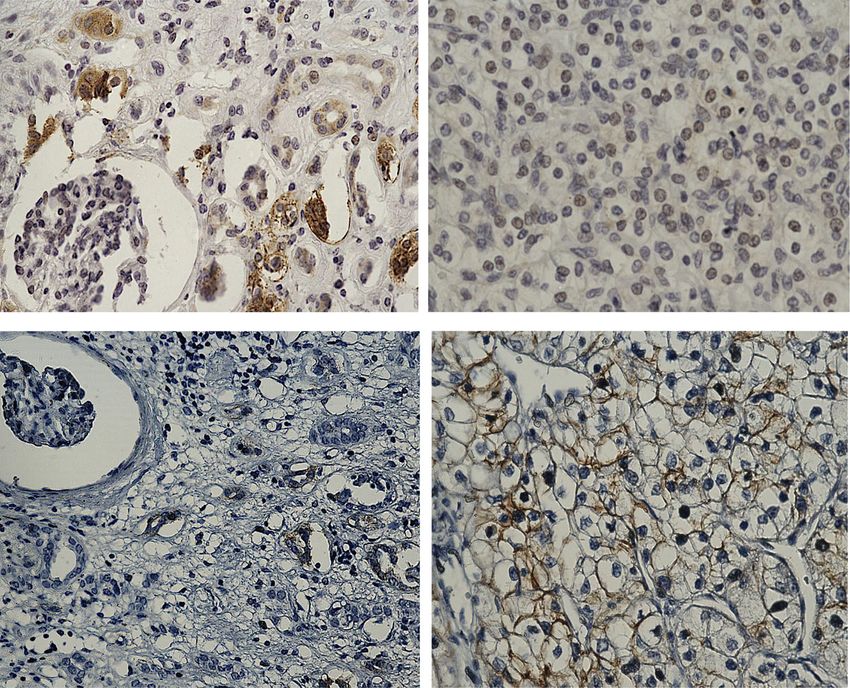
Sharma et al. Journal of Experimental & Clinical Cancer Research    (2021) 40:186                                                   Page 2 of 27

RCC histology                                                             genes is fundamental to the pathogenesis of RCC due to
The gross morphological appearance of RCC varies be-                      their role in promoting angiogenesis, tumour cell sur-
tween tumour types. In general, most RCC presents with                    vival, proliferation, disease progression, glucose metabol-
areas of extensive network of blood vessels with cysts                    ism, and metastatic spread. HIF is also composed of a β
containing watery fluid and areas of cancer cells (Fig. 1).               subunit (ΗΙF-1β). HIF-1β is stable and constitutively
The ccRCC stores glycogen and lipids and the cells con-                   expressed, whereas HIF α subunits are highly unstable
tain clear cytoplasm and a central nucleus encompassed                    and are controlled by cellular oxygen levels. Under nor-
by an intact plasma membrane. Due to the extensive                        moxic conditions, prolyl 2-oxoglutarate-dependent Fe2+
vascular network, the stroma in most cancer cells                         dioxygenases PHD1, PHD2, and PHD3 hydroxylate the
shrinks, the surrounding parenchyma is constricted, and                   two conserved proline residues in HIFα subunits. The
the tumour is confined to capsular structures [6]. Non-                   hydroxylated proline residues are targets of VHL/E3 ubi-
clear cell RCC is a group of diseases, each with different                quitin ligase complex, resulting in the protease-mediated
histologic subtypes and different clinical course and out-                degradation of HIFα. However, during hypoxia, PHDs
comes. The most common are papillary RCC (pRCC)                           cannot hydroxylate HIFα, leading to their stabilisation
and chromophobe RCC (chRCC). pRCC, which show a                           and activation of downstream target genes, which mostly
papillary pattern, although tubular structures or solid                   regulate the expression of angiogenic and tissue remod-
growth patterns can be seen but are rare. In the case of                  elling proteins [11]. Stabilised HIF-1α also enhances the
chRCC, the growth pattern is often solid, comprised of                    expression of glycolytic enzymes lactate dehydrogenase
sheets of tumour cells containing long linear parallel                    (LDH), pyruvate dehydrogenase kinase (PDK), and other
vessels in contrast to the thin delicate network of vessels               glycolysis-related genes, such as glucose transporter-1
of ccRCC [7].                                                             (GLUT-1) and hexokinase (HK), which consequently in-
                                                                          crease glucose uptake and glycolysis, thus reducing the
Genetic alterations in RCC                                                carbon flux through the tricarboxylic acid (TCA) cycle
Genetic alterations are common in RCC and usually in-                     and oxidative phosphorylation in RCC [12, 13]. En-
volve loss of tumour suppressor genes by deletion or                      hanced expression of HIF-1α target genes GLUT1, HK,
functional inactivation or by hypermethylation of the                     LDH, PDK1 and PKM2 in ccRCC compared to matching
gene promoter [8]. Classically, 70% of ccRCC carry a                      adjacent normal kidney tissues are represented in Fig. 2.
mutation in the von Hippel-Lindau (VHL) gene, which                         Hence, HIF-1α is a vital metabolic checkpoint for RCC
encodes VHL protein (pVHL) [9, 10]. pVHL exerts its                       and is essential for the development of RCC in vivo.
tumour suppressing function by binding to and mediat-                     Recent studies have shown that inactivation of both
ing the degradation of hypoxia-inducible factor (HIF).                    HIF-1α and HIF-2α hampers the development of ccRCC
The development of RCC in most cases occurs by dele-                      in mouse models, suggesting that both HIFα genes may
tion or mutation on both alleles of VHL [9]. The loss or                  be crucial for ccRCC initiation and progression [15].
mutation in VHL results in the inactivation of pVHL,                      However, 30–40% of clinically diagnosed ccRCC lack the
leading to the activation and accrual of HIF proteins                     expression of HIF-1α, suggesting that HIF-1α may act as
(HIF-1α, HIF-α and HIF-3α) and transcription of down-                     a tumour suppressor gene in those scenarios where its
stream target genes [4, 10]. The activation of HIF target                 expression may be mandatory for initial development

                                     Normal Kidney                                       Primary RCC

 Fig. 1 Haematoxylin and eosin (H and E) stained paraffin embedded ccRCC section and its corresponding normal kidney tissue. Histopathological
 slides showing H and E images of normal kidney with well-defined glomerulus and tubules, and conventional ccRCC with typical histological
 appearance of epithelial nests of large uniform cells with clear cytoplasm and distinct cell membrane (blue arrow). Delicate branches of blood
 vessels (red arrow) surround the nests of cells. Magnification: 40X, Scale bar: 75 μm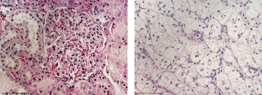
Sharma et al. Journal of Experimental & Clinical Cancer Research (2021) 40:186 Page 3 of 27
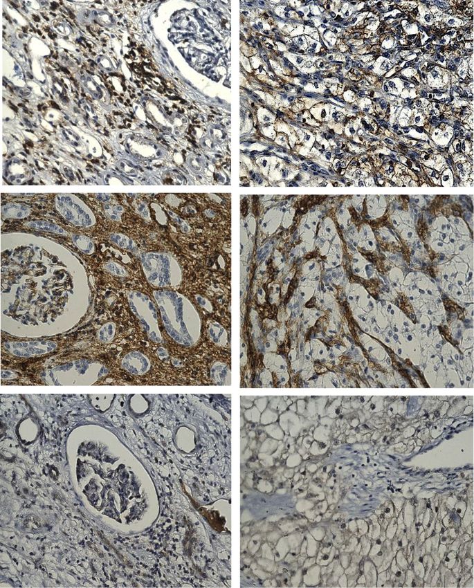
Normal kidney tissue Primary ccRCC Normal kidney tissue Primary ccRCC
PDK1
GLUT1
LDH PKM2
HK
Fig. 2 Representative immunohistochemistry images of RCC patient tumour sections and adjacent normal kidney tissues stained with HIF-1α
target genes GLUT1, HK, LDH, PDK1 and PKM2. Tumours/tissues were stained as described previously [14]. GLUT1 (abcam: ab652): negative
staining in glomerulus and moderate cytoplasmic staining in the tubules of adjacent normal kidney tissue while RCC tissues shows a distinct
membranous staining. HK (OriGene Technologies, Inc.; AM05641PU-S): adjacent normal kidney tissue shows strong cytoplasmic staining in the
tubules and low cytoplasmic staining in the glomerulus while RCC demonstrates a strong cytoplasmic and membranous staining. LDH (abcam;
ab52488): adjacent normal kidney is strongly positive for cytoplasmic staining in the tubules and low cytoplasmic staining in the glomerulus; RCC
tissues presents an intense positive membranous and nuclear staining. PDK1 (GeneTex; GTX60386): adjacent normal kidney is negative in
glomerulus staining and shows very low cytoplasmic staining in the tubules while RCC tissues illustrates distinct membranous and moderate
cytoplasmic staining. PKM2 (abcam; ab150377) staining is negative in the adjacent normal kidney tissue while uniform moderate cytoplasmic
staining is present in the RCC tissue. Magnification: 40X, Scale bar: 75 μm. Representative images of n = 5 tissues for each antibody
but lost as the tumour progresses [16]. By contrast, HIF- increased frequency of p53 mutation reported with in-
2α plays a critical role in RCC progression through its creasing grades and stages of RCC [23]. In this context,
activating effects on c-Myc, epidermal growth factor re- the correlation of p53 mutation with disease-specific
ceptor (EGFR), cyclin D, tumour protein p53, and mam- survival was reported in RCC patients [24].
malian target of rapamycin (mTOR) oncogenes,
resulting in enhanced cell cycle progression and tumour Epithelial-mesenchymal transition in ccRCC
growth [16–18]. Hence, ccRCC cases are divided into Epithelial-mesenchymal transition (EMT) is an embry-
two groups. The first group has both HIF-1α and HIF- onic development process that cancer cells utilize,
2α expressed to drive tumour progression, whereas in whereby epithelial cells lose their epithelial polarity and
the second group, the effect of only HIF-2α prevails, par- attain a mesenchymal phenotype and shape in order to
ticularly in vivo in rapidly proliferating tumours where detach from primary sites to gain entry into surrounding
access to nutrients is limited for tumour cells, resulting tissue vasculatures for re-localization and spread into
in enhanced tumour cell proliferation/angiogenesis and surrounding or distant sites [25]. The process is trig-
poor patient prognosis [16]. gered by various stimuli received by the cancer cells
Next-generation sequencing has identified genes other from the tumour microenvironment, one of which is
than VHL that are commonly altered in RCC. Poly- hypoxia-mediated HIF1-α activation, which plays a crit-
bromo 1 (PBRM1) (41%), BRCA1-associated protein 1 ical role in the initiation and orchestration of EMT [25].
(BAP1) (15%), and SET domain containing 2 histone ly- The dissemination of cancer cells is facilitated by the
sine methyltransferases (SETD2) (19%) have been loss of epithelial cell adhesion molecule E-cadherin and
mapped to chromosome 3p, similar to VHL [19]. PBRM1 upregulation of E-cadherin repressors such as Slug,
is a subunit of SWI/SNF chromatin remodelling com- Snail, ZEB, and Twist, which are the hallmarks of the
plex, BAP1 encodes the histone deubiquitinating enzyme EMT process. An immunohistochemistry study on ZEB2
BRCA1-associated protein, and SETD2 is a histone expression in 116 RCC patients demonstrated high
methyltransferase [20]. Studies have shown that other ZEB2 expression in RCC tumours that correlated with
tumour suppressor genes, such as Wilms tumour 1 gene poor overall survival (OS) and progression-free survival
(WT1), the phosphatase and tensin homolog (PTEN), (PFS) in RCC patients [26]. Similarly, enhanced Snail ex-
and tumour protein p53, are also involved in the path- pression was frequent in high-grade RCC and associated
ology of RCC [21, 22]. Among these genes, p53 mutation with poor OS and PFS in RCC patients [27]. A recent
has been shown as a prognostic indicator for RCC, with study correlated EMT with an increased risk of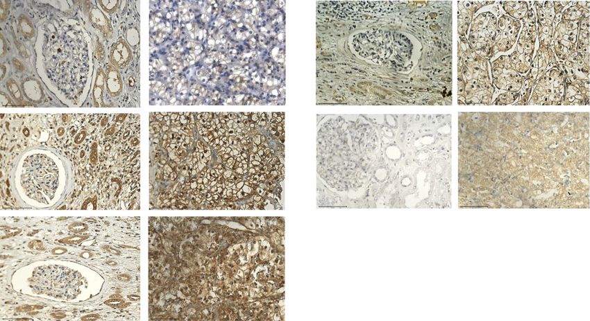
Sharma et al. Journal of Experimental & Clinical Cancer Research      (2021) 40:186                                                     Page 4 of 27

recurrence and poor OS in RCC patients based on the                         of ccRCC into sarcomatoid tumour is regulated by EMT
expression of DCLK1 (a serine/threonine kinase involved                     by triggering N-cadherin expression, dissociation of β-
in microtubule-mediated neuronal migration and mor-                         catenin from the cell membrane, and increased expression
phogenesis) in RCC tumours [28]. In addition, DCLK1                         of Snail and Sparc proteins [32, 33]. A recent study used
was shown to be overexpressed and deregulated in > 93%                      an integration of omics and cellular/molecular biology
of RCC tumours, and its knockdown by siRNA in RCC                           assays on 26 RCC patient samples to demonstrate a
cells resulted in decreased expression of EMT and cancer                    link between fibrosis and EMT correlating that to
stem cell (CSC) markers [29]. Further, the scoring of EMT                   worse patient survival [34]. The above studies clearly
based on the identification of spindle-shaped cells in tu-                  indicate that EMT is a crucial driving force in RCC
mours obtained from 47 RCC patients after nephrectomy                       progression. Besides hypoxia-mediated HIF-1α activa-
was correlated with a shorter OS of 3–6 months in 96.4%                     tion, several cytokines also contribute to the orches-
patients compared to a longer OS of > 6 months in 42.1%                     tration of EMT during RCC progression. Among
patients in whom spindle-shaped cells were absent [30]. A                   these, IL-6, IL-8, IL-15, and tumour necrosis factor α
multivariate analysis of the expression levels of Clusterin,                (TNF-α) play a prominent role in EMT facilitation via
Twist, and C-reactive protein (CRP) in the tumours of 116                   Akt/GSK-3β/β-catenin signalling pathway [35–38].
RCC patients obtained at nephrectomy independently pre-                     Chronic oxidative stress also induces EMT character-
dicted disease recurrence and recurrence-free survival                      istics in RCC cells [39]. Downregulation of the micro-
established by the positive expression of each independent                  RNA (miRNA)-200 family, which includes miR-200a/
factor present in individual patient. Disease recurrence                    b/c, miR-141 and miR-429, is also involved in the
was observed in 7.7% of patients who were negative for                      EMT process in RCC [40]. A recent paper has shown
any risk factor, 31.5% in patients who had one or two risk                  that an immune suppressor cyclosporine in combin-
factors, and 60.9% of patients with three or four risk fac-                 ation with transforming growth factor β (TGFβ) is
tors [31]. RCC tumour stage and histological grade, as well                 able to induce EMT and CSC-like phenotypes in RCC
as sarcomatoid differentiation, are influenced by the ex-                   cells [41]. Figure 3 demonstrates indication of EMT
pression of the transcription factor Snail. The conversion                  in RCC by illustrating enhanced staining of EMT-

                                              Normal kidney                                    Primary ccRCC

                     E -cadherin

                     N-cadherin

 Fig. 3 Representative immunohistochemistry images of RCC patient tumour sections and adjacent normal kidney tissues stained with E-cadherin
 and N-cadherin. Immunohistochemistry was performed as described in Fig. 2. E-cadherin (Cell Signaling; 14,472), images show adjacent normal
 kidney tissue to be slightly positively stained in the tubules while RCC tissue is negative for any staining. N cadherin (Cell Signaling; 13,116):
 increase in the membranous expression of N-cadherin in the RCC tumour compared to normal adjacent kidney tissue. Magnification: 40X, Scale
 bar: 75 μm. Representative images of n = 5 tissues for each antibody
Sharma et al. Journal of Experimental & Clinical Cancer Research   (2021) 40:186                                       Page 5 of 27

related N-cadherin and low expression of E-cadherin                     in individual studies and is mostly based on their func-
in ccRCC tumours compared to adjacent normal kid-                       tional parameters. RCCs have been shown to display di-
ney tissues.                                                            verse CSC markers (such as CD44, CD133, CD105,
                                                                        CXCR-4, Oct4, Nanog, Klf4, and LIN28) and have a high
Cancer stem cells (CSCs) in RCC                                         expression of the ATP-binding cassette family of trans-
CSCs constitute a minor population of cells within tu-                  porter proteins, such as MDR1 (P-glycoprotein) and
mours with a remarkable ability for tumour initiation and               ABCB transporters [53]. However, the proportion of
sustenance through infinite capacity for self-renewal and               cancer cells expressing different CSC markers remains
multi-lineage differentiation towards heterogeneous pro-                uncertain and may not always reflect a true proportion
genies [42]. RCC is known to be a heterogeneous tumour                  of CSCs or have the CSC-like phenotype as described in
with the existence of both intra- and inter-heterogeneity.              other tumours [44, 54]. Furthermore, hypoxia plays a
Heterogenous cell populations are functionally and                      critical role in the conservation of EMT and CSC fea-
phenotypically distinct and therefore display varying de-               tures in solid tumours. In RCC, hypoxia-induced HIF-1α
grees of response and sensitivity to drugs, reducing the                promoted EMT in RCC cell lines through increased ex-
likelihood of treatment success. As such, CSC-like cells                pression of ZEB1 and ZEB2 and E2A immunoglobulin
may be important determinants of clinical resistance and                enhancer-binding factors E12/E47 (TCF3), which sup-
patient outcomes [43, 44]. Further to that, the heterogen-              pressed E-cadherin expression, leading to the attainment
eity in CSCs may be modulated by a diverse range of fac-                of a mesenchymal phenotype in these cells [55].
tors, including genetic mutations, epigenetic changes,                  Tumour-infiltrating macrophages have been shown to
stimulus from the tumour microenvironment due to cell-                  induce EMT and CSC-like phenotypes in RCC cell lines
cell interaction, exposure to different cytokine milieus,               and mouse xenografts [56]. Ectopic expression of retino-
and hypoxia [45, 46].                                                   blastoma binding protein-2 (RBP2) promoted CSC phe-
   The mere observation that cancers can arise long after               notypes through EMT in RCC cells [57]. These
initial exposure to carcinogens implies that the carcino-               observations indicate that the collaboration of EMT and
genic event imposed by oxidative, genotoxic, or cytotoxic               CSC is crucial for RCC progression. Figure 4 demon-
stress leading to damage-associated molecular pattern                   strates presence of CD44, CD105 and CD133 positive
(DAMP) response may persist in the residual long-lived                  CSC staining in RCC tumours and adjacent normal kid-
slowly proliferating stem cell population for an indefinite             ney tissues.
period ranging from months to years. This dormant re-
sponse eventually triggered by unknown mechanism(s)                     Hypoxia and its effect on the tumour
gives rise to generations of daughter and differentiated                microenvironment (TME)
cells, resulting in recurrent tumour masses [47]. Hence,                For cancer to adapt to low oxygen, tumour cells aber-
recurrent or relapsed tumours that arise from CSCs con-                 rantly develop new but defective and leaky blood vessels.
sist of CSCs and a mixed population of cells, which cre-                The abnormal vasculature, together with the hypoxic
ate the full heterogeneous phenotype of the tumour. The                 microenvironment, promotes angiogenesis and inflam-
induction of EMT giving rise to CSC-like cells was first                mation, all of which lead to tumour progression and
shown in breast cancer, in which stimulation by TGFβ                    treatment resistance. Studies have highlighted that ther-
resulted in both EMT and CSC-like cells [48]. Consist-                  apy resistance and cancer progression are not only regu-
ent with this result, the introduction of mesenchymal                   lated by tumour cells but also by the cells and
markers Twist or Snail, responsible for the suppression                 components encompassing the tumour microenviron-
of the epithelial adhesion molecule E-cadherin, led to an               ment (TME) [58]. Hypoxia induces genetic and prote-
increase in the number of CSC-like cells in breast cancer               ome changes in tumours and associated cells in TME
[49]. Hypomethylation of genes specific for the tran-                   leading to accelerated cancer progression and induction
scription stem cell programme leads to EMT in cancer                    of a more resistant tumour phenotype [59]. Hypoxia also
cells [50]. Moreover, E-cadherin transcriptional repres-                decreases drug penetration and increase the expression
sors Snail and Slug enforce CSC-like phenotypes and                     of drug efflux transporters in tumours as well as tumour
chemoresistance in ovarian cancer cells [51]. Recent                    associated endothelial cells (TECs) [59]. In addition, the
studies have shown the existence of a side population                   hypoxic environment promotes tumour cell glycolysis,
(SP) cells in RCC tumours, a distinct type of CSCs, de-                 which enhances lactic acid production, favouring a low
tected by the use of Hoechst 33342 dye (DNA binding                     pH TME that suppresses immune cell functions such as
dye) that displays a unique pattern by fluorescence-                    proliferation and cytotoxicity [60].
activated cell sorting (FACS) [52]. However, no general                   One cell population that thrives under hypoxic condi-
applicable panel of markers for CSCs has been identified                tions in tumours are TECs. These cells are an important
in RCC, and the characterization of putative CSCs varies                component of RCC TME and are key to progression and
Sharma et al. Journal of Experimental & Clinical Cancer Research   (2021) 40:186                                                   Page 6 of 27

                                                normal kidney                                 ccRCC

                       CD44

                       CD105

                       CD133

 Fig. 4 Representative immunohistochemistry images of RCC patient tumour sections and adjacent normal kidney tissues stained with CSC
 markers CD44, CD105 and CD133. Immunohistochemistry was performed as described in Fig. 2. CD44 (abcam; ab51037): normal kidney tissue
 shows negative staining in glomerulus and moderate cytoplasmic staining in the tubules; while RCC tissues shows distinct membranous and
 moderate cytoplasmic staining. CD105 (abcam; ab169545): adjacent normal kidney tissue shows strong cytoplasmic staining in the tubules and
 low cytoplasmic staining in the glomerulus; RCC tissues shows an intense positive membranous staining. CD133 (abcam; ab19898): adjacent
 normal kidney showing negative staining; RCC tumour shows a specific membranous staining. Magnification: 40X, Scale bar: 75 μm.
 Representative images of n = 5 tissues for each antibody

therapy resistance. Higher levels of circulating endothelial             same study showed that aneuploidy could also be induced
cells have been noted in mRCC patients treated with suniti-              in NECs in response to hypoxia, which was inhibited by in-
nib who acquire resistance [61]. Contrary to the popular                 hibitors of VEGFR2 or reactive oxygen species (N-acetyl-L-
belief that TECs are homogenous and cannot proliferate in                cysteine) [64]. These studies indicate that hypoxia modu-
RCC, these cells are capable of hyperproliferation and dis-              lates phenotypic and genotypic characteristics of TECs via
play metabolic, genetic and morphological abnormalities                  VEGF and ROS expression in TME and may transform
compared to normal endothelial cells (NECs) [62]. TECs                   NECs to TECs to favour tumour progression.
isolated from human tumour xenografts of RCC, melanoma                     A recent study has shown TECs isolated from hyp-
and liposarcoma displayed chromosomal irregularities asso-               oxic highly metastatic tumours contained more aneu-
ciated with aneuploidy [63, 64]. These aneuploid TECs in                 ploid cells, had enhanced proliferative and invasive
TME were surrounded by pimonidazole-positive areas, in-                  capacity and had enhanced mRNA expression of pro-
dicating association of hypoxia with aneuploidy [64]. The                angiogenic (VEGF, VEGFR1/2, HIF-1α) and stemness
Sharma et al. Journal of Experimental & Clinical Cancer Research   (2021) 40:186                                          Page 7 of 27

genes than TECs derived from low metastatic tu-                         directly involved in affecting T cell priming and migration,
mours [65]. In addition, TECs from different tumours                    modulating immune cell trafficking by favouring infiltration
(melanoma, liposarcoma, RCC, glioma, breast and he-                     in tumours of immune suppressive (such as Tregs, MSDCs,
patocellular carcinoma) overexpress proangiogenic                       TAMs) rather than immune effector cells (CD8+ and CD4+
growth factors and receptors suggesting an autocrine                    cells) [90]. Hence, it is important to characterise the cancer
loop for sustenance in an activated mode in TME                         promoting functions of hypoxia-modulated TECs to de-
[66]. Compared to NECs, TECs are also more resist-                      velop new strategies targeting TME associated TECs in
ant to serum starvation and cytotoxic drugs [66–69].                    combination with tumour cells.
TECs in RCC express PAX2 and HLA-G, two embry-                             RCC has long been recognised as an immunogenic
onic markers generally expressed in renal tumours                       tumour due to a substantial amount of immune cell infiltra-
[70, 71]. The expression of embryonic markers in                        tion in the tumours [91]. However, the mere presence of
RCC TECs may indicate their dedifferentiation status                    immune cells in the tumours (TILs) does not indicate that
different from adult or tumour stem or progenitor                       these immune cells are active to mount an anti-tumour re-
cells [66]. These functional alteration in TECs may                     sponse. A recent study on the peripheral blood mono-
result from constitutively activated signalling path-                   nuclear cells (PBMCs) of 90 RCC patients showed
ways, such as PI3K/Akt [72], Cox-2 pathways [73]                        increased expression of PD-1 on CD14 bright myelomono-
and downregulation of anti-angiogenic factors such as                   cytic cells, effector T cells and natural killer (NK) cells,
thrombospondin-1 (TSP-1) and endostatin, [72, 74],                      which correlated with disease stage. The PD-1 expression
responsible for the induction of resistance to chemo-                   on immune cells was significantly reduced after surgery of
therapeutic and antiangiogenic drugs.                                   primary tumours [92, 93]. In another study on the PBMC
  Further to that, recent studies have shown that CSCs                  of 40 RCC patients identified CD8+PD-1+TIM-3+Lag3+
in leukemias, breast and ovarian cancer to differentiate                TILs, CD4+ICOS+TILs and CD25+CD127+Foxp3/Helios+
into endothelial cells [75, 76]. In RCC, CD105+ and                     GITR+Tregs phenotype to be associated with high risk of
CD133+ CSCs were noted to generate endothelial cells                    disease progression after nephrectomy within the same year
in vivo [77, 78]. Considering that hypoxic cancer cells                 [94]. In a subsequent study it was shown the patients hav-
are poorly differentiated and express markers of CSCs                   ing high levels of CD8+PD-1+TIM3+LAG3+ in PBMCs
[79, 80], it can be postulated that hypoxic RCC with                    responded significantly well to nivolumab (anti-PD-1) but
CSC phenotype may have the potential to initiate and                    not to everolimus in terms of overall and progression-free
promote vasculogenesis to sustain and accelerate tumour                 survival, suggesting a specific therapeutic role of nivolumab
growth. In that context, hypoxia induced increased ex-                  in these patients [95]. These data suggests the potential of
pression of HIF-1α and HIF-2α has been noted in                         TIM3 and LAG3 as additional checkpoint inhibitors in
neuroblastoma and glioma CSCs [81, 82]. Both HIF-1α                     RCC management. Very recently, tumour-educated B cells
and HIF-2α are also associated with hypoxia-induced                     (TEB) within the RCC TME were shown to play a key role
expression of CD133 and knocking down of either HIF-                    in RCC progression and therapy resistance [96, 97].
1α [83] or HIF-2α [84] was shown to reduce hypoxia-
induced CD133 expression in glioma CSCs.
  Along with irregular blood vessels, hypoxia can also lead             The role of non-coding RNAs (miRNAs) in RCC
to blocked lymphatic drainage with increased interstitial               Recent studies have identified circulating non-coding
pressure [59]. This interstitial fluid pressure within the              RNAs such as miRNAs as potential blood-based bio-
tumour can interfere with tumour cell’s drug uptake by                  markers for early-stage diagnosis, prediction of progno-
counteracting the passage of drug into the tumour cells.                sis and treatment response in RCC [98]. Among the
Cancer-associated fibroblasts (CAFs) in the TME are asso-               different miRNAs described in RCC, miR-210 was
ciated with these pressure forces within the tumour, and                shown by several studies to hold promise as a potential
some studies have successfully demonstrated improved up-                early-stage biomarker as its expression level was signifi-
take of cytotoxic drugs by targeting CAFs [85]. In mRCC,                cantly enhanced in malignant tissues compared to
activation of fibroblast activating protein was shown to in-            healthy adjacent parenchyma, and its level in the serum
duce aggressive phenotype in RCC via CAFs-mediated re-                  of RCC patients was significantly high compared to
cruitment of macrophages leading to remodelling of TME                  healthy controls [14, 99–102]. According to these stud-
[86]. In addition, a recent study has identified a distinct             ies, the serum miR-210 levels could differentiated RCC
angiogenesishighmacrophageslow fingerprint in a cluster of              patients from healthy controls however, both sensitivity
RCC, which may prove crucial for predicting anti-TKI/anti-              and specificity of miR-210 varied substantially between
angiogenesis treatments [87]. Other studies have also linked            the studies. Other studies have shown regulation of
CAFs with resistance to antiangiogenic drugs [88, 89]. In               miR-210 by hypoxia, showing upregulation of miR-210
addition, TECs have immune regulatory roles as they are                 in response to hypoxic conditions in RCC cell lines,
Sharma et al. Journal of Experimental & Clinical Cancer Research            (2021) 40:186                                                           Page 8 of 27

suggesting a close relationship of miR-210 with RCC de-                            Cancer Center (MSKCC) score [106]. However, when
velopment [101–103].                                                               VEGF-TKIs revolutionized the treatment options of
  Apart from miR-210, combination of miR-210 and                                   mRCC, there was a need for a new prognostic score, and
miR-378 provide greater discriminatory ability of identi-                          the International Metastatic RCC Database (IMDC) was
fying RCC patients from healthy individuals [14]. How-                             founded. Based on the median OS, the IMDC prognostic
ever, significant enhancement in the serum levels of                               score has three risk groups: favourable, intermediate,
miR-378 levels have been controversial with some stud-                             and unfavourable. Treatment options for mRCC follow
ies showing its reduced serum levels in RCC patients                               the IMDC prognostic risk factors. Table 1 outlines the
compared to healthy controls [104]. Apart from that,                               IMDC criteria for prognostic evaluation.
combination of miR-378 and miR-451 in serum of RCC                                   Before the development of advanced therapeutics, such
patients could provide sensitivity of 81% and specificity                          as VEGF-TKIs and immunotherapy, the treatment for
of 83% compared to healthy individuals [105].                                      mRCC revolved around the use of cytokines such as inter-
  Further to that, a miR diagnostic signature for RCC                              leukin 2 and IFN-α, which were effective only in 5–15% of
patients based on serum expression of different miRNA                              patients [107]. However, cytokine therapy alone was not
consisting of miR-378, miR-193a-3p, miR-362, miR-572                               enough to overcome the complex vascularization and
and miR-28-5p was developed [104]. However, its clin-                              metastatic biology of RCC regulated by HIF-induced
ical utility in patients could not be conclusively analysed                        downstream angiogenic signalling pathways [108]. Hence,
as the expression of these miRNAs were not deduced in                              there was a need for antiangiogenic treatment that would
corresponding tissues. In addition, the expression of sev-                         target VEGF and mTOR pathways and potentially control
eral other miRs (extensively discussed in [98]) was noted                          angiogenesis to provide better OS and PFS in advanced
to be elevated in the serum of RCC patients compared                               RCC patients. Many observational studies have validated
to control individuals but none showed prognostic utility                          the significant role of anti-angiogenesis therapy in RCC
in a clinical setting.                                                             [109–111]. However, since 2004, the introduction of more
                                                                                   target-specific therapies and immunotherapy has created a
Treatment of metastatic RCC                                                        paradigm shift in the treatment of RCC.
Recent advances in understanding the molecular and gen-
etic characteristics of RCC have led to the development of                         Angiogenesis inhibitors
many novel drugs, leading to improved clinical outcomes.                           Antiangiogenic VEGF-TKIs, such as sunitinib and pazo-
Nivolumab, cabozantinib, and lenvatinib plus everolimus                            panib, are currently used as the first-line treatments in
have gained Food and Drug Administration (FDA) ap-                                 RCC. Sunitinib showed a high response rate of 8.3
proval in the last 2 years. In the following sections, we                          months of PFS in a multicentre phase II trial [112]. The
outline the currently understood innate and acquired drug                          encouraging objective response based on phase I and
resistance mechanisms in RCC and discuss the current                               phase II trials led to a crucial randomized phase III trial
novel approaches used to overcome such resistance.                                 in 750-treatment naïve advanced RCC patients. The re-
  The selection of treatment for RCC patients depends                              sults demonstrated superior efficiency of sunitinib (11
on the prognostic risk factors. These risk factors guide                           months) over IFN-α (5 months) in PFS and supported its
clinical trial design, patient counselling, and risk-specific                      use as the first-line treatment for mRCC [113, 114]. The
treatment decisions. Five prognostic factors, including                            median overall survival in the sunitinib-treated patients
haemoglobin < lower limit of normal (Normal for men:                               was also higher than in the IFN-α group, being 26.4 ver-
13.5–17.5 g/dL and normal for woman: 12–15.5 g/dL),                                sus (vs.) 21.8 months, respectively, and the common tox-
time from diagnosis to systemic treatment < 1 year, cal-                           icity demonstrated in patients included hand and foot
cium > 10 mg/dL, LDH > 1.5x upper limit of normal                                  syndrome, diarrhoea, and hypertension [114]. In phase
were correlated with overall survival (OS) in metastatic                           III trial, pazopanib showed a similar result of a median
RCC (mRCC). These factors were integrated into a prog-                             PFS of 11.1 vs. 2.8 months compared with a placebo in
nostic risk score called the Memorial Sloan- Kettering                             the treatment-naïve subpopulation, and 7.4 vs 4.2

Table 1 IMDC prognostic score risk groups
Number of risk factors                                      Risk Group                                           Median overall survival (months) (95% CI)
0                                                           Favorable/Good                                       43.2 (31.4–50.1)
1 to 2                                                      Intermediate                                         22.5 (18.7–25.1)
3 to 6                                                      Unfavorable/Poor                                     7.8 (6.5–9.7)
The above table describes the criteria for the risk groups depending on the number of risk factors and the median OS of the patients. The six risk factors taken
into consideration include low Karnofsky performance status (< 80%), low serum hemoglobin, high serum calcium level (> 10.2 mg/dL), increased neutrophil and
platelet count (7 × 10⁹/L and 400,000 respectively) and time from diagnosis to the treatment < 1 year
Sharma et al. Journal of Experimental & Clinical Cancer Research (2021) 40:186 Page 9 of 27 months compared to cytokine pre-treated patients [115]. with sorafenib in a phase II trial in 192 patients The overall survival in pazopanib-treated patients was across 13 countries. The primary endpoint was PFS. 22.9 months, and the most experienced adverse effects Axitinib did not demonstrate an increase in the PFS (AEs) were hypertension, vomiting, diarrhoea, anorexia, when compared to sorafenib. Although axitinib did and hair colour changes [116]. Owing to the similar PFS not show any superiority over sorafenib, it is included benefits, two randomized controlled studies COMPARZ as a first-line treatment option in NCCN guidelines trial and PISCES study were undertaken to compare su- (category 2A). The guidelines take into consideration nitinib and pazopanib to find the optimal first-line ther- that axitinib has demonstrated clinical activity and an apy. While the primary endpoint of the COMPARZ trial acceptable safety profile [126, 129]. was PFS, the PISCES study assessed patient preference Cabozantinib is a small molecule oral VEGF-TKI. The between pazopanib and sunitinib as the primary end- FDA first approved its use in November 2012 to treat point. Pazopanib emerged non-inferior to sunitinib in metastatic medullary thyroid cancer. It was approved in terms of PFS and overall survival, with 70% of patients April 2016 as a second-line drug treatment of patients preferring pazopanib to sunitinib [117, 118]. The two with RCC who had previously received antiangiogenic pivotal studies have placed sunitinib and pazopanib at therapy [130]. Cabozantinib is different from other VEGF- par as standard front-line treatments for mRCC across TKIs as it targets multiple tyrosine kinases implicated in the world. In terms of direct transferability of these clin- mRCC in addition to VEGFR, such as mesenchymal- ical trial results in patient care, many recent retrospect- epithelial transition factor (MET), anexelekto (AXL), RET, ive studies have associated sunitinib with better overall KIT, and FLT3 [131]. Pre-clinical studies have shown an survival compared to pazopanib [119–121]. increase in the expression of MET and AXL in RCC tu- Sorafenib and axitinib are the other VEGF-TKIs that mours when exposed to chronic sunitinib therapy; these have been tested as first-line treatments in advanced are important resistance mechanisms in RCC [57, 132]. RCC. First introduced in 2004, sorafenib is an antiprolif- This pre-clinical breakthrough gave a strong rationale for erative and antiangiogenic agent and a multi-target cabozantinib to be studied clinically in the METEOR trial. VEGF-TKI against VEGFRs (1–3), platelet derived The phase III randomised trial compared cabozantinib growth factor-β (PDGRF-β), c-Kit protein (c-Kit), FMS- and everolimus and included 658 patients who progressed related receptor tyrosine kinase 3 (FLT-3), Raf kinases with the cancer after treatment with at least one VEGF- (C-Raf, B-Raf), mutant B-Raf, rearranged during trans- TKI. The primary endpoint was mPFS, whereas the sec- fection (RET), and RET/papillary thyroid carcinomas ondary endpoint was OS and overall response rate (ORR) (PTC) [122, 123]. An open-label phase II trial evaluating and safety. The study achieved its primary endpoint with sorafenib vs. IFN-α for PFS, overall response, and ad- cabozantinib showing a superior outcome to everolimus verse events was conducted in 189 patients with un- (7.4 vs 3.8 months). The rate of progression of the disease treated advanced RCC. It was found that sorafenib did or death was 42% lower in cabozantinib than with everoli- not improve the PFS when compared to IFN-α [124]. mus [133]. A follow-up study after 1 year observed an im- However, according to the TARGET trial, a phase III proved median OS of 21.4 months in cabozantinib-treated randomized placebo-controlled trial in 903 therapy- patients in comparison to 16.5 months with everolimus. failed patients, sorafenib improved progression-free sur- The ORR was 17% with cabozantinib vs 3% with everoli- vival (5.5 vs. 2.8 months) [125]. Even though the Euro- mus. The most common adverse event noted was hyper- pean Society for Medical Oncology (ESMO) guidelines tension [134]. included sorafenib as a first-line treatment, this was not Similar to the METEOR study, the CABOSUN study endorsed by National Comprehensive Cancer Network was undertaken to compare the clinical benefits of cabo- (NCCN) guidelines [126, 127]. A phase III SWITCH trial zantinib with sunitinib in 157 treatment-naïve patients showed that there was no significant difference in the with intermediate to poor IMDC risk. Patients treated PFS between the sequential treatment of sorafenib with cabozantinib showed improved PFS (8.6 vs. 5.3 followed by sunitinib and vice versa. This followed an- months) and ORR (46% vs. 18%) [135]. A superior OS other phase III trial, SWITCH-II, which compared the was achieved in patients treated with cabozantinib; how- total progression-free survival (tPFS) between sorafenib- ever, it was not significant (26.6 vs. 21.2 months). With pazopanib (So-Pa) and pazopanib-sorafenib (Pa-So). cabozantinib, the rate of disease progression or death de- However, So-Pa did not meet the total PFS (8.6 vs 12.9 creased by 34%. Similar grade 3 or 4 adverse events were months) criterion when compared with Pa-So in 377 observed for patients with cabozantinib and sunitinib randomised patients [128]. and included diarrhoea, fatigue, hypertension, palmar- Axitinib, a tyrosine kinase inhibitor of VEGFRs 1–3, plantar erythrodysthesia, and hematologic adverse events is used as a second-line option for mRCC. However, (67% vs. 68%, respectively) [136]. Based on the CABO- it was evaluated as a first-line agent and compared SUN results, NCCN and ESMO recommended
Sharma et al. Journal of Experimental & Clinical Cancer Research   (2021) 40:186                                       Page 10 of 27

cabozantinib as a first-line treatment option for patients              [141]. The primary endpoints were overall survival, ob-
with poor to intermediate IMDC risk (Category 2A).                      jective response rate, and progression-free survival. After
This recommendation was made at a lower level than                      a median follow-up of 25.2 months in intermediate and
the category 1 agents pazopanib, sunitinib, and bevacizu-               poor-risk patients, the 18-month OS rate was 75% with
mab plus IFN-α [126, 137]. Cabozantinib has recently                    the combination immunotherapy and 60% with suniti-
been shown to have enhanced efficacy in a retrospective                 nib. The median overall survival was not reached with
cohort study investigating naïve and refractory meta-                   the nivolumab-ipilimumab combination vs. 26 months
static non-clear RCC belonging to all IMDC model risk                   with sunitinib. The mPFS was 11.6 months in the com-
groups [138].                                                           bined immune checkpoint inhibitors as compared to 8.4
                                                                        months in sunitinib [142]. Further evaluation of patient-
Immune checkpoint inhibitors                                            reported outcomes showed fewer symptoms and im-
Immunotherapy has been an integral part of RCC treat-                   proved health-related quality of life (HRQoL) with com-
ment for decades. RCC is categorized as an immuno-                      bination therapy than sunitinib in intermediate or poor-
genic tumour based on its response to immunotherapy                     risk patients with RCC [143]. The encouraging findings
and high level of T cell infiltration, including dendritic              that suggested a superior efficacy of nivolumab and ipili-
cells, natural killer T cells, macrophages, and memory                  mumab over sunitinib led the FDA to approve this
cells, along with increased cytokine secretion [139].                   double immune checkpoint blockade in April 2018 for
More than a decade ago, treatment of RCC patients                       RCC patients with intermediate or poor-risk features.
heavily depended on interleukin-2 (IL-2) and IFN-α,
which not only yielded a low efficacy and overall re-                   mTOR inhibitors
sponse but also was also associated with significant tox-               The mammalian target of rapamycin (mTOR), a member
icity. Rapid development in immune checkpoint                           of the phosphatidylinositol-3-kinase (PI3K) family, is an
inhibitors in the past decade has helped fill the gaps left             important component of intracellular signalling path-
by IL-2 and IFN-α.                                                      ways that activates growth factors and regulates cellular
                                                                        metabolism, proliferation, and angiogenesis. Although
Nivolumab and Ipilimumab                                                everolimus and temsirolimus have shown to be effective
Recently, Nivolumab, an anti-programmed cell death                      against RCC in patients, only temsirolimus gained ap-
protein 1(PD-1) monoclonal antibody, was the first                      proval to be used as a first-line agent to treat patients
immune checkpoint inhibitor approved by the FDA                         with unfavourable risk factors. Temsirolimus was ap-
in 2015 for RCC patients based on a phase III clin-                     proved after a phase III Global Advanced Renal Carcin-
ical trial CheckMate 025. Nivolumab exploits a nega-                    oma trial, conducted in 626 treatment naive patients
tive co-stimulatory signal meant to mitigate T cell                     with poor prognostic features. This trial showed greater
receptor (TCR) signalling. The trial compared nivo-                     OS (10.9 months) in comparison with IFN- α alone (7.3
lumab with everolimus and was carried out in 821                        months) or as a combination therapy (8.4 months). The
patients previously treated with one or two antian-                     study also reported that temsirolimus had a higher PFS
giogenic therapies. The overall survival of the pa-                     (3.8 months) than IFN- α (1.9 months) [144]. Although
tients treated with nivolumab was significantly                         these phase III trial results were promising, temsirolimus
higher (25 vs.19.6 months) than those treated with                      is not a common treatment method in a regular clinical
everolimus, and the most common adverse event                           setting [127]. Interestingly, there are no clear studies
noted was fatigue. Another less successful checkpoint                   comparing temsirolimus with the existing first-line
inhibitor, ipilimumab, designed to reduce the inhibi-                   VEGF-TKIs. However, RECORD-3, a phase II study that
tory effect of cytotoxic T lymphocyte-associated pro-                   was conducted in 238 patients, compared the sequence
tein 4 (CTLA-4) resulted in significant autoimmune                      of everolimus followed by sunitinib and vice versa. The
toxicities in 61 patients [140].                                        overall survival did not support the use of everolimus-
   Although nivolumab in monotherapy had shown im-                      sunitinib sequential therapy [145].
proved overall survival in the CheckMate 025 trial, the
median PFS (mPFS) was not much superior to everoli-                     Pembrolizumab plus axitinib
mus (4.6 vs. 4.4 months). This observation, along with                  Recently, a phase III trial, was conducted with 861 pa-
the fact that ipilimumab had shown limited efficacy and                 tients randomly receiving axitinib plus pembrolizumab, a
significant toxicity in patients, supported the rationale               PD-1 inhibitor, and sunitinib monotherapy in previously
for combining both nivolumab and ipilimumab. This                       untreated patients with mRCC [111, 146]. The primary
combination produced objective responses in RCC pa-                     endpoint mPFS was significantly higher with the pem-
tients in a pilot study that led to a phase III trial, Check-           brolizumab plus axitinib combination than with suniti-
Mate 214, comprising 1096 treatment-naïve patients                      nib (15.1 vs 11.1). The risk of progression or death was
Sharma et al. Journal of Experimental & Clinical Cancer Research (2021) 40:186 Page 11 of 27
reduced by 47% with the combination therapy. This in the treatment of mRCC. Many combinations were pre-
benefit was observed across all IMDC risk groups. The viously tried that resulted in higher toxicity without add-
most common grade 3 and 4 AEs in both groups were itional antitumour benefits. However, more recently, in a
diarrhoea and hypertension. Based on the benefits and landmark study, a PFS advantage was observed using
tolerability of this combination, FDA approved this ther- VEGFR and mTOR inhibitors in combination. In a phase
apy for treatment-naïve mRCC patients regardless of the II trial, 153 patients previously treated with VEGFR-
IMDC risk stratification or programmed death-ligand 1 VEGF-TKI were assigned to receive lenvatinib and evero-
(PDL1) status. Recently, the investigators published a limus either as a single agent or in combination. The pri-
subgroup study for the combined intermediate/poor risk mary endpoint of prolonged PFS was achieved in the
group and patients with sarcomatoid features. The ob- combination therapy as compared to everolimus alone
served benefits for the subgroup of improved OS, ORR, (14.6 vs. 5.5 months). The most common AEs were
PFS, and complete response were consistent with those fatigue, hyporexia, and vomiting. However, the patients
obtained for the total population [111]. who received combination therapy experienced significant
toxicity as compared to the single agent everolimus (71%
Avelumab plus axitinib vs. 50%). Despite the toxicity issues, the FDA approved
Axitinib also showed encouraging results with avelumab, the combination of lenvatinib plus everolimus in May
a PD-L1 inhibitor, when compared with sunitinib mono- 2016 for the treatment of patients with RCC who have
therapy in a phase III JAVELIN Renal 101 trial. Eight received prior antiangiogenic therapy [149, 150]. The
hundred and eighty six randomised previously untreated current first-line treatment and the subsequent treatment
patients with RCC were assigned to receive the combin- approaches after disease progression are outlined in Ta-
ation therapy or the standard of care sunitinib. The pri- bles 2 and 3. Tables 4 and 5 describe some of the clinical
mary endpoints were PFS and OS among the patients trials ongoing in RCC patients.
with PD-L1-positive tumours. Interestingly, the primary
endpoint of longer PFS was achieved in the combination Drug resistance in RCC
arm irrespective of the PD-L1 expression status (13.8 vs. The last decade has seen tremendous improvement in
8.4 months). In PD-L1-positive tumour patients, the terms of the available treatment options for mRCC. The
ORR was also higher than with sunitinib (55.2% vs. 5-year survival rates have improved in patients with ad-
25.5%). The grades 3 and 4 AEs were similar in both vanced RCC over the last 10 years; yet, a large percent-
arms, with the most common AEs reported being hyper- age of them do not respond due to innate or acquired
tension, diarrhoea, fatigue, and palmar-plantar erythro- resistance. Primary resistance is characterized as an im-
dysesthesia [147]. Based on the positive results of this mediate lack of response to the therapeutic compound,
trial, in May 2019, the FDA approved the combination which occurs when the tumour cells do not express the
of avelumab plus axitinib to be used as a first-line treat- intended target or are intrinsically resistant cells, leading
ment for patients with advanced RCC. to an immediate lack of response to the therapeutic
The treatment approaches for mRCC have been revo- compound. By contrast, acquired resistance occurs while
lutionized twice, once more than a decade ago with the the patient is still on treatment over the course of the
availability of targeted therapy and then in 2015 with the disease, and tumours are able to activate the target path-
advent of immune checkpoint inhibitors. The recent ways by complementary mechanisms. It is characterized
new trials combined the two strategies (VEGF inhibitor as disease progression and cancer relapse after the initial
and immune checkpoint inhibitors), which have proven tumour regression. Over the past few years, many stud-
to have significant benefits. The ongoing clinical trials ies have tried to examine the underlying cause of drug
with new therapeutic approaches have been reviewed in resistance in RCC. A comprehensive evaluation of the
detail in the latter part of the review. In the last decade, mechanisms of VEGF-TKI and ICI resistance will help
the treatment paradigm has shifted, increasing the me- in formulating useful knowledge about developing effect-
dian survival of mRCC patients to about 33 months, and ive treatment strategies for patients with advanced RCC.
the ultimate goal of the new approaches in RCC treat-
ment will be the long-term survival of patients. Hypoxia and drug resistance
RCC is a heterogeneous tumour with widely differing
Lenvatinib and everolimus blood flow conditions across tissues. Two forms of intra-
A combination treatment with approved drugs is typically tumour hypoxic conditions exist in the tumour: chronic
considered to have the potential to improve response rates and transient or acute hypoxia [151]. Acute hypoxia oc-
and overall survival because they often exert a synergistic curs due to temporary blood vessel occlusion in the inner
effect [148]. A combination of a VEGFR and mTOR in- core of tumours, whereas chronic hypoxia results from
hibitor has always been an attractive therapeutic strategy low availability of oxygen in tumour regions at a distanceSharma et al. Journal of Experimental & Clinical Cancer Research (2021) 40:186 Page 12 of 27
Table 2 Current updated front line treatment for mRCC based on the IMDC prognostic score risk factors
Front line therapy Limited Disease Burden/ asymptomatic Substantial disease burden/ symptomatic
IMDC risk
Good/Favourable risk Pazopaniba Nivolumabb plus ipilimumabc
Sunitiniba Pembrolizumabb plus axitiniba
Avelumabd plus axitinib
Intermediate and poor risk Nivolumabb plus ipilimumabc
Pembrolizumabb plus axitiniba
PD1/PD-L1 immune checkpoint inhibitor contraindications
Pazopaniba, Sunitiniba, Cabozantiniba
a
VEGFR-TKI, bAnti-PD-1 antibody, cAnti-CTLA-4 antibody, dAnti-PD-L1
from blood vessels, especially in large tumours. Hypoxia STAT, inositol triphosphate, and diacylglycerol protein
can lead to impaired cellular responses, advanced and kinase C, and aberrant signalling in these pathways is
dysfunctional vascularisation along with metastasis con- linked to the development of sunitinib resistance [155]. In-
tributes to therapy resistance by inducing cell quiescence. hibition of these targets with lenvatinib plus everolimus
Clinical resistance is an important and intricate has demonstrated superior activity in patients previously
phenomenon in RCC resulting from several underlying treated with sunitinib [149]. The Ang/Tie signalling path-
mechanisms. Hypoxia is one of the key factors in RCC, way is another important and alternative angiogenic path-
correlating with poor prognosis in RCC progression and way in RCC. The pathway modulates endothelial cell
affecting activities at the cellular level, resulting in resist- survival and vascular maturation. Ang 2 levels are de-
ance of the tumour cells to VEGF-TKIs and ICIs. creased in patients responding to sunitinib therapy; how-
ICI-mediated antiangiogenic therapy suppresses the ever, it is increased when the patients start showing
production of proangiogenic factors or inhibits their resistance to sunitinib [156]. CovX-bodies (protein-anti-
binding to their respective receptors, subsequently halt- body construct) are a new class of biotherapeutics that
ing angiogenesis; hence, they were approved for the have demonstrated decreased tumour vessel density when
treatment of mRCC [152]. However, sustained treatment combined with sunitinib and sorafenib [157].
with antiangiogenic therapy consequently leads to the Recently, the interaction between the immune system
development of secondary hypoxia caused by decreased and angiogenesis has gained momentum. IL-6, known to
vasculature due to drug treatment. Tumour cells can cause resistance to IFN-α, has also been shown to be an
adapt to sustained hypoxia through coordinated and inducer of VEGF-TKI resistance and a potent activator
complex intracellular signalling responses, resulting in of AKT-mTOR and signal transducer and activator of
several VEGF- and PDGF-independent proangiogenic transcription 3 (STAT3) pathway, along with HIF-2α, all
factors, such as EGFR, PIGF, FGF2, erythropoietin of which leads to VEGF expression [158]. IL-8, an im-
(EPO), TGF-α, ΙL-6, IL-8, which induce acquired resist- portant proangiogenic factor, is known to have high ex-
ance and therapy failure, most importantly, activation of pression in patients resistant to VEGF-TKIs, which
the HIF pathway [153]. enhances angiogenesis via autocrine activation of VEGF
Studies have shown overexpression of FGF 1/2, ephrin R-2 and proliferation of endothelial cells [159]. Both IL-
A1 and A2 (EFNA1/2) and angiopoietin 1 and 2 (Ang 1/2) 6 and IL-8 are poor prognosis indicators in RCC and
as a direct result of hypoxia induced by antiangiogenic can serve as a therapeutic target to reverse VEGF-TKI
treatments. FGF can prompt endothelial cells to prolifer- resistance, as shown in several studies [158–160]. Simi-
ate and form endothelial tubules in the presence of larly, PIGF, a VEGF homolog known to increase angio-
VEGF-TKI [154]. FGF/FGFR pathway regulates intracellu- genesis by binding to VEGFR-1 is expressed by tumour,
lar signalling cascades, such as MAPK/ERK, PI3K/Akt, proangiogenic, inflammatory, stromal, and endothelial
Table 3 Treatment approach for subsequent therapy in patients after the progression of disease with therapy failure
Disease progression with no previous exposure to anti-angiogenic therapy VEGFR Inhibitors- Axitinib, Cabozantinib, Sunitinib, Pazopanib,
Lenvatinib plus everolimus
Nivolumab plus Ipilimumab if no previous exposure Ipilimumab
Disease progression with VEGFR inhibitor plus immunotherapy Cabozantinib, Lenvatinib plus everolimus
Disease progression with VEGFR inhibitor without prior exposure to immune Nivolumab
checkpoint inhibitorsYou can also read