Evolution of the Neocortex Through RNA-Binding Proteins and Post-transcriptional Regulation
←
→
Page content transcription
If your browser does not render page correctly, please read the page content below
REVIEW
                                                                                                                                       published: 10 January 2022
                                                                                                                                  doi: 10.3389/fnins.2021.803107

                                            Evolution of the Neocortex Through
                                            RNA-Binding Proteins and
                                            Post-transcriptional Regulation
                                            Iva Salamon and Mladen-Roko Rasin*
                                            Department of Neuroscience and Cell Biology, Rutgers Robert Wood Johnson Medical School, The State University
                                            of New Jersey, Piscataway, NJ, United States

                                            The human neocortex is undoubtedly considered a supreme accomplishment
                                            in mammalian evolution. It features a prenatally established six-layered structure
                                            which remains plastic to the myriad of changes throughout an organism’s lifetime.
                                            A fundamental feature of neocortical evolution and development is the abundance
                                            and diversity of the progenitor cell population and their neuronal and glial progeny.
                                            These evolutionary upgrades are partially enabled due to the progenitors’ higher
                                            proliferative capacity, compartmentalization of proliferative regions, and specification of
                          Edited by:
                                            neuronal temporal identities. The driving force of these processes may be explained
            Idoia Quintana-Urzainqui,
         European Molecular Biology         by temporal molecular patterning, by which progenitors have intrinsic capacity to
     Laboratory Heidelberg, Germany         change their competence as neocortical neurogenesis proceeds. Thus, neurogenesis
                       Reviewed by:         can be conceptualized along two timescales of progenitors’ capacity to (1) self-renew
           Fernando Garcia-Moreno,
           Achucarro Basque Center
                                            or differentiate into basal progenitors (BPs) or neurons or (2) specify their fate into distinct
             for Neuroscience, Spain        neuronal and glial subtypes which participate in the formation of six-layers. Neocortical
               Fernando Cruz Alsina,
                                            development then proceeds through sequential phases of proliferation, differentiation,
        Duke University, United States
                                            neuronal migration, and maturation. Temporal molecular patterning, therefore, relies on
                  *Correspondence:
                   Mladen-Roko Rasin        the precise regulation of spatiotemporal gene expression. An extensive transcriptional
               roko.rasin@rutgers.edu       regulatory network is accompanied by post-transcriptional regulation that is frequently
                                            mediated by the regulatory interplay between RNA-binding proteins (RBPs). RBPs
                   Specialty section:
         This article was submitted to      exhibit important roles in every step of mRNA life cycle in any system, from splicing,
                        Neurogenesis,       polyadenylation, editing, transport, stability, localization, to translation (protein synthesis).
               a section of the journal
            Frontiers in Neuroscience       Here, we underscore the importance of RBP functions at multiple time-restricted
        Received: 27 October 2021           steps of early neurogenesis, starting from the cell fate transition of transcriptionally
      Accepted: 16 December 2021            primed cortical progenitors. A particular emphasis will be placed on RBPs with mostly
       Published: 10 January 2022
                                            conserved but also divergent evolutionary functions in neural progenitors across different
                             Citation:
                                            species. RBPs, when considered in the context of the fascinating process of neocortical
     Salamon I and Rasin M-R (2022)
  Evolution of the Neocortex Through        development, deserve to be main protagonists in the story of the evolution and
                 RNA-Binding Proteins       development of the neocortex.
  and Post-transcriptional Regulation.
          Front. Neurosci. 15:803107.       Keywords: neocortex, neurogenesis, RNA-binding proteins, post-transcriptional regulation, self-renewal,
     doi: 10.3389/fnins.2021.803107         neuronal differentiation, progenitors, neuronal subtypes

Frontiers in Neuroscience | www.frontiersin.org                                  1                                       January 2022 | Volume 15 | Article 803107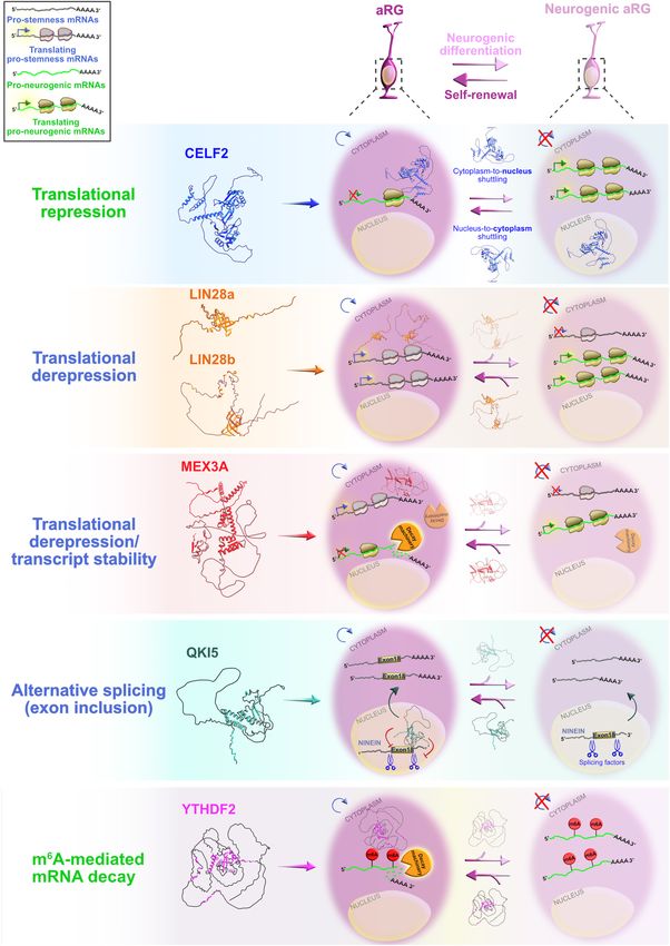
Salamon and Rasin                                                                                                          RBPs in Neocortical Development

INTRODUCTION                                                               BOX 1 | Evolutionary origin of the six-layered neocortex.
                                                                           It was previously thought that the six-layered neocortex arose from the simple,
One of the greatest innovations during the evolution of the                ancient three-layered cortices: the piriform cortex laterally and the
mammalian brain is the cerebral cortex, which has arisen                   hippocampus medially (Molnár, 2011), both of which are commonly present in
from the selective expansion of the dorsal telencephalon in                mammals and reptiles (Naumann et al., 2015). This perspective was
                                                                           challenged by another theory that the neocortex may have evolved from the
the rostral part of the forebrain (Rakic, 2009) and manifests              ancient reptilian telencephalon, the dorsal cortex of reptiles and the
area-specific lamination patterns (Cadwell et al., 2019). The              hyperpallium of birds (Glenn Northcutt and Kaas, 1995; Molnár, 2011).
neocortex (neopallium or isocortex) is considered to be the most           Tosches et al. (2018) tackled this debate by using an unbiased single-cell
recently evolved segment of the cerebral cortex (Gilardi and               sequencing approach to create the neuronal subtype taxonomy of the three
                                                                           layered-cortex of non-avian reptiles (turtles and lizards). To track the evolution
Kalebic, 2021) and is thus assigned the prefix “neo”; in Latin,
                                                                           of glutamatergic and GABAergic neurons, the reptilian transcriptomic maps
neocortex means “new bark” or “new cover” (Box 1). From                    were compared with the transcriptomes from the mammalian ancient cortex
a functional standpoint, the neocortex orchestrates complex                (hippocampus) and evolutionary new six-layered neocortex. Remarkably, the
behavioral repertoires essential to higher cognitive, motor, and           study found clear homology between the reptilian three layered-cortex and the
sensory capabilities, including abstract thinking, metacognition,          mammalian hippocampus (Tosches et al., 2018).
emotional intelligence, and verbal communication, all of which                On the other hand, the mammalian neocortex showed an intricate
                                                                           mosaicism of ancient and evolutionary new neuronal subtypes. For example,
are well-defined abilities in primates. The selective expansion            the major classes of inhibitory GABAergic neurons (e.g., parvalbumin-like,
of the neocortex stems partially from both an increase in                  somatostatin, and serotonin receptor 3A HTR3A) were detected in both
diversity and proliferative capacity of neural progenitors, which          mammals and reptiles, implying that the ancestor-descendant relationship
build an army of most, if not all, neuronal and glial cells                was preserved. In contrast, the correlation of transcription factors specifying
                                                                           glutamatergic fates between reptiles and mammals showed a higher level of
(Lui et al., 2011; Gulden and Šestan, 2014; Taverna et al.,
                                                                           divergence. Since transcription factors that dictate the acquisition of upper-
2014). As Heraclitus said: “Everything flows, and nothing abides,          and lower-layer neuronal identities in mammals mutually repress each other,
everything gives way, and nothing stays fixed,” this symphony              the authors showed clear lineage segregation in mammalian excitatory
of neocortical creation relies on the dynamic and irreversible             neurons. However, these upper- and lower-layer transcription factors were
flow of neurogenesis (Silbereis et al., 2016). Neurogenesis, in            coexpressed in neurons of the turtle three-layered cortex, resembling the
                                                                           broad mammalian neuronal types. Altogether, these findings suggest that
turn, relies on the temporal patterns of gene expression and,
                                                                           diversification of mammalian glutamatergic neurons and appearance of the six
especially, their post-transcriptional regulation. RNA-binding             neocortical layers may have evolved from the novel repressive network that
proteins (RBPs) are certainly workhorses during neurogenesis;              regulates these transcription factors (Tosches et al., 2018). Therefore, the
while they fill roles in neuronal maturation, morphology,                  neocortex appears to be an evolutionary upgrade of the reptilian three-layered
synaptic connectivity, and plasticity (Keene, 2007; Darnell,               neocortex, rather than an upgrade of the reptilian telencephalon.

2013; Glock et al., 2017; Holt et al., 2019), these topics
are outside the scope of this review. Rather, in this work,
we review the recent data that illustrate the contribution of             exhibited large transcriptional differences in human-specific
post-transcriptional regulation via RBPs in the modulation of             genes from non-human primates. This suggest that the
progenitors’ proliferation, differentiation, and specification into       aforementioned regions have undergone changes that have
neuronal and glial progeny, together with the role of RBPs in             led to divergent evolution. Even though these studies imply
neuronal migration.                                                       that differences in transcriptional signatures among primates
   The development of the neocortex starts with the process               have contributed to the structural and functional changes
of neurulation (Pritz, 2005; Dugas-Ford and Ragsdale, 2015;               that enabled the advancement of the human neocortex, the
Werner et al., 2021), during which the flat neural plate                  extent of these variations cannot be explained solely at the
undergoes major morphological transformation to form a                    transcriptional level.
closed neural tube (O’Rahilly and Müller, 2006). Even though                 Recent findings have provided insight into how gene
many histological traits of the neocortex are highly conserved            expression regulation, not only at transcriptional, but also at
across species (Krubitzer, 1995), a quantitative comparison               post-transcriptional (Bolognani and Perrone-Bizzozero, 2008;
of transcriptomes of the prefrontal portion of the neocortex              Alvarez-Castelao and Schuman, 2015; Gardiner et al., 2015;
in humans, chimpanzees, and macaques has revealed that                    Popovitchenko and Rasin, 2017; Sahoo et al., 2018; Biever et al.,
the greatest number of differentially expressed genes (DEGs)              2019; Zahr et al., 2019; Costa et al., 2021; Hoye and Silver, 2021)
are associated with the human neocortex. Thus, the human                  and epigenetic levels (Noack and Calegari, 2018), contributes to
prefrontal cortex exhibits a unique expression profile. Notably,          the evolution and function of the developing neocortex. We refer
the DEGs are mostly related to neocortical laminar specificity            the interested reader to excellent reviews that thoroughly discuss
(He et al., 2017). The divergence of the human neocortex                  the significance of transcriptional programs during neurogenesis
from non-human primates has been further characterized by                 (Tebbenkamp et al., 2014; Andrews and Nowakowski, 2019;
another transcriptional study conducted at the single nuclei              Miller et al., 2019; Molnár et al., 2019; García-Moreno and
level that compared gene expression evolution by simultaneously           Molnár, 2020; Vaid and Huttner, 2020; Oproescu et al., 2021).
examining 33 different brain regions in humans, chimpanzees,              At the post-transcriptional level, despite the fact that mRNA
macaques, and bonobos (Khrameeva et al., 2020). Only                      binding sites are less prone to genetic change than sites on
the primary and secondary cortices, limbic and association                chromatin (Payne et al., 2018), evolutionarily conserved RBPs
cortices, cerebellar white and gray matter, and hypothalamus              exhibit intricate diversification of their developmental functions.

Frontiers in Neuroscience | www.frontiersin.org                       2                                           January 2022 | Volume 15 | Article 803107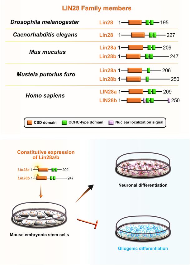
Salamon and Rasin                                                                                                    RBPs in Neocortical Development

For example, more than 1,500 RBPs have been identified in                   timed transcriptional profiles (birthmarks) correspond to the
humans (Gerstberger et al., 2014). A single RBP can potentially             lower- or upper-layer neuronal identities are in turn transmitted
bind to, on average, 22,000 30 untranslated region (30 UTR)-                from mother progenitors to daughter neurons, enabling the
binding sites (Van Nostrand et al., 2016), which translates into            specification of layer-specific subtypes. Passive mother-to-
more than 33 million predicted interactions between human                   daughter transmission of temporal birthmarks is probably
RBPs and targets 30 UTRs (Kim et al., 2021). In addition, the               exploited by the regulatory network at post-transcriptional levels
presence of highly complex regulatory interplay between the                 as transcripts already present in progenitors become translated
same or different RBPs can be competitive, cooperative or                   into proteins or stabilized in differentiating neurons for future
autoregulative in nature (Dassi, 2017), and represents another              actions (Telley et al., 2019).
evolutionary upgrade necessary to control a wide set of                        Taken together, there is a huge potential and great need
mRNA targets during different stages of cortical development                in understanding how an interaction network of RBPs acting
(genesis, migration, localization, and maturation). Moreover,               on available transcripts and their functional heterogeneity
post-transcriptional regulation ensures accurate acquisition of             participate not only in the preservation of progenitors’ fate, but
neuronal identity via delivery of right information regarding               also in the shaping and the expansion of developing neocortex.
protein subcellular localization and abundance over time. These
cell-intrinsic players, together with extrinsic factors, play an
essential role in modulating the information flow from genes                POST-TRANSCRIPTIONAL REGULATION,
to proteins, thereby participating in the diversification of cell           mRNAs POISED FOR TRANSLATION
function from a fixed numbers of genes (Halbeisen et al.,                   AND RNA-BINDING PROTEINS
2008; Ascenzi and Bony, 2017). Such coordinated regulatory
activity of intrinsic and extrinsic patterns is particularly relevant       Transcript abundance can only partially explain exact protein
to instruct the sequential flow of neocortical development                  levels (DeBoer et al., 2013; Kraushar et al., 2014, 2015;
(Kraushar et al., 2015; Yuzwa and Miller, 2017; Park et al.,                Popovitchenko et al., 2020), suggesting that the correlation
2021a,b).                                                                   between mRNA and protein levels depends highly on the state
   The wide range of post-transcriptional regulation can help               of the cell. In a scenario where an mRNA in developing cell
to explain, at least in part, the evolutionary increase in size             is in the stable condition (steady-state level), mRNA-to-protein
and complexity of the neocortex in primates, particularly                   ratios can be predictive of each other-high mRNA levels yield
humans (Figure 1) even without a significant expansion                      high protein levels (Csárdi et al., 2015). However, this is not
of the gene pool. Hence, it is important to uncover and                     always the case. For example, when a neuronal cell is exposed
understand the key regulatory RBPs guiding each step of                     to a changing condition, which is present during development
mRNA metabolism that dictates neocorticogenesis. In this                    (e.g., rapid fate and/or morphological transitions), the mRNA–
review, we explain how RBPs modulate the timely progression                 protein ratio may become perturbed. As a result, transcription
of neurogenesis from the perspective of progenitor temporal                 initiation generally may be too slow to allow the cell to confront
patterning or temporal-identity specification, a process by                 the dynamic changes with rapid and organized agility. Indeed,
which an individual progenitor changes its fate to produce                  it is misleading to rely solely on mRNA steady-state snapshots
a succession of cell types with different identities (Kohwi                 as a reliable proxy of protein abundance (Tahmasebi et al.,
and Doe, 2013). Temporal molecular patterning can further                   2019). This suggests that another type of regulatory network
be subdivided into two parallel timescales, representing one                is necessary after transcription; post-transcriptional regulation
of the two specific fates a progenitor can acquire: the                     provides a more precise, faster, and local reaction to various
general (neurogenic or neuronal) fate, and the specific-cell                developmental demands by modifying, activating, degrading,
fate (Figure 2). The acquisition of general fate represents                 or repressing the functional assortment of already present
the situation when the progenitor stops self-amplifying and                 transcripts. These mRNA processing events, commonly known
switches its fate to producing either neurogenic progenitors                as the ribonome (Mansfield and Keene, 2009), include splicing,
with restricted potency or terminally differentiated neurons.               alternative polyadenylation, editing, stabilization, temporal
The acquisition of the specific cell fate describes a scenario              silencing, targeted localization, and translation. RBPs, together
when progenitors begin to differentiate into either layer-                  with ribosomal proteins and non-coding RNAs [e.g., microRNA
specific neuronal identities or glial cell types during the                 and long non-coding RNA (lncRNA)], are thought to be the
course of neurogenesis, contributing to the layering of the                 key components of the post-transcriptional machinery since they
neocortex and neuronal and glial diversity (Kohwi and Doe,                  shape the final output of the ribonome (Kwan et al., 2012; DeBoer
2013; Oberst et al., 2019). Neuronal diversity may thus be                  et al., 2013; Doxakis, 2014; Mao et al., 2015; Pilaz and Silver, 2015;
pre-defined at the transition from progenitors to neurons,                  Kraushar et al., 2016, 2021; Ceci et al., 2021; Park et al., 2021b).
which is further supported by single-cell profiling of mouse                The array of post-transcriptional network activities indicate that
progenitors and their immediate neuronal descendants at several             dynamic control of the transcriptome is a multifaceted series
developmental time points (Telley et al., 2019). Namely, a                  of events, necessary for the careful orchestration of the cellular
temporal change in progenitor’s behavior (from proliferative,               behavior during neocortical development (Figure 3).
neurogenic to differentiative) is dictated by the sequential                    Despite the fact that RBPs have assigned significant roles
activation of timed, overlapping transcriptional waves. These               during neocortical development, it is largely unclear how each

Frontiers in Neuroscience | www.frontiersin.org                         3                                    January 2022 | Volume 15 | Article 803107
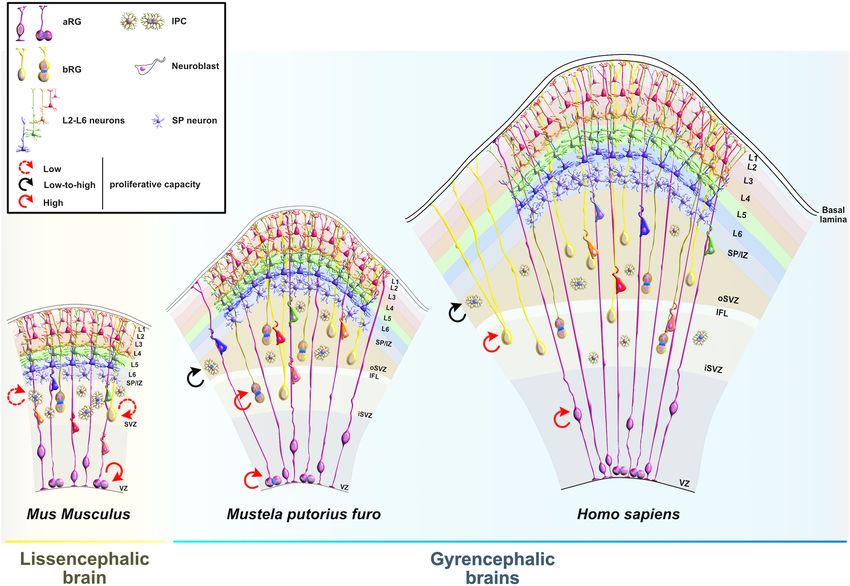
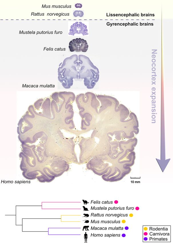
Salamon and Rasin                                                                                                                    RBPs in Neocortical Development

  FIGURE 1 | Comparative anatomy of neocortical expansion. (A) Nissl-stained coronal sections at the level of the anterior commissure from adult brains of Mus
  musculus (mouse), Rattus norvegicus (rat), Mustela putorius furo (ferret), Felis catus (cat), Macaca mulatta (macaque) and Homo sapiens (human). The arrow only
  illustrates the neocortical development (expansion) but does not encapsulate the evolutionary-scale relationship among these mammalian species. Mammals are
  grouped into lissencephalic (e.g., Mus musculus, Rattus norvegicus) and gyrencephalic species (e.g., Mustela putorius furo, Felis catus, Macaca mulatta, and Homo
  sapiens) based on cortical folding. Lissencephalic brains have small and smooth neocortices; the gyrencephalic brains have expanded and convoluted neocortices,
  with considerable variation of gyrification within and between mammalian orders. The images are scaled according to the human brain to demonstrate the notable
  differences in brain size and patterning of surface convolutions that have evolved from ferrets to humans (scale bar: 10 mm). Images of mouse, rat, cat, and rhesus
  macaque are obtained from BrainMaps next-generation atlas (Mikula et al., 2007), the ferret image was adopted from Radtke-Schuller (2018), and the human image
  was acquired from Michigan State University Human Brain Atlas (https://brains.anatomy.msu.edu/). (B) A species phylogenetic tree obtained using examples from
  (A). This simplified representation shows that the ferrets and cats (gyrencephalic cortex) are more evolutionarily distant from humans than mouse and rats
  (lissencephalic cortex).

Frontiers in Neuroscience | www.frontiersin.org                                   4                                         January 2022 | Volume 15 | Article 803107
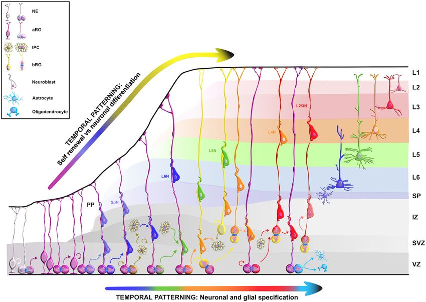
Salamon and Rasin                                                                                                                            RBPs in Neocortical Development

  FIGURE 2 | Temporal molecular patterning during neocortical development. The development of the mammalian neocortex can be conceptualized through the
  evolutionarily conserved mechanism of temporal molecular patterning. Molecular patterning of apical progenitors along two temporal branches provide an overview
  of the intrinsic processes that guide one of the two fate transitions that apical radial glia (aRG) undergo as neurogenesis proceeds: (1) “self-renewal vs. neuronal
  differentiation,” which gives rise to neuronal progeny either directly or indirectly through the generation of basal progenitors, and (2) “neuronal and glial specification,”
  which begins with the sequential production of layer-specific neuronal subtypes, and finishes with the generation of glial progeny during late stages of corticogenesis.
  NE, neuroepithelial cell; aRG, apical radial glia; IPC, intermediate progenitor cell; bRG, basal radial glia; PP, preplate; VZ, ventricular zone; SVZ, subventricular zone;
  IZ, intermediate zone; SP, subplate; SpN, subplate neuron; L1–L6, layers 1–6; L2/3N-L6N, layer 2/3 neuron-layer 6 neuron.

RBP contributes to neocortical development. The main challenge                             regulatory roles during early neurogenesis have recently become
is to identify target mRNAs of RBPs at different developmental                             elucidated (Figure 4).
stages. While spatiotemporal target identification is a challenge,
a subset of transcripts, which often encode functionally related
proteins, can be regulated at multiple levels by virtue of binding                         Embryonic Lethal, Abnormal Vision-Like
to the same RBP or cohort of RBPs, a concept called the RNA                                and CUGBP, ELAVL-Like Family
regulon hypothesis (Keene, 2007; Morris et al., 2010). This is one                         Embryonic lethal, abnormal vision-like (ELAVL) and CUGBP,
way in which regulatory RBP–mRNA interactions can activate                                 ELAVL-like family (CELF) proteins belong to evolutionarily
either general or cell-type specific developmental pathways                                conserved, yet distinct, families of RBPs that display similar
during specific stages of neurogenesis. Various RBPs are already                           domain structures containing two N-terminal RNA recognition
recognized as highly important for the protection of progenitors’                          motifs (RRMs) (RRM1 and RRM2) followed by a divergent
neurogenic potentials (Box 2), such as FMRP, Smaug2, Nanos1,                               linker domain and a third C-terminal RRM3 (Ladd et al.,
Rbfox, and polypyrimidine tract-binding protein 1 (Ptbp1).                                 2001; Figure 5). In mammals, the four members of ELAVL
Since their function during neurogenesis has been previously                               family (ELAVL1 or HuA/R, ELAVL2 or HuB, ELAVL3 or HuC,
reviewed in detail (Pilaz and Silver, 2015; Popovitchenko and                              and ELAVL4 or HuD) are abundantly present in neurons. An
Rasin, 2017; Zahr et al., 2018, 2019; Park et al., 2021b), we will                         exception is the ubiquitously expressed ELAVL1 (Mirisis and
focus on deciphering the function of RBPs whose fascinating                                Carew, 2019). By binding to the AU-rich elements in the 30 UTRs

Frontiers in Neuroscience | www.frontiersin.org                                        5                                            January 2022 | Volume 15 | Article 803107
Salamon and Rasin RBPs in Neocortical Development
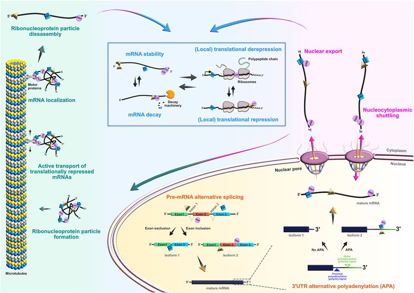
FIGURE 3 | Roles of RNA-binding proteins (RBPs) in the mRNA life-cycle. At the post-transcriptional level, RBPs actively control the entire life cycle of mRNAs in both
progenitors and their neuronal and glial progeny. Posttranscriptional processing begins in the nucleus, where RBPs regulate pre-mRNA alternative splicing, 30 UTR
alternative polyadenylation, and nuclear export of mature mRNAs. RBPs can also act as chaperones of target mRNAs, supporting their nucleocytoplasmic shuttling.
In the cytoplasm, RBPs regulate transcript localization, stability, temporal silencing, and translation, ensuring proper spatiotemporal control of protein abundance.
of their mRNA targets, ELAVL proteins play a pivotal role in example, Itai et al. (2021) have identified for the first-time that
the post-transcriptional regulatory network during neocortical heterozygous CELF2 mutations in unrelated individuals resulted
development and postnatal plasticity (Bolognani et al., 2010; in a range of overlapping clinical symptoms. These symptoms
Ince-Dunn et al., 2012; Dougherty et al., 2013; Perrone-Bizzozero, include neurodevelopmental and epileptic encephalopathy,
2013; DeBoer et al., 2014; Kraushar et al., 2014; Suhl et al., 2015; intellectual disability, and autistic behavior – all to varying
Wang et al., 2015; Dell’Orco et al., 2020; Sena et al., 2021). In severity. This suggests that CELF2, and specifically its dosage, is
human and rodents, the CELF family has six different proteins critical to normal neuronal function (Itai et al., 2021). Another
(CELF1–6) that have the capacity to shuttle between the nucleus recent study has corroborated previous findings by identifying
and cytoplasm to modulate various aspects of mRNA metabolism additional de novo heterozygous missense CELF2 mutations in
at the post-transcriptional level by binding GU-rich elements in RRM3 in patients with neurodevelopmental defects and cortical
the transcripts (Gallo and Spickett, 2010). When compared to malformations. In addition, authors observed that CELF2
other CELF members, CELF1 and CELF2 are phylogenetically exhibits bipartite compartmentalization in mouse embryonic
clustered together due to the highest level of structural topology day 15 (E15) neocortices: while cytoplasmic expression is
and an overlapping, ubiquitous expression pattern. In contrast, dominant in apical radial glia (aRG), nuclear localization
CELF3–6 have more restricted expression, primarily in the is mainly present in IPCs (IPCs) and newborn neurons
nervous system (Dasgupta and Ladd, 2012). (MacPherson et al., 2021). Using a well-designed experimental
The CELF and ELAVL families are linked to neural setup with in vivo and in vitro experiments, the findings point
development and as such the polymorphisms in CELF to a mechanism by which cytoplasmic–nuclear shuttling of
and ELAVL genes, as well as alterations in the functional CELF2 serves as a translational repression–derepression switch
properties of their respective proteins, are associated with between self-renewal and differentiation programs of aRG.
neurodevelopmental disorders (Popovitchenko et al., 2020). For Specifically, the authors revealed that cytoplasmic CELF2 binds
Frontiers in Neuroscience | www.frontiersin.org 6 January 2022 | Volume 15 | Article 803107
Salamon and Rasin                                                                                                                 RBPs in Neocortical Development

  BOX 2 | Neurogenic potential of apical progenitor cells.
                                                                                            showed that only one of two Celf1 isoforms (Celf1 short,
  It is crucial to understand the process of neuronal production and the main               Celf1S) binds the 50 UTRs of specific isoforms of the RBP
  steps of the prenatal neocortical development, known as cortical                          Elavl4 (HuD, -v3, and -v1&4) to induce translational repression
  neurogenesis. Neuroepithelial cells (NEs) are the origin of all excitatory cortical       in aRG during early stages of neurogenesis. Not surprisingly,
  neurons, astrocytes, and oligodendrocytes. NE form a single cell layer of                 Celf1 and its downstream target Elavl4 have opposite protein
  primordial cells in the apical germinative or ventricular zone (VZ). Due to their
  polarized morphology along the apico-basal axis, NE connect the ventricular
                                                                                            expression patterns in both human and mouse neocortical
  (apical) surface with the pia (basal lamina) and are linked together through the          progenitors. The expression of Celf1 radically decreases in
  adherens junction (AJ) belt in the VZ. More importantly, NE behave as neural              the aRG of VZ, and dramatically rises in the CP from
  progenitor cells (NPCs), undergoing extensive symmetric proliferative divisions           early to later stages of neurogenesis. In contrast, Elavl4 (-
  to expand the early progenitor pool (Rakic, 1995). Ultimately, their
                                                                                            v1&4) is expressed only in the post-mitotic neurons in CP
  self-amplifying capacity will enable the expansion of the neocortex in both
  lateral and radial dimensions by influencing the number of neurons generated.
                                                                                            early in development, but its presence becomes noticeable
  NE undergo interkinetic nuclear migration (INM), which is necessary for                   in VZ (-v3), and even more obvious in the IZ and CP
  optimal usage of the limited ventricular surface available for division. During           (-v1&4) at later neurodevelopmental stages (Popovitchenko
  INM, the positioning of the NE nucleus along the apico-basal axis in the VZ               et al., 2020), while corresponding mRNAs are expressed at
  corresponds to stages of the cell cycle. When the nucleus is further from
                                                                                            steady-state across stages. Another single-cell sequencing study
  (when in G1-, S-, G2-phase) or closer to (when in M phase) the ventricular
  surface, the result is a pseudostratified conformation of NE in the VZ                    also showed that Elavl4 mRNA levels are upregulated in
  (Takahashi et al., 1995; Florio and Huttner, 2014).                                       human intermediate progenitors that have a high capacity to
        During early phases of mammalian neocortical development, NE switch to              differentiate into early neurons during neurogenesis (Pollen
  asymmetric consumptive cell divisions to differentiate into another type of               et al., 2015). Silencing of Celf1 in mouse aRG, in which
  apical NPC, called the apical or ventricular radial glia cells (aRG). This event at
                                                                                            Elavl4 protein synthesis is then regularly derepressed, favored
  the early stages of development signals the beginning of the neurogenic
  phase where at least one daughter cell stops dividing by becoming a neuron,               the acquisition of upper layer neuronal identities, at the
  thereby balancing the ratio between proliferation and differentiation (Noctor             expense of lower layer neuronal subtypes, and appeared to
  et al., 2001; Shitamukai and Matsuzaki, 2012). aRG serve two main functions               impair axonal projections reaching the striatum. On the other
  during neocortical development. Firstly, as indicated in their name, glia, which          hand, Cefl1S overexpression (OE) experiments resulted in
  originates from the Greek word “glía” and translates into glue, aRG act as a
  scaffold guiding the migration of early newborn neurons from their place of
                                                                                            a reduced number of upper layer neuronal subtypes and
  birth to their destined position in the neocortex (Kriegstein et al., 2006). Just         ipsilateral atypical accumulation of axonal tracts that should
  like NE, aRG are attached to the VZ by their apical endfeets and project their            have passed the corpus callosum. Similarly, OE of either Elavl4-
  basal processes directly to the pial surface (basal lamina). Secondly, aRG also           v3 and -v4 with their 50 UTRs in mouse aRG promoted the
  express neuroepithelium properties by retaining INM capacity, even though
                                                                                            acquisition of the upper layer neuronal identities but exerted
  their proliferative potential is more restricted than NE (Uzquiano et al., 2018).
  aRG can either self-renew through a series of proliferative symmetric divisions,
                                                                                            opposing effects on the acquisition of the lower layer neuronal
  or divide asymmetrically in a proliferative and consumptive manner.                       subpopulations (Figure 6). Thus, Celf1-guided translational
  Asymmetric proliferative division generates one daughter cell that is identical           repression of Elavl4 isoforms is a key element in determining
  to its mother aRG, and another daughter cell that is either an immature                   the balanced development of upper and lower neuronal
  postmitotic neuron (direct neurogenesis), or one of the two main types of
                                                                                            identities, and also in the establishment of the proper neuronal
  more committed BPs: (1) transit-amplifying progenitors or intermediate
  progenitor cells (IPCs), or (2) outer radial glial cells or basal radial glia (bRG)       connectivity during mouse and potentially human development
  (indirect neurogenesis) (Kriegstein et al., 2006; Lui et al., 2011; Xing et al.,          (Popovitchenko et al., 2020).
  2021). As a result of this enormous accumulation of BP, the neocortex                         To explore the mechanism underlying the directed migration
  becomes even thicker and is comprised of distinct developmental regions: the              of neurons, a recent study used Caenorhabditis elegans and
  VZ, the subventricular zone (SVZ), the intermediate zone (IZ), the subplate
                                                                                            implicated etr-1, a Celf1 homolog, in the regulation of long-
  (SP), the cortical plate (CP), and the marginal zone (MZ).
                                                                                            range migration of the Q neuroblast lineage neurons (AQR
                                                                                            and PQR) in nematode larvae (Ochs et al., 2020). A forward
                                                                                            genetic approach identified a mutation in etr-1(lq61) that is
and recruits proneural factors (such as Neurog2, Neurod1, and                               responsible for the migratory defects of AQR and PQR neurons;
Tbr2) and neurodevelopmental disease-associated mRNAs                                       the etr-1(lq61) mutation is hypomorphic in nature since it
into processing bodies for translational repression, thereby                                induces the premature stop codon in the etr-1 gene. In contrast,
maintaining NPC identity and controlling the NPC fate decision                              silencing of etr-1 in C. elegans causes embryonic lethality and
(MacPherson et al., 2021). Itai et al. (2021) also noticed an                               body wall muscle defects, corroborating previous findings of
aberrant cytoplasmic accumulation of CELF2 after transfecting                               mouse neonatal lethality due to global Celf1 deletion (Kress
human HEK293T cells and African green monkey COS7 cells                                     et al., 2007; Cibois et al., 2012; Popovitchenko et al., 2020).
with plasmids containing disease-associated missense and                                    Both muscle-specific CRISPR/Cas9 genome editing, and etr-
frameshift variants. These results confirm the necessity of                                 1 expression driven only by the body-wall-muscle specific
post-transcriptional regulation, and specifically of cytoplasmic–                           promoter were able to rescue the migratory phenotype. These
nuclear shuttling activity of CELF2, for the maintenance of                                 findings showed that etr-1 influences neuronal migration in a
progenitor self-renewal properties.                                                         non-autonomous manner from body wall muscle, interacting
   On the other hand, another Celf member, Celf1, was found                                 directly or indirectly with the Wnt pathway to generate
to regulate the specification of neocortical neuronal identities                            external factors that modulate AQR and PQR migration
during neurogenesis (Popovitchenko et al., 2020). The study                                 (Ochs et al., 2020). However, the question of whether etr-1

Frontiers in Neuroscience | www.frontiersin.org                                         7                                 January 2022 | Volume 15 | Article 803107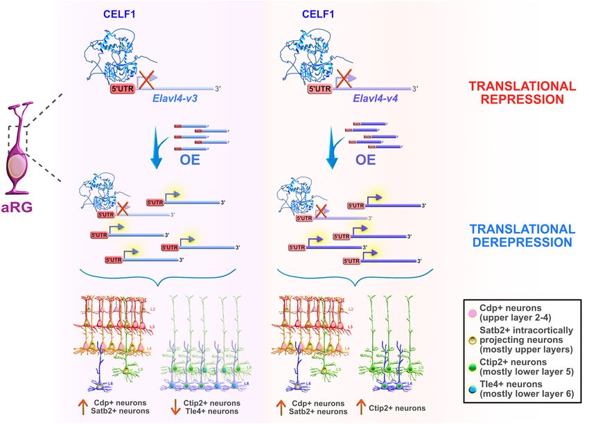
Salamon and Rasin                                                                                                                      RBPs in Neocortical Development

  FIGURE 4 | Post-transcriptional regulation by RNA-binding proteins (RBPs) and transcriptional priming in apical radial glia (aRG). Summary of the functional roles of
  RBPs CELF2, LIN28a/b, MEX3A, QKI5, and YTHDF2 in determining the fate of aRG, which are transcriptionally primed to differentiate into neurons. Cytoplasmic
  CELF2 maintains aRGs in the undifferentiated state by translationally repressing pro-neurogenic mRNAs. LIN28a/b achieves the same outcome by promoting the
  expression of pro-self-renewal transcripts. The regulatory mechanism by which MEX3A contributes to aRG maintenance and controls the appropriate time of aRG
  differentiation is unclear; MEX3A may either act as a translational repressor/derepressor of pro-neurogenic/pro-stemness mRNAs, or it can promote transcript
  stability/decay. The nuclear isoform of QKI (QKI5) controls the aRG-to-neuron transition via pre-mRNA alternative splicing (e.g., inclusion of exon 18 into Ninein
  pre-mRNA protects aRG proliferative capacity). YTHDF2 promotes N6 -methyladenosine (m6 A)-mediated decay of pro-neurogenic transcripts, acting as a pivotal
  regulator of self-renewal capabilities of aRG. The predicted tertiary and secondary full-length protein structures of RBPs in Homo sapiens are adopted from
  https://www.uniprot.org: CELF2 (O95319), LIN28a (Q9H9Z2), LIN28b (Q6ZN17), MEX3A (A1L020), QKI (Q96PU8), and YTHDF2 (Q9Y5A9).

Frontiers in Neuroscience | www.frontiersin.org                                     8                                          January 2022 | Volume 15 | Article 803107
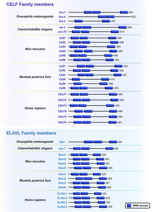
Salamon and Rasin                                                                                                                   RBPs in Neocortical Development

  FIGURE 5 | The CUGBP, ELAVL-like family (CELF) and embryonic lethal, abnormal vision-like (ELAVL) family members are closely related. Similarities and differences
  of RNA-binding domain, RNA Recognition Motif (RRM; blue) which is present in evolutionarily conserved RNA-binding proteins (RBPs) CELF and ELAVL in
  Drosophila melanogaster, Caenorhabditis elegans, Mus musculus, Mustela putorius furo, and Homo sapiens according to UniProt database
  (https://www.uniprot.org) and NCBI (https://www.ncbi.nlm.nih.gov/). The UniProtKB of NCBI accession numbers are indicated below for each member of CELF and
  ELAVL family in D. melanogaster: Bruno (bru)-1 (Q960Z4), bru-2 (Q7K108), bru-3 (Q9VU91), and Embryonic Lethal, Abnormal Vision (Elav) (P16914); C. elegans:
  ELAV-Type RNA binding-protein family (etr)-1 (G5EF03), uncoordinated (unc)-75 (G5EE68), and excretory canal abnormal (exc)-7 (Q20084); M. musculus: Celf1
  (P28659), Celf2 (Q9Z0H4), Celf3 (Q8CIN6), Celf4 (Q7TSY6), Celf5 (A0A5F8MPH2), Celf6 (Q7TN33), and Elavl1 (P70372), Elavl2 (Q60899), Elavl3 (Q60900), Elavl4
  (Q61701); M. putorius furo: Celf1 (M3XXX8), Celf2 (M3YY92), Celf3 (M3XWY8), Celf4 (M3XPL9), Celf5 (M3XX93), Celf6 (XP_004758414.1), and Elavl1 (M3Y9C6),
  Elavl2 (M3YX03), Elavl3 (M3Y100), Elavl4 (M3Y730); and H. sapiens: CELF1 (Q92879), CELF2 (O95319), CELF3 (Q5SZQ8), CELF4 (Q9BZC1), CELF5 (Q8N6W0),
  CELF6 (Q96J87), and ELAVL1 (Q15717), ELAVL2 (Q12926), ELAVL3 (Q14576), ELAVL4 (P26378).

Frontiers in Neuroscience | www.frontiersin.org                                   9                                         January 2022 | Volume 15 | Article 803107
Salamon and Rasin                                                                                                                            RBPs in Neocortical Development

  FIGURE 6 | Celf1 translationally regulates Elavl4 to dictate the development of glutamatergic neuronal subtypes. RNA-binding protein (RBP) Celf1 operates as an
  isoform-specific translational repressor of RBP Elavl4 by binding to its 50 UTRs in apical radial glia (aRG). Translational derepression of Elavl4 isoforms (Elavl4-v3 and
  Elavl4-v4) affected the production of specific neuronal laminar identities as identified using transcription factor profiling (e.g., transcription factors Cdp, Satb2, Ctip2,
  and Tle4 are associated with distinct neuronal subtypes in the neocortex). The conditional overexpression (OE) of 50 UTR Elavl4-v3 in aRG increased the number of
  upper layer Cdp-positive (+) and intracortically projecting Satb2+ neuronal subtypes, while the neuronal density of lower layer Ctip2+ and Tle4+ identities decreased.
  The conditional 50 UTR Elavl4-v4 OE in aRG positively influenced the production of both upper layer (Cdp+ and Satb2+) neuronal identities and specific subtype of
  lower layer (Ctip2+) neurons. These results highlight the importance of studying RBP–RBP interactions to decipher the mechanisms underlying the extraordinary
  diversity of neuronal and non-neuronal cell types in the developing neocortex. The predicted tertiary and secondary full-length protein structure of CELF1 (Q92879) in
  Homo sapiens is adopted from https://www.uniprot.org.

translationally regulates its mRNA targets in muscles, and                                  (Ji et al., 2009). Elavl3 appears to act as a master regulator in
if these targets are shared with mammalian Celf1 remains                                    30 UTR-alternative polyadenylation selection; indeed, silencing
to be addressed.                                                                            of Elavl3 in differentiating inhibitory neurons resulted in the
    Unlike the regulatory roles of Celf2 and Celf1/Elavl4 in neural                         preferential usage of the shorter 30 UTR options. This resulted
generation and specification of glutamatergic excitatory neurons,                           in the downregulation of neural-associated transcripts (such
Elavl3 (HuC) was recently implicated in the differentiation of                              as Tubβ3 and Gad1). Such events indicate aberrations in the
GABAergic inhibitory neurons by participating in alternative                                differentiation process. Interestingly, among all Elavl family
cleavage and polyadenylation, which strongly influence 30 UTR                               members, only Elavl4 was significantly downregulated in the
usage during embryonic neuronal differentiation (Wamsley                                    states of reduced proliferation and early stages of differentiation
et al., 2018; Grassi et al., 2019). Authors used adherent                                   (Grassi et al., 2019), supporting its increased expression at
neural stem cells (ANS) derived from mouse E14 embryonic                                    later stages during neocortical development (Popovitchenko
forebrain that can easily and efficiently differentiate toward                              et al., 2020). Since Elavl3 and Elavl4 share a high degree
an inhibitory lineage. Results confirmed previous findings that                             of sequence homology, it would be interesting to investigate
transcripts preferentially chose widespread lengthening of their                            if Elavl4 plays the same role in alternative polyadenylation-
30 UTRs when the progenitors were undergoing differentiation                                driven differentiation of glutamatergic neurons, and whether

Frontiers in Neuroscience | www.frontiersin.org                                        10                                           January 2022 | Volume 15 | Article 803107
Salamon and Rasin RBPs in Neocortical Development
Celf1 translationally represses Elavl3 in proliferating progenitors differentiation. This indicates that Lin28a/b stimulate the
during neocortical development. symmetric divisions of apical progenitors required for normal
neural tube closure, but are not necessary to trigger the
Lineage Abnormal 28 neuronal differentiation programs that arise later during
Cell lineage abnormal 28 (Lin28) is an RBP that acts as a development (Herrlinger et al., 2019). Hence, Lin28a/b
major translational reprogramming factor (Zhang et al., 2016) are fundamentally important for progenitors’ self-renewal
and has a unique pairing of two RNA-binding domains: the capacity by maintaining the threshold levels that control the
N-terminal cold shock domain (CSD) and two CCHC type transition of apical progenitors from symmetric to asymmetric
zinc finger domains, with the former resembling an RRM divisions. This is consistent with the finding that Lin28a OE
domain that exclusively binds RNA and the latter two which in mouse embryos causes excessive aRG amplification by
participate in the binding of RNA and also DNA (Figure 7A). preventing their cell-cycle exit, and concurrently affecting their
Both binding domains are highly conserved across species: conversion to IPC at the advantage of neuronal production
including worms, flies, frogs, mice, and humans (Moss and (Yang et al., 2015).
Tang, 2003; Faas et al., 2013). Vertebrates have two Lin28 Yang et al. (2015) also showed that Lin28 regulates the
paralogs that have high sequence similarity: Lin28a and Lin28b stemness of apical progenitors through the let-7 independent
(Mayr and Heinemann, 2013). Due to their unique bipartite mechanism by acting as a translational regulator of a subset
structures, Lin28 acts as a master regulator of both miRNAs of mRNAs, including Hmga2, Igf2, Igf1r, Akt1/3, and Imp1.
and mRNAs by inhibiting the biogenesis of let-7 family miRNAs These mRNA targets are mostly involved with the Igf2–
and directly modulating the translation of specific cohort of mTOR signaling pathway that drives progenitor proliferation
mRNAs (Wilbert et al., 2012; Ustianenko et al., 2018). The (Hentges et al., 2001). Other studies have directly linked Lin28a
conservation of Lin28 on-early/off-late expression profile in function with translational regulation and cell division by
the neocortex supports its indispensable role during embryonic showing that Lin28a silencing decreases the levels of its targets
development. Lin28a/b are highly abundant during early stages of Hmga2 and Igf1r, whereas Lin28a OE upregulates Hmga2
neocortical development, especially in NE and aRG, while their and Igf2 in mouse primary cultures of electroporated cortical
expression gradually decreases when neuronal differentiation neurons (Bhuiyan et al., 2013; Jang et al., 2019). Polysome
dominates over progenitor proliferation (Moss and Tang, 2003; profiling analysis of Lin28a/b KO embryonic neocortices
Yang et al., 2015). indicated that transcripts associated with translation, ribosome
Consistent with its distinct expression pattern, Lin28 was biogenesis, mTOR pathway, and cell cycle are decreased,
found to be one of the first heterochronic regulators of cell whereas transcripts involved with neuronal differentiation
fate in C. elegans larvae, in which Lin28 loss-of-function are significantly upregulated in double mutants. Mutant
causes precocious maturation of hypodermal seam cells due phenotype (macrocephaly and an abnormal number of apical
to the absence of progenitors’ symmetric divisions (Ambros progenitors) can be rescued by the ribosomal protein L24
and Horvitz, 1984). In contrast, Lin28 OE at the second larval hypomorphic allele in the background of Lin28a OE mouse
stage causes enormous proliferations due to the reiterations of line, suggesting that Lin28 mainly acts as a translational
progenitors’ symmetric divisions (Moss et al., 1997). Similarly, derepressor in the apical progenitors during early neurogenesis
Yang et al. (2015) showed that Lin28 paralogs are required for (Herrlinger et al., 2019).
the maintenance of the cell-cycle progression and mitotic entry Several lines of evidence also suggest that Lin28 may be
in mouse embryos, which are in turn necessary for the sustained involved in the regulation of temporal-identity specification.
proliferation of progenitors during neocortical development. The To gain better insight into the role that Lin28 plays in
deletion of Lin28a in mouse embryos results in the significant neurogliogenesis, during which Lin28 levels are rapidly
reduction of both aRG and IPC, as reflected in the appearance reduced, Balzer et al. (2010) constitutively expressed Lin28
of mild microcephaly (Yang et al., 2015). This suggest that in differentiating mouse embryonic carcinoma cells. The
RBP dysfunction during neocortical development can cause authors noticed that progression of neuron-to-glia cell fate
severe neurodevelopmental disorders (Kraushar et al., 2014; was severely affected, evidenced by increased neurogenesis
Mao et al., 2015). and decreased gliogenesis. This suggests that Lin28 blocks
On the other hand, Lin28b knockout (KO) embryos do not astroglial differentiation programs and preferentially promotes
exhibit any cellular or morphological phenotypes reminiscent the neuronal-lineage transition in progenitors (Balzer et al.,
of the ones observed in Lin28a KOs (Shinoda et al., 2013; 2010; Figure 7B). Another recent study in vitro confirmed
Herrlinger et al., 2019). Mouse embryos that lack one allele that Lin28 controls the neurogliogenic decision independently
of Lin28b in Lin28a KO background exhibit a more severe of the let-7 mechanism. Namely, Lin28a/b OE in mouse ESC
developmental phenotype, suggesting that Lin28a/b have both increased the Yap1 protein levels, whereas the inhibition of Yap1
essential and partially redundant functions during neocortical in Lin28a/b OE cells partially rescued the glial differentiation
development (Yang et al., 2015). Furthermore, double deletion defect. Lin28a/b directly binds and translationally regulates Yap1
of Lin28a/b in mouse embryos caused the most deleterious mRNA, which seems to be an important regulatory mechanism
morphological phenotype: neural tube defects and embryonic in controlling the cell-fate switch toward astrogliogenesis
lethality. Such developmental consequences are attributed (Luo et al., 2021). These findings show that Lin28 function in
to the reduced proliferation of NE and premature neuronal sequential progression of cell fate is conserved between C. elegans
Frontiers in Neuroscience | www.frontiersin.org 11 January 2022 | Volume 15 | Article 803107Salamon and Rasin RBPs in Neocortical Development FIGURE 7 | Role of lineage abnormal 28 (LIN28) during neocortical development. (A) Schematic presentation of structural domains of evolutionarily conserved RNA-binding protein LIN28 in Drosophila melanogaster, Caenorhabditis elegans, Mus musculus, Mustela putorius furo, and Homo sapiens as per UniProt database (https://www.uniprot.org). Different domains are represented as colored boxes, also in order from left to right: cold shock domain (CSD; orange), CCHC type zinc finger domains (green), nuclear localization signal motif (rose). The UniProtKB accession numbers are indicated below for each member of LIN28 family in D. melanogaster: cell lineage abnormal 28 (Lin28) (Q9VRN5); C. elegans: Lin28 (P92186); M. musculus: Lin28a (Q8K3Y3), Lin28b (Q45KJ6); M. putorius furo: Lin28a (M3YWA5), Lin28b (M3YDK6); and H. sapiens: LIN28a (Q9H9Z2), LIN28b (Q6ZN17). (B) Lin28 is expressed at high levels during early neocortical development. These levels rapidly decrease at later stages of neurogenesis to allow for the sequential generation of neuronal and glial fates. The constitutive expression of Lin28 in undifferentiated stem cells switches off the generation of glial cell fates while supporting the establishment of neuronal fates. Frontiers in Neuroscience | www.frontiersin.org 12 January 2022 | Volume 15 | Article 803107
Salamon and Rasin                                                                                                              RBPs in Neocortical Development

and mammals, and specifically through post-transcriptional                         in nematode embryos, but also plays a redundant role with other
control in both.                                                                   RBPs to promote mitotic proliferations of germline stem cells in
                                                                                   adult nematodes (Ariz et al., 2009; Pagano et al., 2009).
Muscle Excess 3                                                                        In the sea urchin Paracentrotus lividus, the homologous
Muscle excess 3 (Mex3) was first discovered in C. elegans where                    protein to the Mex3 is named RING finger and KH-domain
it is required for the maintenance of germline totipotency. This                   (RKHD); it is also maternally supplied and strongly expressed
RBP is characterized by two K homology (KH) domains and has                        during early zygotic development (Röttinger et al., 2006). The fact
nucleocytoplasmic shuttling ability (Draper et al., 1996; Figure 8).               that RKHD is highly recruited onto polysomes after fertilization
By binding to their targets’ 30 UTRs via conserved KH-domains,                     additionally supports its role in the regulation of mRNA
Mex3 acts as both a translational repressor and as a key regulator                 metabolism during the egg-to-embryo transition (Chassé et al.,
of the asymmetric expression of transcripts encoding critical cell                 2018). Conserved KH domains with RNA-binding capacity are
fate determinants. One such transcript is the maternally supplied                  present in four types of Mex3 orthologs in vertebrates (Mex3A–
transcript Pal-1 (CDX1 homolog) which promotes specification                       D) and are highly similar to Mex3 in nematodes, bolstering
of either hypodermal or muscle precursors during embryogenesis                     evolutionary conservation of its function between invertebrates
in worms (Edgar et al., 2001). The asymmetric distribution of                      and vertebrates (Pagano et al., 2009). Even though the RING
maternal transcripts in early blastomeres serves as a base for                     domain is not a part of the Mex3 structure in nematodes,
proper patterning of nematode embryos. An observed phenotype                       its acquisition in vertebrates is required for control of gene
in nematode embryos with mutated Mex3 was the irregular                            expression at the post-translational level through ubiquitin E3
production of body-wall muscles and hypodermal cells from                          ligase activity (Buchet-Poyau et al., 2007; Bufalieri et al., 2020).
the anterior founder cell, hence the name “muscle excess.” The                     Evolutionary diversification of the Mex3 gene from nematode to
developmental pattern characteristic for the posterior germline                    mammals is reflected in the progression of its function by which
lineage of the wild-type embryo was detected in the anterior                       Mex3 initially acts as a translational repressor in the nematode
blastomere of the Mex3 mutant embryos (Draper et al., 1996).                       lineage and progressively gains additional ubiquitin E3 ligase
Mex3 not only links cell polarity to the specification of cell fates               activity that is required for protein degradation. It is unknown,

  FIGURE 8 | Muscle excess 3 (Mex3) family of evolutionarily conserved RNA-binding proteins. Schematic presentation of binding domains K homology (KH) domain
  (red), and RING domain (yellow) in Mex3 family members in Drosophila melanogaster, Caenorhabditis elegans, Mus musculus, Mustela putorius furo, and Homo
  sapiens adopted by either UniProt database (https://www.uniprot.org) or NCBI (https://www.ncbi.nlm.nih.gov/). The UniProtKB of NCBI accession numbers are
  indicated below for members of Mex3 family in C. elegans: Mex3 (H2L067); M. musculus: Mex3a (NP_001025061.2), Mex3b (Q69Z36), Mex3c (Q05A36), Mex3d
  (Q3UE17); M. putorius furo: Mex3a (XP_012904401.1), Mex3b (XP_004763707.1), Mex3c (XP_012904006.1); and H. sapiens: MEX3A (A1L020), MEX3B (Q6ZN04)
  MEX3C (Q5U5Q3), MEX3D (Q86XN8).

Frontiers in Neuroscience | www.frontiersin.org                               13                                       January 2022 | Volume 15 | Article 803107
Salamon and Rasin RBPs in Neocortical Development
however, whether Mex3 can regulate developmental processes which differ only in their C-terminal tail: nuclear Qki5,
post-translationally by acting as E3 ubiquitin ligase. nuclear and cytoplasmic Qki6, and predominantly cytoplasmic
A Mex3A homolog was first identified as a potential regulator Qki7 (Fagg et al., 2017). The protein expression profile of
of adult neurogenesis in Nothobranchius furzeri, or killifish, two of the isoforms, Qki5 and Qki6, shows cell-type and
which is a powerful vertebrate model to study age-related subcellular localization specificity in the VZ during early
changes. In situ hybridization data showed that Mex3A has mouse neocortical development. Even though both isoforms
high expression in neurogenic niches of zebrafish embryos and are exclusively coexpressed in aRG during the earliest stages
young N. furzeri animals, which exponentially decrease with of embryonic neurogenesis, their abundance rapidly decreases
age (Baumgart et al., 2014). For the first time, the same group in IPC (Hardy et al., 1996; Wu et al., 1999; Hayakawa-Yano
revealed that Mex3A indeed plays a role in embryonic vertebrate et al., 2017). These findings imply that Qki5 and Qki6 play an
nervous system development using Xenopus laevis as a model important role in modulating the progenitor proliferative state
system. Silencing, OE, and phenotypic rescue experiments in during neurogenesis.
X. laevis showed that Mex3A disables neuronal differentiation Similarly, the evolutionary orthologs of mammalian Qki from
during neurogenesis by maintaining neural progenitors in an other species, held out wings (HOW) in Drosophila melanogaster
undifferentiated, proliferative state. The proposed mechanism of and germline defective-1 (GLD-1) in Caenorhabditis elegans,
Mex3A regulation takes place through the induction of Sox2 and play crucial roles during embryogenesis. In Drosophila embryos,
Musashi-1 expression, both of which support the self-renewal one of two known isoforms, HOW(L), transiently blocks cell-
of neural progenitors, and a simultaneous downregulation cycle progression to enable mesoderm invagination during the
of elrC (Elavl3 homolog), which is commonly used as an beginning of gastrulation. Mechanistically, this isoform promotes
early marker of neuronal differentiation. Furthermore, in situ the degradation of string/Cdc25 transcripts, known to positively
hybridization of mouse embryos at E18 showed intense Mex3A regulate the timing of highly patterned cell divisions (Nabel-
signal in the proliferative regions of the VZ and SVZ, suggesting Rosen et al., 2005). In the next stage of early mesoderm
the conserved function of Mex3A in the maintenance of development, HOW(L) downregulates the levels of various
progenitors’ stemness competence (Naef et al., 2020). Mex3A maternal mRNAs that enable uniform mesoderm spreading
seems to be an important post-transcriptional regulator during over the ectoderm, an event necessary for the acquisition
neocortical development, but the exact mechanism by which of specific mesodermal cell-fates at later stages (Toledano-
Mex3A regulates its targets remains elusive. The future studies Katchalski et al., 2007). In nematode embryos, GLD-1 levels
should clarify whether Mex3A operates as a translational are high only in the distal part of the gonads. GLD-1 represses
derepressor/repressor of pro-neurogenic transcripts/pro- the translation of maternally supplied transcripts (such as Pal-
neuronal transcripts, or if the underlying mechanism goes 1), possibly immediately after the ribosomes have loaded on
through the stabilization/degradation of its target transcripts. the mRNAs, to maintain the germ cell identity and block the
Even though the exact role of MEX3A in human prenatal propagation of maternal transcripts into early embryos. Also,
neurogenesis is yet to be uncovered, its regulation of a GLD-1 simultaneously represses translation of RBP Mex through
stemness state seems to be a recurrent topic within the its 30 UTR, supporting Mex expression and repressive function
human MEX3 family members. For example, MEX3A OE in only in the proximal part of the gonads (Mootz et al., 2004;
human gastrointestinal 2D and 3D cultures strongly represses Albarqi and Ryder, 2021).
the expression of the CDX2 intestinal transcriptional factor To better understand the developmental function of Qki
(Pereira et al., 2013), which functions as both a lineage-specific proteins, Hayakawa-Yano et al. (2017) performed transcriptomic
transcriptional enhancer of trophectoderm genes and a repressor profiling of Qki knockdown neural stem cells and revealed
of inner cell mass pluripotency genes during early embryonic that the nuclear isoform Qki5 preferentially binds introns of
development (Jedrusik et al., 2008; Huang et al., 2017). The various pre-mRNAs involved in cellular organization. Thus, Qki5
binding of MEX3A to 30 UTRs of CDX2 results in a reduction can bidirectionally control three types of alternative splicing to
of differentiation and polarity features, which might be the suppress pro-neuronal transcripts. Specifically, exon skipping
turning point that enables a permissive environment for the occurs when Qki5 binds to the 30 end of intronic regions
maintenance of stemness (Pereira et al., 2013). The BrainSpan immediately upstream of regulated exon, whereas exon inclusion
Atlas of the developing human brain (Miller et al., 2014) occurs upon binding to the 50 or 30 end intronic regions
provides a comprehensive transcriptome map across the key downstream of the alternative exon. The splicing function of
stages of human development. In BrainSpain, transcripts of Qki5 was further confirmed in the Qki conditional KO (cKO)
MEX3 homologs show the highest expression profile during mouse which displayed several cellular defects. The protein
the earliest embryonic stages and their expression gradually γ-tubulin, which is required for microtubule nucleation from
decreases toward the postnatal stages, implying that MEX3 might the centrosome, was mislocalized from the ventricular surface
indeed regulate the stemness/differentiation decision during into the VZ. The authors also noticed ectopic neurogenesis, as
human embryonic development. observed by the incorrect localization of immature neurons in
the VZ, and M-phase and S-phase aRG in the VZ and SVZ.
Quaking These results further suggested that Qki proteins regulate cell
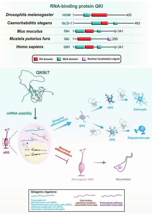
Mammalian Quaking (QKI) is another RBP with a KH-type cycle-dependent INM and inhibit neurogenesis by maintaining
domain (Figure 9A) that has three major spliced isoforms stemness-related genes in aRG. In particular, Qki5 positively
Frontiers in Neuroscience | www.frontiersin.org 14 January 2022 | Volume 15 | Article 803107You can also read