Electrochemical Biosensors in Food Safety: Challenges and Perspectives - MDPI
←
→
Page content transcription
If your browser does not render page correctly, please read the page content below
molecules
Review
Electrochemical Biosensors in Food Safety: Challenges
and Perspectives
Antonella Curulli

                                          Istituto per lo Studio dei Materiali Nanostrutturati (ISMN) CNR, Via del Castro Laurenziano 7, 00161 Roma, Italy;
                                          antonella.curulli@cnr.it

                                          Abstract: Safety and quality are key issues for the food industry. Consequently, there is growing
                                          demand to preserve the food chain and products against substances toxic, harmful to human health,
                                          such as contaminants, allergens, toxins, or pathogens. For this reason, it is mandatory to develop
                                          highly sensitive, reliable, rapid, and cost-effective sensing systems/devices, such as electrochemical
                                          sensors/biosensors. Generally, conventional techniques are limited by long analyses, expensive
                                          and complex procedures, and skilled personnel. Therefore, developing performant electrochemical
                                          biosensors can significantly support the screening of food chains and products. Here, we report some
                                          of the recent developments in this area and analyze the contributions produced by electrochemical
                                          biosensors in food screening and their challenges.

                                          Keywords: food; safety; electrochemical biosensors; bacteria; toxins; pesticides; antibiotics; contaminants

                                          1. Introduction

                                        Food safety is an important critical issue for the modern food industry. Contaminants,
                                          bacteria, toxins, etc., can enter the food chain during the production of different steps. For
Citation: Curulli, A. Electrochemical
Biosensors in Food Safety: Challenges
                                          example, they can accumulate in food during storage and/or are produced in the food by
and Perspectives. Molecules 2021, 26,
                                          reaction with chemical compounds [1].
2940. https://doi.org/10.3390/                 A preventative approach to food safety is the hazard analysis critical control point
molecules26102940                         (HACCP), which attempts to avoid the contamination of unwanted substances into the food
                                          chain [2,3]. On the other hand, some rigid guidelines are defined by the regulatory agencies,
Academic Editor: Marek Trojanowicz        such as United States Food and Drug Administration (USFDA) and the European Food
                                          Safety Authority (EFSA). These guidelines indicate the maximum levels for contaminants
Received: 29 March 2021                   in foods to preserve consumer health [4,5].
Accepted: 12 May 2021                          Food analysis is carried out at the end of the production process using conventional
Published: 15 May 2021                    techniques, such as chromatography, mass spectrometry, ultraviolet detection, or fluores-
                                          cence techniques either individually or combined with other separation techniques [6,7].
Publisher’s Note: MDPI stays neutral      These traditional approaches have several limitations. First, since the analysis is performed
with regard to jurisdictional claims in   at the end of the process, contaminated products can pass through the entire production
published maps and institutional affil-   chain or even be placed on the market before contamination is noticed. Second, these
iations.                                  analysis methods are laborious and complex, expensive, time-consuming, require large
                                          sample volumes and skilled personnel [8].
                                               In this context, biosensors can offer a possible alternative to allow the screening of food
                                          samples before the end of the production process [8]. Furthermore, biosensors also provide
Copyright: © 2021 by the author.          rapid and on-site monitoring and real-time information about the production process [9].
Licensee MDPI, Basel, Switzerland.        Among various biosensors, electrochemical biosensors have been widely used due to their
This article is an open access article    well-understood biointeraction mechanisms and detection process [10]. Electrochemical
distributed under the terms and           biosensors can represent smart detection tools for food commodities as part of an accurate,
conditions of the Creative Commons        sensitive, specific, and rapid analysis system [11,12].
Attribution (CC BY) license (https://          In this review, we consider recently developed electrochemical biosensors applied
creativecommons.org/licenses/by/
                                          for food analysis and safety. We illustrate recent advances in biosensing technologies
4.0/).

Molecules 2021, 26, 2940. https://doi.org/10.3390/molecules26102940                                          https://www.mdpi.com/journal/molecules
Molecules 2021,
Molecules 26,26,
2021, 2940
2940 2 of 63 2 of 62
In this review, we consider recently developed electrochemical biosensors applied
and evaluate their related weaknesses and drawbacks. We include some future ideas and
for food analysis and safety. We illustrate recent advances in biosensing technologies and
challenges that
evaluate their electrochemical
related weaknesses andbiosensors
drawbacks.must overcome
We include sometofuture
be new and
ideas smart
and chal- tools for
food
lengesanalysis and safety. biosensors must overcome to be new and smart tools for food
that electrochemical
analysis and safety.
2. Electrochemical Biosensors
2. Electrochemical
A biosensor Biosensors
is an analytical device used to determine the amount of a molecule in
a sample. Generally,
A biosensor it is characterized
is an analytical device usedby a bioreceptor
to determine (enzyme,
the amount of a whole-cell,
molecule in aantibody,
sample. Generally,
aptamer, it is characterized
nucleic acid) connected tobya asuitable
bioreceptor (enzyme,The
transducer. whole-cell,
specificantibody, ap- between
interaction
tamer, nucleic acid) connected to a suitable transducer. The specific interaction between
the target molecule and the biocomponent generates a physicochemical or biological signal,
the target molecule
converted and the biocomponent
into a measurable property by generates a physicochemical
the transducer. The choice orof
biological sig-
the bioreceptor and
nal, converted into a measurable property by the transducer. The choice of the bioreceptor
the transducer depends on the sample’s characteristics and the type of measurable property
and the transducer depends on the sample's characteristics and the type of measurable
being considered. The bioreceptor represents the biosensor key element, responding only
property being considered. The bioreceptor represents the biosensor key element, re-
to a particular analyte and not to the interferences eventually present in the sample under
sponding only to a particular analyte and not to the interferences eventually present in
analysis [13–15].
the sample under analysis [13–15].
2.1. Electrochemical Biosensors Classification
2.1. Electrochemical Biosensors Classification
Biosensorscan
Biosensors canbebeclassified
classified
byby type
type of recognition
of recognition element
element or type
or type of signal
of signal trans-transduc-
tion [16], as indicated in Figure 1.
duction [16], as indicated in Figure 1.
Figure 1.1.Classification
Figure Classificationof of
biosensors based
biosensors on various
based bioreceptors
on various and transducers
bioreceptors used [16].used [16].
and transducers
In
Inthis
thisreview,
review,wewe focus
focuson on
electrochemical
electrochemical biosensors. Very interesting
biosensors. and recent
Very interesting and recent
reviews have illustrated the characteristics and performances of the other
reviews have illustrated the characteristics and performances of the other biosensors biosensors with with
different transducer systems, such as optical, piezoelectric, calorimetric [1,3,17–19]
different transducer systems, such as optical, piezoelectric, calorimetric [1,3,17–19].
Among the different typologies of biosensors, electrochemical ones combine the sen-
Among the different typologies of biosensors, electrochemical ones combine the
sitivity, as indicated by low detection limits, of electrochemical transducers with the high
sensitivity,
specificity ofasbiorecognition
indicated by processes
low detection limits,devices
[10]. These of electrochemical transducers
contain a biological with the high
recognition
specificity
element, like ofthe
biorecognition
other biosensors processes
(enzymes,[10]. These antibodies,
proteins, devices contain
nucleicaacids,
biological recognition
cells, tis-
element, like the other
sues, or receptors), biosensors
reacting (enzymes,
specifically with the proteins, antibodies,
target analyte nucleican
and producing acids, cells, tissues,
electrical
or receptors),
signal related to reacting specificallyofwith
the concentration the target
the analyte. analyte and
A schematic producing an
representation electrical
of an elec- signal
Molecules 2021, 26, 2940 related to the
trochemical concentration
biosensor is shown ofin
the analyte.
Figure 2. A schematic representation of an electrochemical
3 of 63
biosensor is shown in Figure 2.
Figure 2. Scheme of a biosensor with an electrochemical transducer. Reprinted with permission
Figure 2. Scheme of a biosensor with an electrochemical transducer. Reprinted with permission
from [10] Copyright (2010) Royal Society of Chemistry (RSC).
from [10] Copyright (2010) Royal Society of Chemistry (RSC).
Electrochemical biosensors can be divided into two main categories based on the na-
ture of the biological recognition process, i.e., biocatalytic devices and affinity biosensors
[2,20,21]. Biocatalytic devices incorporate enzymes, whole cells or tissue slices that recog-
nize the target analyte and produce electroactive species.
Affinity biosensors are based on a selective binding interaction between the analyte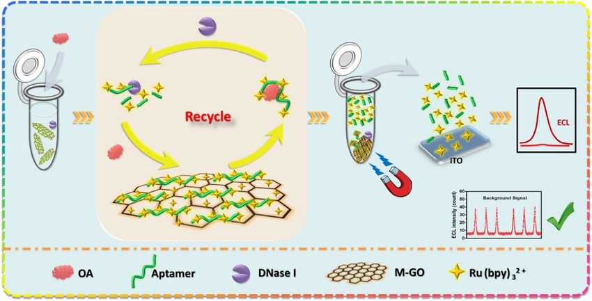
Molecules 2021, 26, 2940                                                                                            3 of 62

                                 Electrochemical biosensors can be divided into two main categories based on the
                           nature of the biological recognition process, i.e., biocatalytic devices and affinity biosen-
                           sors [2,20,21]. Biocatalytic devices incorporate enzymes, whole cells or tissue slices that
                           recognize the target analyte and produce electroactive species.
                                 Affinity biosensors are based on a selective binding interaction between the analyte
                           and a biological component, such as an antibody, nucleic acid, or receptor. Immunosensors
                           and DNA hybridization biosensors with electrochemical detection are considered examples
                           of affinity sensors.
                                 In the first case, the recognition element can be characterized by enzymes, whole-
                           cells (bacteria, fungi, cells, yeast), cells organelle and plant or animal tissue slices; the
                           catalytic sensors have the most consolidated tradition in the field of biosensors: historically,
                           glucose biosensors are the most cited examples, including a wide commercial success and
                           diffusion [20].
                                 If the recognition key is an enzyme, it is the most critical component of the biosensor
                           since it provides the selectivity for the sensor and catalyzes forming the electroactive
                           product to be detected. The electroactive product or, alternatively, the disappearance of the
                           redox-active reactant in an enzyme-catalyzed reaction can be monitored by the electrode
                           using an electrochemical technique. The activity of the immobilized enzyme depends on
                           solution parameters and electrode design.
                                 Some biocatalytic sensors can use as recognition element cellular materials (whole-
                           cells or tissue slices). These biocatalytic electrodes act similarly to the conventional enzyme
                           electrodes since enzymes present in the tissue or cell can produce or consume electrochem-
                           ically detectable species. Whole cells and tissue slices are sometimes a better source of
                           enzymatic activity than isolated enzymes as some enzymes are expensive or not commer-
                           cially available as purified enzymes. In addition, many isolated enzymes have limited
                           stability and lifetime compared to enzymes in their native environment. However, the
                           sensor response may be slower for these sensors because of a more difficult diffusion of the
                           substrate through a thick tissue material.
                                 Unfortunately, many biochemical analytes of interest are not suitable to be detected
                           by enzyme electrodes because of the lack of selectivity for the analyte or the analyte not
                           being commonly found in living/biological systems. For these reasons, affinity biosensors
                           are considered a good option.
                                 Considering the affinity-based biosensors, the biomolecule can be represented by
                           chemoreceptors, antibodies, nucleic acids, and they provide selective interactions with the
                           analyte [2,21].
                                 Affinity sensors use the selective and strong binding of biomolecules, such as anti-
                           bodies (Ab), membrane receptors, or oligonucleotides, with a target analyte to produce
                           a measurable electrical signal. The molecular recognition is mainly determined by the
                           complementary size and shape of the binding site for the analyte of interest. The high affin-
                           ity and specificity of the biomolecule for its ligand make these sensors very sensitive and
                           selective. The binding process, such as DNA hybridization or antibody–antigen (Ab–Ag)
                           complexation, is governed by thermodynamic considerations and rules.
                                 Immunosensors are Ab-based affinity biosensors where the detection of an analyte,
                           an antigen or hapten, is induced by its binding to a region/site of an Ab.
                                 The electrochemical transducer reacts to the binding event and converts the electrical
                           response to an easily handled output. Complementary regions of the Ab bind to an Ag,
                           used for producing the antibodies in a host organism with high specificity and affinity.
                           Such polyclonal Abs are heterogeneous concerning their binding domain and may be
                           refined by a selection process to yield monoclonal Abs-MAbs. All of the members of a
                           particular MAb clone are identical. Abs and MAbs can be developed for a wide range
                           of substances.
                                 Immunosensors are well-known for their extremely low detection limits. For this
                           reason, immunosensors can be used to detect trace levels (ppb, ppt) of bacteria, viruses,
                           drugs, hormones, pesticides, and numerous other chemical compounds. Examples of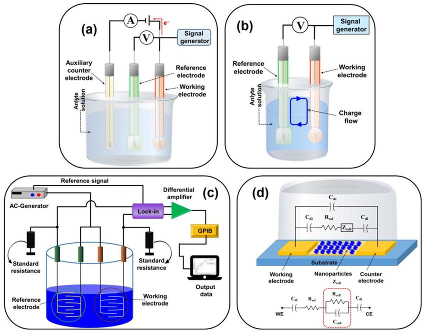
Molecules 2021, 26, 2940                                                                                                                                   4 of 62

                                   immunosensor applications, including monitoring food safety detecting toxins, bacteria
                                   allergens, contaminants, such as pesticides, endocrine disruptors, and drugs, are included
                                   in this review (see Section 3).
                                         Nucleic acids have been commonly used as the biorecognition element in affinity
                                   sensors. Biorecognition using DNA or RNA nucleic acid fragments is based on either
                                   complementary base-pairing between the sensor nucleic acid sequence and the analyte of
                                   interest or generating nucleic acid structures, known as aptamers, recognizing and binding
                                   to three-dimensional surfaces, such as those of proteins. Nucleic acids are now becoming
                                   of greater importance as the biorecognition agent in sensors since a recent rapid expansion
                                   in knowledge of their structure and how to manipulate them. The corresponding affinity
                                   probes are commonly indicated as aptasensors and nowadays are widely applied in food
                                   analysis, as shown in Section 3.
                                         As concerns the electrochemical biosensors, measurements of signals from biologi-
                                   cal samples are generally linked to an electrochemical reaction involving a bio element
                                   electrochemically active. Usually, biological reactions can generate either a change in
                                   conductance or impedance, a measurable current, or charge accumulation, measured by
                                   conductometric, potentiometric, or amperometric techniques [22,23]. Investigated reactions
                                   are normally detected near the electrode surface, and the detection techniques are generally
                                   chosen considering the electrochemical properties of the electrode surface. Electrochemical
                                   techniques involve reference, auxiliary, and working/sensing electrodes.
                                         The working/sensing electrode acts as a transduction element, whereas a counter elec-
                                   trode establishes a connection between the solution and the sensing electrode surface [23].
                                         Electrochemical techniques have been considered useful tools for food safety analysis.
                                   They are cheap, portable, easy to handle, and fast. Thus they can be preferred to the
                                   other analytical techniques. For more details about theories underlying the different
                                   electrochemical approaches used in the biosensing area, several books and reviews in the
                                   literature are well-known [22,24–29].
                                         Classified on the transduction principle and then on the corresponding electrochemical
        Molecules 2021, 26, 2940
                                   technique, the electrochemical biosensors are categorized as (a) Potentiometric,
                                                                                                           5 of 63
                                                                                                                    (b) amperometric,
                                   (c) impedimetric, (d) conductometric, and (e) voltammetric, as shown in Figure 3.

                                   Figure 3. Schematic diagram of (a) amperometric/voltammetric, (b) potentiometric, (c) conducto-
                                   Figure    3. Schematic diagram of (a) amperometric/voltammetric, (b) potentiometric, (c) conductomet-
                                   metric biosensors, and (d) impedimetric biosensor with the relative equivalent circuit [16] (Cdl =
                                   ric biosensors,
                                   double-layer       and (d)
                                                 capacitance      impedimetric
                                                               of the               biosensor
                                                                      electrodes, Rsol = resistancewith
                                                                                                    of thethe  relative
                                                                                                           solution, Cde equivalent    circuit [16] (Cdl = double-
                                                                                                                         = capacitance of
                                   the electrode,
                                   layer          Zcell = impedance
                                          capacitance                  introducedRsol
                                                           of the electrodes,      by the= bound  nanoparticles,
                                                                                           resistance             and Rcell and
                                                                                                         of the solution,    CdeCcell are
                                                                                                                                  = capacitance   of the electrode,
                                   the resistance and capacitance in parallel).
                                   Zcell = impedance introduced by the bound nanoparticles, and Rcell and Ccell are the resistance and
                                   capacitance     in parallel).
                                         Potentiometric    biosensors used ion-selective electrodes for measuring the change in
                                   potential at the surface of the working electrode upon specific analyte–bioreceptor inter-
                                   action. These biosensors are widely used for different bioanalytes, such as glucose, triglyc-
                                   erides, and pesticides. However, potentiometric transducers generally lack sensitivities
                                   when compared to amperometric transducers.
                                        Potentiometry (PM) measures the potential of a solution between two electrodes is
Molecules 2021, 26, 2940                                                                                           5 of 62

                                 Potentiometric biosensors used ion-selective electrodes for measuring the change in po-
                           tential at the surface of the working electrode upon specific analyte–bioreceptor interaction.
                           These biosensors are widely used for different bioanalytes, such as glucose, triglycerides,
                           and pesticides. However, potentiometric transducers generally lack sensitivities when
                           compared to amperometric transducers.
                                 Potentiometry (PM) measures the potential of a solution between two electrodes is
                           used in electroanalytical chemistry to measure the electrochemical potential of charged
                           species. However, a highly stable and accurate reference electrode is required, limiting
                           applying PM in bioanalysis.
                                 In an amperometric biosensor, the current produced at the working electrode through
                           the conversion of electroactive moieties is measured when a certain potential is applied
                           concerning the reference electrode. The current so produced is directly related to oxidation
                           or reduction of the analyte species after its specific interaction with the bioreceptor in
                           proportion to the concentration of target components. The amperometric biosensors are
                           relatively simple and easy to use while also offering relatively high sensitivities.
                                 Compared with the potentiometric biosensors, this method allows sensitive, fast,
                           precise, and linear response, resulting in more suitable for mass production. However,
                           poor selectivity and interferences from other electroactive substances are the disadvantages
                           of these sensors.
                                 Generally, the widest used amperometric techniques are constant potential amperom-
                           etry and chronoamperometry.
                                 Constant potential amperometry (CPA) is an electrochemical technique in which the
                           current is measured, while the potential difference between the sensing and reference
                           electrodes is held at a constant value sufficient to oxidize or reduce the analyte. This
                           potential value is generally evaluated from the CV or LSV experiment
                                 Chronoamperometry (CA) is a potentiostatic technique, where the current is recorded
                           as a time function, and it is useful to determine the concentration of the analyte once
                           its identity is known using other techniques, such as CV, chromatography and/or other
                           separation techniques.
                                 Voltammetric biosensors detect analytes by measuring the current during the con-
                           trolled variation of the applied potential. Advantages of these sensors include highly
                           sensitive measurements and possible simultaneous detection of multiple analytes.
                                 Voltammetry is an analytical technique in which the current is measured under a
                           potential sweep. In a voltammogram, the intensity of a peak is directly proportional to
                           the concentration of the analyte, while the position of the peak maximum depends on
                           the chemical species involved in the redox reactions. The type of voltammetry depends
                           on the potential control. Differential pulse voltammetry (DPV), cyclic voltammetry (CV)
                           and square wave voltammetry (SWV) are the most commonly used to detect pathogenic
                           bacteria in food. Cyclic voltammetry is also often used to characterize the surface and the
                           various functionalization steps in all types of biosensors.
                                 Conductometric biosensors quantify the change in the conductance between the pair
                           of electrodes due to an electrochemical reaction (change in conductivity properties of
                           the analyte). Conductometric and impedimetric biosensors are usually used to monitor
                           metabolic processes in living biological systems.
                                 Impedimetric biosensors measure the electrical impedance produced at the elec-
                           trode/electrolyte interface when a small sinusoidal excitation signal is applied.
                                 Electrochemical impedance spectroscopy (EIS) is an effective technique for detecting
                           the interaction between bioreceptor immobilized on an electrode surface and the analyte
                           by testing the electrode/electrolyte interface and following the change in the impedance of
                           the electrode/solution interface.
                                 In general, more comprehensive and complete information about the biosensing
                           system can be obtained from EIS when compared to that one obtained from the more
                           usual voltammetric techniques. EIS can distinguish between two or more electrochemical
                           reactions occurring simultaneously and can identify diffusion-limited reactions. The im-
Molecules 2021, 26, 2940                                                                                          6 of 62

                           pedimetric technique makes biosensors label-free, highly sensitive, and miniaturized. In
                           these biosensors, the interaction of the analyte–bioreceptor is correlated with the change
                           in impedance (Z) across the surface of the working electrode. The Z values are studied to
                           determine the change in frequency concerning time. The impedance data are generally
                           represented in the form of a Nyquist plot, in which the real component of impedance
                           (Z’) and the imaginary component of impedance (Z”) are plotted on the x- and y-axes,
                           respectively. Note that the impedance is made of two major parts, i.e., resistance (R) and
                           capacitance (C). At high frequency, the solution resistance accounts for the impedance. In
                           contrast, at low-frequency regions, the charge transfer resistance (Rct) or the resistance to
                           the flow of electrons is the source of impedance.

                           2.2. Biorecognition Keys: Bioreceptors
                                 The bioreceptors must interact specifically with the target analyte to generate a mea-
                           surable signal by the transducer. As mentioned above, when the classification of biosen-
                           sors is reported, they are enzymes, antibodies, nucleic acids, and aptamers. In addition,
                           other recognition elements, such as synthetic aptamers, DNA, proteins, and viruses, have
                           improved the selectivity of sensors for food analysis. Further, developing innovative
                           bioconjugation approaches for stable immobilization of biomolecules on the electrode
                           surface has enhanced the stability of biosensors. The introduction of nanomaterials in
                           biosensors has impacted the sensitivity of sensors with a high surface area to volume ratio
                           to strengthen the loading capacity of biomolecules relative to the biosensors assembled
                           without nanomaterials.

                           2.3. Sensing Materials and Electrodes
                                The transducer is the most important component of a biosensor because it directly
                           affects the sensor performances, such as sensitivity and response time. The chemical
                           reaction occurring in the sensing layer near the electrode surface is transformed into
                           an electrochemical signal. The rate and the quality of signal production are directly
                           related to the surface properties of the electrode, the rate of electron transfer, and mass
                           transfer. Thus, selecting electrode material highly affects the rate of electron transfer in
                           electrochemical biosensors. Various types of electrodes used in electrochemical biosensors
                           are reported below.
                                The peculiar properties of gold (e.g., biocompatibility, stability, and conductivity)
                           have promoted its use as electrodes in electrochemistry. The gold electrode sensitivity and
                           functionality can be enhanced by modifying their surface, introducing suitable molecules
                           and polymers.
                                For example, long-chain organic compounds, such as thiol, have been employed to
                           modify gold surfaces using self-assembling monolayers (SAM), which can be used as
                           anchoring/immobilizing platforms of enzymes or specific bioreceptors. Such modified
                           electrodes have been applied preferentially in several examples of biosensors.
                                In addition, gold nanomaterials were employed in the electrochemical biosensing area
                           not only for their high conductivity, their compatibility but also for their high surface to
                           volume ratio [30,31].
                                Carbon has been recognized as one of the most common electrode materials used in
                           electrochemical biosensing. The most common forms of carbon used as electrode materials
                           are carbon paste, glassy carbon, carbon nanotubes, and graphene electrodes. All these
                           carbon materials are cheaper than noble metals.
                                Carbon paste is made of graphite powder and an organic binder, immiscible with water
                           and is useful in insulating graphite from aqueous solutions. This carbon-based electrode
                           presents several advantages, such as low cost, low background current, regenerability, and
                           various operating potentials. Moreover, different compounds can be easily incorporated
                           into the carbon paste for bioanalytical applications.
                                Similarly, glassy carbon electrodes have also been employed for electrochemical
                           biosensors using ad hoc modifications. However, apart from their high cost, glassy car-
Molecules 2021, 26, 2940                                                                                             7 of 62

                           bon electrodes need an accurate pretreatment procedure, constraining their use in many
                           electrochemical applications.
                                Carbon nanotubes (CNTs) present several properties associated with their structure,
                           functionality, morphology, and flexibility and can be classified as single-walled nanotubes
                           (SWNTs), double-walled nanotubes (DWNTs), and multi-walled nanotubes (MWNTs)
                           depending on the number of graphite layers. [32] Functionalized CNTs have been used in
                           several application fields. The chemical functionalities for their application in biosensing
                           can easily be designed and tuned through tubular structure modification.
                                Graphene is one of the most applied nanomaterials in the sensing area. Different
                           graphene-based materials have been produced (e.g., electrochemically and chemically
                           modified graphene) using many procedures [33]. Graphene shows properties, such as high
                           conductivity, speeding up electron transfer, and a large surface area, very similar indeed to
                           the corresponding properties of CNTS, so it is considered a good candidate for assembling
                           sensors to determine several target molecules.
                                Graphene oxide (GO) is hydrophilic and can be dispersed in water solution because
                           of the presence of hydrophilic functional groups (OH, COOH and epoxides). On the other
                           hand, GO has a lower conductivity than graphene, so reduced GO (rGO) is more employed
                           as the electrode modifier in the electrochemical biosensing area [33].
                                Finally, nonconventional sensing platforms, such as paper and/or screen-printed
                           electrodes (SPE), frequently modified with different nanomaterials and/or nanostructures,
                           are employed in assembling electrochemical biosensors.
                                Screen-printing technology offers several advantages for assembling electrochemical
                           biosensors, including a wide range of geometries, mass production, disposability, and
                           portability. [34] These properties are very important for commercializing biosensors.
                                Recent developments in the fabrication of screen-printing electrodes (SPEs) were the
                           topic not only of numerous original research papers but also of interesting reviews, [35,36]
                           analyzing selecting support material, ink composition, and methods of surface modification
                           or functionalization. Finally, in all the reviews mentioned above, methods of obtaining well-
                           defined geometries and microelectrode arrays are discussed and compared for assembling
                           smart electrochemical biosensors.

                           3. Application of Electrochemical Biosensors in Food Analysis
                                This review focused on the electrochemical biosensors as smart analytical tools to
                           detect some of the most important bacteria, toxins, pesticides, antibiotics, and contaminants
                           in foods.

                           3.1. Toxins
                                Toxins are present in a natural environment. They are produced by microbes and
                           algae. According to their origin, toxins are commonly classified into bacterial toxins, fungal
                           toxins, and algal toxins. [37] Toxins contamination is unforeseeable and inevitable. In fact,
                           it can take place during the food production chain, including processing, transport, and
                           storage, so causing severe economic losses and public health problems. Based on the survey
                           from the World Health Organization (WHO), humans are exposed to toxins through the
                           ingestion of contaminated foods, causing severe poisoning [38,39].
                                Herein, this review investigates the state-of-art of the electrochemical biosensors to
                           detect toxins with a particular focus on several typical toxins, such as shellfish toxins, algae
                           toxins, and mycotoxins, and Table 1 summarizes the analytical characteristics of recent
                           electrochemical biosensors for toxins reported in the review.

                           3.1.1. Shellfish Toxins
                               Most shellfish toxins are small molecules, usually produced by toxic algae and accu-
                           mulated in shellfish [40].
Molecules 2021, 26, 2940                                                                                         8 of 62

                                 Wu et al. reported an overview of the different and widely used approaches in
                           biosensing for shellfish toxins detection [41], emphasizing the importance of electrochemi-
                           cal biosensors and of impedimetric ones.
                                 Herein, some interesting examples of innovative approaches to determining shellfish
                           toxins, such as saxitoxin (STX), domoic acid (DA), and okadaic acid (OA), are reported.
                                 The European Safety Authority (EFSA) [42] indicated provisional acute reference doses
                           (ARfDs) for the OA, STX, and DA toxins 0.33 mg/kg, 0.7 mg/kg and 100 mg/kg, respectively.
                                 The acute reference dose is the estimate of the amount of substance in food, normally
                           expressed on a body–weight basis (mg/kg or µg/kg of body weight), that can be ingested
                           in a period of 24 h or less without appreciable health risk to the consumer based on all
                           known facts at the time of evaluation [42].
                                 Wang and coworkers reported a label-free electrochemical aptasensor assembled with
                           nanotetrahedron and aptamer triplex for sensitive detection of saxitoxin [43].
                                 The aptamer technology, DNA nanotetrahedron, DNA triplex, and electrochemistry
                           were combined for the first time to construct a label-free electrochemical aptasensor for the
                           sensitive detection of small molecules.
                                 A typical small molecule, saxitoxin, was chosen as a model target, considering its low
                           molecular weight and high toxicity. STX is one of the major toxins of paralytic shellfish
                           poison (PSP) and can cause shock, asphyxia and even death to fisheries and humans [44].
                                 Some concepts such as aptasensors, DNA nanotetrahedron must be introduced.
                                 Aptamers are binding oligonucleotides molecules generated by systematic evolution
                           of ligands by exponential enrichment (SELEX), showing the high affinity and high selectiv-
                           ity towards their specific molecular targets. Aptamers have attracted particular attention,
                           especially in the research areas targeting small molecules, owing to aptamers’ advantages,
                           such as in vitro selection, rapid chemical synthesis, and easy chemical modification [45].
                           Many aptamers showing high affinity and selectivity vs. small molecules have been se-
                           lected, such as aptamers towards marine toxins [46], mycotoxins [47], pesticides [48], etc.
                           Various aptasensors were developed in the past decades [49]. The electrochemical aptasen-
                           sors can involve easy handling and rapid response [50,51], allowing a direct capture of the
                           molecule target. The applicability of electrochemical aptasensors towards small molecules
                           is constantly evolving and developing, and it is still under investigation [52].
                                 To overcome some limitations of electrochemical aptasensors for small molecules,
                           the aptamer and DNA triplex were combined and assembled with the nanotetrahedron
                           to form one DNA structure, followed by immobilization on the surface of screen-printed
                           electrodes [43].
                                 Nanotetrahedron (NTH), a rigid DNA nanostructure assembled by four single-stranded
                           monomers, is a spacer for the oriented immobilization of DNAs on surfaces. With the
                           assistance of nanotetrahedron, the absorption of the immobilized DNAs was eliminated,
                           and the target’s access to the immobilized DNAs was facilitated.
                                 DNA triplex is formed by a third DNA strand composed of homopurine or homopy-
                           rimidine bonded to a DNA duplex.
                                 The nanotetrahedron assisted the oriented immobilization of the aptamer triplex on
                           the surface of screen-printed electrodes, protecting the aptamer triplex from absorption and
                           assisting the aptamer to show full accessibility to STX. The developed aptasensor provided
                           high sensitivity with a LOD of 0.92 nM and showed good applicability to detect STX in real
                           seawater samples, with a recovery ranging from 94.4% to 111%, good selectivity, stability,
                           and repeatability. The authors suggested this kind of aptasensor to detect small molecules,
                           but application and validation on real food samples should be highly recommended.
                                 Nelis et al. proposed an enzyme-linked immunomagnetic electrochemical (ELIME) as-
                           say to detect domoic acid (DA) as a model target, utilizing screen-printed carbon electrodes
                           (SPCEs), modifying with carbon black (CB) [53].
                                 We remind that domoic acid (DA) is a marine toxin produced by phytoplankton
                           species, Nitzschia pungens, and the main toxic agent associated with incidents of amnesic
                           shellfish poisoning (ASP) on the east and west coasts of North America [54].
Molecules 2021, 26, 2940                                                                                          9 of 62

                                A comparison with SPCE pretreated by anodization (pre-SPCEs) and with SPCEs
                           modified with other nanomaterials, such as gold nanospheres (GNS) and gold nanostars
                           (GNST), was performed.
                                A competitive chronoamperometric immunosensor for the domoic acid (DA) was
                           assembled using the differently modified SPCEs. Hapten-functionalized magnetic beads
                           were used to avoid the individual SPCEs functionalization with antibodies. By comparison
                           among the different modified electrodes, the CB-SPCE biosensor exhibited the best electro-
                           analytical performances. DA was determined with a detection limit that is tenfold lower
                           compared to pre-SPCE (4 vs. 0.4 ng mL−1 ). These results show very good agreement with
                           HPLC data when analyzing contaminated scallops.
                                The method applied for detecting DA, using CB-SPCEs, showed great potential for
                           the antibody-based determination of small molecules in a complex matrix.
                                Another known marine biotoxin produced by various dinoflagellates is okadaic acid.
                           Chemically, OA is a polyether fatty acid derivative and exists in seafood, such as shellfish.
                           The consumption of contaminated shellfish with OA leads to diarrheic shellfish poisoning
                           (DSP), which results in the inhibition of protein phosphatase enzymes in humans.
                                Singh and coworkers have described the performances of an electrochemical microflu-
                           idic biochip to detect OA [55].
                                The screen-printed carbon electrode (SPCE) was modified by phosphorene–gold (BP–
                           Au) nanocomposite, and an aptamer specific to OA was immobilized on it.
                                BP–Au nanocomposites were synthesized by an in situ, one-step method without
                           using a reducing agent.
                                To improve the performances, a microfluidic platform was realized. The integrated
                           system consisted of a microfluidic chip housing an aptamer modified SPCE as a single
                           detection module for okadaic acid. The nanomaterials and the microfluidic channels
                           prepared were spectroscopically and electrochemically analyzed. A detection limit of 8 pM
                           and a linear range between 10 and 250 nM were obtained. Selectivity studies were also
                           performed with mussel samples in the presence of interfering species. The aptasensor did
                           not show any cross-reactivity with other types of food toxins.
                                Singh et al. developed a sensor based on a naphthalimide–gold nanocomposite to
                           detect okadaic acid [56].
                                In this work, a composite for detecting OA using a naphthalimide-based receptor
                           and gold nanoparticles were synthesized. The organic receptor was transformed into
                           nanoparticles (ONPs) via the reprecipitation method. These ONPS were then coated on
                           gold nanoparticles (Au@ONPs). The obtained composite was used to detect okadaic acid.
                           UV-visible absorption spectroscopy, fluorescence spectroscopy and cyclic voltammetry
                           techniques were used as the detection techniques, and a detection limit of 20 nM was
                           obtained from UV-vis data.
                                The developed sensor maintained its sensing ability in the pH range of 5–9 and in
                           high salt concentrations and was used for the OA detection in water samples.
                                As the most recent example of the detection of OA, we introduce an aptasensor
                           developed by Lin [57].
                                A magnetic graphene-oxide (M-GO)-assisted homogeneous electrochemiluminescence
                           (ECL) aptasensor was developed for sensitive detection of okadaic acid (OA). The aptamer
                           and Ru(bpy)3 2+ were adsorbed in M-GO to prepare the ECL probe. The principle of
                           M-GO-assisted homogeneous ECL aptasensor is illustrated in Figure 4.
                                When OA disassociated aptamer from M-GO, Ru(bpy)3 2+ was proportionally re-
                           leased from M-GO to generate the ECL signal. With the cooperation of deoxyribonuclease
                           I (DNase I), the cyclic dissociation and degradation of aptamers induced much more avail-
                           able Ru(bpy)3 2+ for signal amplification. On the other hand, the unreleased Ru(bpy)3
                           2+ were still adsorbed in M-GO and magnetically separated. Hence, the background sig-

                           nal decreased, and the sensitivity was further improved. Results showed that the ECL
                           intensity enhanced with the increasing logarithmic concentration of OA in the range of
                           0.01–10.0 ng mL−1 , and the limit of detection was 4 pg mL−1 .
high salt concentrations and was used for the OA detection in water samples.
                               As the most recent example of the detection of OA, we introduce an aptasensor de-
                           veloped by Lin [57].
                               A magnetic graphene-oxide (M-GO)-assisted homogeneous electrochemilumines-
Molecules 2021, 26, 2940   cence (ECL) aptasensor was developed for sensitive detection of okadaic acid (OA).
                                                                                                          10 ofThe
                                                                                                                62
                           aptamer and Ru(bpy)3 2+ were adsorbed in M-GO to prepare the ECL probe. The principle
                           of M-GO-assisted homogeneous ECL aptasensor is illustrated in Figure 4.

                            Figure4.4.Schematic
                           Figure     Schematicdiagram
                                                diagramofofthe
                                                            theM-GO-assisted
                                                                M-GO-assistedhomogeneous
                                                                              homogeneousECL ECL  aptasensor
                                                                                                aptasensor forfor
                                                                                                                OAOA determi-
                                                                                                                   determina-
                            nation. Reprinted with permission  from [57] Copyright 2021 Elsevier.
                           tion. Reprinted with permission from [57] Copyright 2021 Elsevier.

                                  When
                                 The      OA disassociated
                                       aptasensor   has been aptamer
                                                              used for from   M-GO, Ru(bpy)
                                                                        OA detection                was proportionally
                                                                                               3 2+sample
                                                                                        in a real                       re-
                                                                                                           of mussels and
                            leased from
                           represents     M-GO to generate
                                        a cost-effective      the ECL
                                                         approach      signal. With
                                                                   for sensitive    the cooperation
                                                                                 detection of marine of  deoxyribonuclease
                                                                                                       toxins.
                            I (DNase I), the cyclic dissociation and degradation of aptamers induced much more avail-
                           3.1.2. Mycotoxins
                            able Ru(bpy)   3 2+ for signal amplification. On the other hand, the unreleased Ru(bpy)3 2+

                            wereThe
                                  stillmost
                                        adsorbed
                                             commonin M-GO   and magnetically
                                                        and abundant             separated.
                                                                        toxins present      Hence,are
                                                                                        in nature    themycotoxins.
                                                                                                         background signal
                                                                                                                     They
                           are produced by fungi [58] and can contaminate crops and foods, inducing teratogenic,
                           mutagenic, carcinogenic, immunosuppressive, and endocrine-disrupting effects on humans
                           and animals. To ensure food safety and prevent contamination risks in the agro-food
                           sector, authorized levels for the most common mycotoxins in foods were established
                           by the European Commission [59]. Therefore, various electrochemical biosensors using
                           different analyzing techniques have been developed for mycotoxins monitoring at the
                           required concentrations.
                                 Zhang et al. [60] reviewed the newly released mycotoxin aptasensors, intending to
                           provide indications concerning practical applications and tailored design of aptasensors
                           for mycotoxins and other analytes.
                                 More recently, Kong [61] reported the recent advances of different new immunosensors
                           for mycotoxin determination over the past five years. The real application possibility, the
                           advantages, and drawbacks, together with current challenges and future perspectives of
                           these mycotoxin immunosensors, are evidenced.
                                 Among the 400 different mycotoxins identified, aflatoxins presented high toxicity and
                           carcinogenicity, and they are responsible for around 25% of animal mortality [58].
                                 You et al. [62] reviewed the recent advances in electrochemical biosensors for aflatox-
                           ins detection, emphasizing the innovative sensing strategies based on electrochemistry,
                           photoelectrochemistry, and electrochemiluminescence.
                                 In the present review, some interesting examples of novel approaches and strategies
                           to determine aflatoxins are reported and discussed.
                                 Aflatoxins are detected in corn, peanuts, cottonseeds, nuts, almonds, figs, pistachios,
                           spices, milk, and cheese and in various other food and beverages; they are stable at high
                           temperatures. Consequently, they may resist the cooking processes [58]. Four types of
                           aflatoxins were identified: AFB1, AFB2, AFG1, AFG2, plus two additional metabolites:
                           AFM1 and AFM2, being AFB1 classified as the most abundant and toxic.
                                 Among these, aflatoxin B1 (AFB1) is highly toxic, carcinogenic, mutagenic, genotoxic,
                           and immunosuppressive, and classified as a group 1 carcinogen by International Agency
                           for Research on Cancer (IARC) and a dose of more than 20 mg/kg bw (bw body weight)
                           per day was associated with acute aflatoxicosis in adults [63].
Molecules 2021, 26, 2940 11 of 62
An innovative electrochemical sensing strategy [64] was developed to detect AFB1
using aptamer (Apt)-complementary strands of aptamer (CSs) complex and exonuclease I
(Exo I). A π-shape structure is organized on the surface of the electrode. The presence of
π-shape structure as a double-layer physical barrier allowed the detection of AFB1 with
high sensitivity. In the absence of AFB1, the π-shape structure remained intact, so only
a weak peak current was recorded. Upon the addition of AFB1, the π-shape structure
collapsed, and a strong current was recorded following the addition of Exo I. Under optimal
conditions, a linear range between 7 and 500 pg mL−1 and a limit of detection of 2 pg mL−1
were observed. The developed aptasensor was also used to analyze AFB1 in spiked human
serum and grape juice samples, and the recoveries were 95.4–108.1%.
Another strategy based on a competitive immunoassay using a secondary antibody
conjugated with alkaline phosphatase enzyme as a tag was applied for the voltammetric
detection of mycotoxins, ochratoxin (OTA) and AFM1, metabolite of AFB1, using modified
gold screen-printed electrodes (AuSPEs) [65]. The biosensor was validated in red wine
and milk samples with no need for pretreatment or preconcentration of the sample. The
analytical signal was proportional to the toxin concentration in a wide linear range, showing
a good limit of detection at the ng mL−1 level.
A magnetically assembled aptasensor [66] has been designed for label-free determina-
tion of AFB1 by employing a disposable screen-printed carbon electrode (SPCE) covered
with a polydimethylsiloxane (PDMS) film as a micro electrolytic cell. The resulting label-
free aptasensor has been developed using electrochemical impedance spectroscopy as an
electroanalytical technique after the biorecognition between aptamers and the targets. The
aptasensor showed a linear range from 20 pg mL−1 to 50 ng mL−1 with a detection limit of
15 pg mL−1 and was applied to detect AFB1 in spiked samples of peanuts. This sensing
strategy seems to be a promising approach also for determining other targets.
An interesting AFB1 biosensor [67] is assembled by using a porous anodized alumina
membrane modified with graphene oxide and an aptamer of AFB1. Briefly, the aptamer
is immobilized on the surface of the porous anodized alumina nanochannels by covalent
bonding. Graphene oxide is then immobilized on the surface by π–π stacking with the
aptamer. On the addition of AFB1, graphene oxide is detached from the alumina surface
because of the specific binding between AFB1 and the aptamer, resulting in the increased
current response. The increase in current is proportional to the concentration of AB1. The
detection limit of the aptasensor is about 0.13 ngmL−1, and the linear range is from 1 to
20 ng mL−1 . Furthermore, a good selectivity towards AFB1 was observed, but applying
real food samples should be important for an effective sensor validation.
An electrochemical enzyme-linked oligonucleotide sensor for rapid detection of afla-
toxin B1 (AFB1) is developed by Marrazza and coworkers [68].
The assay is based on a competitive format and disposable screen-printed cells (SPCs).
Aflatoxin B1 conjugated with bovine serum albumin (AFB1-BSA) was immobilized by cova-
lent binding on electropolymerized poly-(aniline–anthranilic acid) copolymer (PANI–PAA).
After performing the affinity reaction between AFB1 and the biotinylated DNA-aptamer,
the solution was dropped on the modified SPCs, and the competition occurred. The biotiny-
lated complexes formed onto the sensor surface were coupled with a streptavidin–alkaline
phosphatase conjugate. 1-naphthyl phosphate was used as an enzymatic substrate, and
the electroactive product was detected by differential pulse voltammetry (DPV). A dose–
response curve was obtained between 0.1 ng mL−1 and 10 ng mL−1, and a limit of detection
of 0.086 ng mL−1 was achieved. Finally, the sensor was applied for detecting AFB1 in
maize flour samples.
Another electrochemical aptasensor achieving rapid detection of aflatoxin B1 (AFB1)
was designed and developed by Zhao [69]. A short anti-AFB1 aptamer with a methylene
blue (MB) as redox tag was immobilized on the surface of a gold electrode. Under optimized
conditions, an AFB1 dynamic concentration range from 2 nM to 4 µM was obtained. The
sensor could be well regenerated and reused. This sensor could detect AFB1 spiked in
20-fold diluted beer and 50-fold diluted white wine, respectively.Molecules 2021, 26, 2940 12 of 62
An electrochemiluminescence (ECL) platform based on a screen-printed bipolar elec-
trode (BPE) was developed by Chen et al. [70] for sensitive detection of aflatoxin B1
in cereals.
The sensor included a cathode of closed BPE as a sensing interface and an anode as a
signal collection interface. The BPE-ECL combination approach avoids the direct contact
between the reaction/sensing system and the signal measurement system. In other words,
the sensing system is physically separated from the signal measurements system. The
authors argued that in this way, it is possible to eliminate the problem of false-positive and
false-negative.
After mixing the test sample with a known concentration of horseradish peroxidase-
labeled AFB1 (HRP-AFB1), a competition for binding to monoclonal antibodies occurred.
The sensor showed a good analytical performance for AFB1 with a linear range from 0.1
to 100 ng mL−1 and a detection limit of 0.033 ng mL−1 . Different kinds of cereals (rice,
wheat, corn, sorghum, barley, and buckwheat) were selected as model grains to be tested.
The results demonstrated that the recovery rate and accuracy of this sensor are at least
comparable with those from ELISA.
A peculiar and innovative biosensor for the toxicity assessment of AFB1 and zear-
alenone (ZEN), another mycotoxin, was fabricated by Ghao et al. [71].
The International Agency for Research on Cancer (IARC) has classified ZEN as a
group 3 substance (not carcinogenic to humans) [63], and the EFSA Panel on Contaminants
in the Food Chain stated a tolerable daily intake (TDI) for ZEN of 0.25 mg/kg bw [72].
The proposed biosensor combines the advantages of both the electrochemical method
and the peculiar characteristics of bacteria (E. coli) as the biorecognition element. The
toxicity of mycotoxin AFB1 and ZEN are evaluated by the inhibition of E. coli metabolic
activity. The combined toxic effect of the two mycotoxins was investigated, and synergistic
biotoxicity was observed.
Under optimized experimental conditions, a linear concentration range of AFB1 and
ZEN in the range of 0.01–0.3 and 0.05–0.5 mg mL−1 , with the detection limits of 1 and
6 ng mL−1 , respectively.
The recovery experiments in real oil samples (peanut and corn oils) indicated that the
biosensor is applicable for the real sample mycotoxin detection.
An interesting strategy for AFB1 detection in grains [73] was based on DNA
nanotetrahedron-structured probe (DTP), and horseradish peroxidase (HRP) triggered
polyaniline (PANI) deposition. Briefly, the DNA nanotetrahedron was assembled on a gold
electrode. Its carboxylic group was conjugated with the AFB1 monoclonal antibody (mAb)
to form DTP. The test sample and a known set concentration of HRP-labeled AFB1 were
mixed, and they compete for binding to DTP. The HRP assembled on the gold electrode
catalyzed the polymerization of aniline on DTP. AFB1 in grains could be determined by
using PANI, which could be detected using the electrochemical method. The dynamic AFB1
concentration range was from 0.05 to 20 ng mL−1 . The detection limit was 0.033 ng mL−1 .
Rice, wheat, corn, sorghum, barley, and buckwheat were selected as model grains to be
tested. The results showed that the recovery rate and accuracy of this sensor are compa-
rable with those of ELISA. In fact, considering compared recovery data coming from the
proposed method and the ELISA method, it can be deduced that the relative standard
deviation ranged from −9.3% to 9.8%, which indicated there is no clear difference between
the two data set.
Layer-by-layer self-assembly technology was used to assemble an electrochemical
EIS aptasensor to detect AFB1 [74]. A multilayered sandwich structured electrode was
obtained, depositing alternately positively charge layers (modified graphene nanosheets)
and negatively charge layers (carboxylated polystyrene nanospheres). In this way, many
electrochemical active sites and high conductivity were produced. The aptamer of AFB1
was immobilized on the positively charged layer via an amide bond. The optimized
electrochemical aptasensor showed a limit of detection of 0.002 ng mL−1 and good stabilityMolecules 2021, 26, 2940 13 of 62
after 30 days. The electrochemical aptasensor was applied to detect AFB1 in oil and soy
sauce, yielding recovery values in the range of 94.5 and 103.3%.
A glassy carbon electrode (GCE) modified with a nanocomposite composed of poly-
(4-aminobenzoic acid) (PABA), graphene oxide (GO), and gold nanoparticles (AuNps)
was used for detecting AFB1 [75]. The carboxyl groups are used to bind covalently AFB1
antibodies via self-assembly of the antibody on AuNPs surface, enhancing the binding sites
for the capture probe molecule and electrochemical signal. The obtained immunosensor
showed a good linear range from 0.01 to 1 ng mL−1 and from 1 to 25 ng mL−1 , and its
detection limit is determined to be 0.001 ng mL−1 . This immunosensor also demonstrated
satisfactory reproducibility, selectivity, and stability. Moreover, the immunosensor could
detect AFB1 in vegetable oil samples.
An electrochemical sensor based on a modified gold electrode to detect aflatoxin B1
(AFB1) [76] was assembled by using a 26-mer DNA aptamer with methylene blue (MB)
label on an internal thymine (T) site (e.g., 18th T) and a thiol moiety at 50 terminal. This
sensor showed a detection limit of 6 pM and enabled detection of AFB1 in wine, milk, and
corn flour samples. This sensor can be regenerated and shows good stability.
Fusarium mycotoxins are a general term for indicating the secondary metabolites
produced by Fusarium species, and fumonisins is one the most representative family of this
kind of mycotoxins.
Approximately 15 different derivatives of fumonisins have been discovered, including
fumonisin A1 (FA1), FA2, FB1, FB2, FB3, FB4, FC1, FC2, FC3, FC4 and FP1 [77].
Fumonisin B1 (FB1) is the most toxic compound in this family, exhibiting hepato-,
nephro-, and immunotoxicity in many animal species. It is also classified as group 2B
carcinogen (possibly carcinogenic to humans) by the International Agency for Research
on Cancer [63], and the EFSA Panel on Contaminants in the Food Chain stated a tolerable
daily intake (TDI) for FB1 of 1.7 mg/kg bw [78].
Guo [79] reviewed the advances in biosensors, chemosensors, and assays based on the
classical and novel recognition elements, such as antibodies, aptamers, and molecularly
imprinted polymers. Application to food analysis, limits and time of the detection were
also analyzed and discussed.
Some interesting examples of novel approaches and strategies to determine FB1 are
reported and discussed in the following. We would like to underline that few examples of
sensors to determine FB1 include electrochemical biosensors, probably because they are lim-
ited to dedicated applications because of the instability of their bioreceptors and fabrication
difficulties. In this regard, there is still much room for improving FB1 determination.
Escarpa and his group [80] developed an electrochemical magneto immunosensor
involving magnetic beads and disposable screen-printed carbon electrodes (SPCE) for
fumonisins (FB1, FB2 and FB3). Once the immunochemical reactions took place on the
magnetic beads, they were confined on the surface of SPCE, where electrochemical detec-
tion is achieved through the addition of suitable substrate and mediator for enzymatic
tracer (Horseradish peroxidase, HRP). A detection limit of 0.33 µg L−1 , good repeatability,
reproducibility, and accuracy with a recovery rate of 85–96% was obtained. The mag-
neto immunosensor was applied to fumonisin in beer samples with a good recovery rate
of 87–105%.
Gunasekaran et al. [81] report an electrochemical immunosensing method for rapid
and sensitive detection of two mycotoxins, fumonisin B1 (FB1) and deoxynivalenol (DON).
A disposable screen-printed carbon electrode (SPCE) was used as a sensing platform.
The working electrode was modified by gold nanoparticles (AuNPs) and polypyrrole
(PPy)-electrochemically reduced graphene-oxide (ErGO) nanocomposite film. It can be
considered a suitable platform for an effective anti-toxin antibody immobilization, with
enhanced conductivity and biocompatibility.
Under optimized conditions, the limit of detection and linear range achieved for FB1
was 4.2 ppb and 0.2 to 4.5 ppm; and the corresponding values for DON were 8.6 ppb and
0.05 to 1 ppm. The immunosensor can specifically detect the two target toxins, even ifMolecules 2021, 26, 2940 14 of 62
present in the same sample. The sensor exhibited high sensitivity and low matrix interfer-
ence when tested on spiked corn samples. Hence, this electrochemical immunosensing
approach can be employed for the rapid detection of different mycotoxins present at the
same in food.
As a more recent example, we would like to introduce a sensitive and selective
electrochemical sensor using molecularly imprinted polymer nanoparticles (nanoMIPs)
for FB1 recognition [82]. It is an electrochemical sensor, not properly a biosensor, but the
detection strategy is very interesting and effective.
NanoMIPs were prepared by free-radical polymerization using the solid-phase syn-
thesis method. The sensor was assembled in two steps. First, a film of the conducting
polypyrrole–zinc porphyrin composite was deposited on a Pt electrode by electropoly-
merization. Then, nanoMIPs were covalently attached to this film. Both electrochemical
impedance spectroscopy (EIS) and differential pulse voltammetry (DPV) were used for the
sensor analytical characterization. The linear concentration range for FB1 was from 1 fM to
10 pM. The limit of detection was 0.03 and 0.7 fM, respectively. This electrochemical sensor
showed no cross-reactivity vs. other mycotoxins. The FB1 recovery considering the FB1
spiked maize analysis samples was between 96 and 102%.
The last mycotoxins family we considered is that of ochratoxins, secondary metabolites
secreted by fungi species (e.g., Aspergillus and Penicillium) during their growth. They are
present in different crops and beverages, including coffee, wine, grape juice, and dried
fruits [78]. Among them, ochratoxin A (OTA) is classified as a possible carcinogen by the
International Agency for Research on Cancer (IARC) due to its severe toxicity [60], and the
EFSA Panel on Contaminants in the Food Chain stated a tolerable daily intake (TDI) for
OTA of 0.4 mg/kg bw [83]. In addition, OTA is chemically stable, so that it is metabolized
very slowly with a half-life of more than 30 days in the body. With the recognition of
its serious threat, developing smart sensing platforms for OTA plays a crucial role in
food safety.
In a recent review [84], Wang reported an overview of the conventional and novel
methods of OTA detection. The latest research progress and related applications of novel
OTA electrochemical biosensors are mainly described with a new perspective. Furthermore,
a summary of the current limitations and future challenges in OTA analysis is included,
providing references for further research and applications.
Nevertheless, we reported and discussed some recent and interesting examples of
OTA electrochemical detection.
As a first example, a label-free electrochemical impedimetric aptasensor for rapid
detection and quantitation of OTA in cocoa beans is reported [85]. The anti-OTA aptamer
was immobilized on screen-printed carbon electrodes (SPCEs) via a diazonium-coupling
reaction. The aptasensor exhibited a limit of detection of 0.15 ng/mL, showed good
selectivity and reproducibility. The increase in electron transfer resistance was linearly
proportional to the OTA concentration in the range 0.15–2.5 ng mL−1 , with a recovery
percentage of 91–95%, obtained in cocoa samples. The analysis can be performed on-site
employing a portable EIS setup.
Another impedimetric aptasensor able to directly detect OTA without any amplifica-
tion procedure has always been developed by Marty and his group [86]. This aptasensor
was assembled by coating the surface of a gold electrode with a film of polypyrrole (PPy),
modified with covalently bound polyamidoamine dendrimers of the fourth generation
(PAMAM G4). Finally, DNA aptamers binding, specifically OTA, were covalently bound to
the PAMAM G4. The OTA detection was performed using electrochemical impedance spec-
troscopy (EIS), and the results indicated that the presence of OTA led to the modification of
the electrical properties of the PPy film due to the aptamers’ conformational changes after
the OTA-specific binding. The aptasensor had a dynamic range of up to 5 mg L−1 of OTA
and a detection limit of 2 ng L−1 of OTA, which is below the OTA concentration authorized
in food by the European legislation. The efficient detection of OTA by this electrochemicalYou can also read