Episiectomy and partial vaginectomy with urethroplasty for excision of vulvar mast cell tumour in a female dog
←
→
Page content transcription
If your browser does not render page correctly, please read the page content below
Case Report                                                                      Veterinarni Medicina, 66, 2021

                                                                     https://doi.org/10.17221/94/2020-VETMED

Episiectomy and partial vaginectomy
with urethroplasty for excision of vulvar
mast cell tumour in a female dog
Daniele Lira dos Santos1*, Jaese Farias Chaves1, Marcella Pinheiro
Souto1, Geyse Maria Machado Lima1, Julyanne de Sousa Siqueira2,
Simon Silva de Sousa1, Gabriele Sousa Silva1, Marcio Alan Oliveira
Moura2, Priscila dos Santos Ribas1, Katiane Schwanke1, Pedro Soares
Bezerra Junior2, Pedro Paulo Maia Teixeira1
1
    Veterinary Hospital, Federal University of Pará, Castanhal – Pará, Brazil
2
    Pathology Animal Lab, Federal University of Pará, Castanhal – Pará, Brazil
*Corresponding author: danielelira.ta@gmail.com

Citation: Santos DL, Chaves JF, Souto MP, Lima GMM, Siqueira JS, Sousa SS, Silva GS, Moura MAO, Ribas PS, Schwanke K,
Bezerra Junior PS, Teixeira PPM (2021): Episiectomy and partial vaginectomy with urethroplasty for excision of vulvar
mast cell tumour in a female dog. Vet Med-Czech 66.

Abstract: This case report describes the surgical treatment of a grade II mast cell tumour in the vulvar region
of a 4-year-old female Miniature Pinscher dog. The patient weighed 2 kg, and the tumour measured 2 cm in diameter.
The surgery involved an episiotomy, a partial vaginectomy, and a subsequent urethroplasty. Due to the patient’s
small size, the surgical margins were set at 2 cm laterally and 2 cm deep, which meant that the entire vulva, as well
as the ventral part of the vaginal canal had to be removed. Afterwards, the urethral ostium was elliptically fixed
to the skin at the end of the urethroplasty. The follow-up evaluations revealed an excellent recovery without
metastasis or recurrence over the following two years.

Keywords: canine; neoplasia; reconstructive surgery; surgical margin

   As the life expectancy of animals has increased,           The common clinical presentation is a single,
it has resulted in an increased rate of neoplasia,          alopecic, and non-ulcerated tumour; however, pa-
known as a major cause of death in dogs (Gonzalez-          tients usually do not present systemic clinical signs
Chavez et al. 2015). The most common cutane-                (Takeuchi et al. 2013; Miller et al. 2016).
ous malignant neoplasm is the mast cell tumour,               It is diagnosed through a fine-needle aspiration
which is characterised by the neoplastic prolifera-         (FNA) cytology and biopsy, and it can be graded
tion of mastocytes. It accounts for approximate-            through the histopathology into different grades
ly 7–21% of the skin tumours in canine species              that attempt to predict the malignancy and be-
(Arendt et al. 2015). Although the aetiology is un-         haviour.
known, it is recognised that the Bulldog breed, ad-           There are two recognised grading systems for
vanced age (Pratschke et al. 2013; Silva et al. 2014),      mast cell tumours; one, as defined by Patnaik et al.
receptor tyrosine kinase (c-KIT) defects and muta-          (1984), who classified the tumour into grades I, II,
tions (Takeuchi et al. 2013), and genetic mutations         and III; and another, more recently by Kiupel et al.
(Arendt et al. 2015) are possible predisposing fac-         (2011) and Silva et al. (2014), who classified the
tors for this neoplasm (Pratschke et al. 2013).             tumour into high and low grades.

Supported by Federal University of Pará, Brazil (PROAP UFPA/UFRA Reproamazon 2020).

1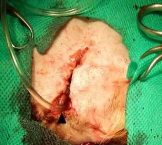
Case Report                                                                     Veterinarni Medicina, 66, 2021

                                                                     https://doi.org/10.17221/94/2020-VETMED

  A surgical excision is the treatment of choice,              A fine-needle aspiration (FNA) was performed
often times combined with radiotherapy, chemo-              to collect samples of the vulvar mass and the pop-
therapy, and or electrochemotherapy. Any adjuvant           liteal lymph nodes, which were sent to the Animal
treatment will depend on the grade of the tumour,           Pathology Laboratory of UFPA for a cytological ex-
whether or not the surgical margins are tumour free         amination. The material collected from the patient’s
on the histopathological examination, and the pres-         vulva consisted of individualised round cells with
ence or absence of a metastatic disease. In oncologi-       moderate anisocytosis and an abundant cytoplasm
cal surgery, the broad margins are typically made           containing granules and nuclei with moderate an-
laterally and deeply, which can cause extensive sur-        isokaryosis and evident nucleoli. The cytopatho-
gical wounds and even lead to the total or partial          logical findings were compatible with a mast cell
amputations of the affected organs (Spugnini et al.         tumour diagnosis. The lymph node material pre-
2011; Warland et al. 2015).                                 sented no evidence of metastasis. The blood count
  Consequently, reconstructive surgery in veteri-           and biochemical tests were unremarkable.
nary medicine has been utilised to repair these                Subsequently, the animal was scheduled for the
wounds (Pratschke et al. 2013), and has become im-          surgical removal of the vulvar nodule. The pre-
portant as it improves the function and aesthetics,         anaesthetic medication included promethazine
and increases the animal survival. Here, we report          hydrochloride 0.2 mg/kg, i.v. (Pamergan; Cristália,
a case of vulvar mast cell tumour removal associ-           São Paulo, Brazil); tramadol hydrochloride 4 mg/kg,
ated with reconstructive surgery.                           i.v.; (Tramadon; Cristália, São Paulo, Brazil), melox-
                                                            icam 0.2 mg/kg, i.v. (Maxican 2%; Ourofino, São
                                                            Paulo, Brazil), and amoxicillin trihydrate 20 mg/kg,
Case report                                                 i.v. (Agemoxi; Agener União, São Paulo, Brazil).
                                                            Propofol 4 mg/kg (Provive 1%; União Química, São
  The patient, a 4-year-old sterilised female Min-          Paulo, Brazil) was used for the induction and isoflu-
iature Pinscher dog, weighing 2 kg, was presented           rane (Isoforine; Cristália, São Paulo, Brazil) 1.5 V%
to the Veterinary Hospital of the Federal University        for the maintenance. The urethra was catheterised
of Pará (UFPA). The owners reported the presence            with a number 6 PVC urethral catheter.
of a firm, round nodule measuring approximately                Subsequently, a triangular incision was made
2 cm in the vulvar region, progressing in size rath-        around the vulva (Figure 2); the relevant tissue was
er quickly over approximately 1 month (Figure 1).           dissected and a monopolar diathermy was used for
On physical examination, the animal was alert,              the tissue haemostasis. The area affected by the
had a good nutritional status and the heart rate,
respiratory rate, and temperature were within                Figure 2
the physiological range for the species; however,
a palpation revealed bilaterally enlarged popliteal
lymph nodes.

 Figure 1

Figure 1. Perivulvar cutaneous mast cell tumour in a dog.
The nodule well delineated and not ulcerated, measuring     Figure 2. Transoperative procedure – triangular excision
approximately 2 cm (black arrow)                            around the vulva (dashed triangle)

2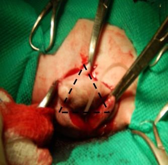
Case Report Veterinarni Medicina, 66, 2021
https://doi.org/10.17221/94/2020-VETMED
Figure 3 Figure 4
Figure 4. Immediate post-procedure aspect of the episi-
Figure 3. Intraoperative procedure – removal of the otomy and partial vaginectomy with the urethroplasty
vulva and the vaginal canal and fixation of the urethra for the excision of the vulvar mast cell tumour. The ure-
to the skin thral canal with a catheter (black arrowhead)
tumour was removed with a minimum surgical nucleoli, with two to three mitotic figures in ten
margin of approximately 2 cm. high-power fields. Additionally, there were ar-
This involved removing the entire vulva and eas of necrosis and occasional eosinophils among
part of the vaginal canal by following the tech- the neoplastic cells. The neoplasm was classified
nique of Salomon et al. (2004) and a technique as a grade II or low-grade mast cell tumour accord-
adapted from Nelissen and White (2012) (Figure 3). ing to Patnaik et al. (1984) and Kiupel et al. (2011),
To obtain the stipulated 2 cm deep margin, a vagi-
nectomy was carried out by using a vestibular ap- Figure 5
proach through access from an episiotomy. The
caudal part of the vagina was dissected and separated
from the vestibule at the vestibulovaginal junction,
just cranial to the urethral tubercle. The muscles
were dissected from the vestibule, and the remnant
tissue was apposed with a continuous suture pattern
to obliterate the dead space. The transected urethra
was pulled caudally and then sutured to the distal
vestibule with a nylon suture material, using an in-
terrupted pattern.
The subcutaneous space was reduced, and the
urethral ostium was elliptically fixed to the skin
at the end of the urethroplasty (Figure 4).
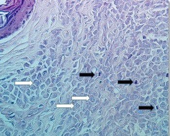
The tumour sample was sent for a histopatho- Figure 5. Vulvar cutaneous mast cell tumour in a dog.
logical examination, and the results showed that The neoplasm with round cells arranged in strands
it consisted of round cells arranged in strands or solid islands with a small amount of stroma. The cells
or solid islands with a small amount of fibrous stro- present moderate anisocytosis, abundant basophilic
ma extending from the superficial to deep dermis and granular cytoplasm, and round or oval nuclei with
(Figure 5). The neoplastic cells showed an abun- moderate anisokaryosis, loose chromatin, and evident
dant basophilic cytoplasm, metachromatic granules nucleoli (white arrows). Occasional eosinophils (black
stained in toluidine blue, and nuclei with moder- arrows) are visualised among the neoplastic cells.
ate anisokaryosis, loose chromatin, and evident Haematoxylin&Eosin (H&E), × 40 magnification
3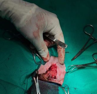
Case Report                                                                     Veterinarni Medicina, 66, 2021

                                                                     https://doi.org/10.17221/94/2020-VETMED

Figure 6                                                     Figure 7

Figure 6. Healing progress of the surgical wound 10 days
after the vulvar mast cell tumour removal

respectively. The patient was given a guarded prog-
nosis based on the neoplasm morphology, and had             Figure 7. Follow-up 8 months after surgery to remove the
a low risk for tumour recurrence and metastasis.            vulvar mast cell tumour
  The dog recovered well in the postoperative
period, and the following drugs were prescribed:              Miniature Pinschers are not mentioned in the lit-
amoxicillin + potassium clavulanate (Agemoxi                erature among the breeds highly predisposed to cu-
CL; Agener União, São Paulo, Brazil; 20 mg/kg               taneous mast cell tumours. The more predisposed
b.i.d.) v.o., for 10 days, ketoprofen (Ketojet; Agener      breeds are the Boxer, English Setter, Dachshund,
União, São Paulo, Brazil; 1 mg/kg s.i.d.) v.o., for         Weimaraner, Boston Terrier, Bullmastiff, and Golden
3 days, dipyrone sodium (Dipirona gotas; Biovet,            Retriever, which may have genetic components in-
São Paulo, Brazil; 25 mg/kg t.i.d.) v.o. for 3 days,        volved (Dobson 2013; Arendt et al. 2015; Smiech
tramadol hydrochloride (Tramadon; Cristália, São            et al. 2017). In addition to the genetic assumptions,
Paulo, Brazil; 4 mg/kg t.i.d.) v.o., for 7 days, and pro-   cutaneous mast cell tumours can be associated with
methazine hydrochloride (Pamergan; Cristália, São           chronic dermatitis or the use of or exposure to skin-
Paulo, Brazil; 0.2 mg/kg b.i.d.) v.o., for 3 days. The      irritating substances, which can also affect other
patient returned after 5 days for the evaluation and        breeds and even mixed-breeds (Gonzalez-Chavez
removal of the urethral catheter, and after 5 more          et al. 2015; Miller et al. 2016).
days for the removal of the stitches (Figure 6).              Mast cell tumours located in the mucocutaneous
Subsequently, the patient was seen for a follow-up          and perineal regions tend to be more aggressive
at the clinic after 40 days, and again 8 months after       (Warland et al. 2015). In this case, the nodule lo-
the surgery (Figure 7), and more than 2 years after         cated in the vulvar region was classified as grade II
the event, no tumour recurrence was noted.                  according to Patnaik et al. (1984), and as low-
                                                            grade according to Kiupel et al. (2011) (Silva et al.
                                                            2014). Although tumours with this classification
DISCUSSION                                                  are not the most aggressive ones, they have a meta-
                                                            static potential of between 5–22%, mainly involving
  Genitourinary tract reconstruction surgeries are          the regional lymph nodes (Pratschke et al. 2013;
necessary in the treatment of tumours in the vulvar         Silva et al. 2014). For the metastasis assessment,
region, each presenting particularities depending           especially in the regional lymph nodes, the FNA cy-
on the location and type of the neoplasia (Nelissen         tology technique has shown high sensitivity (Mutz
and White 2012).                                            et al. 2017). The FNA samples collected from the

4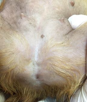
Case Report                                                                      Veterinarni Medicina, 66, 2021

                                                                    https://doi.org/10.17221/94/2020-VETMED

popliteal lymph nodes, in our case, did not show           Conflict of interest
any metastasis during the clinical follow-up.
  The lateral excision margin recommended for the            The authors declare no conflict of interest.
treatment of a grade II mast cell tumour is 3 cm,
and it must be deep enough to reach the deep fas-
cial plane (Pratschke et al. 2013). The European           REFERENCES
consensus on mast cell tumour removal in dogs
and cats recommends margins of 3 cm for mast               Arendt ML, Melin M, Tonomura N, Koltookian M, Courtay-
cell tumour grades III and 2–3 cm for grades II              -Cahen C, Flindall N, Bass J, Boerkamp K, Megquir K,
(Blackwood et al. 2012). However, it has already             Youell L, Murphy S, McCarthy C, London C, Rutteman
been described that the surgical margins differ              GR, Starkey M, Lindblad-Toh K. Genome-wide associa-
from the histological margins in mast cell tumour            tion study of Golden Retrievers identifies germ-line risk
samples (Risselada et al. 2015). Regardless of the           factors predisposing to mast cell tumours. PLoS Genet.
small size of the patient in this case, the margins          2015 Nov 20;11(11):e1005647.
were sufficient.                                           Blackwood L, Murphy S, Buracco P, De Vos JP, De Fornel-
  The combined abdominal and vestibular ap-                  Thibaud P, Hirschberger J, Kessler M, Pastor J, Ponce F,
proach for the subtotal vaginectomy allowed a com-           Savary-Bataille K, Argyle DJ. European consensus docu-
plete resection of the extensive vaginal lesions, was        ment on mast cell tumours in dogs and cats. Vet Comp
not associated with any major complications, and             Oncol. 2012 Sep;10(3):e1-e29.
the outcome was favourable (Nelissen and White             Dobson JM. Breed-predispositions to cancer in pedigree
2012). Antihistamines were used in the pre- and              dogs. ISRN Vet Sci. 2013 Jan 17;2013:941275.
post-operative period, as the manipulation of the          Gonzalez-Chavez MT, Gonzalez BP, Rodriguez YF, Aur-
tumour causes the mast cells to release inflamma-            recochea JCR, Seoane LC, Alvarez MM, Montalvo YZ,
tory mediators such as histamines, proteases and             Garcia JLR, Romero JAM, Socarras CC. Frecuencia de
vasoactive amines. These can act locally to cause            presentacion de neoplasias en caninos del municipio del
itching and swelling, resulting in delayed healing           Padron, La Habana, Cuba [Frequency of neoplasm pre-
(Warland et al. 2015).                                       sentation in canines of the municipality of San Miguel
  However, recent studies have shown that mast               del Padron, Havana, Cuba]. Rev Salud Anim. 2015;37(1):
cell vasodilation is mediated by two receptors, H1           39-46.
and H 2, in addition to a prostaglandin called D2,         Kiupel M, Webster JD, Bailey KL, Best S, DeLay J, Detrisac
which is also released by canine mast cells. The             CJ, Fitzgerald SD, Gamble D, Ginn PE, Goldschmidt MH,
used antihistamines act only on the H1 receptor,             Hendrick MJ, Howerth EW, Janovitz EB, Langohr I, Lenz
leaving their action ineffective and/or unsatisfac-          SD, Lipscomb TP, Miller MA, Misdorp W, Moroff S, Mul-
tory in dogs (Sanchez et al. 2017). However, in this         laney TP, Neyens I, O’Toole D, Ramos-Vara J, Scase TJ,
case, it occurred satisfactorily, with positive results.     Schulman FY, Sledge D, Smedley RC, Smith K, W Sny-
  We conclude that oncological surgeries of the              der P, Southorn E, Stedman NL, Steficek BA, Stromberg
genitourinary tract can result in the complete               PC, Valli VE, Weisbrode SE, Yager J, Heller J, Miller R.
surgical removal, specifically in the case of a mast         Proposal of a 2-tier histologic grading system for canine
cell tumour, and especially in cases of small pa-            cutaneous mast cell tumors to more accurately predict
tients. However, in cases where it is difficult to at-       biological behavior. Vet Pathol. 2011 Jan;48(1):147-55.
tain a margin, our technique has proved effective          Miller RL, Van Lelyveld S, Warland J, Dobson JM, Foale RD.
as a treatment, while importantly managing to pre-           A retrospective review of treatment and response of high-
serve the urinary function, so this case can serve           risk mast cell tumours in dogs. Vet Comp Oncol. 2016 Dec;
as a guide for future similar cases.                         14(4):361-70.
                                                           Mutz ML, Boudreaux BB, Royal A, Merchant S, Pucheu-
                                                             Haston C, Griffith EH, Gieger TL. Cytologic comparison
Acknowledgement                                              of the percentage of mast cells in lymph node aspirate
                                                             samples from clinically normal dogs versus dogs with
  The authors would like to thank the UFPA                   allergic dermatologic disease and dogs with cutaneous
Veterinary Hospital team for all their cooperation           mast cell tumors. J Am Vet Med Assoc. 2017 Aug 15;251(4):
in solving the case.                                         421-8.

5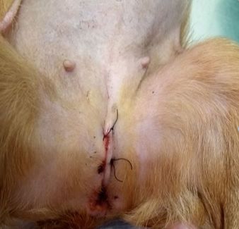
Case Report                                                                          Veterinarni Medicina, 66, 2021

                                                                        https://doi.org/10.17221/94/2020-VETMED

Nelissen P, White RA. Subtotal vaginectomy for manage-         Silva ALDA, Queiroz RP, Szabo MPJ, Medeiros AA. Degree
  ment of extensive vaginal disease in 11 dogs. Vet Surg.        of malignancy of the canine cutaneous mast cell tumor
  2012 May;41(4):495-500.                                        as for location according to the classifications of Patnaik
Patnaik AK, Ehler WJ, MacEwen EG. Canine cutaneous               et al. (1984) and Kiupel et al. (2011). Rev Bras de Ciência
  mast cell tumor: Morphologic grading and survival time         Veterinária. 2014;21(3):183-7.
  in 83 dogs. Vet Pathol. 1984 Sep;21(5):469-74.               Smiech A, Slaska B, Lopuszynski W, Jasik A, Szczepanik M,
Pratschke KM, Atherton MJ, Sillito JA, Lamm CG. Evalu-           Wilkolek P. Epidemiological study of canine mast cell
  ation of a modified proportional margins approach for          tumours according to the histological malignancy grade.
  surgical resection of mast cell tumors in dogs: 40 cases       Pol J Vet Sci. 2017 Sep 26;20(3):455-65.
  (2008–2012). J Am Vet Med Assoc. 2013 Nov 15;243(10):        Spugnini EP, Vincenzi B, Citro G, Dotsinsky I, Mudrov T,
  1436-41.                                                       Baldi A. Evaluation of Cisplatin as an electrochemother-
Risselada M, Mathews KG, Griffith E. Surgically planned          apy agent for the treatment of incompletely excised mast
  versus histologically measured lateral tumor margins for       cell tumors in dogs. J Vet Intern Med. 2011 Mar-Apr;25(2):
  resection of cutaneous and subcutaneous mast cell tu-          407-11.
  mors in dogs: 46 cases (2010–2013). J Am Vet Med Assoc.      Warland J, Brioschi V, Owen L, Dobson J. Canine mast cell
  2015 Jul 15;247(2):184-9.                                      tumours: Decision-making and treatment. In Pract. 2015;
Salomon JF, Deneuche A, Viguier E. Vaginectomy and ure-          37(7):315-32.
  throplasty as a treatment for non-pedunculated vaginal       Takeuchi Y, Fujino Y, Watanabe M, Takahashi M, Naka-
  tumours in four bitches. J Small Anim Pract. 2004 Mar;         gawa T, Takeuchi A, Bonkobara M, Kobayashi T, Ohno K,
  45(3):157-61.                                                  Uchida K, Asano K, Nishimura R, Nakayama H, Sugano S,
Sanchez A, Valverde A, Sinclair M, Mosley C, Singh A,            Ohashi Y, Tsujimoto H. Validation of the prognostic value
  Mutsaers AJ, Hanna B, Johnson R, Gu Y, Beaudoin-Kim-           of histopathological grading or c-kit mutation in canine
  ble M. Antihistaminic and cardiorespiratory effects of di-     cutaneous mast cell tumours: A retrospective cohort
  phenhydramine hydrochloride in anesthetized dogs               study. Vet J. 2013 Jun;196(3):492-8.
  undergoing excision of mast cell tumors. J Am Vet Med
  Assoc. 2017 Oct 1;251(7):804-13.                                                               Received: April 23, 2020
                                                                                            Accepted: November 10, 2020

6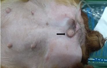
You can also read