High resolution optical coherence elastography of retina under prosthetic electrode
←
→
Page content transcription
If your browser does not render page correctly, please read the page content below
Original Article

High resolution optical coherence elastography of retina under
prosthetic electrode
Runze Li1,2#, Zhaodong Du2#, Xuejun Qian1,2, Yan Li3,4, Juan-Carlos Martinez-Camarillo2, Laiming Jiang2,
Mark S. Humayun1,2, Zhongping Chen3,4, Qifa Zhou1,2
1
    Department of Biomedical Engineering, University of Southern California, Los Angeles, CA, USA; 2USC Roski Eye Institute, University of
Southern California, Los Angeles, CA, USA; 3Beckman Laser Institute, University of California, Irvine, CA, USA; 4Department of Biomedical
Engineering, University of California, Irvine, CA, USA

#
These authors contributed equally to this work.

Correspondence to: Zhongping Chen. University of California, 1002 Health Sciences Road East, Irvine, CA 92617, USA. Email: z2chen@uci.edu;
Qifa Zhou. 1042 Downey Way, LA, CA 90089-1111, USA. Email: qifazhou@usc.edu.

                  Background: Quantitatively investigating the biomechanics of retina with a retinal prosthetic electrode, we
                  explored the effects of the prosthetic electrode on the retina, and further supplemented data for a potential
                  clinical trial.
                  Methods: Biomechanical properties were assessed with a high resolution optical coherence tomography
                  (OCT) based elastography (OCE) system. A shaker was used to initiate elastic waves and an OCT system was
                  used to track axial displacement along with wave propagation. Rabbits received surgery to implant the retinal
                  prosthetic electrode, and elastic wave speed was measured before and after implantation; anatomical B-mode
                  images were also acquired.
                  Results: Spatial-temporal maps of each layer in retina with and without prosthetic electrodes were
                  acquired. Elastic wave speed of nerve fiber to inner plexiform layer, inner nuclear to outer nuclear layer,
                  retinal pigmented epithelium layer and choroid to sclera layer without prosthetic electrode were found to be
                  3.66±0.36, 5.33±0.07, 6.85±0.37, and 9.69±0.24 m/s, respectively. With prosthetic electrode, the elastic wave
                  speed was found to be 4.09±0.26, 5.14±0.11, 6.88±0.70, and 9.99±0.73 m/s, respectively in each layer.
                  Conclusions: Our results show that the elastic wave speed in each layer of retina is slightly faster with the
                  retinal electrode, and further demonstrate that the retinal prosthetic electrode does not affect biomechanical
                  properties significantly. In the future, we expect OCE technology to be used by clinicians where it could
                  become part of routine testing and evaluation of the biomechanical properties of the retina in response to
                  long term use of prosthetic electrodes in patients.

                  Keywords: Optical coherence elastography; elastic wave propagation; mechanical shaker; retinal prosthetic
                  electrode

                  Submitted Oct 08, 2020. Accepted for publication Dec 15, 2020.
                  doi: 10.21037/qims-20-1137
                  View this article at: http://dx.doi.org/10.21037/qims-20-1137

Introduction                                                                   are affected by RP worldwide. Initially, patients retain
                                                                               their central visual field with gradual peripheral vision
Diseases of the posterior segment of the eye, including
age-related macular degeneration (AMD) and retinitis                           loss, however, eventually they will go on to lose both areas
pigmentosa (RP), damage photoreceptors and their                               of vision with progression of the disease (2). AMD is the
supporting epithelial cells (1). More than 1 million patients                  leading cause of blindness in patients aged 65 or older in

© Quantitative Imaging in Medicine and Surgery. All rights reserved.    Quant Imaging Med Surg 2021;11(3):918-927 | http://dx.doi.org/10.21037/qims-20-1137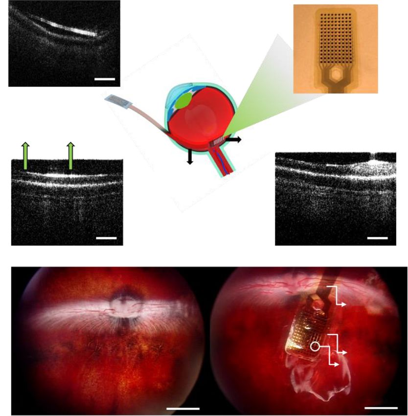
Quantitative Imaging in Medicine and Surgery, Vol 11, No 3 March 2021                                                                                       919

developed countries, with more than 8 million Americans                              techniques, OCE is the most promising imaging modality in
suffering from the disease (3). Currently, there is no known                         the field of ophthalmology owing to the advantages of high
cure to reverse the progressive loss of photoreceptors due to                        resolution (
920                                                                              Li et al. High resolution OCE of retina under prosthetic electrode

                                                                                     CO         OA              M

                                                    Isolator           OC
                                      SLD
                                                                                     CO                   GM
                                                         CO

                                                        G
                                                                                                           L1
                                                        L2

                                                               CCD

                                                   Digitizer                          FG                   RFA

Figure 1 Schematic of shaker based OCE system. OCE, optical coherence tomography based elastography; SLD, superluminescent diode; OC,
optical coupler; CO, collimator; OA, optical attenuator; M, mirror; GM, galvanometer mirrors; L1/L2, lens; RFA, radiofrequency amplifier;
FG, function generator; G, grating.

of the OCT scanning beam with real time Doppler OCT                                intensity data and phase-resolved Doppler OCT data
imaging. A step size of 6 µm was used for the scanning                             were first obtained. To calculate the elastic wave speed,
galvanometer mirror positioning system (galvo) and a total                         the spatial-temporal map was reconstructed at each lateral
of 500 positions were scanned.                                                     position and time. The elastic wave speed can be estimated
   To precisely induce and track elastic wave propagation, all                     with known propagation distance and time. To precisely
components of the system were synchronized. A baseband                             calculate the wave speed, linear regression was used for all
signal generated by the personal computer (PC) triggered                           successive peak axial displacements at each lateral position
an arbitrary function generator (AFG 3252C, Tektronix,                             in the corresponding spatial-temporal map. In this study,
Beaverton, OR, USA). A 0.6 ms pulse-width impulse signal                           the elastic wave speed in each layer was measured before
was used to generate detectable axial displacement with                            and after the implant of prosthetic electrode, the Shapiro-
broad bandwidth (25). The impulse signal was amplified                             Wilk test was performed to evaluate the normality of the
using a power amplifier (Type 2718, Bruel & Kjaer, Duluth,                         speed distribution, and the paired t-test was performed to
Georgia, USA) and finally transmitted to the shaker. A series                      evaluate the statistical significance.
of 400 A-lines was acquired at every position corresponding
to 8.8 ms before the galvo translated to the next position.
                                                                                   Implant surgery
Inter-A-line analysis was performed with a 20 μs time interval
to obtain axial displacements for further processing (24).                          Surgery protocol was approved by the University of
Inter-A-line analysis was insensitive to motion artifacts                          Southern California Institutional Animal Care and Use
because the data acquisition frequency was much faster                             Committee (IACUC). Prior to surgery, Dutch Belted
than the motion frequency, and adequate anesthesia was                             Pigmented rabbits (~2 kg) were anesthetized with
administrated to reduce the motion artifact.                                       ketamine (35 mg/kg) and xylazine (5 mg/kg) through
                                                                                   subcutaneous injection. Additional anesthesia was induced
                                                                                   using 2.5% sevoflurane through a facial mask. One drop of
Post-processing and data analysis
                                                                                   0.5% proparacaine hydrochloride ophthalmic solution was
After scanning in each lateral position, raw data was saved                        administrated topically for ocular anesthesia, and 1 drop
to disk for off-line processing. Depth-resolved OCT                                of 1% tropicamide and 2.5% phenylephrine was applied

© Quantitative Imaging in Medicine and Surgery. All rights reserved.        Quant Imaging Med Surg 2021;11(3):918-927 | http://dx.doi.org/10.21037/qims-20-1137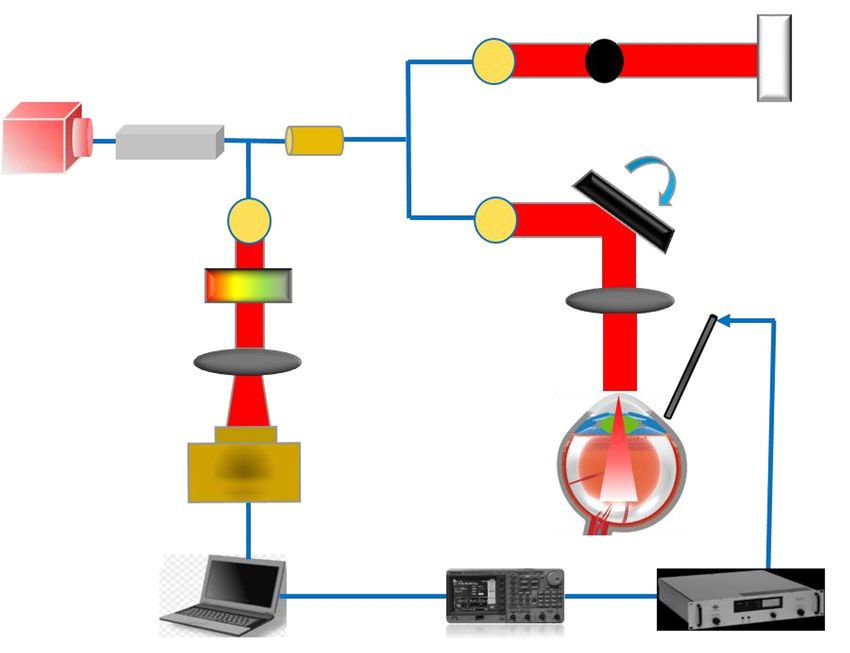
Quantitative Imaging in Medicine and Surgery, Vol 11, No 3 March 2021 921
A B
14×9 150 μm diameter
elements
3.5 mm
Natural response
to gravity
37 mm
2 mm
C D
Bend >90° Bend >90°
2 mm 2 mm
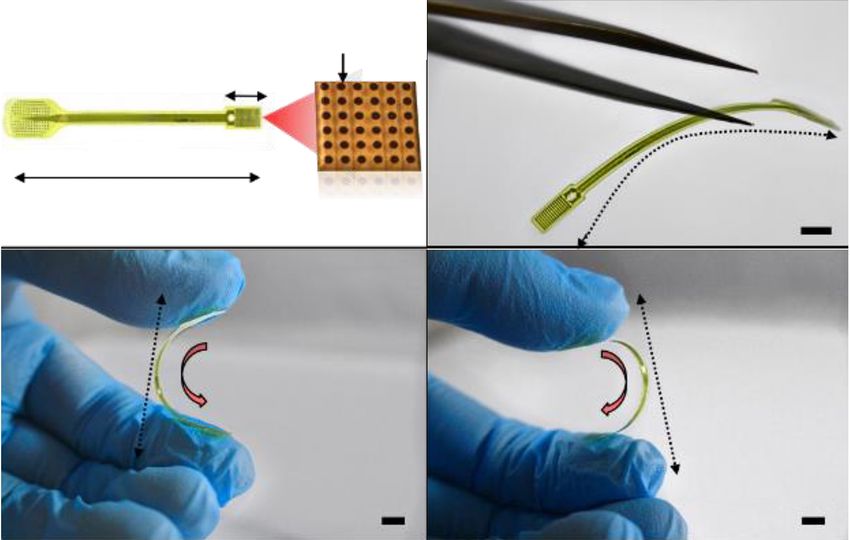
Figure 2 Characterizations of retinal prosthetic electrode. (A) Structural image of the prosthesis; (B,C,D) optical images of the flexible
prosthesis when it stands freely, bends convexly and concavely with more than 90°.
in the same way for eye dilation. Heart-rate, respiration, Results
oxygen saturation, body temperature, non-invasive blood
Prosthetic electrode characterization
pressure, oxygen flow and end tidal carbon dioxide (when
sevoflurane was used), were recorded every 5 minutes while The requirements for retinal prosthetic electrodes mainly
the animals were unconscious. First, phacoemulsification include high electrode density, biocompatibility, and
was performed for all three animals, and the OCE imaging flexibility (29,30). A high-density prosthetic electrode
was conducted for 3 times in each animal. Intra-vitreous allows high resolution stimulation and hence improves
injection of 0.3 mL sterile air was performed and then visual acuity. Due to the natural curvature of retina,
the eyes were treated with antibiotic ointment and 1% flexibility enables the prosthetic electrode to adhere to the
atropine immediately to prevent infection and to maintain surface of the retina seamlessly for a prolonged period of
postoperative cycloplegia. One week later, a 3-port 23-G time. Polyimide has the advantages of flexibility, mechanical
pars plana vitrectomy (PPV) was performed with the Stellaris stability, biocompatibility, and thermal stability, and was
PC platform (Bausch & Lomb, Rochester, NY, USA) in a therefore used as the structural material in the prosthetic
sterile environment. A microelectrode was then inserted electrode. The three-dimensional platinum (Pt)-pillar
through a temporal sclerotomy (approximately 4 mm in coating method was used to increase the electrode density
width) and was placed onto the retina below the optic nerve and constrain the electrode size while maintaining flexibility
head (ONH) and then flattened using perfluorocarbon liquid of the entire structure (31).
to keep it securely attached to the retina after vitrectomy. Figure 2A shows the structure of the prosthetic electrode.
The extraocular portion of the cable was sutured to the The multi-electrode array is connected with a high lead
sclera below the conjunctiva. At the end of the procedure, count cable for retinal stimulation. The array has 14×9
the sclerotomies and conjunctiva were sutured. After the elements with a diameter of 150 µm each. The length of
operation, topical antibiotic eye drops were used 4 times the prosthetic electrodes and the array is 37 and 3.5 mm,
daily. And OCE imaging was performed 3 times in each respectively. Figure 2B,C,D demonstrates the prosthetic
animal with the implant. No complications related to surgery electrode’s flexibility which facilitates seamless bonding to
were observed during or after surgery. the complex surface of the retina.
© Quantitative Imaging in Medicine and Surgery. All rights reserved. Quant Imaging Med Surg 2021;11(3):918-927 | http://dx.doi.org/10.21037/qims-20-1137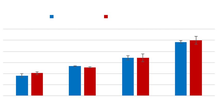
922                                                                                  Li et al. High resolution OCE of retina under prosthetic electrode

                                       Cable

                                                    500 μm
                      A
                                                                                                                   Multielectrode

                          Sbustrate    Electrode
                            Edge                                                                 Multielectrode
                                                                                                     Array

                                                                       Highly Flexible
                                                                           Cable

                                                    500 μm                                                                          500 μm
                      B                                                                             C

                                                             ONH
                                                                                                    RV    ONH
                                                                                                                             fexible cable

                                               RV

                                                                                                                              substrate edge
                                                                                                                           electrode

                                                                            2 mm                                                      2 mm
                     D                                                                   E
Figure 3 OCT cross-section and RetCam images of retina with prosthetic electrode. (A,B,C) Cross-sectional OCT images of retina with
prosthetic electrode; (D,E) RetCam images of retina after phacoemulsification procedure and implantation procedure respectively. OCT,
optical coherence tomography; ONH, optic nerve head; RV, retina vessels.

Surgery characterization                                                                 phacoemulsification and implantation respectively.

Post-surgery evaluation is essential in this study. Failure
of implantation is common in phacoemulsification and                                     Biomechanical response of retina to the prosthetic electrode
implantation procedures. Hemorrhage and iris bleeding                                    To investigate the influence of the prosthetic electrode
are also rarely observed during vitrectomy due to the high                               on the retina, OCE imaging was performed after
viscosity of vitreous humor (32,33). Retinal detachment                                  phacoemulsification and implantation. As with our previous
can occur during prosthetic electrode implantation when                                  work (25), here we segment the raw measured data to five
vitreous humor is not fully removed prior to the procedure.                              different layers according to the anatomy of the retina and
Figure 3 demonstrates successful implantation and bonding                                assign them respective numerals i through v for efficient
of the prosthetic electrode to the retina. Figure 3A,B,C                                 classification. Layer i to layer v correspond to the nerve
show cross-sectional OCT images with the prosthetic                                      fiber, ganglion cell, and inner plexiform layer; inner nuclear,
electrode. Figure 3D,E show the en face RetCam (Natus                                    outer plexiform, and outer nuclear layer; retinal pigmented
Medical Incorporated, Pleasanton, CA, USA) images after                                  epithelium layer; choroid layer; choroid and sclera layer

© Quantitative Imaging in Medicine and Surgery. All rights reserved.           Quant Imaging Med Surg 2021;11(3):918-927 | http://dx.doi.org/10.21037/qims-20-1137
Quantitative Imaging in Medicine and Surgery, Vol 11, No 3 March 2021 923
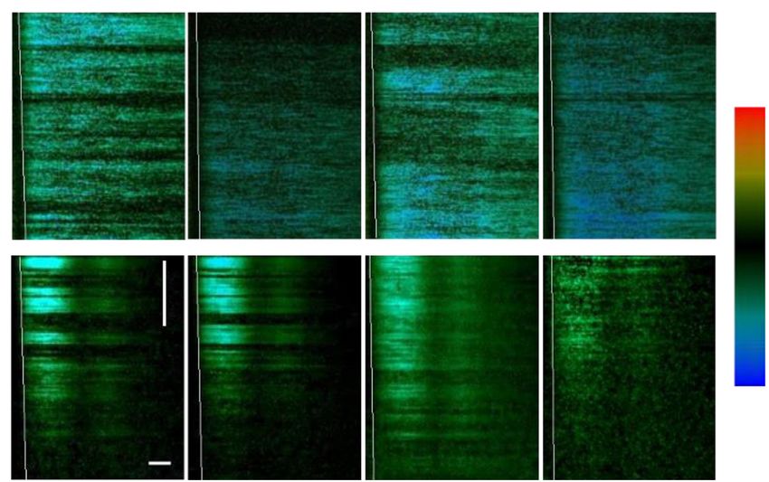
Layer i Layer ii Layer iii Layer v
W/O Prosthesis
300 nm
0.5 mm
W/ Prosthesis
–300 nm
1.5 ms
Figure 4 Spatial-temporal maps in each layer of retina with and without prosthetic electrode. The slope of the white line represents the
corresponding elastic wave speed. Color bar represents the axial displacement.
Elastic wave speed distribution
w/o Prosthesis w/ Prosthesis
12
9.99
9.69
Elastic wave speed (m/s)
10
8 6.85 6.88
6 5.33 5.14
4.09
3.66
4
2
0
Layer i Layer ii Layer iii Layer v
Figure 5 Statistical analysis of elastic wave speed in each layer of the retina with and without prosthetic electrode.
respectively. Spatial-temporal maps were constructed in electrode. The slope of the wavefront is the elastic wave
each layer along with lateral positions and time except for speed in the corresponding retinal layer. Figure 5 shows
layer iv due to its low OCT signal which is similar to our the statistical analysis of elastic wave speed in each layer
previously reported study (25). Figure 4 shows the spatial- with and without the prosthetic electrode; the error bar
temporal map in each layer with and without the prosthetic represents the standard deviation. Without the prosthetic
© Quantitative Imaging in Medicine and Surgery. All rights reserved. Quant Imaging Med Surg 2021;11(3):918-927 | http://dx.doi.org/10.21037/qims-20-1137924 Li et al. High resolution OCE of retina under prosthetic electrode electrode, the elastic wave speed of layers i, ii, iii, and v biomechanical properties of the eye in the presence of the is 3.66±0.36, 5.33±0.07, 6.85±0.37, and 9.69±0.24 m/s, implanted electrode. In the current study, we investigated respectively. With the prosthetic electrode, the elastic wave how the prosthetic electrode affects the biomechanical speed in each corresponding layer is 4.09±0.26, 5.14±0.11, properties of the retina to help further knowledge and 6.88±0.70, and 9.99±0.73 m/s, respectively. The difference general understanding of these important effects. between the results in each group and each layer were In this study, we measured the elastic wave speed before evaluated using a Shapiro-Wilk test and a paired t-test. and after prosthetic electrode implantation. Results showed The statistical analysis showed that the elastic wave speed that the measured biomechanical properties of the retina distributes normally with P
Quantitative Imaging in Medicine and Surgery, Vol 11, No 3 March 2021                                                                                925

resulting in intraocular blurring. Last, the metal electrode                  uniform disclosure form (available at http://dx.doi.
blocked the optical path of the OCT system, with the                          org/10.21037/qims-20-1137). The special issue “Advanced
beam only able to penetrate through the gap between                           Optical Imaging in Biomedicine” was commissioned by
electrodes which led to a decrease in the OCT signal. All                     the editorial office without any funding or sponsorship.
of these factors degraded the OCT image quality in this                       Dr. ZC reports personal fees from OCT Medical Imaging
study. While these issues are currently difficult to resolve                  Inc., outside the submitted work; In addition, Dr. ZC has a
with our current system and setup, in future work we                          patent US 16/402,591 pending. The authors have no other
plan to use improved surgical techniques and a new high                       conflicts of interest to declare.
power OCT system to improve the image quality. This
study is also limited by the lack of electrical stimulation                   Ethical Statement: This study was approved by the University
during OCE imaging. However, the aim of this study was                        of Southern California Institutional Animal Care and Use
to explore the effects of the prosthetic electrode on the                     Committee (IACUC).
biomechanics of the retina. Further studies are needed
to apply a range of electrical stimuli and investigate the                    Open Access Statement: This is an Open Access article
corresponding biomechanical effects.                                          distributed in accordance with the Creative Commons
                                                                              Attribution-NonCommercial-NoDerivs 4.0 International
                                                                              License (CC BY-NC-ND 4.0), which permits the non-
Conclusions
                                                                              commercial replication and distribution of the article with
In summary, our work demonstrated surgical implantation of                    the strict proviso that no changes or edits are made and the
a prosthetic electrode in rabbit eye, and the biomechanical                   original work is properly cited (including links to both the
response of the retina to the prosthetic electrode. Based on                  formal publication through the relevant DOI and the license).
the elastic wave speed from the measured data in each layer                   See: https://creativecommons.org/licenses/by-nc-nd/4.0/.
of the retina with and without the prosthetic electrode, we
conclude that the prosthetic electrode does not affect the
                                                                              References
biomechanical properties of the retina significantly. We
hope that this technology can be further translated into the                  1.   Humayun MS, deJuan E, Dagnelie G, Greenberg RJ,
clinical use so that it can evaluate retinal biomechanics in                       Prost RH, Phillips DH. Visual perception elicited by
patients with retinal prosthetic electrodes for longitudinal                       electrical stimulation of retina in blind humans. Arch
study over an extended period of time.                                             Ophthalmol 1996;114:40-6.
                                                                              2.   Hartong DT, Berson EL, Dryja TP. Retinitis pigmentosa.
                                                                                   Lancet 2006;368:1795-809.
Acknowledgments
                                                                              3.   Jager RD, Mieler WF, Miller JW. Medical progress:
We thank Dr. Yi Zhang for prosthetic electrode preparation,                        Age-related macular degeneration. N Engl J Med
and Dr. Robert Wodnicki for proofreading.                                          2008;358:2606-17.
Funding: This work was partially supported by the                             4.   Humayun MS, Weiland JD, Fujii GY, Greenberg R,
NIH NEI grant R01EY028662, R01EY026091, NIH                                        Williamson R, Little J, Mech B, Cimmarusti V, Van
P30EY029220, and an Unrestricted Departmental Grant                                Boemel G, Dagnelie G, de Juan E. Visual perception in
from Research to Prevent Blindness, New York, NY 10017.                            a blind subject with a chronic microelectronic retinal
ZD was a visiting scholar at Roski Eye Institute, University                       prosthesis. Vision Res 2003;43:2573-81.
of Southern California.                                                       5.   Luo YHL, da Cruz L. The Argus (R) II Retinal
                                                                                   Prosthesis System. Progress in Retinal and Eye Research
                                                                                   2016;50:89-107.
Footnote
                                                                              6.   Zhou DD, Dorn JD, Greenberg RJ, Grp AIS. The Argus
Provenance and Peer Review: With the arrangement by the                            (R) Ii Retinal Prosthesis System: An Overview. Electronic
Guest Editors and the editorial office, this article has been                      Proceedings of the 2013 Ieee International Conference on
reviewed by external peers.                                                        Multimedia and Expo Workshops (Icmew) 2013.
                                                                              7.   da Cruz L, Dorn JD, Humayun MS, Dagnelie G, Handa
Conflicts of Interest: All authors have completed the ICMJE                        J, Barale PO, Sahel JA, Stanga PE, Hafezi F, Safran AB,

© Quantitative Imaging in Medicine and Surgery. All rights reserved.   Quant Imaging Med Surg 2021;11(3):918-927 | http://dx.doi.org/10.21037/qims-20-1137
926 Li et al. High resolution OCE of retina under prosthetic electrode
Salzmann J, Santos A, Birch D, Spencer R, Cideciyan AV, functional Ultrasonic Micro-elastography Imaging System.
de Juan E, Duncan JL, Eliott D, Fawzi A, de Koo LCO, Sci Rep 2017;7:1230.
Ho AC, Brown G, Haller J, Regillo C, Del Priore LV, 19. Shih CC, Qian XJ, Ma T, Han ZL, Huang CC, Zhou
Arditi A, Greenberg RJ, Grp AIS. Five-Year Safety and QF, Shung KK. Quantitative Assessment of Thin-Layer
Performance Results from the Argus II Retinal Prosthesis Tissue Viscoelastic Properties Using Ultrasonic Micro-
System Clinical Trial. Ophthalmology 2016;123:2248-54. Elastography With Lamb Wave Model. IEEE Trans Med
8. Yue L, Falabella P, Christopher P, Wuyyuru V, Dorn J, Imaging 2018;37:1887-98.
Schor P, Greenberg RJ, Weiland JD, Humayun MS. Ten- 20. Qian X, Ma T, Shih CC, Heur M, Zhang J, Shung
Year Follow-up of a Blind Patient Chronically Implanted KK, Varma R, Humayun MS, Zhou Q. Ultrasonic
with Epiretinal Prosthesis Argus I. Ophthalmology Microelastography to Assess Biomechanical Properties of
2015;122:2545-+. the Cornea. IEEE Trans Biomed Eng 2019;66:647-55.
9. Stronks HC, Dagnelie G. The functional performance of 21. Jin Z, Khazaeinezhad R, Zhu J, Yu JX, Qu YQ, He
the Argus II retinal prosthesis. Expert Rev Med Devices YM, Li Y, Alvarez-Arenas TEG, Lu F, Chen ZP. In-
2014;11:23-30. vivo 3D corneal elasticity using air-coupled ultrasound
10. Dagnelie G, Christopher P, Arditi A, da Cruz L, Duncan optical coherence elastography. Biomed Opt Express
JL, Ho AC, Olmos de Koo LC, Sahel JA, Stanga PE, 2019;10:6272-85.
Thumann G, Wang Y, Arsiero M, Dorn JD, Greenberg 22. Li Y, Moon S, Chen JJ, Zhu Z, Chen Z. Ultrahigh-
RJ, Argus IISG. Performance of real-world functional sensitive optical coherence elastography. Light Sci Appl
vision tasks by blind subjects improves after implantation 2020;9:58.
with the Argus (R) II retinal prosthesis system. Clin Exp 23. Qu Y, Ma T, He Y, Zhu J, Dai C, Yu M, Huang S, Lu F,
Ophthalmol 2017;45:152-9. Shung KK, Zhou Q, Chen Z. Acoustic Radiation Force
11. Rabinowitz YS. Keratoconus. Surv Ophthalmol Optical Coherence Elastography of Corneal Tissue. IEEE
1998;42:297-319. J Sel Top Quantum Electron 2016;22:6803507.
12. Sigal IA, Flanagan JG, Ethier CR. Factors influencing 24. Li Y, Zhu J, Chen JJ, Yu JX, Jin Z, Miao YS, Browne
optic nerve head biomechanics. Invest Ophthalmol Vis Sci AW, Zhou QF, Chen ZP. Simultaneously imaging and
2005;46:4189-99. quantifying in vivo mechanical properties of crystalline
13. Braunsmann C, Hammer CM, Rheinlaender J, Kruse lens and cornea using optical coherence elastography
FE, Schaffer TE, Schlotzer-Schrehardt U. Evaluation with acoustic radiation force excitation. APL Photonics
of Lamina Cribrosa and Peripapillary Sclera Stiffness in 2019;4:106104.
Pseudoexfoliation and Normal Eyes by Atomic Force 25. Qian X, Li R, Li Y, Lu G, He Y, Humayun MS, Chen Z,
Microscopy. Invest Ophthalmol Vis Sci 2012;53:2960-7. Zhou Q. In vivo evaluation of posterior eye elasticity using
14. Quigley HA. The contribution of the sclera and lamina shaker-based optical coherence elastography. Exp Biol
cribrosa to the pathogenesis of glaucoma: diagnostic and Med (Maywood) 2020;245:282-8.
treatment implications. New Trends in Basic and Clinical 26. Du Z, Li R, Qian X, Lu G, Li Y, He Y, Qu Y, Jiang L,
Research of Glaucoma: A Neurodegenerative Disease of Chen Z, Humayun MS, Chen Z, Zhou Q. Quantitative
the Visual System, Pt A 2015;220:59-86. confocal optical coherence elastography for evaluating
15. He Y, Qu Y, Zhu J, Zhang Y, Saidi A, Ma T, Zhou Q, biomechanics of optic nerve head using Lamb wave model.
Chen Z. Confocal Shear Wave Acoustic Radiation Neurophotonics 2019;6:041112.
Force Optical Coherence Elastography for Imaging and 27. Wang S, Li JS, Manapuram RK, Menodiado FM, Ingram
Quantification of the In Vivo Posterior Eye. IEEE J Sel DR, Twa MD, Lazar AJ, Lev DC, Pollock RE, Larin KV.
Top Quantum Electron 2019;25:10. Noncontact measurement of elasticity for the detection of
16. Pavlatos E, Pan XL, Clayson K, Hart RT, Weber P, Liu soft-tissue tumors using phase-sensitive optical coherence
J. ONH Deformation in Human Eyes Using Ultrasound tomography combined with a focused air-puff system. Opt
Speckle Tracking. Invest Ophthalmol Vis Sci 2017;58. Lett 2012;37:5184-6.
17. Zhou B, Chen JJ, Kazemi A, Sit AJ, Zhang X. An 28. Ambroziński Ł, Song SZ, Yoon SJ, Pelivanov I, Li D, Gao
Ultrasound Vibro-Elastography Technique for Assessing L, Shen TT, Wang RKK, O'Donnell M. Acoustic micro-
Papilledema. Ultrasound Med Biol 2019;45:2034-9. tapping for non-contact 4D imaging of tissue elasticity. Sci
18. Qian X, Ma T, Yu M, Chen X, Shung KK, Zhou Q. Multi- Rep 2016;6:38967.
© Quantitative Imaging in Medicine and Surgery. All rights reserved. Quant Imaging Med Surg 2021;11(3):918-927 | http://dx.doi.org/10.21037/qims-20-1137Quantitative Imaging in Medicine and Surgery, Vol 11, No 3 March 2021 927
29. Rodger DC, Weiland JD, Humayun MS, Tai YC. Scalable pars plana vitrectomy. Ophthalmic Surg Lasers Imaging
high lead-count parylene package for retinal prostheses. 2007;38:270-5.
Sensors and Actuators B-Chemical 2006;117:107-14. 34. Kirby MA, Pelivanov I, Song SZ, Ambrozinski L, Yoon SJ,
30. Zeng Q, Zhao SS, Yang HG, Zhang Y, Wu TZ. Micro/ Gao L, Li D, Shen TT, Wang RK, O'Donnell M. Optical
Nano Technologies for High-Density Retinal Implant. coherence elastography in ophthalmology. J Biomed Opt
Micromachines (Basel) 2019;10:419. 2017;22:1-28.
31. Xia K, Sun B, Zeng Q, Wu TZ, Humayun MS. 35. Zvietcovich F, Pongchalee P, Meemon P, Rolland JP,
Surface Modification of Neural Stimulating/Recording Parker KJ. Reverberant 3D optical coherence elastography
Microelectrodes with High-Performance Platinum-Pillar maps the elasticity of individual corneal layers. Nat
Coatings. 2017 Ieee 12th International Conference on Commun 2019;10:4895.
Nano/Micro Engineered and Molecular Systems (Nems) 36. Parker KJ, Ormachea J, Zvietcovich F, Castaneda B.
2017:291-4. Reverberant shear wave fields and estimation of tissue
32. Park SS, Marcus DM, Duker JS, Pesavento RD, Topping properties. Phys Med Biol 2017;62:1046-61.
TM, Frederick AR, Damico DJ. Posterior Segment 37. Han Z, Aglyamov SR, Li J, Singh M, Wang S, Vantipalli
Complications after Vitrectomy for Macular Hole. S, Wu C, Liu CH, Twa MD, Larin KV. Quantitative
Ophthalmology 1995;102:775-81. assessment of corneal viscoelasticity using optical
33. Gupta OP, Weichel ED, Regillo CD, Fineman coherence elastography and a modified Rayleigh-Lamb
MS, Kaiser RS, Ho AC, McNamara JA, Vander JF. equation. J Biomed Opt 2015;20:20501.
Postoperative complications associated with 25-gauge
C i t e t h i s a r t i c l e a s : L i R , D u Z , Q i a n X , L i Y,
Martinez-Camarillo JC, Jiang L, Humayun MS, Chen Z,
Zhou Q. High resolution optical coherence elastography of
retina under prosthetic electrode. Quant Imaging Med Surg
2021;11(3):918-927. doi: 10.21037/qims-20-1137
© Quantitative Imaging in Medicine and Surgery. All rights reserved. Quant Imaging Med Surg 2021;11(3):918-927 | http://dx.doi.org/10.21037/qims-20-1137You can also read