IDENTIFICATION OF A SULFATASE THAT DETOXIFIES GLUCOSINOLATES IN THE PHLOEM-FEEDING INSECT BEMISIA TABACI AND PREFERS INDOLIC GLUCOSINOLATES - MPG.PURE
←
→
Page content transcription
If your browser does not render page correctly, please read the page content below
ORIGINAL RESEARCH
                                                                                                                                             published: 04 June 2021
                                                                                                                                      doi: 10.3389/fpls.2021.671286

                                             Identification of a Sulfatase that
                                             Detoxifies Glucosinolates in the
                                             Phloem-Feeding Insect Bemisia
                                             tabaci and Prefers Indolic
                                             Glucosinolates
                                             Abinaya Manivannan 1 , Bhawana Israni 1† , Katrin Luck 1† , Monika Götz 2† , Elena Seibel 1 ,
                                             Michael L. A. E. Easson 1 , Roy Kirsch 1 , Michael Reichelt 1 , Beate Stein 2 , Stephan Winter 2 ,
                                             Jonathan Gershenzon 1 and Daniel Giddings Vassão 1*
                                             1
                                              Max Planck Institute for Chemical Ecology, Jena, Germany, 2 Leibniz Institute DSMZ-German Collection of Microorganisms
                                             and Cell Cultures, Braunschweig, Germany

                          Edited by:
                        Jens Rohloff,        Cruciferous plants in the order Brassicales defend themselves from herbivory using
      Norwegian University of Science
            and Technology, Norway
                                             glucosinolates: sulfur-containing pro-toxic metabolites that are activated by hydrolysis
                        Reviewed by:
                                             to form compounds, such as isothiocyanates, which are toxic to insects and other
                           Inga Mewis,       organisms. Some herbivores are known to circumvent glucosinolate activation with
         Humboldt University of Berlin,      glucosinolate sulfatases (GSSs), enzymes that convert glucosinolates into inactive
                              Germany
                          Shaoli Wang,       desulfoglucosinolates. This strategy is a major glucosinolate detoxification pathway in
  Insititute of Vegetables and Flowers       a phloem-feeding insect, the silverleaf whitefly Bemisia tabaci, a serious agricultural
                         (CAAS), China
                                             pest of cruciferous vegetables. In this study, we identified and characterized an
                   *Correspondence:
                Daniel Giddings Vassão
                                             enzyme responsible for glucosinolate desulfation in the globally distributed B. tabaci
                   vassao@ice.mpg.de         species MEAM1. In in vitro assays, this sulfatase showed a clear preference for indolic
     † These   authors have contributed      glucosinolates compared with aliphatic glucosinolates, consistent with the greater
                    equally to this work
                                             representation of desulfated indolic glucosinolates in honeydew. B. tabaci might use
                   Specialty section:
                                             this detoxification strategy specifically against indolic glucosinolates since plants may
         This article was submitted to       preferentially deploy indolic glucosinolates against phloem-feeding insects. In vivo
                      Plant Metabolism
                                             silencing of the expression of the B. tabaci GSS gene via RNA interference led to lower
                  and Chemodiversity,
               a section of the journal      levels of desulfoglucosinolates in honeydew. Our findings expand the knowledge on the
            Frontiers in Plant Science       biochemistry of glucosinolate detoxification in phloem-feeding insects and suggest how
        Received: 23 February 2021           detoxification pathways might facilitate plant colonization in a generalist herbivore.
           Accepted: 21 April 2021
          Published: 04 June 2021            Keywords: whitefly, phloem-feeder, glucosinolates, sulfatase, pre-emptive detoxification, Bemisia tabaci MEAM1
                             Citation:
      Manivannan A, Israni B, Luck K,
     Götz M, Seibel E, Easson MLAE,         INTRODUCTION
        Kirsch R, Reichelt M, Stein B,
         Winter S, Gershenzon J and         Plants rely on a complex arsenal of toxic chemicals to defend themselves against herbivores
  Vassão DG (2021) Identification of a      and pathogens. One successful defense strategy used to safely accumulate large concentrations
              Sulfatase that Detoxifies
                                            of defensive compounds while preventing auto-toxicity is the production of two-component
Glucosinolates in the Phloem-Feeding
    Insect Bemisia tabaci and Prefers
                                            activated defenses, such as cyanogenic glucosides and glucosinolates (Halkier and Gershenzon,
               Indolic Glucosinolates.      2006; Mithöfer and Boland, 2012). These glucosylated pro-toxins are stable and only become toxic
         Front. Plant Sci. 12:671286.       after enzymatic activation coincident with herbivore damage. Multi-component plant defenses like
      doi: 10.3389/fpls.2021.671286         this, however, offer herbivores multiple targets for counter-adaptation, including mechanisms to

Frontiers in Plant Science | www.frontiersin.org                                   1                                          June 2021 | Volume 12 | Article 671286
Manivannan et al. Bemisia tabaci Glucosinolate Sulfatase
prevent or redirect activation, or detoxify the activated poisons of glucosinolates that are only activated into more chemically
(Pentzold et al., 2014; Jeschke et al., 2016b). Piercing–sucking, reactive toxins upon herbivory, this two-component system
phloem-feeding insects, such as aphids and whiteflies, are serves as an important chemical defense against most herbivores
thought to lessen plant defensive responses by causing only (Hopkins et al., 2009; Jeschke et al., 2016a; Schweizer et al., 2017).
minimal damage during feeding, stealthily penetrating plant Several species of insect herbivores, however, have developed
tissues to feed on the sugar-rich phloem sap. Nevertheless, means to detoxify glucosinolates and successfully feed on
even piercing–sucking feeding leads to some activation of crucifer plants with relative impunity (Jeschke et al., 2016b).
glucosinolates (Kim et al., 2008; Danner et al., 2018), so For example, the isothiocyanate hydrolysis products can be
that detoxification strategies preventing activation of two- converted to hydrophilic glutathione–isothiocyanate conjugates
component defenses can be greatly advantageous to phloem- (Schramm et al., 2012; Jeschke et al., 2016a). This reaction, which
feeding insects. Here, we examine the biochemical basis of takes place in many organisms including humans, can occur
one such detoxification in the silverleaf whitefly Bemisia tabaci very efficiently in certain insects adapted to crucifers. On the
(Gennadius) (Hemiptera): the desulfation of glucosinolates to other hand, the larvae of some crucifer-feeding Pieris species
form derivatives that can no longer be activated. use a “nitrile specifying protein” for directing glucosinolate
Glucosinolates are sulfur-containing defensive metabolites hydrolysis toward the formation of less toxic nitriles rather
restricted to the plant order Brassicales. The family Brassicaceae than isothiocyanates (Wittstock et al., 2004). Another enzymatic
of this order (mustard family) includes many economically detoxification mechanism is the action of glucosinolate sulfatases
important oilseed, vegetable, condiment, and fodder crops, such (GSSs) in the insect gut. These enzymes are used by crucifer
as rapeseed, cabbages, and mustards, as well as the model plant specialists, such as the diamondback moth larvae [Plutella
Arabidopsis thaliana (L.) Heynh. These plants have distinctive xylostella (L.)] (Ratzka et al., 2002) and the cabbage stem
tastes and smells that are conferred in great part by hydrolysis flea beetle [Psylliodes chrysocephala (L.)] (Ahn et al., 2019),
products from glucosinolates, also called mustard oils (Halkier, as well as the generalist herbivores desert locust [Schistocerca
2016). The core chemical structure of glucosinolates (Figure 1) gregaria (Forskål)] (Falk and Gershenzon, 2007) and the silverleaf
consists of a β-thioglucose residue, a sulfonated oxime moiety, whitefly (B. tabaci) (Malka et al., 2016). GSSs cleave the sulfate
and a variable amino acid-derived side chain group R (Halkier group of glucosinolates to form desulfoglucosinolates (Figure 1),
and Gershenzon, 2006). A recent review lists up to 137 natural essentially reversing the last step in the biosynthesis of the
glucosinolates described based on the variability at the R group glucosinolate core structure. As desulfoglucosinolates can no
(Blažević et al., 2020), with these being classifiable according to longer be used as substrates by myrosinases, no toxic downstream
the precursor amino acid from which they are derived, such as products are produced.
aliphatic (e.g., from Met, Ala, and Val), benzenic (Phe and Tyr), Among the insects that can desulfate glucosinolates is
and indolic (Trp). B. tabaci, a piercing-sucking, phloem-feeder that inhabits mainly
In addition to glucosinolates, cruciferous plants produce tropical and subtropical regions. B. tabaci is a cryptic species
thioglucosidase enzymes known as myrosinases that catalyze complex, existing as a conglomerate of dozens of species (Perring,
glucosinolate hydrolysis (Rask et al., 2000). Myrosinases and 2001) that are difficult to distinguish even with molecular
glucosinolates are physically segregated in the intact plant (Grob markers (Boykin et al., 2012, 2018). The most invasive and
and Matile, 1979; Höglund et al., 1991). While the localization geographically widespread species are MED (Mediterranean,
of each component is still a topic of study, myrosinases have formerly named Q) and MEAM1 (Middle East–Asia Minor
been reported in protein-accumulating idioblasts called myrosin 1, formerly B). B. tabaci species are altogether extreme
cells present in the phloem parenchyma. Glucosinolates, on generalists with a wide host plant range, colonizing more
the other hand, are enriched in sulfur-containing S-cells and than 600 plant species including food and ornamental crops
would thus be separated from myrosinases at a cellular level (Oliveira et al., 2001), and are, therefore, one of the most
(Koroleva et al., 2000). However, in other spatial separation serious agricultural pests in the world. B. tabaci has been
models, glucosinolates are confined into the vacuoles, whereas recently reported to desulfate glucosinolates with the resulting
myrosinases are present in the cytoplasm of the same cells, desulfoglucosinolate products excreted in honeydew (Malka
enabling compartmentalization at a subcellular level (Lüthy and et al., 2016). These transformation products were not observed
Matile, 1984). Upon tissue disruption, such as during herbivory, in honeydew from other phloem feeders examined [the green
this compartmentalization is compromised, and myrosinases peach aphid Myzus persicae (Sulzer) and the cabbage whitefly
hydrolyze the glucose moiety of the glucosinolate forming Aleyrodes proletella (L.) – unpublished data], suggesting that
a mixture of toxic products (Figure 1). Isothiocyanates are desulfation is not widespread in hemipteran insects. Indeed,
considered the most toxic components of the mixture. This is among the insects currently known to detoxify glucosinolates by
attributed to their polarity and reactivity. The lipophilic nature desulfation, B. tabaci is the only phloem-feeder. This specialized
of the isothiocyanate side chains allows them to easily cross feeding mode minimizes mechanical damage during feeding,
cellular membranes, and the electrophilic nature of the –S=C=N but still leads to detectable glucosinolate activation (Danner
group promotes reaction with nucleophilic targets (Kawakishi et al., 2018), especially of indolic glucosinolates (Kim et al.,
and Kaneko, 1987). The release of toxic isothiocyanates from 2008). Pre-emptive deactivation of glucosinolates, such as via
glucosinolates is often referred to as the “mustard oil bomb.” By GSS activity, may therefore confer benefits to phloem-feeding
allowing the accumulation of large (millimolar) concentrations insects as well.
Frontiers in Plant Science | www.frontiersin.org 2 June 2021 | Volume 12 | Article 671286Manivannan et al. Bemisia tabaci Glucosinolate Sulfatase
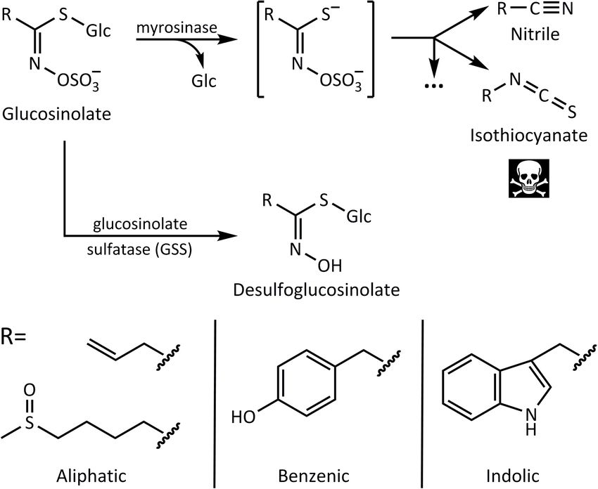
FIGURE 1 | Glucosinolate activation by myrosinase leads to nitriles and toxic isothiocyanates, whereas desulfation by glucosinolate sulfatase (GSS) forms
desulfoglucosinolates that cannot be activated. Below are examples of side chains of aliphatic, benzenic, and indolic glucosinolates derived from different amino acid
precursors.
In this study, we aimed to identify and characterize the Chemicals
enzyme(s) performing glucosinolate desulfation in B. tabaci. Solvents used were HPLC grade and were obtained from
Several genes encoding potential GSSs were cloned and VWR (Darmstadt, Germany). Glucosinolates were obtained from
heterologously expressed, and the resulting proteins were tested Sigma-Aldrich (Munich, Germany) and Phytoplan (Heidelberg,
for activity. However, only a single GSS was observed to Germany). Arylsulfatase from Helix pomatia was obtained from
efficiently desulfate glucosinolates in B. tabaci, and this enzyme Sigma-Aldrich and further processed as in Graser et al. (2000)
had a strong preference for indolic glucosinolates. Silencing and was used for preparation of desulfoglucosinolate standards
of BtGSS gene expression in vivo reduced the proportion by incubation of pure glucosinolates.
of desulfoglucosinolates in the excreted honeydew relative
to intact glucosinolates, confirming its role in glucosinolate Bemisia tabaci Sulfatase Candidate
metabolism in this insect.
Selection and Their Tissue-Specific
Expression Levels
MATERIALS AND METHODS The gene sequences of the putative B. tabaci MEAM1
GSSs were obtained from the Whitefly Genome Database
Plants and Insects (whiteflygenomics.org, downloaded January 26, 2017) and
Bemisia tabaci (MEAM1, formerly known as biotype B) was NCBI (downloaded July 11, 2018). B. tabaci arylsulfatase gene
reared in the Leibniz Institute DSMZ (German Collection of candidates were picked based on BLAST similarities to the
Microorganisms and Cell Cultures), Braunschweig, Germany sequence of the previously published P. xylostella GSS (PxGSS,
and in the Hebrew University of Jerusalem, Israel on eggplant Px0181041 ). The initial screening of B. tabaci sulfatase candidates
[Solanum melongena (L.)] and Brussels sprouts [Brassica oleracea included the estimation of ratios of gene expression between
(L.)] plants. A. thaliana Col-0 was cultivated in a controlled-
environment growth chamber under short day conditions 1
http://iae.fafu.edu.cn/DBM/genePage.php?id=Px018104, downloaded on
(9.5:13.5 h, L:D, 140 µmol/m2 /s photosynthetic photon flux 01/15/2017, and http://www.insect-genome.com/search/gene_detail1.php?id=
density, 21◦ C, 50–60% relative humidity). 790752, downloaded on 05/18/2021
Frontiers in Plant Science | www.frontiersin.org 3 June 2021 | Volume 12 | Article 671286Manivannan et al. Bemisia tabaci Glucosinolate Sulfatase
dissected B. tabaci gut tissues and whole insect bodies, using were performed in duplicates for screening and triplicates
previously published data (Ye et al., 2014; Luan et al., 2015) for product quantification. Enzymatic assays with recombinant
SRR835757 and SRR1523522, previously also used for gene P. xylostella GSS were used as a positive control. The formation
expression analysis in Jing et al. (2016). Expression levels in of desulfated glucosinolate products was quantified by high-
FPKM were estimated using DNAStar QSeq with the software performance liquid chromatography mass spectrometry (HPLC–
parameters used in Vogel et al. (2014). MS/MS) using external calibration curves.
Phylogenetic Analysis of GSSs and Polyhistidine-Tagged Protein Purification
Arylsulfatase Candidates The polyhistidine tagged, heterologously produced B. tabaci
A multiple alignment of the nucleotide sequences of the GSS BtGSS was affinity-purified from the extracellular culture
putative B. tabaci sulfatases and P. xylostella GSS was performed medium. First, the medium was collected and concentrated to
using the MUSCLE algorithm (Edgar, 2004) in the Geneious 1 ml using centrifugal filter units (Amicon Ultra Centrifugal
Prime software (default parameters, eight maximum iterations). Filters; Merck, Darmstadt, Germany) with a molecular weight
The phylogenetic relationships among sequences were inferred cut-off of 10,000 Da. Purification over Ni-NTA agarose resin
using the neighbor-joining method and the Tamura-Nei genetic (Qiagen, Hilden, Germany) followed the protocol of Eakteiman
distance model, with the reliability of the tree branching tested et al. (2018).
with 1,000 bootstrap replicates, using the Geneious software.
Determination of B. tabaci GSS Optimal
Primer Design, PCR Amplification, and Temperature and pH
Cloning of Candidate Genes For determination of optimal reaction temperature, BtGSS was
The gene sequences of the putative B. tabaci MEAM1 GSSs incubated with allyl glucosinolate (2 mM) in Tris buffer (100 mM,
(obtained as described above) were used for the design of pH 7.5) for 10 min at 10, 15, 25, 35, 45, 55, 65, and 75◦ C.
primers for the amplification of the respective open reading The enzyme reactions were stopped using acetic acid (10% v/v).
frames (ORFs). Primer design was carried out using the Enzymatic assays were performed in duplicate. The desulfated
Geneious software (version 10.0.5; Biomatters Ltd., Auckland, allyl glucosinolate product formed was quantified by HPLC–
New Zealand). Full-length ORFs were amplified using Phusion MS/MS.
HF Polymerase (Thermo Scientific, Darmstadt, Germany) For determination of optimum pH, BtGSS was incubated
using B. tabaci cDNA as template for amplification. DNA with allyl glucosinolate (2 mM) in each of the following buffers:
fragments were then extracted from agarose gels using either broad range: phosphate citrate (0.2 M phosphate, 0.1 M citrate;
QIAquick Gel Extraction Kit (Qiagen, Hilden, Germany) or pH 3.0, 4.0, 5.0, 6.0, 7.0), Tris–HCl (0.1 M; pH 7.0, 8.0), and
the Zymoclean Gel DNA Recovery Kit (Zymo Research, glycine–NaOH buffer (0.2 M; pH 8.0, 9.0); narrow range: sodium
Freiburg im Breisgau, Germany) as per the manufacturer’s phosphate (0.1 M; pH 6.25, 6.50, 6.75, 7.00, 7.25, 7.50, 7.75,
instructions and cloned into the pIB/V5-His TOPO TA 8.00). Reactions were carried out for 10 min at 25◦ C and
expression vector (Thermo Scientific). Final plasmids were stopped using acetic acid (10% v/v). Assays were performed in
transfected into Sf9 cells by lipid-mediated transfection using duplicate. The desulfated allyl glucosinolate product formed was
FuGENE Transfection Reagent (Promega, Mannheim, Germany) quantified by HPLC–MS/MS.
according to the manufacturer’s instructions for recombinant
protein expression. A pIB/V5-His TOPO TA plasmid containing Quantitative Real-Time PCR Analysis of
the corresponding full-length PxGSS from P. xylostella was also Basal BtGSS Expression
transfected and used as a positive expression and activity control. To check for the expression levels of BtGSS, quantitative
Spodoptera frugiperda Sf9 cells (Life Technologies, Darmstadt, real-time PCR (qPCR) analyses were performed using cDNA
Germany) were cultured in Sf-900 II serum-free medium (Life generated from four biological replicates of B. tabaci reared
Technologies). Adherent cell cultures were maintained at 27◦ C on eggplant (non-glucosinolate-containing diet) and Brussels
and subcultured every 3–4 days. Collected cells and media were sprouts (glucosinolate-containing diet). Ribosomal protein L-13
separated by gentle centrifugation at 500 × g for 5 min, with cell (Bta04282) was used as a reference gene. Primers were designed
pellets then re-suspended and homogenized to generate crude with an optimal melting temperature of 60◦ C. The list of primers
cell-free protein extracts. is available in Supplementary Table 1. Each reaction was done
in triplicate. Expression levels of BtGSS were relatively quantified
Screening Bemisia tabaci Arylsulfatase using the 2−11CT method (Pfaffl, 2001) and are presented as
Candidates for GSS Activity means ± standard errors.
To identify B. tabaci GSSs, in vitro enzyme assays were performed
with crude protein extracts and media of cells heterologously Silencing of BtGSS Expression in vivo
expressing B. tabaci arylsulfatases. Reactions were performed in and Resulting Metabolic Changes
Tris buffer (100 mM, pH 7.5), to which 2–5 mM of glucosinolates Silencing experiments were carried out using artificial diets
being tested was added. The reactions were incubated at 25◦ C containing sucrose, 4msob glucosinolate, and two double-
for 30 min and stopped using acetic acid (10% v/v). Assays stranded RNases (dsRNAs), one targeting dsRNase2 and the
Frontiers in Plant Science | www.frontiersin.org 4 June 2021 | Volume 12 | Article 671286Manivannan et al. Bemisia tabaci Glucosinolate Sulfatase
other BtGSS, or a scrambled BtGSS-derived sequence used as a as a control for non-enzymatic glucosinolate degradation. The
negative control (synthesis and cloning into pUC57 backbone intact glucosinolates remaining in each reaction sample were
by GenScript, Leiden, Netherlands). Initial amplification was quantified by HPLC–MS/MS.
carried out from B. tabaci cDNA (or pUC57 plasmid for the
scrambled sequence) as template, using gene-specific primers Quantitative Tests of BtGSS Substrate
containing the T7 polymerase site (Supplementary Table 2). The Preference With Selected Pure
PCR products were used as templates for in vitro transcription
reactions using the MEGAscript kit (Thermo Fisher Scientific).
Glucosinolates
The products obtained were quantified using Nanodrop (Thermo To test the substrate preference of the B. tabaci GSS, enzyme
Fisher Scientific), and the integrity of the dsRNA was confirmed assays were performed with selected pure glucosinolates: 4msob-,
on a 1.2% denaturing agarose gel. Feeding experiments were i3m-, allyl-, and pOHBn-glucosinolates. Purified BtGSS enzyme
carried out in glass vials, with 50–100 whiteflies per vial. Aqueous (1 µg) was added to Tris buffer (0.1 M, pH 7.5, 100 µl total
artificial diets were enclosed between two thin layers of stretched volume) and was incubated with 2 mM glucosinolate extract at
parafilm and contained 0.5 µg/µl BtGSS (or scrambled BtGSS) 25◦ C. Reactions proceeded for 10 min and were then stopped
dsRNA, 0.5 µg/µl dsRNase2 dsRNA, 0.29 M sucrose, and 5 mM with acetic acid (10% v/v). Enzymatic assays were performed in
4msob glucosinolate. Silencing was carried out over a period of triplicate, and no-enzyme reactions served as a control for non-
48 h. At the end of the feeding period, whiteflies and honeydew enzymatic glucosinolate degradation. The desulfoglucosinolates
were collected for gene expression and metabolite analysis, formed from 4msob and i3m glucosinolates were quantified
respectively. Whiteflies were homogenized in 500 µl TRIzol relative to external calibration curves of the respective products
reagent (Thermo Fisher Scientific) for total RNA isolation, and by HPLC–MS/MS, as described below.
cDNA synthesis was carried out with 0.5 µg total RNA using
SuperScript IV Reverse Transcriptase (Thermo Fisher Scientific) High-Performance Liquid
according to the manufacturer’s protocol. Honeydew samples Chromatography Mass Spectrometry
were processed by washing the glass vials twice with 500 µl Analyses of desulfated and intact glucosinolates in honeydew
methanol, concentrating under nitrogen flow and re-suspending and enzyme assays were performed on an Agilent Technologies
in 200 µl water. Honeydew samples were used for quantification (Santa Clara, CA, United States) 1200 Series HPLC using
of 4msob desulfoglucosinolate and non-metabolized 4msob a Nucleodur Sphinx RP column (250 × 4.6 mm × 5 µm;
glucosinolate by HPLC–MS/MS. Macherey-Nagel, Düren, Germany) coupled to an API 3200
triple-quadrupole mass spectrometer (Applied Biosystems,
Glucosinolate Extraction From Darmstadt, Germany). Formic acid (0.2%) in water and
Arabidopsis thaliana acetonitrile were employed as mobile phases A and B,
Leaves from A. thaliana Col-0 rosette stage plants were harvested, respectively. The flow rate was 1.1 ml/min. The elution
flash frozen using liquid N2 , and freeze-dried (Alpha 1-4 LDplus profile was as follows: 0–2.5 min, 1.5% B; 2.5–5 min, 1.5–10% B;
freeze dryer; Martin Christ, Osterode am Harz, Germany). 5–12.5 min, 10–40% B; 12.5–17.5 min, 40–70% B; 17.6–20 min,
Intact glucosinolates were extracted with 10 ml 80% (v/v) 100% B; and 20.1–24 min, 1.5% B. The ion spray voltage was
methanol:water per g dry weight (DW) on ice under continuous maintained at 4,500 eV. The turbo gas temperature was set to
shaking for 5 min, followed by the addition of metal beads 700◦ C. Nebulizing gas was set at 70 psi, curtain gas at 20 psi,
(3 mm) and vigorous shaking for 10 min in a paint shaker. heating gas at 60 psi, and collision gas at 10 psi. Multiple reaction
The supernatant collected after centrifugation (4,000 × g at monitoring (MRM, Supplementary Tables 3, 4) was used to
4◦ C for 15 min) was then processed through centrifugal filter monitor parent ion to fragment ion conversion (Malka et al.,
units (Amicon Ultra Centrifugal Filters) with a molecular 2016). Analyst 1.5 software (Applied Biosystems) was used for
weight cut-off of 10,000 Da to separate glucosinolates from data acquisition and processing.
the plant myrosinase. The flow-through was then treated on Analyses of desulfated and intact glucosinolates during RNAi
a rotatory evaporator for removal of the solvent and was silencing experiments were performed on an identical HPLC
resuspended in water. setup as those above, but coupled to an API 6500 triple-
quadrupole mass spectrometer (Applied Biosystems). Formic
acid (0.05%) in water and acetonitrile were employed as mobile
Tests of Bemisia tabaci Sulfatase phases A and B, respectively. The elution profile was identical
Substrate Preference With Arabidopsis to the abovementioned but with a flow rate of 1.0 ml/min. Ion
Glucosinolates spray voltages were maintained at 4,500 eV (positive mode for
To test the substrate preference of the B. tabaci GSS, enzyme desulfoglucosinolates) and -4,500 eV (negative mode for intact
assays were performed with the glucosinolate extract obtained glucosinolates). The turbo gas temperature was set to 650◦ C.
from A. thaliana Col-0 leaves. Purified BtGSS was added to Curtain gas was set at 40 psi, collision gas at medium, ion source
Tris buffer (100 mM, pH 7.5) and incubated with glucosinolate gas 1 at 60 psi, and ion source gas 2 at 60 psi. MRM was used to
extract at 25◦ C. Aliquots were taken at different time points (0– monitor parent ion to fragment ion conversion (Supplementary
300 min) and mixed with acetic acid (10% v/v). Enzymatic assays Table 5). Analyst 1.6.3 software (Applied Biosystems) was used
were performed in duplicate, and no-enzyme reactions served for data acquisition and processing.
Frontiers in Plant Science | www.frontiersin.org 5 June 2021 | Volume 12 | Article 671286Manivannan et al. Bemisia tabaci Glucosinolate Sulfatase
RESULTS insects grown on both plants. BtGSS was constitutively and
highly expressed in insects grown on both plant species tested
Identification of Putative Bemisia tabaci (Supplementary Figure 2).
GSS
The protein sequence of the previously identified P. xylostella
Silencing of BtGSS Supports Its Role as
GSS (Ratzka et al., 2002) was used as a query to identify the Major GSS in Bemisia tabaci
B. tabaci sulfatases (BtSulf) in the published B. tabaci MEAM1 To examine whether BtGSS acts to desulfate glucosinolates
draft genome (Chen et al., 2016) that might serve as GSSs. in vivo, we manipulated the expression of its encoding gene BtGSS
Candidates were sought that had high similarity to PxGSS and using an RNAi approach. For this purpose, dsRNA targeting
a high expression level of the encoding genes in B. tabaci BtGSS was synthesized using in vitro transcription and fed to
guts relative to the whole insect based on publicly available B. tabaci adults in artificial diets. Since non-specific dsRNases
expression data (Table 1 and Figure 2). Most candidates had been in B. tabaci could cause dsRNA degradation in the whiteflies,
previously annotated as arylsulfatases. Selected sequences were artificial diets also contained dsRNA targeting dsRNase2, which
cloned from B. tabaci (species MEAM1) whole-insect cDNA and had been previously shown to enhance the silencing of genes
heterologously expressed in Sf9 cells, and the produced protein of interest (Luo et al., 2017). This strategy led to a decrease of
was subsequently screened for GSS activity. 43% in the expression of BtGSS in B. tabaci compared with a
scrambled dsRNA control (Figure 4A). After dsRNA treatment,
Bemisia tabaci BtSulf9 Encodes a GSS the proportion of 4msob-desulfoglucosinolate relative to non-
metabolized intact 4msob-glucosinolate in honeydew samples
Enzyme (BtGSS)
was 48% lower in BtGSS-silenced adults than in scrambled
Previous reports showed that PxGSS produced in Escherichia
dsRNA-fed control insects (Figure 4B). Both the extent of
coli had no sulfatase activity (Ratzka et al., 2002), suggesting
BtGSS gene silencing and the differences in desulfoglucosinolate
that those cells lacked the post-translational modification
formation indicated a high variability among experimental
machinery necessary to generate an active GSS. Hence, insect
groups. However, these two factors were linearly correlated, with
cells (S. frugiperda Sf9 cells) were used here for expressing
higher BtGSS expression corresponding to higher proportions of
the B. tabaci GSS candidates. The full-length ORFs of selected
desulfoglucosinolate products in honeydew (Figure 4C).
B. tabaci sulfatases (BtSulf1, 3, 5–9, and 11) were successfully
expressed to produce V5-/His-tagged proteins. The resulting
BtGSS Shows Preference Toward Indolic
B. tabaci enzymes were screened for their activity toward
pure glucosinolates: allyl-(sinigrin), p-hydroxybenzyl-(pOHBn, Glucosinolates
sinalbin), and 4-methylsulfinylbutyl-(4-msob, glucoraphanin) To compare BtGSS activity toward glucosinolates with different
glucosinolates. Heterologously produced PxGSS and commercial side chains as substrates, the enzyme was incubated for different
H. pomatia sulfatase were used as positive controls for GSS periods with pure glucosinolates and with a glucosinolate
activity. The resulting desulfoglucosinolate products were extract obtained from A. thaliana Col-0 leaves, which contained
detected by HPLC–MS/MS. Intact glucosinolates also gave rise both aliphatic and indolic glucosinolates (Brown et al., 2003).
to MS signals corresponding to desulfated glucosinolates, due BtGSS desulfated all glucosinolates offered, both as pure
to in-source fragmentation (spontaneous sulfate loss) in the compounds and within the A. thaliana extract (Figures 3A–
mass spectrometer. However, these artifactual desulfated D and Supplementary Figure 1A–H). However, indolic
glucosinolates were readily distinguished from genuine glucosinolates were degraded much more rapidly than aliphatic
desulfoglucosinolates existing before analysis due to different glucosinolates (Figure 3E). All glucosinolates were stable under
retention times. These analyses revealed that BtSulf9 had non-enzymatic control conditions (Supplementary Figure 3).
GSS activity toward all tested glucosinolates (Figure 3 and This confirmed that BtGSS acts on different glucosinolate classes
Supplementary Figure 1) and this was henceforth named BtGSS. and suggested additionally that it might have distinct substrate
Both BtGSS and PxGSS were secreted by the Sf9 cells into the affinities/catalytic efficiencies toward them. We confirmed this
extracellular culture medium. BtGSS purified by metal affinity using two selected pure glucosinolates for in vitro incubations
chromatography had an optimum temperature around 50–55◦ C with BtGSS, 4msob- and i3m-glucosinolates. BtGSS desulfated
and an optimum pH between 7.5 and 7.8. None of the other i3m-glucosinolate, the most abundant indolyl glucosinolate in
produced sulfatases tested displayed detectable GSS activity A. thaliana Col-0, with a catalytic efficiency of 466 nmol/mg
in vitro, including BtSulf1 (the closest homolog to PxGSS) and BtGSS/min. This was 2.93 times more efficient than the
those in the same phylogenetic branch as BtGSS (BtSulf7, 8, 11). conversion of 4msob-glucosinolate (159 nmol/mg BtGSS/min),
its most abundant aliphatic counterpart in A. thaliana and
BtGSS Expression Is Independent of several Brassica crops.
Glucosinolate Ingestion
To compare the relative expression levels of BtGSS in B. tabaci DISCUSSION
reared on non-glucosinolate diet (eggplant) and those reared
on a glucosinolate-rich diet (Brussels sprouts), we analyzed Although cruciferous plants possess a sophisticated
the expression of BtGSS by qPCR in B. tabaci MEAM1 adult glucosinolate–myrosinase defense system to thwart their
Frontiers in Plant Science | www.frontiersin.org 6 June 2021 | Volume 12 | Article 671286Manivannan et al. Bemisia tabaci Glucosinolate Sulfatase
TABLE 1 | Predicted Bemisia tabaci sulfatase (“BtSulf”) candidates, ordered based on sequence similarity scores (e-values) to Plutella xylostella GSS (PxGSS).
MEAM MED
NCBI entry Bta entry Name e-value MEAM gut MEAM adults Expression MED gut MED
(vs. PxGSS) (FPKM) (FPKM) ratios, gut vs. (FPKM) adults
whole adults (FPKM)
XM 019054398 Bta06756 BtSulf1 2,60E-122 0 216 0 0 138
XM 019062599 Bta04898 BtSulf2 4,21E-112 0 81 0 0 20
XM 019061487 Bta03550 BtSulf3 1,18E-107 0 70 0 3 488
XM 019062598 Bta04899 BtSulf4 2,09E-107 0 248 0 1 275
XM 019051255 Bta02222 BtSulf5 1,50E-106 460 675 1,5 lower 370 945
XM 019052592 Bta04774 BtSulf6 1,11E-101 16 277 17,3 lower 5 103
XM 019059032 Bta14665 BtSulf7 5,36E-100 48 272 5,7 lower 81 201
XM 019058912 Bta14669 BtSulf8 1,02E-97 0 20 0 0 93
XM 019059016 Bta14666 BtSulf9 4,87E-96 8526 1203 7,1 higher 7296 135
XM 019049969 Bta01141 BtSulf10 2,88E-83 10 39 3,9 lower 96 62
XM 019059017 Bta14667 BtSulf11 1,76E-65 10 0 higher 2 0
XM 019042567 Bta08750 BtSulf12 5,73E-21 2627 9434 3,6 lower 1587 9732
XM 019054362 Bta06730 BtSulf13 7,94E-17 18 468 26,0 lower 12 746
XM 019049823 Bta01054 BtSulf14 3,13E-13 40 207 5,2 lower 15 55
XM 019053003 Bta05280 BtSulf15 3,59E-08 51 300 5,9 lower 38 360
Genes are denoted according to their accession numbers in NCBI and the whitefly genome databases. Estimated expression levels (FPKM) for these genes in gut tissues
and whole insect bodies were calculated from publicly available data for B. tabaci MEAM1 insects and, as a reference, also for B. tabaci MED insects.
herbivores, several insects possess mechanisms to detoxify gut lumen, where it can efficiently detoxify potentially harmful
these plant defensive metabolites. One such strategy is to glucosinolates after ingestion. This modus operandi mimics that
employ a GSS enzymatic system to convert glucosinolates to of the P. xylostella GSS, which is secreted from gut epithelial cells
their corresponding desulfated forms, and thus prevent them and is present in high amounts in the larval gut lumen (Ratzka
from being broken down to toxic downstream products by et al., 2002). Nevertheless, the B. tabaci BtGSS we identified is
myrosinases (Ratzka et al., 2002; Falk and Gershenzon, 2007). not the sulfatase with closest homology to the P. xylostella GSS in
Interestingly, while all previously described GSS activities the whitefly. The sulfatase designated BtSulf1 is phylogenetically
had come from leaf-chewing herbivores, this detoxification closest to PxGSS but did not have GSS activity in vitro. These
mechanism was recently reported from the phloem-feeding data suggest that GSS activity evolved independently in whiteflies
herbivore B. tabaci (Malka et al., 2016). It was once believed and lepidopterans from sulfatases performing different innate
that phloem-feeding insects, by delicate piercing of plant functions in these distant insect lineages. In fact, BtGSS was the
tissues and stealthy sucking of the plant sap, do not strongly only sulfatase for which GSS activity was detected among all
activate the glucosinolate–myrosinase system (Walling, 2008; BtSulf candidates tested in this study. These included BtSulf 7,
Pentzold et al., 2014). However, more recent studies showed 8, and 11, whose sequences were most closely related to BtGSS,
that phloem feeding indeed leads to glucosinolate activation but did not show GSS activity in vitro. Additionally, RNAi-
and production of isothiocyanates (Walling, 2008; Danner et al., mediated silencing of BtGSS in artificial diet-fed whiteflies led to a
2018). These recent results clarify the advantage of efficiently and decrease in the proportion of desulfoglucosinolates in honeydew
pre-emptively detoxifying glucosinolates in piercing–sucking relative to unmetabolized intact glucosinolates. This suggests that
insects, such as whiteflies. As the B. tabaci species complex is a BtGSS is indeed the major sulfatase responsible for glucosinolate
major agricultural pest, it was of interest to identify the GSS(s) desulfation in B. tabaci MEAM1.
performing glucosinolate detoxification in this insect. We found that BtGSS exhibits a clear preference in vitro
We successfully identified one GSS (BtGSS) in B. tabaci toward indolic glucosinolates rather than their aliphatic
MEAM1 that actively desulfated different glucosinolates in vitro. counterparts in A. thaliana Col-0. This result is in accordance
The heterologously produced enzyme displayed optimum GSS with the higher proportion of indolic glucosinolates found to be
activity at pH 7.5–7.8 and was secreted into the extracellular desulfated relative to their aliphatic counterparts in honeydew
culture medium by Sf9 cells, in agreement with the presence of of B. tabaci feeding on Brussels sprouts and A. thaliana (Malka
a native signal peptide guiding BtGSS for secretion. Additionally, et al., 2016). Thus, B. tabaci might employ its GSS detoxification
available transcriptomic data suggested an elevated expression machinery to specifically combat the induction and activation of
of this enzyme in the B. tabaci gut tissues relative to the indolic glucosinolates that takes place during phloem sap-feeding
rest of the body in both MEAM1 and MED B. tabaci species (Kim et al., 2008). This substrate preference also complements
(formerly named “B” and “Q” biotypes, respectively). These data well another preemptive glucosinolate detoxification mechanism
together suggest that this enzyme is secreted into the B. tabaci in B. tabaci, the formation of glucosylated glucosinolates.
Frontiers in Plant Science | www.frontiersin.org 7 June 2021 | Volume 12 | Article 671286Manivannan et al. Bemisia tabaci Glucosinolate Sulfatase FIGURE 2 | Phylogenetic relationships of predicted Bemisia tabaci sulfatases (BtSulf), including BtSulf9 (BtGSS, italicized) and P. xylostella GSS (PxGSS). Gene names are as described in Table 1, and bootstrap values (%) are shown next to each node. Analysis of honeydew from B. tabaci feeding on A. thaliana production was increased above natural levels in transgenic lines, showed that glucosylation was found to occur mostly to aliphatic B. tabaci development and performance was negatively affected glucosinolates and few glucosylated indolic glucosinolate (Elbaz et al., 2012; Markovich et al., 2013). Moreover, myb51, derivatives were detected (Malka et al., 2020). a positive regulator of indolic glucosinolate production, is up- The existence of efficient pre-emptive detoxification pathways regulated in A. thaliana after infestations by the phloem-feeding in B. tabaci can help explain the apparent lack of preference insects B. tabaci, M. persicae, and Brevicoryne brassicae (Foyer of MEAM1 and MED whiteflies for plants with lower vs. et al., 2015). Taken together, these findings suggest that plants higher glucosinolate contents (Yang et al., 2020); that is, as specifically induce the production of indolic glucosinolates as these B. tabaci species efficiently deactivate glucosinolates, a response against phloem-feeding insects. These glucosinolates they can feed on plants producing natural concentrations of could be activated by one of the recently characterized non- glucosinolates with impunity. However, when glucosinolate canonical glucosinolate-β-thioglucosidases, such as PEN2 or Frontiers in Plant Science | www.frontiersin.org 8 June 2021 | Volume 12 | Article 671286
Manivannan et al.                                                                                                                  Bemisia tabaci Glucosinolate Sulfatase

  FIGURE 3 | BtGSS has activity with a variety of glucosinolates. (A–D) LC–MS extracted MRM chromatograms of desulfated glucosinolates produced in vitro by
  BtGSS. The early eluting peaks in each chromatogram indicate genuine desulfoglucosinolates formed during the enzyme assay, whereas the later eluting peaks
  show desulfoglucosinolates formed from intact glucosinolates via in-source fragmentation during MS analysis and so actually indicate the presence of intact sulfated
  glucosinolates used as substrates. (E) The time-dependent degradation of a mixture of intact glucosinolates from Arabidopsis thaliana Col-0, with indolic
  glucosinolates being depleted more quickly than aliphatic ones (amount of each glucosinolate present at 0 min was taken as 100%, N = 2 independent reactions).
  “ds-” = desulfated glucosinolates; 3msop = 3-methylsulfinylpropyl; 4msob = 4-methylsulfinylbutyl; 4mtb = 4-methylthiobutyl; 5msop = 5-methylsulfinylpentyl;
  7msoh = 7-methylsulfinylheptyl; 8msoo = 8-methylsulfinyloctyl; i3m = indol-3-ylmethyl; 4moi3m = 4-methoxyindol-3-ylmethyl.

  FIGURE 4 | BtGSS silencing in vivo. (A) Dietary administration of dsRNA (N = 3 per treatment; statistical differences determined via t-tests) targeting BtGSS led to
  diminished expression of this gene (relative to rpl-13) compared with a treatment using a scrambled dsRNA control. (B) Silencing reduced the proportion of
  desulfo-4msob glucosinolate relative to intact glucosinolate present in whitefly honeydew. (C) BtGSS expression was positively correlated to the proportions of
  desulfo-4msob glucosinolate:intact glucosinolate in honeydew.

PYK10 (Bednarek et al., 2009; Clay et al., 2009; Nakano et al.,                         said to have strong antifeedant and toxic effects on crucifer-
2017), which may be more specific to indolic glucosinolates.                            consuming insects (Kim and Jander, 2007; Agerbirk et al., 2008;
Plants can also convert indol-3-ylmethyl glucosinolate into more                        Kim et al., 2008).
bioactive methoxylated forms as a defensive response (Kim                                  Quantitative real-time PCR analysis showed that the gene
and Jander, 2007; Wiesner et al., 2013; Pfalz et al., 2016). The                        encoding the identified B. tabaci GSS (BtGSS) is highly and
enzymatic breakdown products of indolic glucosinolates are                              constitutively expressed irrespective of whether the insect

Frontiers in Plant Science | www.frontiersin.org                                    9                                             June 2021 | Volume 12 | Article 671286
Manivannan et al.                                                                                             Bemisia tabaci Glucosinolate Sulfatase

consumed glucosinolates or not. This suggests that this GSS                 of glucosinolates (especially indolic glucosinolates) upon
could have other function(s) for the insect, perhaps serving as a           infestation by phloem feeders. Therefore, the strategies used by
more general arylsulfatase. While the in vivo functions of insect           phloem-feeding insects that feed on glucosinolate-containing
(aryl)sulfatases are still not well-defined, one of their proposed          plants to circumvent these defensive metabolites deserve
roles is during the molting process, for example, of southern               further attention.
armyworm larvae (Yang et al., 1973). It is known that sulfate                  The whitefly B. tabaci is one such crucifer-colonizing,
conjugates of ecdysone, the steroidal insect molting hormone,               phloem-feeding insect, which has been reported to efficiently
are less bioactive than non-sulfated analogs and arylsulfatase              detoxify glucosinolates by conversion to their corresponding
titers increase upon molting (King, 1972; Karlson and Koolman,              desulfated forms. This species complex is a group of serious
1973; Yang et al., 1973). Arylsulfatases could, therefore, be               agricultural pests that causes extensive damage to crops
involved in converting ecdysone to its active form. However,                including cruciferous vegetables, and also vectors many plant
ecdysteroid localization is typically limited to ecdysteroidogenic          pathogenic viruses. Here, we have successfully identified
glands and hemolymph (Gilbert et al., 2002). According to                   and characterized a B. tabaci MEAM1 sulfatase that is
previous studies of the P. xylostella GSS and S. gregaria GSS,              responsible for this metabolic reaction. This enzyme exhibits
and in accordance with what we observed for B. tabaci, GSS                  a strong preference in vitro toward indolic glucosinolates in
is secreted into the gut lumen (Ratzka et al., 2002; Falk and               comparison with aliphatic glucosinolates, implying that B. tabaci
Gershenzon, 2007). Therefore, it seems unlikely that BtGSS is               employs this GSS detoxification mechanism to cope with
involved in ecdysone metabolism, and its other potential roles              the negative effects of indolic glucosinolate breakdown. This
in vivo remain unknown.                                                     strategy seems to complement well the other glucosinolate
    In this work, we have characterized the BtGSS gene expression           detoxification pathways present in B. tabaci (glucosylation and
levels and protein activities in B. tabaci of the MEAM1                     glutathione conjugation) and helps explain why this insect
species. Desulfoglucosinolates had, however, been previously                can feed widely on cruciferous plants. However, the relative
reported in B. tabaci MED whiteflies (Malka et al., 2016),                  importance of the individual pathways, their costs, and how
another aggressive, generalist B. tabaci species. Whether MED               each contributes to the success of B. tabaci still remains
and other B. tabaci species might have different GSS activity               to be determined.
levels, and how this might affect their establishment on
crucifers, is not yet known. Nevertheless, it seems clear that
B. tabaci uses several different biochemical strategies to deal with        DATA AVAILABILITY STATEMENT
glucosinolates. Besides the serial glucosylation of glucosinolates
                                                                            The datasets presented in this study can be found in online
(Malka et al., 2020), a glutathione-S-transferase enzyme has also
                                                                            repositories. The names of the repository/repositories
been reported in B. tabaci (Eakteiman et al., 2018) that can
                                                                            and accession number(s) can be found in the
metabolize the hydrolysis products of both major aliphatic and
                                                                            article/Supplementary Material.
indolic glucosinolates present in A. thaliana. This latter activity
might help to mitigate the potential negative effects caused
by the small amounts of glucosinolates successfully activated               AUTHOR CONTRIBUTIONS
during and after ingestion by neutralizing the toxic hydrolysis
products. Whether other strategies might also be employed by                DV designed this study with input from AM, SW, and JG. AM,
this whitefly, as in another generalist phloem-feeding insect,              KL, BI, MG, ES, MLAEE, and BS performed the data collection
M. persicae (Kim and Jander, 2007; Kim et al., 2008), remains               and analysis. RK and MR provided additional materials,
to be determined.                                                           expertise, and technical help. SW, JG, and DV supervised the
                                                                            study. AM and DV drafted the manuscript with input from all
                                                                            coauthors. All authors contributed to the article and approved the
CONCLUSION                                                                  submitted version.

The glucosinolate–myrosinase system is a sophisticated
two-component chemical defense strategy present in plants                   ACKNOWLEDGMENTS
of the order Brassicales. The constituents of this binary
                                                                            We would like to acknowledge the Max Planck Society for
defense system are kept spatially segregated, but during
                                                                            funding this research and the publication of this manuscript. We
herbivory, myrosinases catalyze the hydrolysis of glucosinolates
                                                                            thank Osnat Malka and Shai Morin (The Hebrew University of
forming toxic breakdown products. In contrast to leaf-chewing
                                                                            Jerusalem) for generously providing B. tabaci cDNA.
insects, which cause extensive damage during herbivory
and activate the glucosinolate–myrosinase system, it was
long assumed that phloem-feeding insects did not cause                      SUPPLEMENTARY MATERIAL
sufficient damage for activation. However, glucosinolate
hydrolysis products have been detected during attack by                     The Supplementary Material for this article can be found
aphids; and these hydrolysis products negatively affect phloem-             online at: https://www.frontiersin.org/articles/10.3389/fpls.2021.
feeding insects. Moreover, plants induce the production                     671286/full#supplementary-material

Frontiers in Plant Science | www.frontiersin.org                       10                                     June 2021 | Volume 12 | Article 671286
Manivannan et al. Bemisia tabaci Glucosinolate Sulfatase
REFERENCES Höglund, A. S., Lenman, M., Falk, A., and Rask, L. (1991). Distribution of
myrosinase in rapeseed tissues. Plant Physiol. 95, 213–221. doi: 10.1104/pp.95.
Agerbirk, N., De Vos, M., Kim, J. H., and Jander, G. (2008). Indole glucosinolate 1.213
breakdown and its biological effects. Phytochem. Rev. 8:101. doi: 10.1007/ Hopkins, R. J., van Dam, N. M., and van Loon, J. J. A. (2009). Role of glucosinolates
s11101-008-9098-0 in insect-plant relationships and multitrophic interactions. Annu. Rev. Entomol.
Ahn, S. J., Betzin, F., Gikonyo, M. W., Yang, Z. L., Kollner, T. G., and Beran, F. 54, 57–83. doi: 10.1146/annurev.ento.54.110807.090623
(2019). Identification and evolution of glucosinolate sulfatases in a specialist Jeschke, V., Gershenzon, J., and Vassão, D. G. (2016a). A mode of action of
flea beetle. Sci. Rep. 9:15725. doi: 10.1038/s41598-019-51749-x glucosinolate-derived isothiocyanates: detoxification depletes glutathione and
Bednarek, P., Piślewska-Bednarek, M., Svatoš, A., Schneider, B., Doubský, J., cysteine levels with ramifications on protein metabolism in Spodoptera littoralis.
Mansurova, M., et al. (2009). A glucosinolate metabolism pathway in living Insect Biochem. Mol. Biol. 71, 37–48. doi: 10.1016/j.ibmb.2016.02.002
plant cells mediates broad-spectrum antifungal defense. Science 323, 101–106. Jeschke, V., Gershenzon, J., and Vassão, D. G. (2016b). Insect detoxification of
doi: 10.1126/science.1163732 glucosinolates and their hydrolysis products. Adv. Bot. Res. 80, 199–245. doi:
Blažević, I., Montaut, S., Burčul, F., Olsen, C. E., Burow, M., Rollin, P., et al. 10.1016/bs.abr.2016.06.003
(2020). Glucosinolate structural diversity, identification, chemical synthesis and Jing, X., White, T. A., Luan, J., Jiao, C., Fei, Z., and Douglas, A. E. (2016).
metabolism in plants. Phytochemistry 169:112100. doi: 10.1016/j.phytochem. Evolutionary conservation of candidate osmoregulation genes in plant phloem
2019.112100 sap-feeding insects. Insect Mol. Biol. 25, 251–258. doi: 10.1111/imb.12215
Boykin, L. M., Armstrong, K. F., Kubatko, L., and De Barro, P. (2012). Species Karlson, P., and Koolman, J. (1973). On the metabolic fate of ecdysone and 3-
delimitation and global biosecurity. Evol. Bioinform. 8, 1–37. doi: 10.4137/EBO. dehydroecdysone in Calliphora vicina. Insect Biochem. 3, 409–417. doi: 10.1016/
S8532 0020-1790(73)90074-7
Boykin, L. M., Kinene, T., Wainaina, J. M., Savill, A., Seal, S., Mugerwa, H., et al. Kawakishi, S., and Kaneko, T. (1987). Interaction of proteins with allyl
(2018). Review and guide to a future naming system of African Bemisia tabaci isothiocyanate. J. Agric. Food Chem. 35, 85–88. doi: 10.1021/jf00073a020
species. Syst. Entomol. 43, 427–433. doi: 10.1111/syen.12294 Kim, J. H., and Jander, G. (2007). Myzus persicae (green peach aphid) feeding on
Brown, P. D., Tokuhisa, J. G., Reichelt, M., and Gershenzon, J. (2003). Variation Arabidopsis induces the formation of a deterrent indole glucosinolate. Plant J.
of glucosinolate accumulation among different organs and developmental 49, 1008–1019. doi: 10.1111/j.1365-313X.2006.03019.x
stages of Arabidopsis thaliana. Phytochemistry 62, 471–481. doi: 10.1016/S0031- Kim, J. H., Lee, B. W., Schroeder, F. C., and Jander, G. (2008). Identification of
9422(02)00549-6 indole glucosinolate breakdown products with antifeedant effects on Myzus
Chen, W., Hasegawa, D. K., Kaur, N., Kliot, A., Pinheiro, P. V., Luan, J., et al. persicae (green peach aphid. Plant J. 54, 1015–1026. doi: 10.1111/j.1365-313X.
(2016). The draft genome of whitefly Bemisia tabaci MEAM1, a global crop pest, 2008.03476.x
provides novel insights into virus transmission, host adaptation, and insecticide King, D. S. (1972). Ecdysone metabolism in insects. Am. Zool. 12, 343–345.
resistance. BMC Biol. 14:110. doi: 10.1186/s12915-016-0321-y Koroleva, O. A., Davies, A., Deeken, R., Thorpe, M. R., Tomos, A. D., and Hedrich,
Clay, N. K., Adio, A. M., Denoux, C., Jander, G., and Ausubel, F. M. (2009). R. (2000). Identification of a new glucosinolate-rich cell type in Arabidopsis
Glucosinolate metabolites required for an Arabidopsis innate immune response. flower stalk. Plant Physiol. 124, 599–608. doi: 10.1104/pp.124.2.599
Science 323, 95–101. doi: 10.1126/science.1164627 Luan, J. B., Chen, W. B., Hasegawa, D. K., Simmons, A. M., Wintermantel, W. M.,
Danner, H., Desurmont, G. A., Cristescu, S. M., and van Dam, N. M. (2018). Ling, K. S., et al. (2015). Metabolic coevolution in the bacterial symbiosis of
Herbivore-induced plant volatiles accurately predict history of coexistence, whiteflies and related plant sap-feeding insects. Genome Biol. Evol. 7, 2635–
diet breadth, and feeding mode of herbivores. New Phytol. 220, 726–738. doi: 2647. doi: 10.1093/gbe/evv170
10.1111/nph.14428 Luo, Y. A., Chen, Q. G., Luan, J. B., Chung, S. H., Van Eck, J., Turgeon, R., et al.
Eakteiman, G., Moses-Koch, R., Moshitzky, P., Mestre-Rincon, N., Vassão, D. G., (2017). Towards an understanding of the molecular basis of effective RNAi
Luck, K., et al. (2018). Targeting detoxification genes by phloem-mediated against a global insect pest, the whitefly Bemisia tabaci. Insect Biochem. Mol.
RNAi: a new approach for controlling phloem-feeding insect pests. Insect Biol. 88, 21–29. doi: 10.1016/j.ibmb.2017.07.005
Biochem. Mol. Biol. 100, 10–21. doi: 10.1016/j.ibmb.2018.05.008 Lüthy, B., and Matile, P. (1984). The mustard oil bomb: rectified analysis of the
Edgar, R. C. (2004). MUSCLE: multiple sequence alignment with high accuracy and subcellular organisation of the myrosinase system. Biochem. Physiol. Pflanzen
high throughput. Nucleic Acids Res. 32, 1792–1797. doi: 10.1093/nar/gkh340 179, 5–12. doi: 10.1016/s0015-3796(84)80059-1
Elbaz, M., Halon, E., Malka, O., Malitsky, S., Blum, E., Aharoni, A., et al. (2012). Malka, O., Easson, M. L. A. E., Paetz, C., Götz, M., Reichelt, M., Stein, B.,
Asymmetric adaptation to indolic and aliphatic glucosinolates in the B and et al. (2020). Glucosylation prevents plant defense activation in phloem-feeding
Q sibling species of Bemisia tabaci (Hemiptera: Aleyrodidae. Mol. Ecol. 21, insects. Nat. Chem. Biol. 69, 1795–1797. doi: 10.1038/s41589-020-00658-6
4533–4546. doi: 10.1111/j.1365-294X.2012.05713.x Malka, O., Shekhov, A., Reichelt, M., Gershenzon, J., Vassão, D. G., and Morin, S.
Falk, K. L., and Gershenzon, J. (2007). The desert locust, Schistocerca gregaria, (2016). Glucosinolate desulfation by the phloem-feeding insect Bemisia tabaci.
detoxifies the glucosinolates of Schouwia purpurea by desulfation. J. Chem. Ecol. J. Chem. Ecol. 42, 230–235. doi: 10.1007/s10886-016-0675-1
33, 1542–1555. doi: 10.1007/s10886-007-9331-0 Markovich, O., Kafle, D., Elbaz, M., Malitsky, S., Aharoni, A., Schwarzkopf, A.,
Foyer, C. H., Verrall, S. R., and Hancock, R. D. (2015). Systematic analysis of et al. (2013). Arabidopsis thaliana plants with different levels of aliphatic- and
phloem-feeding insect-induced transcriptional reprogramming in Arabidopsis indolyl-glucosinolates affect host selection and performance of Bemisia tabaci.
highlights common features and reveals distinct responses to specialist and J. Chem. Ecol. 39, 1361–1372. doi: 10.1007/s10886-013-0358-0
generalist insects. J. Exp. Bot. 66, 495–512. doi: 10.1093/jxb/eru491 Mithöfer, A., and Boland, W. (2012). Plant defense against herbivores: chemical
Gilbert, L. I., Rybczynski, R., and Warren, J. T. (2002). Control and biochemical aspects. Annu. Rev. Plant Biol. 63, 431–450. doi: 10.1146/annurev-arplant-
nature of the ecdysteroidogenic pathway. Annu. Rev. Entomol. 47, 883–916. 042110-103854
doi: 10.1146/annurev.ento.47.091201.145302 Nakano, R. T., Pislewska-Bednarek, M., Yamada, K., Edger, P. P., Miyahara, M.,
Graser, G., Schneider, B., Oldham, N. J., and Gershenzon, J. (2000). The methionine Kondo, M., et al. (2017). PYK10 myrosinase reveals a functional coordination
chain elongation pathway in the biosynthesis of glucosinolates in Eruca sativa between endoplasmic reticulum bodies and glucosinolates in Arabidopsis
(Brassicaceae). Arch. Biochem. Biophys. 378, 411–419. doi: 10.1006/abbi.2000. thaliana. Plant J. 89, 204–220. doi: 10.1111/tpj.13377
1812 Oliveira, M. R. V., Henneberry, T. J., and Anderson, P. (2001). History, current
Grob, K., and Matile, P. (1979). Vacuolar location of glucosinolates in horseradish status, and collaborative research projects for Bemisia tabaci. Crop Protection
root-cells. Plant Sci. Lett. 14, 327–335. doi: 10.1016/S0304-4211(79)90281-5 20, 709–723. doi: 10.1016/S0261-2194(01)00108-9
Halkier, B. A. (2016). General introduction to glucosinolates. Adv. Bot. Res. 80, Pentzold, S., Zagrobelny, M., Rook, F., and Bak, S. (2014). How insects overcome
1–14. doi: 10.1016/bs.abr.2016.07.001 two-component plant chemical defence: plant β-glucosidases as the main target
Halkier, B. A., and Gershenzon, J. (2006). Biology and biochemistry of for herbivore adaptation. Biol. Rev. 89, 531–551. doi: 10.1111/brv.12066
glucosinolates. Annu. Rev. Plant Biol. 57, 303–333. doi: 10.1146/annurev. Perring, T. M. (2001). The Bemisia tabaci species complex. Crop Protection 20,
arplant.57.032905.105228 725–737. doi: 10.1016/S0261-2194(01)00109-0
Frontiers in Plant Science | www.frontiersin.org 11 June 2021 | Volume 12 | Article 671286You can also read