Imaging of Insulinlike Growth Factor Type 1 Receptor in Prostate Cancer Xenografts Using the Affibody Molecule
←
→
Page content transcription
If your browser does not render page correctly, please read the page content below

Imaging of Insulinlike Growth Factor Type 1 Receptor in
Prostate Cancer Xenografts Using the Affibody Molecule
111In-DOTA-Z
             IGF1R:4551

Vladimir Tolmachev1, Jennie Malmberg2, Camilla Hofström3, Lars Abrahmsén4, Thomas Bergman4, Anna Sjöberg4,
Mattias Sandström5, Torbjörn Gräslund3, and Anna Orlova2
1Division of Biomedical Radiation Sciences, Department of Radiology, Oncology and Clinical Immunology, Rudbeck Laboratory,

Uppsala University, Uppsala, Sweden; 2Preclinical PET Platform, Department of Medicinal Chemistry, Uppsala University, Uppsala,
Sweden; 3Division of Molecular Biotechnology, School of Biotechnology, AlbaNova University Center, Royal Institute of Technology,
Stockholm, Sweden; 4Affibody AB, Stockholm, Sweden; and 5Medical Physics, Department of Oncology, Uppsala University Hospital,
Uppsala, Sweden

One of the pathways leading to androgen independence in
prostate cancer involves upregulation of insulinlike growth
                                                                           T     he insulinlike growth factor (IGF) axis includes 2
                                                                           insulinlike growth factors (IGF-1 and IGF-2), 2 trans-
factor type 1 receptor (IGF-1R). Radionuclide imaging of IGF-              membrane tyrosine kinase receptors (IGF-1R and IGF-
1R in tumors might be used for selection of patients who would
most likely benefit from IGF-1R–targeted therapy. The goal of
                                                                           2R), and 6 IGF binding proteins (IGFBP-1 to IGFBP-6)
this study was to evaluate the feasibility of in vivo radionuclide         (1,2). An increasing amount of evidence implicates that
imaging of IGF-1R expression in prostate cancer xenografts                 this axis, in particular IGF-1R signaling, plays a role in
using a small nonimmunoglobulin-derived binding protein called             malignant cell transformation, cancer progression, and
an Affibody molecule. Methods: The IGF-1R-binding ZIGF1R:4551              metastatic spread of different types of tumors. Increased
Affibody molecule was site-specifically conjugated with a mal-             IGF-1R signaling has been shown to result in activation of
eimido derivative of DOTA and labeled with 111In. The binding
                                                                           growth-promoting intracellular signaling pathways, in-
of radiolabeled ZIGF1R:4551 to IGF-1R–expressing cells was eval-
uated in vitro and in vivo. Results: DOTA-ZIGF1R:4551 can be               cluding the ras-raf-MAPK and PI3K cascades (1–3). Ele-
stably labeled with 111In with preserved specific binding to               vated IGF-1R expression has also been shown to confer
IGF-1R–expressing cells in vitro. In mice, 111In-DOTA-                     resistance to different anticancer therapies in preclinical
ZIGF1R:4551 accumulated in IGF-1R–expressing organs (pan-                  studies (4–6)—an effect that can be reversed by inhibition
creas, stomach, lung, and salivary gland). Receptor saturation             of IGF-1R signaling. For these reasons, IGF-1R signaling
experiments demonstrated that targeting of DU-145 prostate                 inhibitors have been developed to be used for cancer ther-
cancer xenografts in NMRI nu/nu mice was IGF-1R–specific.
                                                                           apy (7,8).
The tumor uptake was 1.1 6 0.3 percentage injected dose
per gram, and the tumor-to-blood ratio was 3.2 6 0.2 at 8 h                   In prostate cancer, upregulation and activation of IGF-1R
after injection. Conclusion: This study demonstrates the feasi-            is considered as a part of outlaw signaling pathway, leading
bility of in vivo targeting of IGF-1R–expressing prostate cancer           to activation of androgen receptors and transcription of
xenografts using an Affibody molecule. Further development of              target genes in the absence of or at insufficient concen-
radiolabeled Affibody molecules might provide a useful clinical            trations of androgens—that is, androgen independence
tool for stratification of patients with prostate cancer for IGF-
                                                                           (9–11). Accordingly, IGF-1R inhibition using monoclonal
1R–targeting therapy.
                                     111In;
                                                                           antibodies, small-molecule tyrosine kinase inhibitors, or
Key Words: Affibody molecule;                 molecular imaging; IGF-
                                                                           antisense oligonucleotides is regarded as a promising strat-
1R; DU145 xenograft
                                                                           egy for treatment of androgen-independent prostate cancer
J Nucl Med 2012; 53:90–97
                                                                           (12,13). Both preclinical and clinical data suggest that an
DOI: 10.2967/jnumed.111.090829
                                                                           elevated expression level of IGF-1R in tumors is a prereq-
                                                                           uisite for response to IGF-1R–targeting therapy (14–17).
                                                                           Monitoring of the IGF-1R expression level in androgen-
                                                                           independent prostate cancer lesions is therefore important
                                                                           for the selection of patients who would most likely benefit
  Received Mar. 20, 2011; revision accepted Aug. 17, 2011.                 from anti–IGF-1R treatment.
  For correspondence or reprints contact: Vladimir Tolmachev, Biomedical
Radiation Sciences, Rudbeck Laboratory, Uppsala University, SE-751 85,
                                                                              Histologic evaluation of tumor samples would be the
Uppsala, Sweden.                                                           most accurate approach to stratify patients for anti–IGF-1R
  E-mail: Vladimir.Tolmachev@bms.uu.se
  Published online Dec. 15, 2011.
                                                                           therapy. However, this method is associated with invasive
  COPYRIGHT ª 2012 by the Society of Nuclear Medicine, Inc.                acquisition of multiple biopsies from a patient. In addition,

90      THE JOURNAL      OF   NUCLEAR MEDICINE • Vol. 53 • No. 1 • January 2012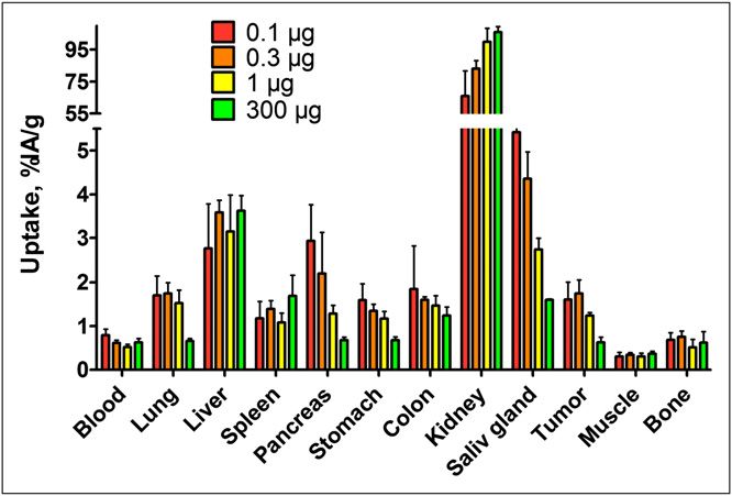
the accuracy of biopsy-based methods is limited: samplings        Characterization of Affibody Molecule ZIGF1R:4551 and
may be nonrepresentative because of intratumoral expres-          Conjugation with DOTA
sion heterogeneity, and molecular target level may be dis-           ZIGF1R:4551 was obtained by affinity maturation of the Affibody
cordant in primary tumors and metastases. Radionuclide            molecule Z4:40. The affinity maturation will be described else-
                                                                  where. A single cysteine was introduced at the C-terminus of
molecular imaging offers an alternative and allows repeated
                                                                  ZIGF1R:4551 to enable site-specific labeling using thiol-directed
noninvasive evaluation of tyrosine kinase expression both
                                                                  conjugation chemistry. The identity of ZIGF1R:4551 was confirmed
for patient stratification and for monitoring expression-level    by high-performance liquid chromatography with mass spectrom-
changes in response to therapy (18). This approach would          etry, CD spectra were recorded, and melting point was determined,
apparently be attractive for in vivo determination of IGF-1R      as described earlier (27).
status of disseminated malignant tumors.                             For affinity determination, Oregon green (OG) was site-
   Recently a new generation of molecular imaging                 specifically conjugated to ZIGF1R:4551 as described by Lundberg
probes, made by the coupling of a radiolabel to a scaf-           et al. (28). The affinity of ZIGF1R:4551 for IGF-1R–expressing
fold-based affinity protein, has been shown to give               MCF-7 cells was determined by fluorescence-activated cell sorting
promising results (19). One such class of scaffold proteins       by making a 3-fold dilution series comprising 12 different con-
is the Affibody molecules, which are structurally based on        centrations of ZIGF1R:4551-OG, ranging between 50 mM and 2.8
                                                                  pM. The fluorescence signal was plotted as a function of
one of the independently folding IgG-binding domains of
                                                                  ZIGF1R:4551-OG concentration, and the equilibrium association
staphylococcal protein A (20,21). Thirteen surface-ex-
                                                                  constant was retrieved from the inflection point of the curve.
posed amino acids on one face of the molecule have been              For site-specific labeling, maleimide-monoamide-DOTA (MMA-
subjected to randomization, resulting in a library from           DOTA; 1,4,7,10-tetraazacyclododecane-1,4,7-tris-acetic acid-10
which variants with desired specificity can be selected           maleimidoethylacetamide) (Macrocyclics) was conjugated to the
by biopanning. This process has led to the isolation of           C-terminal cysteine, as described by Ahlgren et al. (27). The
several high-affinity binders to different target antigens        conjugated Affibody molecule, designated DOTA-ZIGF1R:4551,
(21,22). One example is Affibody molecules binding to             was lyophilized.
HER2 with high affinity; these molecules have demon-
strated high-contrast imaging of HER2-expressing xeno-            Radiolabeling and In Vitro Evaluation
grafts in preclinical models (23) and imaging of HER2-            of 111In-DOTA-ZIGF1R:4551
expressing metastases in the clinic (24). Direct preclinical         DOTA-ZIGF1R:4551 was reconstituted in 0.2 M ammonium ace-
                                                                  tate buffer, pH 5.5 (purified of metal impurities using Chelex 100
comparison of biodistribution data for radiolabeled Affi-
                                                                  resin [Bio-Rad Laboratories]) to a concentration of 0.75 mg/mL.
body molecules and the anti-HER2 monoclonal antibody              An aliquot, containing 60 mg of DOTA-ZIGF1R:4551, was mixed
trastuzumab showed that Affibody molecules provide                with 60 MBq of 111In-chloride (Covidien). The mixture was in-
much better contrast and specificity of radionuclide mo-          cubated for 30 min either at 50C or at 90C and then analyzed
lecular imaging of HER2 expression (25).                          using 50-771 Dark Green Tec-Control Chromatography instant
   We have recently reported on the development of a new          thin-layer chromatography strips (Biodex Medical Systems) eluted
Affibody ligand, Z4:40, which specifically binds to IGF-1R        with 0.2 M citric acid. To assess labeling stability, 111In-DOTA-
with a high affinity of 1.9 6 0.2 nM (26). This high af-          ZIGF1R:4551 (labeled at 90C) was incubated with a 1,000-fold
finity would be sufficient for imaging of molecular targets       excess of ethylenediaminetetraacetic acid (EDTA) for 4 h and then
that are abundantly expressed. However, the level of IGF-         analyzed using instant thin-layer chromatography.
1R, which is associated with tumor response to anti-IGF-             The specificity of 111In-DOTA-ZIGF1R:4551 for binding to IGF-
                                                                  1R–expressing cells after labeling at 60C and 90C was evaluated
1R therapy, is fairly low (10,000–30,000 receptors per
                                                                  using 2 prostate cancer cell lines, DU-145 and PC-3, and the
cell) (14,15). Therefore, affinity maturation was under-          cervical carcinoma A431 cell line. All cell lines were purchased
taken (Graslund, unpublished data, 2010), resulting in            from American Type Tissue Culture Collection via LGC Promo-
the Affibody molecule ZIGF1R:4551 having enhanced affin-          chem. An in vitro specificity test was performed according to the
ity for IGF-1R.                                                   methods described earlier (27). Briefly, a solution of 111In-DOTA-
   The goal of this study was to investigate the feasibility      ZIGF1R:4551 (at 1 nM) was added to 6 Petri dishes (;106 cells in
of radionuclide-based IGF-1R imaging in vivo using                each). For blocking, a 100-fold excess of nonlabeled DOTA-
ZIGF1R:4551.                                                      ZIGF1R:4551 was added 15 min before radiolabeled conjugates to
                                                                  saturate the receptors. The cells were incubated during 2 h in
                                                                  a humidified incubator at 37C. Thereafter, the medium was col-
MATERIALS AND METHODS
                                                                  lected, the cells were detached by trypsin-EDTA solution, and the
Instrumentation                                                   radioactivity in cells and medium was measured, to enable calcu-
   Radioactivity was measured using an automated g-counter        lation of the fraction of cell-bound radioactivity.
with a 7.6 cm (3-in) NaI(Tl) detector (1480 Wizard; Wallac           An internalization of 111In-DOTA-ZIGF1R:4551 by DU-145 cells
Oy). Distribution of radioactivity along the thin-layer chroma-   was performed according to the method described earlier (27).
tography strips was measured on a Cyclone Storage Phosphor        Briefly, the cells (;106 per dish) were incubated with the labeled
System and analyzed using the OptiQuant image analysis soft-      compound (1.5 nM) at 37C, 5% CO2. At predetermined time
ware (PerkinElmer). Cells were counted using an electronic cell   points (1, 2, 3, and 4 h after incubation start), the medium from
counter (Beckman Coulter).                                        a set of 3 dishes was removed. To collect the membrane-bound

                                                      IGF-1R–TARGETING AFFIBODY MOLECULE • Tolmachev et al.                      91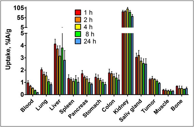
radioactivity, the cells were treated with 0.2 M glycine buffer        mL of PBS. At 1, 2, 4, 8, and 24 h after injection, mice were
containing 4 M urea, pH 2.5, for 5 min on ice. To collect cells        sacrificed and analyzed as described.
containing internalized radioactivity, a treatment with 1 M NaOH          g-Camera Imaging. The imaging experiment was performed
at 37C for 0.5 h was used. A percentage of internalized radioac-      8 h after injection. Three mice with DU-145 xenografts (0.9–1.0
tivity was calculated for each time point.                             cm3) were injected with 111In-DOTA-ZIGF1R:4551 (1 MBq, 1 mg, in
   To quantify the IGF-1R expression in the DU-145 cell line, the      100 mL of PBS). Immediately before imaging, the animals
cells were incubated for 4 h at 4C with the radiolabeled Affibody     were sacrificed by overdosing Ketalar–Rompun. After euthanasia,
molecule at concentrations in the range of 0.08–27 nM in com-          the urine bladders were excised. The imaging experiment was
plete medium. For each data point, 4 dishes were used, including 1     performed using an Infinia g-camera (GE Healthcare) equipped
presaturated with unlabeled DOTA-ZIGF1R:4551 at a 3.7-mM con-          with a medium-energy general purpose collimator. Static images
centration to determine specific binding. After incubation, the        (30 min) were obtained with a zoom factor of 2 in a 256 · 256
medium was aspirated, and the cells were detached by trypsin-          matrix. The evaluation of the images was performed using Osiris
EDTA solution and counted. Radioactivity of the samples was            4.19 software (University Hospital of Geneva, Switzerland). In
measured. The data were analyzed using Prism (version 5.04             each animal, a region of interest was drawn around the tumor.
[GraphPad Software] for Windows [Microsoft]).                          The same region was copied to a contralateral thigh. Calculation
                                                                       of tumor–to–contralateral thigh ratios was based on average
In Vivo Studies                                                        counts in the regions of interest.
   All animal experiments were planned and performed in
accordance with national legislation on laboratory animals’ pro-       Statistics
tection and were approved by the Ethics Committee for Animal              Data on cellular uptake and biodistribution were analyzed by
Research in Uppsala.                                                   unpaired, 2-tailed t test using Prism to determine any significant
   Animal Model Validation: Biodistribution in Normal NMRI             differences (P , 0.05).
Mice. Subsets of cells in colon, lung, pancreas, salivary gland, and
stomach express IGF-1R (http://www.proteinatlas.org/index.php).        RESULTS
To determine the cross-reactivity of 111In-DOTA-ZIGF1R:4551 with       Characterization of Affibody Molecule ZIGF1R:4551 and
murine IGF-1R in healthy tissues, NMRI mice (average weight,           Conjugation with DOTA
32.9 6 1.5 g) were intravenously injected (tail) with 25 kBq of           The probe to be used for imaging of IGF-1R–expressing
               IGF1R:4551 in 100 mL of phosphate-buffered saline
111In-DOTA-Z
                                                                       cells was based on the Affibody molecule ZIGF1R:4551. The
(PBS). The injected protein dose was adjusted by dilution with
                                                                       affinity for binding to IGF-1R–expressing MCF-7 cells was
nonlabeled DOTA-ZIGF1R:4551 to 0.03, 1, or 10 mg per mouse. A
group of 4 mice was used for each protein dose. At 4 h after           500 6 79 pM by flow cytometry (Fig. 1A). The melting
injection, mice were sacrificed by injection of a lethal dose of       point of the protein was 62C. To prepare the imaging
anesthesia (20 mL of Ketalar–Rompun per gram of body                   probe, MMA-DOTA was coupled to ZIGF1R:4551. After
weight: Ketalar [50 mg/mL; Pfizer], 10 mg/mL; Rompun [20               high-performance liquid chromatography purification
mg/mL; Bayer]), followed by heart puncture and exsanguina-             of DOTA-ZIGF1R:4551, no unconjugated ZIGF1R:4551 could
tion with a 1-mL syringe rinsed with heparin (5,000 IE/mL; Leo         be detected.
Pharma). Samples of blood, colon, lung, liver, spleen, pancreas,
salivary gland, stomach, kidney, muscle, and bone were col-            Labeling and In Vitro Evaluation
lected and weighed, and their radioactivity was measured.              of 111In-DOTA-ZIGF1R:4551
Tissue uptake was calculated as percentage of injected radio-             Labeling provided a radiochemical purity of 98.9% 6
activity per gram.                                                     0.2% (at 50C) or 99.7% 6 0.1% (at 90C), which excluded
   Biodistribution in NMRI nu/nu Mice Bearing DU-145 Prostate
                                                                       the need for additional purification. For biologic experi-
Cancer Xenografts: Influence of Protein Dose on Uptake in Tumors
                                                                       ments, the conjugates were diluted with PBS. Challenge
and Normal Tissues. For xenografting, 5 · 106 DU-145 cells (in
Matrigel [BD Biosciences]) were subcutaneously implanted in            with a 1,000-fold molar excess of EDTA did not cause
the right hind leg. At the time of the experiment, the average         any detectable release of 111In, showing that the label was
animal weight was 32.7 6 1.6 g, and the average tumor size was         stable.
0.44 6 0.19 g. The mice were randomly distributed into 4 groups           Binding specificity tests were performed to assess that
and injected with 25 kBq of 111In-DOTA-ZIGF1R:4551 in 100 mL           the binding of 111In-DOTA-ZIGF1R:4551 to living IGF-1R–
of PBS. The injected protein dose was adjusted by dilution with        expressing cells was receptor-mediated. Saturation of the
nonlabeled DOTA-ZIGF1R:4551 to 0.1, 0.3, 1, or 300 mg per              receptors by preincubation with nonlabeled DOTA-
mouse. A group of 4 mice was used for each protein dose. At            ZIGF1R:4551 significantly (P , 0.05) decreased the binding
4 h after injection, mice were sacrificed and analyzed as de-          of the radiolabeled Affibody molecule, suggesting that the
scribed.
                                                                       binding was specific (Fig. 1B). Importantly, binding was
   Biodistribution of 111In-DOTA-ZIGF1R:4551 in NMRI nu/nu Mice
                                                                       receptor-specific also after labeling at 90C, extending the
Bearing DU-145 Prostate Cancer Xenografts at Different Time
Points. DU-145 xenografts were implanted in male NMRI nu/nu            range of methods that might be used for labeling of
mice as described. At the time of the experiment, the average          ZIGF1R:4551. According to the saturation assay, the DU-
animal weight was 32.6 6 2.3 g, and average tumor size was             145 cells expressed 25,000 6 3,000 receptors per cells.
0.55 6 0.22 g. The mice were randomly distributed into 5 groups           Data concerning binding and internalization of 111In-
and injected with 25 kBq (1 mg) of 111In-DOTA-ZIGF1R:4551 in 100       DOTA-ZIGF1R:4551 by the prostate cancer DU-145 cell line

92      THE JOURNAL     OF   NUCLEAR MEDICINE • Vol. 53 • No. 1 • January 2012
of the total cell-associated radioactivity after 1 h incubation
                                                                         to 38% 6 3% after 4 h.

                                                                         In Vivo Studies
                                                                            Biodistribution in Normal NMRI Mice. Biodistribution of
                                                                         111In-DOTA-Z
                                                                                          IGF1R:4551 in normal NMRI mice was per-
                                                                         formed to investigate whether the animal model would re-
                                                                         flect the situation in humans, in whom the target receptor is
                                                                         expressed in several tissues. Data obtained at 4 h after in-
                                                                         jection showed that 111In-DOTA-ZIGF1R:4551 was rapidly
                                                                         cleared from the blood irrespective of the amount of added
                                                                         cold tracer (Fig. 2). The clearance was predominantly renal,
                                                                         as radioactivity in intestines with content was below 2% at
                                                                         this time point. Renal clearance was accompanied by an
                                                                         appreciable reabsorption of radioactivity in the kidneys.
                                                                         The accumulation of radioactivity in the pancreas, stomach,
                                                                         lung, and salivary gland was lower at higher injected pro-
                                                                         tein doses (P , 0.05 for uptake values after injection of
                                                                         0.03 and 10 mg per animal). This finding demonstrates that
                                                                         the uptake in these IGF-1R–expressing tissues is saturable,
                                                                         indicating that it is, at least partially, receptor-mediated. In
                                                                         contrast, the amount of radioactivity in the liver was in-
                                                                         dependent of the protein dose.
                                                                            Biodistribution in NMRI nu/nu Mice Bearing DU-145
                                                                         Prostate Cancer Xenografts: Influence of Protein Dose on
                                                                         Uptake in Tumors and Normal Tissues. The results of the
                                                                         previous experiment suggest that uptake of 111In-DOTA-
                                                                         ZIGF1R:4551 is receptor-mediated for several tissues. Our pre-
                                                                         vious studies with targeting of epidermal growth factor
                                                                         receptor (29) showed that it was possible to partially saturate
                                                                         receptors in normal tissues to a higher extent than in tumors,
                                                                         leading to an increase in tumor-to-organ uptake ratio. To
                                                                         evaluate whether this was possible for IGF-1R, the biodis-
                                                                         tribution as a function of injected dose at 4 h after injection
                                                                         in male outbreed NMRI nu/nu mice bearing DU-145 prostate
                                                                         cancer xenografts was studied (Fig. 3). In agreement with the
FIGURE 1. In vitro characterization of ZIGF1R:4551. (A) Affinity de-
termination of ZIGF1R:4551 binding to MCF-7 cells. Cells were stained
with different concentrations of ZIGF1R:4551-OG, followed by analysis
by flow cytometry. Mean fluorescence intensity (MFI) after back-
ground subtraction was recalculated on scale from 0 (only back-
ground intensity) to 1 (maximum difference between MFI and
background) and was plotted as a function of concentration of
ZIGF1R:4551. The experiment was repeated 3 times, and a represen-
tative curve is shown. (B) Binding specificity of 111In-DOTA-
ZIGF1R:4551 Affibody molecule to IGF-1R–expressing cells. For pre-
saturation of IGF-1R, a 100-fold molar excess of nonradioactive
IGF-1R–binding Affibody molecule was added. Data are mean val-
ues from 3 cell dishes 6 SD. (C) Binding and internalization of 111In-
DOTA-ZIGF1R:4551 by prostate cancer DU-145 cell line. Cells were
incubated with 111In-DOTA-ZIGF1R:4551 at 37C. Data are mean val-
ues from 3 cell dishes 6 SD; some error bars are not seen because
they are smaller than the symbols.

are presented in Figure 1C. The binding was rapid, reaching
                                                                         FIGURE 2. Biodistribution of 111In-DOTA-ZIGF1R:4551 in NMRI mice
a plateau after 1 h of incubation. The internalization                   at 4 h after injection. Values are presented as mean percentage of
of 111In-DOTA-ZIGF1R:4551 was relatively inefficient, with               injected radioactivity per gram of tissue (%IA/g) 6 SD (n 5 4).
a slow increase of the internalized fraction from 27% 6 2%               Saliv 5 salivary.

                                                            IGF-1R–TARGETING AFFIBODY MOLECULE • Tolmachev et al.                       93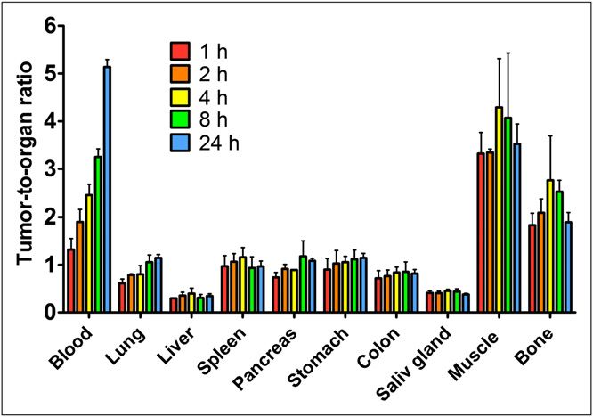
study in non–tumor-bearing mice, the accumulation of radio-
activity in the lung, stomach, pancreas, and salivary gland
significantly decreased (P , 0.05) with increasing protein
dose, supporting the hypothesis that saturation occurred in
these tissues. In contrast, no significant difference (P , 0.05)
was seen in blood, spleen, liver, muscle, or bone at different
injected protein doses, suggesting that ligand–receptor inter-
action did not play any significant role in accumulation of
radioactivity in those organs. The lungs showed a separate
pattern of radioactivity accumulation. A large decrease was
observed when the protein dose was increased from 1 to
300 mg (P , 0.05), whereas no significant differences could
be seen between the lowest 3 doses. There was no significant
difference in the tumor uptake after injection of 0.1–1 mg of
protein per mouse; however, an injection of 300 mg caused
                                                                         FIGURE 4. Tumor-to-organ ratios for 111In-DOTA-ZIGF1R:4551 in
a highly significant (P , 0.0005) decrease of radioactivity              male NMRI nu/nu mice with subcutaneous DU-145 xenografts at
accumulation in tumors.                                                  4 h after injection. Values are presented as mean value 6 SD (n 5
   The influence of injected protein dose on tumor-to-organ              4). Saliv 5 salivary.
ratios is presented in Figure 4. An increase of the injected
protein dose from 0.3 to 1 mg resulted in an increase of the
tumor-to-pancreas ratio by 1.8-fold and of the tumor–to–                 was slow washout of radioactivity from tumors and normal
salivary gland ratio by 1.5-fold (P , 0.005 in both cases).              IGF-1R–expressing organs (colon, lung, pancreas, salivary
In contrast, the tumor-to-liver ratio decreased when the                 gland, and stomach). The washout rate was somewhat slower
dose was increased. With most organs and tissues, no sig-                from the tumor than from tissues, which increased the tumor-
nificant difference in tumor-to-organ ratios could be seen—              to-organ ratios for blood, lung, and pancreas with time (Fig. 6).
that is, the dependence on the amount of injected protein                The tumor-to-bone ratio reached its maximum at 8 h after
was the same as for the tumor.                                           injection.
   Biodistribution in NMRI nu/nu Mice Bearing DU-145                        g-Camera Imaging. Images acquired at 8 h after the
Prostate Cancer Xenografts: Results for Different Time                   administration of 111In-DOTA-ZIGF1R:4551 to immunodefi-
Points. The biodistribution as a function of time was also               cient mice bearing subcutaneous DU-145 are presented in
investigated, and the results are presented in Figure 5. The             Figure 7. The tumor xenografts were visualized with appre-
results demonstrate a rapid clearance from blood and lungs.              ciably higher accumulation of radioactivity than in contra-
The concentration of radioactivity in liver, salivary gland, and         lateral sites. The tumor–to–contralateral site ratio was 2.9
spleen was rather constant over the course of the experiment.            6 0.1 (average 6 SD). Also, as predicted from the biodis-
Similarly, no significant difference was observed in muscles             tribution studies, there was substantial accumulation of ra-
and bones over the course of the experiment (except that the             dioactivity in the kidneys, liver, and salivary gland, which
level at 1 h after injection was higher than at 8 and 24 h). There       were thereby clearly visualized.

                                                                         FIGURE 5. Biodistribution of 111In-DOTA-ZIGF1R:4551 (injected
FIGURE 3. Dose-escalation study of 111In-DOTA-ZIGF1R:4551 in male        dose, 1 mg) in male NMRI nu/nu mice with subcutaneous DU-145
NMRI nu/nu mice with subcutaneous DU-145 xenografts at 4 h after         xenografts at different time points. Values are presented as mean
injection. Values are presented as mean percentage of injected ra-       percentage of injected radioactivity per gram of tissue (%IA/g) 6 SD
dioactivity per gram of tissue (%IA/g) 6 SD (n 5 4). Saliv 5 salivary.   (n 5 4). Saliv 5 salivary.

94      THE JOURNAL      OF   NUCLEAR MEDICINE • Vol. 53 • No. 1 • January 2012
imaging of molecular targets is possible only several days
after injection, which is an essential clinical disadvantage.
The applicability of radiolabeled antibodies for molecular
imaging is also limited by the unspecific uptake of proteins
with a molecular weight of more than 45 kDa in tumors, the
so-called enhanced permeability and retention effect (34),
because this can lead to false-positive findings. A possible
advantage of monoclonal antibodies is that a therapeutic
antibody with known targeting efficiency and safety profile
can be radiolabeled, providing a facile way to develop the
imaging agent. Recently, the use of radiolabeled anti–IGF-
1R R1507 antibody for immunoSPECT and immunoPET of
triple-negative breast cancer xenografts was reported, dem-
onstrating the feasibility of in vivo IGF-1R detection (30).
As expected, a good imaging contrast was not obtained
FIGURE 6. Tumor-to-organ ratios for 111In-DOTA-ZIGF1R:4551
until 3 d after the administration.
(injected dose, 1 mg) in male NMRI nu/nu mice with subcutaneous
DU-145 xenografts at different time points. Values are presented as The use of IGF-1 and its mutated variants (31–33) for in
mean value 6 SD (n 5 4). vivo targeting of IGF-1R–overexpressing or IGF-1R–trans-
fected tumor xenografts has shown promising results. The
major issue with IGF-1–based probes is their agonistic ac-
DISCUSSION
tion, potentially eliciting a broad range of physiologic
In this work, we have investigated the feasibility of using responses. Scaffold-based affinity proteins, such as Affi-
Affibody molecules for radionuclide molecular imaging of body molecules, may have agonistic, antagonistic, or no
IGF-1R–expressing tumors in vivo. Imaging of IGF-1R can effect on their target receptor, much like antibodies. In fact,
potentially provide essential information influencing man- the parental IGF-1R–binding Affibody molecule Z4:40 is an
agement of several different tumor types. For example, this IGF-1R antagonist (26).
method has a potential to allow for stratification of patients In this study, we have for the first time evaluated the use
with androgen-independent prostate cancer for IGR-1R– of Affibody molecules for in vivo imaging of IGF-1R
targeting therapy and for monitoring the response to such expression in a prostate cancer model. We have shown that
therapy. In principle, several classes of radiolabeled molec- conjugation of DOTA to ZIGF1R:4551 and subsequent label-
ular targeting probes can be considered for imaging of ing with 111In provides a conjugate with preserved binding
IGF-1R overexpression—such as anti–IGF-1R monoclonal specificity (Fig. 1B). Biodistribution in normal mice dem-
antibodies or fragments thereof; a natural ligand, IGF-1, or onstrated specific (dose-dependent) binding of 111In-
its mutated variants (30–33); or scaffold proteins such as DOTA-ZIGF1R:4551 to IGF-1R–expressing organs and
Affibody molecules. tissues (Fig. 2). This finding suggests that 111In-DOTA-
An issue with monoclonal antibodies for radionuclide ZIGF1R:4551 possesses cross-reactivity to murine IGF-1R,
molecular imaging is their large size, resulting in slow making a mouse model adequate for evaluation of IGF-
blood clearance and slow tumor accumulation. Thus, 1R targeting in vivo because a high expression of the target
in normal organs might appreciably influence the imaging
results, especially when the target level in the tumor is low.
Injection of a high 111In-DOTA-ZIGF1R:4551 protein dose
caused a significant decrease of uptake in tumors, suggest-
ing a saturability of IGF-1R targeting in vivo—a strong
indication of targeting specificity. Further, after optimiza-
tion of the injected dose, an increase in tumor-to-blood,
tumor–to–salivary gland, and tumor-to-pancreas ratio could
be achieved. This study suggests that the optimal time point
for imaging aquisition is between 8 and 24 h after injection.
Experimental g-camera imaging confirmed the feasibility
of visualization of IGF-1R–expressing prostate cancer xen-
ografts. A limitation of the tested conjugate is the high
accumulation of radioactivity in the liver and kidneys. Sev-
eral approaches can be explored to overcome this limita-
FIGURE 7. Imaging of IGF-1R expression in DU-145 prostate can-
cer xenografts in NMRI nu/nu mice using111In-DOTA-ZIGF1R:4551. tion. For example, the probe investigated in this study
Planar g-camera images were acquired at 8 h after injection. Arrows contained a hexahistidine tag at the N-terminus to facilitate
point to tumor (T), kidney (K), liver (L), and salivary gland (SG). immobilized metal ion affinity chromatographic purifica-
IGF-1R–TARGETING AFFIBODY MOLECULE • Tolmachev et al. 95tion of the construct, as we have recently shown to be to trastuzumab resistance of breast cancer cells. Cancer Res. 2005;65:
11118–11128.
associated with elevated liver uptake of radioactivity 5. Thomas F, Holly JM, Persad R, Bahl A, Perks CM. Fibronectin confers survival
(27,35). Using alternative purification schemes that do not against chemotherapeutic agents but not against radiotherapy in DU145 prostate
require the use of hexahistidine tags might reduce liver cancer cells: involvement of the insulin like growth factor-1 receptor. Prostate.
2010;70:856–865.
uptake of IGF-1R–targeting Affibody molecules. The use 6. Wu JD, Haugk K, Coleman I, et al. Combined in vivo effect of A12, a type 1
of a nonresidualizing radiohalogen label is a complementary insulin-like growth factor receptor antibody, and docetaxel against prostate can-
approach to appreciably reduce renal retention of radioac- cer tumors. Clin Cancer Res. 2006;12:6153–6160.
7. Sachdev D, Yee D. Inhibitors of insulin-like growth factor signaling: a therapeu-
tivity in comparison with radiometal labels (36). Our recent tic approach for breast cancer. J Mammary Gland Biol Neoplasia. 2006;11:
data have also demonstrated that low renal radioactivity reten- 27–39.
tion can be obtained using a cysteine-containing peptide-based 8. Rodon J, DeSantos V, Ferry RJ Jr, Kurzrock R. Early drug development of
inhibitors of the insulin-like growth factor-I receptor pathway: lessons from
GGGC chelator for 99mTc, which may be considered as a che-
the first clinical trials. Mol Cancer Ther. 2008;7:2575–2588.
lator in future studies of IGF-1R imaging (37). 9. Krueckl SL, Sikes RA, Edlund NM, et al. Increased insulin-like growth factor I
The possibility of visualizing IGF-1R expression in vivo receptor expression and signaling are components of androgen-independent pro-
gression in a lineage-derived prostate cancer progression model. Cancer Res.
is not only of value for treatment optimization of prostate
2004;64:8620–8629.
cancer. There is strong evidence that expression of IGF-1R 10. Pienta KJ, Bradley D. Mechanisms underlying the development of androgen-
and cross-talk between HER2 and IGF-1R signaling is a po- independent prostate cancer. Clin Cancer Res. 2006;12:1665–1671.
11. Culig Z, Hobisch A, Cronauer MV, et al. Androgen receptor activation in pros-
tential reason for trastuzumab resistance in disseminated
tatic tumor cell lines by insulin-like growth factor-I, keratinocyte growth factor,
HER2-positive breast cancer (4,38). Thus, determination and epidermal growth factor. Cancer Res. 1994;54:5474–5478.
of IGF-1R status in those tumors could also be of value 12. Chi KN, Bjartell A, Dearnaley D, et al. Castration-resistant prostate cancer: from
in the clinic. Furthermore, IGF-1R signaling has been sug- new pathophysiology to new treatment targets. Eur Urol. 2009;56:594–605.
13. Antonarakis ES, Carducci MA, Eisenberger MA. Novel targeted therapeutics for
gested to be involved in development of gefitinib resistance metastatic castration-resistant prostate cancer. Cancer Lett. 2010;291:1–13.
in non–small cell lung cancer (39,40). Thus, further re- 14. Zha J, O’Brien C, Savage H, et al. Molecular predictors of response to a human-
search regarding applications of the anti–IGF-1R Affibody ized anti-insulin-like growth factor-I receptor monoclonal antibody in breast and
colorectal cancer. Mol Cancer Ther. 2009;8:2110–2121.
molecule is warranted. 15. Cao L, Yu Y, Darko I, et al. Addiction to elevated insulin-like growth factor I
receptor and initial modulation of the AKT pathway define the responsiveness of
rhabdomyosarcoma to the targeting antibody. Cancer Res. 2008;68:8039–8048.
CONCLUSION 16. Gong Y, Yao E, Shen R, et al. High expression levels of total IGF-1R and
sensitivity of NSCLC cells in vitro to an anti-IGF-1R antibody (R1507). PLoS
This study has for the first time demonstrated the ONE. 2009;4:e7273.
feasibility of in vivo targeting of IGF-1R–expressing pros- 17. de Bono JS, Attard G, Adjei A, et al. Potential applications for circulating tumor
tate cancer xenografts using Affibody molecules. Further cells expressing the insulin-like growth factor-I receptor. Clin Cancer Res.
2007;13:3611–3616.
development of the radiolabeled probe might result in a use- 18. Tolmachev V, Stone-Elander S, Orlova A. Current approaches to the use of
ful clinical tool for stratification of patients with prostate radiolabeled tyrosine kinase-targeting drugs for patient stratification and treat-
cancer for IGF-1R–targeting therapy. ment response monitoring: prospects and pitfalls. Lancet Oncol. 2010;11:992–
1000.
19. Miao Z, Levi J, Cheng Z. Protein scaffold-based molecular probes for cancer
molecular imaging. Amino Acids. 10.1007/s00726-010-0503-9.
DISCLOSURE STATEMENT
20. Grönwall C, Ståhl S. Engineered affinity proteins: generation and applications.
The costs of publication of this article were defrayed in J Biotechnol. 2009;140:254–269.
21. Nygren PA. Alternative binding proteins: Affibody binding proteins developed
part by the payment of page charges. Therefore, and solely from a small three-helix bundle scaffold. FEBS J. 2008;275:2668–2676.
to indicate this fact, this article is hereby marked “adver- 22. Löfblom J, Feldwisch J, Tolmachev V, Carlsson J, Ståhl S, Frejd FY. Affibody
tisement” in accordance with 18 USC section 1734. molecules: engineered proteins for therapeutic, diagnostic and biotechnological
applications. FEBS Lett. 2010;584:2670–2680.
23. Ahlgren S, Tolmachev V. Radionuclide molecular imaging using Affibody mol-
ACKNOWLEDGMENTS ecules. Curr Pharm Biotechnol. 2010;11:581–589.
24. Baum RP, Prasad V, Müller D, et al. Molecular imaging of HER2-expressing
This research was financially supported by grants from malignant tumors in breast cancer patients using synthetic 111In- or 68Ga-labeled
the Swedish Cancer Society (Cancerfonden), the Swedish Affibody molecules. J Nucl Med. 2010;51:892–897.
25. Orlova A, Wållberg H, Stone-Elander S, Tolmachev V. On the selection of
Research Council (Vetenskapsrådet), and O.E. and Edla a tracer for PET imaging of HER2-expressing tumors: direct comparison of
Johansson Foundation. No other potential conflict of inter- 124I-labelled Affibody molecule and trastuzumab in a murine xenograft model.
est relevant to this article was reported. J Nucl Med. 2009;50:417–425.
26. Li J, Lundberg E, Vernet E, Larsson B, Höidén-Guthenberg I, Gräslund T. Se-
lection of affibody molecules to the ligand-binding site of the insulin-like growth
REFERENCES factor-1 receptor. Biotechnol Appl Biochem. 2010;55:99–109.
27. Ahlgren S, Orlova A, Rosik D, et al. Evaluation of maleimide derivative of
1. Pollak MN, Schernhammer ES, Hankinson SE. Insulin-like growth factors and DOTA for site-specific labeling of recombinant Affibody molecules. Bioconjug
neoplasia. Nat Rev Cancer. 2004;4:505–518. Chem. 2008;19:235–243.
2. Werner H, Bruchim I. The insulin-like growth factor-I receptor as an oncogene. 28. Lundberg E, Höidén-Guthenberg I, Larsson B, Uhlén M, Gräslund T. Site-spe-
Arch Physiol Biochem. 2009;115:58–71. cifically conjugated anti-HER2 Affibody molecules as one-step reagents for
3. Frasca F, Pandini G, Sciacca L, et al. The role of insulin receptors and IGF-I target expression analyses on cells and xenograft samples. J Immunol Methods.
receptors in cancer and other diseases. Arch Physiol Biochem. 2008;114:23–37. 2007;319:53–63.
4. Nahta R, Yuan LX, Zhang B, Kobayashi R, Esteva FJ. Insulin-like growth factor-I 29. Tolmachev V, Rosik D, Sjöberg A, et al. Imaging of EGFR expression in murine
receptor/human epidermal growth factor receptor 2 heterodimerization contributes xenografts using site-specifically labelled anti-EGFR [111In-MMA-Cys]Z2377
96 THE JOURNAL OF NUCLEAR MEDICINE • Vol. 53 • No. 1 • January 2012Affibody molecule: aspect of injected amount. Eur J Nucl Med Mol Imaging. and shows improved biodistribution with reduced hepatic radioactivity accumu-
2010;37:613–622. lation. Bioconjug Chem. 2010;21:2013–2022.
30. Heskamp S, van Laarhoven HW, Molkenboer-Kuenen JD, et al. ImmunoSPECT 36. Tolmachev V, Mume E, Sjöberg S, Frejd FY, Orlova A. Influence of valency and
and immunoPET of IGF-1R expression with the radiolabeled antibody R1507 in labelling chemistry on in vivo targeting using radioiodinated HER2-binding
a triple-negative breast cancer model. J Nucl Med. 2010;51:1565–1572. Affibody molecules. Eur J Nucl Med Mol Imaging. 2009;36:692–701.
31. Sun BF, Kobayashi H, Le N, et al. Biodistribution of 125I-labeled des(1-3) in- 37. Wållberg H, Orlova A, Altai M, et al. Molecular design and optimization of
sulin-like growth factor I in tumor-bearing nude mice and its in vitro catabolism. 99mTc-labeled recombinant Affibody molecules improves their biodistribution
Cancer Res. 1997;57:2754–2759. and imaging properties. J Nucl Med. 2011;52:461–469.
32. Sun BF, Kobayashi H, Le N, et al. Effects of insulin-like growth factor binding 38. Nahta R, Shabaya S, Ozbay T, Rowe DL. Personalizing HER2-targeted therapy
proteins on insulinlike growth factor-I biodistribution in tumor-bearing nude in metastatic breast cancer beyond HER2 status: what we have learned from
mice. J Nucl Med. 2000;41:318–326. clinical specimens. Curr Pharmacogenomics Person Med. 2009;7:263–274.
33. Cornelissen B, McLarty K, Kersemans V, Reilly RM. The level of insulin growth 39. Morgillo F, Kim WY, Kim ES, Ciardiello F, Hong WK, Lee HY. Implication
factor-1 receptor expression is directly correlated with the tumor uptake of 111In- of the insulin-like growth factor-IR pathway in the resistance of non-small
IGF-1(E3R) in vivo and the clonogenic survival of breast cancer cells exposed in cell lung cancer cells to treatment with gefitinib. Clin Cancer Res. 2007;
vitro to trastuzumab (Herceptin). Nucl Med Biol. 2008;35:645–653. 13:2795–2803.
34. Wester HJ, Kessler H. Molecular targeting with peptides or peptide-polymer 40. Knowlden JM, Jones HE, Barrow D, Gee JM, Nicholson RI, Hutcheson IR.
conjugates: just a question of size? J Nucl Med. 2005;46:1940–1945. Insulin receptor substrate-1 involvement in epidermal growth factor receptor
35. Tolmachev V, Hofström C, Malmbeg J, et al. HEHEHE-tagged Affibody mole- and insulin-like growth factor receptor signalling: implication for Gefitinib
cule may be purified by IMAC, is conveniently labeled with [99mTc(CO)3](1), (‘Iressa’) response and resistance. Breast Cancer Res Treat. 2008;111:79–91.
IGF-1R–TARGETING AFFIBODY MOLECULE • Tolmachev et al. 97You can also read