Impact Factor: 7.569 Volume 10, Issue 7, July 2021 - ijirset
←
→
Page content transcription
If your browser does not render page correctly, please read the page content below

Volume 10, Issue 7, July 2021

Impact Factor: 7.569

International Journal of Innovative Research in Science, Engineering and Technology (IJIRSET)

                        | e-ISSN: 2319-8753, p-ISSN: 2320-6710| www.ijirset.com | Impact Factor: 7.569|

                                            || Volume 10, Issue 7, July 2021 ||
                                           |DOI:10.15680/IJIRSET.2021.1007092|

        A Review: Drosophila melanogaster as A
          Model Organism to Study Memory
          Archana N. Moon1, Apurva S. Mendulkar2 , Sushil G. Hedau3, Nikita R. Nashikkar4,
 Prerna R. Ukey5, Payal S. Ahir6 Pratiksha A. Gajbhiye 7, Sampada P. Pendse8, Kanchan A. Darode9,
                         Shubham R. Murai10, Aniket P. Randal11, Ninad Nagarakar12
   Post Graduate Teaching, Department of Biochemistry Rashtrasant Tukadoji Maharaj Nagpur University Nagpur,
                                                      Maharashtra, India1-12

ABSTRACT: The purpose of the article is to show Drosophila melanogaster as a model organism in studying and
understanding memory. We had covered types of memory, an aspect that affects & nourishes the memory and its
consequences along with the pathways. In this we explored how circadian rhythm, ageing, sleep deprivation, oxidative
stress, and alcohol (ethanol) consumption affect memory and how neurodegenerative diseases leads to disruption of
different types of memory in Drosophila. Also factors studied in this article may show similar effects on human as 75%
of genes responsible for human diseases have homologs in flies.

KEYWORDS: - Drosophila Melanogaster, Memory, Sleep, Circadian Rhythm.

                                                      I. INTRODUCTION
“The power of the mind to remember things” or “something remembered from the past like a recollection”, these
several interrelated ideas denotes term memory. It refers to the processes that are used to acquire, store, retain, and later
retrieve information. Encoding, storage, and retrieval are three major processes involved in memory.

Short term memory (STM), long term memory (LTM) and middle term memory (MTM) are the three parts of memory
in Drosophila. Short-term memory also known as working memory allows recall for a period of several seconds to a
minute without rehearsal. Its capacity is very limited. STM temporarily records the succession of events in our lives.
Short-term memory has a storage capacity of only about seven items and lasts only a few dozen seconds. It may register
a face that we see in the street, or a telephone number that we overhear someone giving out, but this information will
quickly disappear forever unless we make a conscious effort to retain it. STM includes: sensory memories, short term
storage, working memory, long term working memory.

Much larger quantities of information for potentially unlimited duration (sometimes a whole life span) can be store in
long-term memory (LTM). It lets us retain the meanings of words and the physical skills that we have learned. LTM
stores all the significant events that mark our lives. Its capacity seems unlimited, and it can last days, months, years, or
even an entire lifetime! But it is far from infallible. It includes: olfactory, visual, explicit, Middle term memory (MTM)
lies between STM and LTM containing anesthesia resistant memory (ARM) and anesthesia sensitive memory (ASM)
(1).

                             II. FACTORS AFFECTING MEMORY OF DROSOPHILA

 Factors that affect different types of memory and its function either directly or indirectly in Drosophila are listed
below.
Circadian Rhythm:
Circadian rhythms are a series of endogenous autonomous oscillators generated by the molecular circadian clock which
acting on co-ordinating internal time with the external environment in a 24 hour daily cycle. It is a natural cycle of
physical, mental and behaviour changes that the body goes through a 24 hour cycle. Drosophila circadian rhythm have
paved the way for understanding circadian behaviour and diseases related to sleep-wake condition in other animals,
including humans. Abnormal circadian rhythms may be uplinked to obesity, diabetes, depression, bipolar disorder,

IJIRSET © 2021                                    |     An ISO 9001:2008 Certified Journal |                           9239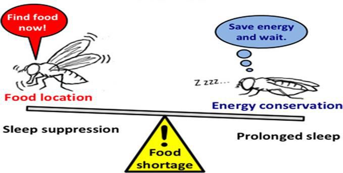
International Journal of Innovative Research in Science, Engineering and Technology (IJIRSET)

                        | e-ISSN: 2319-8753, p-ISSN: 2320-6710| www.ijirset.com | Impact Factor: 7.569|

                                            || Volume 10, Issue 7, July 2021 ||
                                           |DOI:10.15680/IJIRSET.2021.1007092|

seasonal affective disorder, and sleep disorders such as insomnia. Circadian rhythm is sometimes called the body’s
clock. It reflects direct response to the changing temperature or light. Drosophila melanogaster (fruit fly) shows many
different and easily in large scale measurable circadian patterns of behaviour than any other model organisms. Also, it
has the reduced gene number that means there is less redundancy & less compensation and its genome is the closest to
that of mammals.
Drosophila for instance is only active when it is not too hot and dry on the one hand & when it’s not too cold on the
other hand. Fruit flies hatch in greatest numbers just at dawn. Drosophila melanogaster shows two activity peaks – one
in a morning and one in the evening. It also shows other clock related behaviour that is eclosion, olfactory sensitivity,
egg laying, courtship, gustatory sensitivity or learning and memory, especially the simplicity of its neuronal network
organizations (4). About 150 clock cells per brain hemisphere are yet manageable and permit dissection of the function
of single cells or cell clusters in the brain. The late Seymour Benzer in early 1970s revealed 'clock gene’ in which he
discovered abnormal circadian behaviour. The gene responsible for the same is located on X-chromosome (2). To adapt
to different environments and photoperiods, circadian clock need to be flexible, otherwise it would lack an important
ability. Therefore, circadian clock evolved sophisticated molecular mechanisms to react to new environments. Some
input factors called ‘Zeitgebers’ harmonize clock neurons to environmental light, temperature, social cues or even
magnetism influences circadian rhythm. Light is the most important input factor. The response to variable light is
mainly caused by degradation of Tim protein (6).
In Drosophila, as there are two circadian oscillator i.e. the morning oscillator which accelerates by light and evening
oscillator which shows inducing activity in evening and is slowed down by light. Both oscillators are coupled. The
reason for this bimodal activity is the adaptation to different photoperiods. Molecular genetic analysis has made
circadian rhythms in Drosophila one of the best understood behaviours at molecular level.
The Circadian Clock in Drosophila:- A number of genes in Drosophila are turned on when the animal is exposed to
light:
1) Effector genes whose products mediate the animal’s responses (e.g. hatching or molting)
2) Clock genes whose products regulate the circadian clock
    Two key members of this group are: Period (per) & Timeless (Tim)
Activation of all of these genes requires that their promoters are bound by the protein transcription factors (3).
CLOCK encoded by the gene clock (clk)
The Mechanism:
                                        CYCLE encoded by the gene cycle (cyc)

IJIRSET © 2021                                   |   An ISO 9001:2008 Certified Journal |                            9240
International Journal of Innovative Research in Science, Engineering and Technology (IJIRSET)

                        | e-ISSN: 2319-8753, p-ISSN: 2320-6710| www.ijirset.com | Impact Factor: 7.569|

                                          || Volume 10, Issue 7, July 2021 ||
                                         |DOI:10.15680/IJIRSET.2021.1007092|

The PER and TIM proteins (synthesized on ribosomes in the cytoplasm) form dimers. When the concentration of these
gets high enough (early evening), they dissociate and are transported into the nucleus (3).
Here PER Binds to the CLK/CYC transcription factors, removing them from the promoters of the genes they activate;
thus shutting off transcription. Because these genes include PER and TIM, the result is a negative feedback loop; that
is, the product of the PER gene inhibit its own synthesis (as well as that of TIM). Just as the heat of a furnace turns
through the thermostat – its own production off, so a rising level of PER/TIM dimers turns off further synthesis of
them. As the level then falls, this inhibition is lifted and PER/TIM activity begins anew to turn on clock gene
expression. The time required for the different effects results in the levels of PER/TIM and CLOCK oscillating in
opposite phases with a circadian (~24 hr) rhythm (3).
Setting the Clock:-
Even without any external cues (e.g., alternating light and dark), the cycles persist although they tend to drift away
from environmental time. The clocks are precise under natural conditions light is one of the most important because
they are "set" (synchronized) by environmental cues.The protein cryptochrome (CRY) absorbs blue light in
Drosophila. This causes an allosteric change in its conformation enabling it to bind to TIM and PER. this causes to
break down (in proteasomes) TIM and PER ending their inhibition of gene transcription.. If this happens when
PER/TIM levels are rising (late in the day), it sets the clock back. But if it happens when PER/TIM levels are declining
(late in the night), it sets the clock ahead (3).
Circadian Rhythm Disorder:-
Impairment in normal rhythmicity favors the activation of gene expression and pro-inflammatory responses, which may
play a role in neurodegeneration. Through the modulation of oxidative stress responses a proposed pathway associates
the master clock to neurodegeneration. However other mechanisms such as neuroinflammation or an impaired
degradation of pathogenic protein aggregates might also be involved. There are some evidences of a link between
circadian system and psychiatric diseases. Environmental or genetic disruption of the circadian system leads to an
increased liability to psychiatric disease. Disruption of clock genes or clock network might be related to etiology of
these pathologies. Through clock-independent pleiotropy some genes known for their circadian clock functions might
be associated to mental illness. In Psychiatric patients, sleep and memory are often impaired. A majority of patients
with schizophrenia, bipolar and depressive disorders refer sleep disturbances (insomnia or sleepiness). They may also
be characterized by impairments in the selection of positive or negative information from working memory and in their
capability to access episodic memories. In addition, patients may also display impairments in interpersonal interactions.
Several data suggests an involvement of circadian clock or genes in determination of these complex behaviours can
also be seen in Drosophila (5).Circadian rhythm clocks participate in regulation of neurogenesis. Higher incidences of
brain diseases such as sleep disorder and depression were observed in a population with interrupted circadian rhythm
People suffering from psychiatric disorders can induce emotional & psychotic attacks in individuals through circadian
dysregulation. Most of the researchers suggest that circadian disorder is a symptom of neurodegenerative diseases
including Alzheimer’s, Parkinson’s and Huntington’s disease (8). The impairment formation of memories and
manifestation of other cognitive deficits are due to disruptions in circadian regulation. Circadian dysfunction markedly
affects cognitive functions (7).

IJIRSET © 2021                                  |   An ISO 9001:2008 Certified Journal |                           9241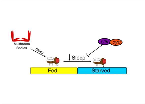
International Journal of Innovative Research in Science, Engineering and Technology (IJIRSET)

                        | e-ISSN: 2319-8753, p-ISSN: 2320-6710| www.ijirset.com | Impact Factor: 7.569|

                                           || Volume 10, Issue 7, July 2021 ||
                                          |DOI:10.15680/IJIRSET.2021.1007092|

The Drosophila and Mammalia circadian oscillator are comparable and many components are evolutionary related.
Also, it has complex nervous system which resembles much with the cell and neurobiological level with humans. Many
neuropsychiatric diseases can be studied and understood well by using Drosophila melanogaster as a model. Hence,
Drosophila might be informative in the study of possible relationship between circadian clock and human psychiatric
diseases. The complex behaviour such as sleep as well as memory appears to be influenced in some way by circadian
system or gene, even if the mechanistic relationship is still unclear. That is why this area of research has to be explored
more in future to get a clearer picture.
Sleep:
For performing several brain functions and maintaining memory sleep is very crucial and it promotes consolidation of
memory. Irregular cycle of sleep can impair memory be it directly or indirectly. Sleep is interrelated to many of the
brain functions and it is regulated by the circadian clock, which ensures appropriate timing of sleep, but the amount and
quality of sleep are also determined by other mechanisms that ensure a homeostatic balance between sleep and wake .
Sleep is also regulated by factors such as food availability, stress, and social environment.
In Drosophila, memory formation is regulated in circadian manner. Circadian clock modulates short-term olfactory
memory peak memory levels in early to mid-evening & lowest memory levels during mid-day in both light-dark cycles
(LD) and constant darkness. Circadian mutants for core clock genes; period (PER) & timeless (TIM) failed to show any
rhythms in short-term memory (STM). Circadian rhythm in STM appears to be modulated by central brain circadian
oscillators and not the peripheral circadian clock in the antennae as cryptochrome mutants exhibited strong circadian
rhythms in STM (3). Sleep is important for optimal cognitive performance, and sleep disruptions lead to defects in
learning and memory. It is said that enhanced amount of sleep can reverse memory meaning if the sleep cycle is
disturbed it may lead to memory loss.
Sleep Deprivation:
If you don't get enough sleep a condition occurs termed as sleep deprivation. Sleep deficiency is a broader concept. It
occurs if you don't get enough sleep due to any medical or other reasons. Sleep plays a key role in learning and
memory.in Human and other vertebrate. Because of sleep deprivation hippocampal plasticity is seen, it's result in
impaired subsequent retention. In Drosophila loss of sleep caused learning and memory impairment. It also affect
waking experience in Drosophila, All these studies shows that sufficient sleep is very important in learning and
memory. Several experiments are carried out for study the impact of sleep deprivation on memory and learning.
To study the impact of sleep deprivation on memory and learning, sleep deprivation experiment performed to show that
how flies silence transmission of mushroom bodies in brain due to that for some hour memory in Drosophila get
impaired. 1day sleep deprivation effect of slpD on learning and memory was evaluated using Pavlovian olfactory
conditioning in Drosophila and its locomotor activity was measured, using Drosophila activity monitoring system in
12:12 light dark cycle. And it shows that slpD specifically impaired 1 hour memory in wild type canton's flies, and the
effect remain at least for 2 hours. Mechanisms studies shows that flies with either silence transmission of mushroom
bodies (MB) during slpD or down regulate the level of cAMP in the MB demonstrate no slpD induced 1 hour memory
impairment and results show that slpD specifically impaired 1 hour memory in Drosophila and either silencing of MB
transmission during slpD or down regulating of the cAMP level in the MB protect the flies from slpD induced
impairment (9). Due to sleep deprivation in major depressive disorders working memory and learning, sleep get
affected. In deprivation disorders 2-pore domain potassium channel TREK-1 play major role and its relation with sleep
and memory were shown in the following experiment.
In major depressive disorder (MDDs), sleep and cognitive dysfunction are common. Recently, the 2-pore domain
potassium channel twik-related K (+) channel 1 (TREK-1) has been identified to be closely related to the etiology of
MDD Function of TREK-1 is still unknown either is involved in the regulation of sleep and cognition. For this one
more experiment was carried out to study the role of outwardly rectifying K+ channel-1 (ORK1) (TREK-1 homolog in
Drosophila) in sleep and cognition in Drosophila. The mutant and over-expressed lines of ork1 were generated using
Drosophila genetics. To test sleep time and short-term memory of the mutant and over-expressed ORK1 lines, Sleep
analysis and short-term memory experiments were used. Result showed that the learning index of ork1mutant lines was
increased compared to wild type. Ork1 mutant could obviously decrease sleep time in Drosophila. They also found that
ORK1 over-expression could increase sleep time and decrease learning index in Drosophila, contrary to the ork1
mutant lines. This study suggests that ORK1 might play an important role in the regulation of sleep time and short-term
memory in Drosophila (10). This studies show that sleep deprivation majorly affect short term memory in Drosophila.

IJIRSET © 2021                                   |   An ISO 9001:2008 Certified Journal |                            9242
International Journal of Innovative Research in Science, Engineering and Technology (IJIRSET)
| e-ISSN: 2319-8753, p-ISSN: 2320-6710| www.ijirset.com | Impact Factor: 7.569|
|| Volume 10, Issue 7, July 2021 ||
|DOI:10.15680/IJIRSET.2021.1007092|
Aging:
Aging could be a phenomenon which is related to functional senescence in most organisms. Drosophila melanogaster
has short life span and numerous age- related functional senescence similarities with human subjects (11).Drosophila
melanogaster is useful model for studying the normal aging process and its associated cognitive changes, and cause
disturbance in memory, sleep, locomotion and other behaviours in aging flies (12). Decline in cognitive function in old
flies with respect to both short lived and consolidated form of olfactory memory. During aging memory impairment is
to decline in neuronal function and increase in neurodegeneration (11).Flies had long, uninterrupted bouts of sleep
when they were young sleep become fragmented as they aged (13).Young flies were able to fully restore the stable
memory for 3 hours. Post extinction while aging flies were failed to do so. Only stable but not the labile memory
component was suppressed by extinction results in higher memory loss in aging flies. Young flies restored memory
reduction while it lacks in aging flies (14).Age related defect in olfactory memory is identical to those of MTM mutant
amnesiac (Amn). Aging may also affect motor activity. Aging is known to be associated with a decrease of learning
and memory. Some regions of the brain are more sensitive to aging than others. ARM is less sensitive to aging as LTM
formation is highly sensitive (16).LTM that is independent of protein synthesis remain unaffected by age, whereas that
form of LTM requiring protein synthesis become impaired therefore there is mechanistic specificity in effect of aging
on LTM. Aging disrupts specific temporal form of memory, including ITM (Intermediate Term Memory) and protein
synthesis LTM, but not STM and protein synthesis independent LTM (17).An increase in oxidative stress disrupts
sleep-wake cycle in a manner similar to that of aging as sleep become fragmented with age and rate at which this
fragmentation occurs depends on ambient temperature and life span, it is an integral part of physiological aging process
(13).There will be decrease in expression of ETC gene as well as increase in expression of immune and stress gene of
all this been reported in aging studies. Co-expression module have been identified i.e., enriched in gene known to affect
learning and memory in flies (12). Decreased IIS signalling promotes learning and has been implicated in memory and
aging. TOR signalling blocks LTM and LTF while reduced TORC2 activity causes LTM deterioration. Dietary
restrictions affects learning performance during aging as young DR flies show enhanced MTM but their STM is not
affected while mid aged flies has poor STM that of flies grew on rich diet. Inhibition of cdk5 kinase decreases
autophagy causes defect in brain regions associated with olfactory learning and memory (18).
Starvation:
Starvation is the result of a severe or total lack of nutrients needed for the maintenance of life. Drosophila
melanogaster is model organism that is straightforward for studying a number of biological investigations including
feeding behaviours under various conditions. Drosophila is considered as an herbivore, At times, Drosophila larvae
may encounter nutritional stress that could be transient or chronic and therefore must learn to adapt to it for survival
(19). Experiments have been done to find out the survival response of Drosophila under starved conditions, which
resulted in finding out that under resource shortage in order to survive starvation the brain prioritizes the activation of
only the crucial mechanisms of the body and has been found to shut down the long term memory (LTM) in order to
save the energy.
Brain uses almost 20% of body energy which is known for regulating the energy equilibrium in the body. Studies show
that when Drosophila is kept under the situation of starvation it shuts down the long term memory (LTM) but keeps the
anesthesia resistant memory (ARM) functioning, to save the energy (22).Why so? It’s because the long term memory
(LTM) forms through the de novo protein synthesis which requires metabolic machinery and will lead to huge amount
of energy. Consumption and under the condition of food deprivation it will not be possible for the body to generate that
much amount of energy. Thus in contrast it activates the anesthesia-resistant memory (ARM) a memory process which
results in the formation of consolidated memory resistant to disruption of the patterned activity of the brain, without
requiring protein synthesis which will save a lot amount of energy and prevent non vital energy expenses also will help
an organism to survive under the severe food deprivation conditions. Researchers have also experimented forcing the
long term memory (LTM) on Drosophila instead of anesthesia resistant memory (ARM) but that resulted in death of an
organism, thus it shows that starvation can affect the memory of a Drosophila.
Another studies show that starvation in Drosophila induces sleep loss in them, that means starvation suppresses sleep.
This sleep insufficiency or sleeplessness signal towards sleep deprivation which is known to impair learning and
memory in Drosophila (20). As mentioned above that starvation induces sleep loss in Drosophila that sleep
suppression is done by the circadian clock genes Clock (Clk) and cycle (cyc). These both are critical for proper sleep
suppression during starvation. Studies suggest that the homeostatic regulation of feeding and sleep involves
functionally interconnected mechanisms. To determine whether flies suppress sleep during starvation, they measured
IJIRSET © 2021 | An ISO 9001:2008 Certified Journal | 9243International Journal of Innovative Research in Science, Engineering and Technology (IJIRSET)
| e-ISSN: 2319-8753, p-ISSN: 2320-6710| www.ijirset.com | Impact Factor: 7.569|
|| Volume 10, Issue 7, July 2021 ||
|DOI:10.15680/IJIRSET.2021.1007092|
their locomotor activity in the Drosophila activity monitor system (DAMS) and analysed sleep. They monitored
changes in sleep during 24 hr. of food deprivation and found that similar to mammals, both male flies dramatically
suppressed their sleep during starvation compared to control flies fed ad libitum. Determining the percentage change in
sleep within individual animals revealed that starved flies robustly suppressed sleep compared to baseline control
levels. Flies housed with sucrose did not suppress their sleep, indicating that caloric intake with no amino acids is
sufficient to support normal levels of sleep. Flies housed with the nonmetabolizable sweetener sucralose suppressed
sleep confirming that the effects of starvation on sleep are due to a caloric deficit rather than sensory systems detecting
the absence of food in an environment.
Courtesy: Keene AC, Duboué ER, McDonald DM, Dus M, Suh GS, Waddell S, Blau J. Clock and cycle limit
starvation-induced sleep loss in Drosophila. Curr Biol. 2010 Jul 13; 20(13):1209-15.
Fig: Clock and cycle Limit Starvation-Induced Sleep Loss in Drosophila
Courtesy: Miura, M, Takahashi, A. Starvation tolerance associated with prolonged sleep bouts upon starvation in a
single natural population of Drosophila melanogaster.https://doi.org/10.1111/jeb.13514
Fig: Foraging strategy
IJIRSET © 2021 | An ISO 9001:2008 Certified Journal | 9244International Journal of Innovative Research in Science, Engineering and Technology (IJIRSET)
| e-ISSN: 2319-8753, p-ISSN: 2320-6710| www.ijirset.com | Impact Factor: 7.569|
|| Volume 10, Issue 7, July 2021 ||
|DOI:10.15680/IJIRSET.2021.1007092|
Along with sleep suppression Drosophila (fruit flies) follows one more strategy to cope up with the situation of
starvation which is called as foraging, foraging is a food seeking behaviour which animals possess when there is
shortage of food, Drosophila tends to forage when they are starved (21).Foraging involves food searching behaviour
that increases locomotor activity which results in sleep loss this shows that foraging also indirectly contributes in
impairing memory.
Sleep suppression during starvation is a generalizable phenomenon in Drosophilidae; and observed it in multiple D.
melanogaster laboratory lines and Drosophila species when tested with the same (20).also foraging increases
locomotor activity which adds in losing sleep. Thus this shows that starvation impacts memory in Drosophila by
suppressing sleep which is known as cause of sleep deprivation.
Alcohol Induced Effects on Neurotransmitters in Drosophila:
To understand the behavioural responses of Drosophila to alcohol, the study of specific neurotransmitters could even
be a critical topic. Neurotransmitters are both highly conserved from the mammal to the fly, which they’re critically
involved within the neurobiological activity of alcohol. Excitatory, inhibitory, or modulatory effects can be exerted by
neurotransmitters. Flies and mammals share many conserved similarities but there are some differences between their
neurotransmitter systems. In mammals, glutamate is primary excitatory neurotransmitter within the CNS which they
use Acetylcholine (ACh) at synapse. Conversely, flies use glutamate at the synapse and their primary excitatory
neurotransmitter is ACh (23).In vertebrates and flies, GABA and glycine both function as inhibitory neurotransmitters
and thus the two classes of organisms also share many neuromodulators (24).In flies, neurotransmitters show an
oversized form of behaviours impacted by alcohol, which are summarized in Table 1.
Sr.
Neurotransmitters Drosophila behaviours impacted by alcohol
No.
1 Dopamine Associative learning (25), Sleep and arousal (26), Olfactory learning and memory (25)
2 Serotonin Associative learning (27), Long-term memory formation (27),Sleep (28)
Associative olfactory learning (29), Sleep length and onset (30), Sleep and memory
3 GABA
consolidation (31)
4 Acetylcholine Sleep promotion (32)
5 Glutamate Olfactory learning and memory (33), Wake promotion (34)
Table1. Drosophila behaviours associated with each neurotransmitter
The fly brain and the mammalian brain do not show any structural resemblance but Drosophila has neural circuits
which fulfil roles like those of the vertebrate brain. Like in mammals, alcohol impacts the Drosophila dopaminergic
(DAergic system) (23) and plenty of DA-related behaviours are linked to and affected by alcohol. Several DA mediated
behaviours in Drosophila, such as locomotion, sedation, and reward are affected by alcohol. Serotonin is critical for
memory formation in Drosophila (27). Serotonin exerts behavioural and physiological effects on processes like sleep
(28).GABA activity impacts numerous behaviours. In Drosophila, these behaviours include olfactory learning (29), and
sleep regulation (42).In mammals; glutamatergic activation likely has circadian fluctuations (36) which is additionally
true for flies. Reducing glutamatergic release from glutamatergic neurons decreased wakefulness, and increased
glutamatergic activity promoted wakefulness (34).This implies that glutamate is wake-active in Drosophila (34).
Learning and Memory:
DA is incredibly important for complex behaviours like learning and memory. In Drosophila, blocking DA inhibits the
acquisition of aversive memories (37). DAergic inputs to the Mushroom Body (MB) are vital for olfactory learning and
memory. Serotonin affects numerous behaviours in Drosophila, and manipulation of the fly serotonergic system has
recapitulated symptoms of neuropsychiatric disorders like depression and anxiety. Glutamate likely plays an awfully
important role in the antennal lobe (AL). Both ionotropic and metabotropic glutamate receptors are present in the AL
(33). Optogenetic studies indicate that the dorsal population of circadian clock neurons uses glutamate as an inhibitory
transmitter to push sleep. Dopamine (DA) plays a dual role in learning and forgetting. Researchers propose that as
dopamine neurons (DANs) provide unconditioned (US) signal to the MBs predominantly through the dopamine
IJIRSET © 2021 | An ISO 9001:2008 Certified Journal | 9245International Journal of Innovative Research in Science, Engineering and Technology (IJIRSET)
| e-ISSN: 2319-8753, p-ISSN: 2320-6710| www.ijirset.com | Impact Factor: 7.569|
|| Volume 10, Issue 7, July 2021 ||
|DOI:10.15680/IJIRSET.2021.1007092|
receptor dDA1 and fulfil their role in the acquisition of memory, they still release dopamine onto the MBs that signals
through the DAMB receptor to cause forgetting of recently acquired labile memories (38). Memory expression is
enhanced by learning on blocking the output from DANs, while stimulating DANs accelerated memory decay. The
dopamine-based forgetting mechanism preferentially removes labile memories, because a blockade of DAN synaptic
activity enhances labile but not cold resistant, consolidated memories. Forgetting of consolidated memories can be
induced due to excessive stimulation of the mechanism by Trp1. TrpA1-mediated stimulation results in overall higher
levels of dopamine signalling that renders consolidated memory, formed for either aversive or appetitive conditioning,
vulnerable to forgetting (38).
Neurodegenerative Disease:
Alzheimer’s disease:
The most common hypothesis is the formation of beta amyloid plaque which leads to neurodegeneration. By the action
of γ secretase & BACE1 (β–site APP cleaving enzyme–1) the intramembrane amyloid precursor protein APP (in
Drosophila it is amyloid precursor protein–like (APPL)) get cleaved to generate Aβ42 which is pathogenic while the α–
secretase cleaves the APP in non- pathogenic manner (39) (40) (41).Amyloid aggregates generation also involves tau
protein. Normally the tau proteins are bounded to microtubule but when it is hypo phosphorylated it get detached which
destabilize the microtubule & decreases neurotransmission.
The models of Alzheimer’s disease in Drosophila are basically divided into three groups. One is by mutation in
Drosophila ortholog of human disease gene, models used for studying environmental stresses on toxicity of Aβ,
transgenic constructs which carry alleles of human disease causing gene. The third category which uses transgenic
constructs is also used to study tau involvement in Alzheimer’s disease (42). Drosophila homolog of Alzheimer’s
disease revealed about human gene which functions in the pathways related to disease. The draper gene (MEGF10 in
humans) functions in engulfment of Aβ by glial cell which reduces neurotoxicity in Alzheimer’s disease of Drosophila
(55). Another study which includes nearly 87 Drosophila genes, all with homologs in human of which by genome wide
association study it is fond that nine genes impact more on toxicity of tau. Those 9 genes are XYLT1 (oxt), CELF1
(aret), PTPRD (Lar), CD2AP (cindr), ITGA9 (scb), FERMT2 (Fit 1, Fit 2), SNRPN (SmB), MAST4 (CG6498),
ITGAM (scb) (56). The FERMT2 & CD2AP which is association with integrin plays role in cell adhesion. Also,
ITGA9 7 & ITGAM generates α along with PTPRD & XYLT1 function in cell adhesion. (44) (45) (46) (47).
Lewy body dementia- Parkinson’s disease:
In this disease the aggregation of α-synuclein occurs in brain cell because of which there is loss of posture stability,
tremor due to degeneration of dopaminergic (DA) neurons in midbrain which help in transferring dopamine to basal
ganglia. Other areas in brain like olfactory tubercle, cerebral cortex, post commissural putamen which leads to more
different type of symptoms (48).Parkinson disease homolog gene is present in Drosophila which are LRRK2, PINK1,
PARK2, UCH L1, HtrA2, Tau, GBA, DJ-1
In Drosophila model PINK 1 is very necessary for mitochondrial functioning, mutations in PINK 1 have less DA
neurons so exhibit dysfunctions. The overexpression of DA neuron in Drosophila model shows caspase dependent
death of imaginal disc in eye, disruption in patterning of pupal retina (49).Removal of HtrA2 which shows protease
activity & associate with apoptosis can decrease the ommatidium number, motor ability & life span. (50). When the tau
is overexpressed in mushroom body of Drosophila it leads to memory & learning defects. As the tau forms neurotoxic
inclusion that are seen in Alzheimer’s & Parkinson’s disease. (51).Both under & over expression of LRRK2 can lead to
loss of tyrosine hydroxylase (TH)-immunoreactive neurons. (51).If both the α–Syn and Tau are expressed
simultaneously in Drosophila can harm the function of cytoskeleton which leads to neurodegeneration (52) .
Amyotrophic lateral sclerosis (ALS) & frontotemporal dementia:
In this disease basically motor neurons are degenerated there are seven genes which is responsible for this which are
studied in Drosophila. These are VAPB, VCP, C90RF72, FUS, TDP-43, SOD-1, UBQLN-2. In the gene C9ORF72
which contain intronic hexanucleotide repeat (G4C2)n get repeatedly expanded which is most common factor affecting
ALS (53)(54). The aggregates commonly contain both ubiquitin and TDP–43, thereby uniting multiple ALS genes in a
common. TDP–43 encodes the transactive response (TAR) DNA–binding protein, which can bind to both DNA and
RNA. There is possibility that TDP53 incorporates the cryptic exons in the mRNA which make abrupt proteins which
disrupts process of degradation leading to aggregates which are neurotoxic.
IJIRSET © 2021 | An ISO 9001:2008 Certified Journal | 9246International Journal of Innovative Research in Science, Engineering and Technology (IJIRSET)
| e-ISSN: 2319-8753, p-ISSN: 2320-6710| www.ijirset.com | Impact Factor: 7.569|
|| Volume 10, Issue 7, July 2021 ||
|DOI:10.15680/IJIRSET.2021.1007092|
Huntington’s disease:
This is caused due to generation of trinucleotide repeats which forms polyglutamine segment of 36 or more units in the
Huntingtin (Htt) protein (55)(56). Despite of studying entire polyQ protein scientist focus on large poly Q domains.
Some studies in Drosophila utilized fragments containing 12 exons & in some models only first three exons where used
(59) (61). When the HDAC receptors are inhibited it declines the toxicity induced by polyglutamine. Also, the factors
like photoreceptor quantification, motor performance, survival, circadian rhythm are studied for deficiency mutation &
treatment strategy. (58) (59).
Ataxia Telangiectasia:
If the ataxia telangiectasia gene is mutated, in human it can cause problem in motor movements, increased infection
frequency, increased cancer possibility & neurodegeneration (62). An atypical protein kinase is expressed as gene
product of ATM. In Drosophila the homolog of ATM are telomerase fusion (tefu) & dATM. When telomerase function
and dATM functions are studied, lot of information is obtained which elucidated the mechanism of ataxia
telangiectasia. If the kinase activity is lost then it activates the innate immune response (63).
Aggression:
The term Aggression came from Latin word ‘aggressio’ means to attack. Aggression means the feeling of anger,
vicious behaviour, or ready to attack someone. In physiology and social and behavioural sciences, the term aggression
refers to the type of behaviour intending to cause physical or mental harm. Anger is a feeling and aggression is
behaviour. The whole animal kingdom has militant behaviour which is necessary for their survival and reproduction.
Aggressive gestures are used to acquire territory, food, or mates and in defense of the individual or its progeny against
predators or conspecific rivals. Additionally, in some species, these behaviours are necessary to establish a social
hierarchy. Several factors such as neurotransmitters, hormones, pheromones, sex, and individual anatomical differences
have been studied in a variety of species that have an impact on aggression (64).
Drosophila melanogaster shows aggressive behaviour by some modules (behavioural patterns) which are: approaching
- where one fly lowers his body and moves in the direction of the other; wing threats - where one fly speedily raises his
wings toward its opponent; lunging - where one fly push himself on his opponent; boxing - where both flies raise on
their hind legs and hit each other with their forelegs; tussling - where both flies tumble over each other; fencing or
kicking; chasing and holding (64).
Several experiments have been performed to study the fighting behaviour in male and female Drosophila melanogaster
independently, they were induced to fight. About behavioural patterns, it was observed that male and female
Drosophila melanogaster share five common behavioural patterns that are high posture fencing, low posture fencing,
lunge, walking retreat, approach. Whereas several gender selective patterns are seen – in females thrust with a wing
threat, high posture fencing with wing threat, wing threat, head butt. In male’s wing threat, hold, chase, running or
flying retreat, wing flicking, boxing, and tussling (65), (66)
Most studies of learning and memory in Drosophila show that fights have used controlled classical or operant
conditioning paradigms in which flies try a behavioural strategy and if it works they use that strategy frequently during
subsequent meetings. In a variety of classical or operant conditioning paradigms, fruit flies generate learned
behavioural responses that they associate with visual, tactile, or olfactory signals. Learning and memory are illustrated
by the experiment performed in 2006, in the form of fight strategy adaptation by winners and losers and status-
dependent behavioural changes; accompany aggression between pairs of socially naive male fruit flies. The study
emphasises whether flies adopt distinct winning and losing strategies as hierarchical relationships are established, it
showed that as soon as hierarchical relationships were established, behavioural strategies changed: lunging is the
preferred strategy of winners and retreating of losers. Winners gradually lunged more and retreated less, whereas losers
gradually lunged less and retreated more. At this point, behavioural changes last up to 30 min in the absence of an
adversary. The time period examined here, which places them in the range of short- or mid-term memory. Whether flies
remembered previous fights, do their fighting strategies change in subsequent fights; the result showed that flies were
re-paired with familiar or unfamiliar adversaries, after 30 min of separation. In familiar pairings, fewer encounters were
observed during the first 10 min of fighting than in unfamiliar pairings, and former losers fought differently against
familiar winners than unfamiliar winners. Winner/winner, loser/loser, and naive/naive are paired which revealed that
losers used low-intensity strategies in later fights and were unlikely to form new hierarchical relationships, compared
with winners or socially naive flies. These results firmly support the idea that learning and memory accompany the
IJIRSET © 2021 | An ISO 9001:2008 Certified Journal | 9247International Journal of Innovative Research in Science, Engineering and Technology (IJIRSET)
| e-ISSN: 2319-8753, p-ISSN: 2320-6710| www.ijirset.com | Impact Factor: 7.569|
|| Volume 10, Issue 7, July 2021 ||
|DOI:10.15680/IJIRSET.2021.1007092|
changes in social status resulting from fights between pairs of male fruit flies. An unusual aggression behaviour
different from other animals is seen in Drosophila that is losing flies continually reengage winners. This tendency may
be because flies have no dangerous weapons that threaten the health of a returning loser (67).
Environmental Factors:
The environmental sensitivity of insect brain development varies depending on specific brain regions and the type of
environmental input or stress. Brain development and behaviour are sensitive to a variety of environmental influences
including social interactions and physicochemical stressors. Multiple environmental factors interact to affect brain
anatomical structures, circuits and cognitive function. Environmental factors also play important roles in sculpting and
refining neural circuitry and consequent behaviour. Alternatively, disruption of central nervous system (CNS)
development by environmental stress exposure (nutritive, chemical, electromagnetic and thermal) has been shown in
every model system studied to date, including humans. Environmentally induced neuronal and behavioural plasticity is
not limited to vertebrates, as both social and physicochemical cues affect insect brain development and function (70).
Learning is defined as an adaptive response to change in environment, which results in the formation of memory due to
molecular changes in neurons. Mushroom Bodies (MB) in D. melanogaster are the primary location for memory
formation, including associative learning (68).According to research, adult D. melanogaster in response to heat stress,
the mushroom bodies were the most volumetrically impaired among all of the brain structures. The effect is highly co-
related with reduced odor-learning performance. Experimental studies revealed that ecologically relevant thermal stress
affects development of MB in Drosophila. Daily hyperthermic episode throughout larval & pupal development highly
disrupts MB anatomy by reducing intrinsic Kenyon cells (KC) neuron numbers. It also greatly impairs associative
odour-learning in adults. Therefore, the duration & intensity of heat stress can impair brain development and learning
potential (69).
Long Term Memory (LTM) is stored as functional modifications of relevant neural circuits in brain. The process
known as Memory Consolidation requires learning-dependent transcriptional activation and de novo protein synthesis.
But, till date it is not well understood that how this consolidated memory is maintained for longer period in brain,
despite constant turnover of molecular substrates. Researchers demonstrate that environmental light play an important
role in LTM maintenance. LTM is impaired when flies are kept in constant darkness (DD) during memory maintenance
phase. Light activates brain neurons expressing neuropeptide pigment-dispersing factor (Pdf). Temporal activation of
Pdf neurons compensated for DD-dependent LTM impairment and temporal knockdown of Pdf during memory
maintenance phase impaired LTM in light/dark cycles. The transcription factor cAMP response element-binding
protein (CREB) is required in memory center i.e. MBs for LTM maintenance and Pdf signalling regulates light-
dependent transcription via CREB protein. Temporary memory can be consolidated into LTM through de novo protein
synthesis and functional modifications of neuronal circuits in brain. And then, LTM requires continual maintenance to
keep it for longer period against molecular turnover and cellular reorganization which may disrupt memory traces.
However, mechanistically it is not well understood yet, but environmental light affects LTM by activating the CREB
via peptidergic signalling (70). Another research demonstrates that when kept in dark, Drosophila loses the Long Term
Memory of a traumatic event. As LTMs are very difficult to erase, this discovery may result in a crucial role to treat the
patients suffering from trauma (Post-traumatic stress disorder). It may be beneficial in erasing life-altering traumatic
Memories. But the mechanism of this process is still not understood and it opens up for the new discoveries in this area
for future researchers (72).
Recent researches and findings revealed that memory retention is disrupted by copper (Cu2+) exposure in both young
and adult flies. Old flies show more memory and learning impairments as compared to young flies. As copper is one of
the essential element to all living organisms, but the repeated use of metal-enriched chemicals, fertilizers and organic
substances may cause contamination at large scale. Altered levels of Cu2+ results in harmful effects and can be
associated with memory and cognitive dysfunction.
Exposure of Drosophila eggs to Cu2+ increased mortality of larvae, pupae and adults and decreased memory retention
in adults. Also, it is demonstrated that male flies were more susceptible to Cu2+ toxicity than females (71). As this
research is very recent, more investigation is needed in upcoming future to confirm the facts and findings entirely.
More precise knowledge in this area of research is awaited.
IJIRSET © 2021 | An ISO 9001:2008 Certified Journal | 9248International Journal of Innovative Research in Science, Engineering and Technology (IJIRSET)
| e-ISSN: 2319-8753, p-ISSN: 2320-6710| www.ijirset.com | Impact Factor: 7.569|
|| Volume 10, Issue 7, July 2021 ||
|DOI:10.15680/IJIRSET.2021.1007092|
Fig: copper (cu2+) Exposure
III. CONCLUSION
Drosophila has been proposed as a model organism in biological research since very long time because of its short life
cycle, genetic similarity to mammals and also it is easy to culture. Abundance of work had done on Drosophila
melanogaster to study memory and learning. In addition many factors like sleep, sleep deprivation, circadian rhythm,
ageing, alcoholism, aggressiveness, starvation, environmental factors, oxidative stress have an impact on learning and
memory either positively or negatively. All the above mentioned factors are in relation to memory and they are inter
connected with each other effect of this factors give rise to many neurobiological disorder which can be studied in
Drosophila as it has genetically similar neural circuit and functioning.
In this article we have tried to congregate all the factors affecting and influencing memory to study the memory in
deep. This article assembled discrete research on memory in one single paper which will be helpful for upcoming
researches and scholar.
REFERENCES
1. Learning and memory: a comprehensive reference second edition by john.h.byrne
2. Nicolai Peschel Charlotte Helfrich-Förster / FEBS Letters 585 (2011) 1435–1442 Setting the clock – by nature:
Circadian rhythm in the fruit fly Drosophila melanogaster
3. Kimball, John W. Circadian Rhythms in Drosophila and Mammals. Tufts University & Harvard, 1 Jan. 2021,
4. Joan C. Hendricks, VMD, PhD; Amita Sehgal, PhD SLEEP, Vol. 27, No. 2, 2004. Why a Fly? Using Drosophila
to Understand the Genetics of Circadian Rhythms And Sleep
5. Mauro Agostino Zordan and Federica Sandrelli Front. Neurol., 20 April 2015. Circadian clock dysfunction and
psychiatric disease: could fruit flies have a say?
6. Wangjie Yu, Paul E. Hardin Journal of Cell Science 2006 119: 4793-4795; doi: 10.1242/jcs.03174. Circadian
oscillators of Drosophila and mammals
7. Harini C. Krishnan and Lisa C. Lyons, doi: 10.1101/lm.038877.115 Learn. Mem. 2015. © 2015 Krishnan and
Lyons; Published by Cold Spring Harbor Laboratory Press, Synchrony and desynchrony in circadian clocks:
impacts on learning and memory
8. Yanling Xie, Qingming Tang, Guangjin Chen, Mengru Xie, Shaoling Yu, Jiajia Zhao and Lili Chen, Front.
Physiol., 25 June 2019, New Insights Into the Circadian Rhythm and Its Related Diseases
https://doi.org/10.3389/fphys.2019.00682
9. The involvement of potassium channel ORK1 in short-term memory and sleep in Drosophila Xiaoyan Zhang,
Yabin Zheng, Qingguo Ren, and Hong Zhou Medicine 96 (27), 2017).
10. Dissel S. Drosophila as a Model to Study the Relationship between Sleep, Plasticity, and Memory. Front Physiol.
2020; 11:533.Published2020May28. doi:10.3389/fphys.2020.00533
IJIRSET © 2021 | An ISO 9001:2008 Certified Journal | 9249International Journal of Innovative Research in Science, Engineering and Technology (IJIRSET)
| e-ISSN: 2319-8753, p-ISSN: 2320-6710| www.ijirset.com | Impact Factor: 7.569|
|| Volume 10, Issue 7, July 2021 ||
|DOI:10.15680/IJIRSET.2021.1007092|
11. Haddadi M, Jahromi SR, Sagar BK, Patil RK, Shivanandappa T, Ramesh SR. Brain aging, memory impairment
and oxidative stress: a study in Drosophila melanogaster. Behav Brain Res. 2014;259:60–9.Epub2013/11/05.
https://doi.org/10.1016/j.bbr.2013.10.036 PMID: 24183945.
12. Pacifio R, MacMullen C, Walkinshaw E, Zhang X, Davis R. L. Brain transcriptome changes in the aging
Drosophila melanogaster accompany olfactory memory performance deficits. (2018)
http://doi.org/10.1371/journal.pone.0209405.
13. Koh K, Evans JM, Hendricks JC, Sehgal A. A Drosophila model for age-associated changes in sleep: wake cycles.
Proceedings of the National Academy of Sciences of the United States of America. 2006;103(37):13843–
7.Epub2006/08/3 https://doi.org/10.1073/pnas.0605903103 PMID: 16938867; PubMed Central PMCID:
PMCPMC1564207.
14. Chen N, Guo A, Li Y. Aging accelerates memory extinction and impairs memory restoration in Drosophila
melanogaster. (2015) 944-948.
15. Tamura T, Chiang AS, Ito N, Liu HP, Horiuchi J, Tully T, et al. aging specifically impairs amnesiac- dependent
memory in Drosophila. Neuron. 2003; 40(5):1003–11. Epub 2003/12/09. PMID: 14659098
16. F. Mery, Aging and its differential effects on consolidated memory forms in Drosophila, Exp. Gerontol. 42 (2007)
99e101.
17. Tonoki, R.L. Davis, Aging impairs protein-synthesis-dependent long-term memory in Drosophila, J. Neurosci. 35
(2015) 1173e1180.
18. Gkikas I., Petratou D., Tavernarakis N. Longevity pathways and memory aging. 2014 Doi:
10.3389/fgene.2014.00155
19. Ahmad, M., Chaudhary, S., Afzal, A. et al. Starvation-Induced Dietary Behaviour in Drosophila melanogaster
Larvae and Adults.SciRep5, 14285 (2015). https://doi.org/10.1038/srep14285
20. Keene AC, Duboué ER, McDonald DM, Dus M, Suh GS, Waddell S, Blau J. Clock and cycle limit starvation-
induced sleep loss in Drosophila. Curr Biol. 2010 Jul 13; 20(13):1209-15.
21. Foraging alters resilience/vulnerability to sleep disruption and starvation in Drosophila, Jeffrey Donlea, Averi
Leahy, Matthew S. Thimgan, Yasuko Suzuki, Bryon N. Hughson, Marla B. Sokolowski, Paul J. Shaw, Proceedings
of the National Academy ofSciencesFeb2012,109(7)26132618;DOI:10.1073/pnas.1112623109
22. Plaçais, Pierre-Yves, and Thomas Preat. "To Favor Survival under Food Shortage, the Brain Disables Costly
Memory." Science, v. 339,.6118 pp. 440-442. doi: 10.1126/science.1226018
23. Colombo, M. N., and Francolini, M. (2019). Glutamate at the vertebrate neuromuscular junction: from modulation
to neurotransmission. Cells 8:996. doi: 10.3390/cells8090996
24. Frenkel, L., Muraro, N. I., Beltrán González, A. N., Marcora, M. S., Bernab,ó, G., Hermann-Luibl, C., et al.
(2017). Organization of circadian behaviour relies on glycinergic transmission.Cell.Rep.19,72–
85.doi:10.1016/j.celrep.2017.03.034
25. Riemensperger, T., Völler, T., Stock, P., Buchner, E., and Fiala, A. (2005). Punishment prediction by
dopaminergic neurons inDrosophilaCurr.Biol.15,1953–1960.doi: 10.1016/j.cub.2005.09.042
26. Foltenyi, K., Andretic, R., Newport, J. W., and Greenspan, R. J. (2007). Neurohormonal and neuromodulatory
control of sleep in Drosophila. Cold Spring Harb. Symp. Quant. Biol. 72, 565–571. doi: 10.1101/sqb.2007.72.045
27. Sitaraman, D., Zars, M., LaFerriere, H., Chen, Y.-C., Sable-Smith, A., Kitamoto, T., et al. (2008). Serotonin is
necessary for place memory in Drosophila. Proc. Natl. Acad. Sci. U.S.A. 105, 5579–5584. doi:
10.1073/pnas.0710168105
28. Liu, C., Meng, Z., Wiggin, T. D., Yu, J., Reed, M. L., Guo, F., et al. (2019). Serotonin-modulated circuit controls
sleep architecture to regulate cognitive function independent of total sleep in Drosophila. Curr. Biol. 29, 3635–
3646.e5. doi: 10.1016/j.cub.2019.08.079
29. Liu, X., Krause, W. C., and Davis, R. L. (2007). GABAA receptor RDL inhibits Drosophila olfactory associative
learning. Neuron 56, 1090–1102. doi: 10.1016/j.neuron.2007.10.036
30. Agosto, J., Choi, J. C., Parisky, K. M., Stilwell, G., Rosbash, M., and Griffith, L. C. (2008). Modulation of
GABAA receptor desensitization uncouples sleep onset and maintenance in Drosophila. Nat. Neurosci. 11, 354–
359. doi: 10.1038/nn2046
31. Haynes, P. R., Christmann, B. L., and Griffith, L. C. (2015). Single pair of neurons links sleeps to memory
consolidation in Drosophilamelanogaster.Elife4:e03868.doi: 10.7554/eLife.03868
32. Y., Sitaraman, D., Ichinose, T., Kaun, K. R., Vogt, K., Belliart-Guérin, G., et al. (2014b). Mushroom body output
neurons encode valence and guide memory-based action selection in Drosophila. Elife 3:e04580. doi:
10.7554/eLife.04580
33. Xia, S., Miyashita, T., Fu, T.-F., Lin, W.-Y., Wu, C.-L., Pyzocha, L., et al. (2005). NMDA receptors mediate
olfactory learning and memory in Drosophila. Curr. Biol. 15, 603–615. doi: 10.1016/j.cub.2005.02.059
IJIRSET © 2021 | An ISO 9001:2008 Certified Journal | 9250International Journal of Innovative Research in Science, Engineering and Technology (IJIRSET)
| e-ISSN: 2319-8753, p-ISSN: 2320-6710| www.ijirset.com | Impact Factor: 7.569|
|| Volume 10, Issue 7, July 2021 ||
|DOI:10.15680/IJIRSET.2021.1007092|
34. Zimmerman, J. E., Chan, M. T., Lenz, O. T., Keenan, B. T., Maislin, G., and Pack, A. I. (2017). Glutamate is a
wake-active neurotransmitter in Drosophila melanogaster. Sleep 40:zsw046. doi: 10.1093/sleep/zsw046
35. Bainton, R. J., Tsai, L. T.-Y., Singh, C. M., Moore, M. S., Neckameyer, W. S., and Heberlein, U. (2000).
Dopamine modulates acute responses to cocaine, nicotine and ethanol in Drosophila. Curr. Biol. 10, 187–194.doi:
10.1016/S0960-9822(00)00336-5
36. Prosser, R. A. (2001). Glutamate blocks serotonergic phase advances of the mammalian circadian pacemaker
through AMPA and NMDA receptors’. Neurosci. 21, 7815–7822. doi: 10.1523/JNEUROSCI.21-19-07815.2001
37. Honjo, K., and Furukubo-Tokunaga, K. (2009). Distinctive neuronal networks and biochemical pathways for
appetitive and aversive memory in Drosophila larvae. J. Neurosci. 29, 852–862. doi: 10.1523/JNEUROSCI.1315-
08.2009
38. Jacob A. Berry, Isaac Cervantes-Sandoval, Eric P. Nicholas, and Ronald L. Davis (2012).Dopamine is required for
learning and forgetting in Drosophila. Neuron 74(3): 530–542. doi:10.1016/j.neuron.2012.04.007
39. De Strooper, B.; Annaert, W. Proteolytic processing and cell biological functions of the amyloid precursor protein.
J. Cell. Sci. 2000, 113 Pt 11, 1857–1870
40. O’Brien, R.J.; Wong, P.C. Amyloid precursor protein processing and Alzheimer’s disease. Annu. Rev. Neurosci.
2011, 34, 185–204.
41. Liu, P.P.; Xie, Y.; Meng, X.Y.; Kang, J.S. History and progress of hypotheses and clinical trials for Alzheimer’s
disease. Signal Transduct. Target Ther. 2019, 4, 29
42. Crowther, D.C.; Kinghorn, K.J.; Miranda, E.; Page, R.; Curry, J.A.; Duthie, F.A.; Gubb, D.C.; Lomas, D.A.
Intraneuronal Abeta, non-amyloid aggregates and neurodegeneration in a Drosophila model of Alzheimer’s
disease. Neuroscience 2005, 132, 123–135
43. Ray, A.; Speese, S.D.; Logan, M.A. Glial Draper Rescues Abeta Toxicity in a Drosophila Model of Alzheimer’s
disease. J. Neurosci. 2017, 37, 11881–11893
44. Shulman, J.M.; Imboywa, S.; Giagtzoglou, N.; Powers, M.P.; Hu, Y.; Devenport, D.; Chipendo, P.; Chibnik, L.B.;
Diamond, A.; Perrimon, N.; et al. Functional screening in Drosophila identifies Alzheimer’s disease susceptibility
genes and implicates Tau-mediated mechanisms. Hum. Mol. Genet. 2014, 23, 870–877
45. Wolf, G.; Stahl, R.A. CD2-associated protein and glomerular disease. Lancet 2003, 362, 1746–1748
46. Lai-Cheong, J.E.; Parsons, M.; McGrath, J.A. The role of kindlins in cell biology and relevance to human disease.
Int. J. Biochem. Cell. Biol. 2010, 42, 595–603
47. Chagnon, M.J.; Uetani, N.; Tremblay, M.L. Functional significance of the LAR receptor protein tyrosine
phosphatase family in development and diseases. Biochem. Cell. Biol. 2004, 82, 664–675.
48. Galvan, A.; Wichmann, T. Pathophysiology of Parkinsonism. Clin. Neurophysiol. 2008, 119, 1459–1474.
49. Thao, D.T.P. Drosophila Model in the Study Role of UCH-L1, Drosophila melanogaster. In Model for Recent
Advances in Genetics and Therapeutics; Perveen, F.K., Ed.; Intech Open: London, UK, 2018
50. M’Angale, P.G.; Staveley, B.E. The HtrA2 Drosophila model of Parkinson’s disease is suppressed by the pro-
survival Bcl-2 Buffy. Genome 2017, 60, 1–7
51. Mershin, A.; Pavlopoulos, E.; Fitch, O.; Braden, B.C.; Nanopoulos, D.V.; Skoulakis, E.M.C. Learning and
memory deficits upon TAU accumulation in Drosophila mushroom body neurons. Learn. Memory 2004, 11, 277–
287.
52. Bardai, F.H.; Ordonez, D.G.; Bailey, R.M.; Hamm, M.; Lewis, J.; Feany, M.B. Lrrk promotes tau neurotoxicity
through dysregulation of actin and mitochondrial dynamics. PLoS Biol. 2018, 16, e2006265.
53. Roy, B.; Jackson, G.R. Interactions between Tau and alpha-synuclein augment neurotoxicity in a Drosophila
model of Parkinson’s disease. Hum. Mol. Genet. 2014, 23, 3008–3023.
54. Casci, I.; Pandey, U.B. A fruitful endeavour: Modelling ALS in the fruit fly. Brain Res. 2015, 1607, 47–74
55. Majounie, E.; Renton, A.E.; Mok, K.; Dopper, E.G.; Waite, A.; Rollinson, S.; Chio, A.; Restagno, G.; Nicolaou,
N.; Simon-Sanchez, J.; et al. Frequency of the C9orf72 hexanucleotide repeat expansion in patients with
amyotrophic lateral sclerosis and frontotemporal dementia: A cross-sectional study. Lancet Neurol. 2012, 11, 323–
330.
56. Macdonald, M.E.; Ambrose, C.M.; Duyao, M.P.; Myers, R.H.; Lin, C.; Srinidhi, L.; Barnes, G.; Taylor, S.A.;
James, M.; Groot, N.; et al. A Novel Gene Containing a Trinucleotide Repeat That Is Expanded and Unstable on
Huntingtons-Disease Chromosomes. Cell 1993, 72, 971–983
57. Rubinsztein, D.C.; Leggo, J.; Coles, R.; Almqvist, E.; Biancalana, V.; Cassiman, J.J.; Chotai, K.; Connarty, M.;
Crauford, D.; Curtis, A.; et al. Phenotypic characterization of individuals with 30-40 CAG repeats in the
Huntington disease (HD) gene reveals HD cases with 36 repeats and apparently normal elderly individuals with
36-39 repeats. Am. J. Hum. Genet. 1996, 59, 16–22.
IJIRSET © 2021 | An ISO 9001:2008 Certified Journal | 9251You can also read