In silico model of infection of a CD4(+) T-cell
←
→
Page content transcription
If your browser does not render page correctly, please read the page content below
In silico model of infection of a CD4(+) T-cell
                                           by a human immunodeficiency type 1 virus, and a
arXiv:2102.03876v1 [q-bio.MN] 7 Feb 2021

                                             mini-review on its molecular pathophysiology.
                                               Vivanco-Lira, A. (a.vivancolira@ugto.mx)1,2 and Nieto-Saucedo,
                                                                             J.R.1
                                           1
                                               Department of Medicine and Nutrition, University of Guanajuato,
                                                                       Leon, Mexico.
                                                 2
                                                   Department of Exact Sciences and Engineering, Open and
                                                Distance Learning University of Mexico, Mexico City, Mexico.

                                                                     Submitted on February 2021

                                                                                    Abstract
                                                      Introduction. Infection by the Human Immunodeficiency Virus can
                                                  be defined as a chronic viral infection which mainly affects T-cells; this
                                                  virus displays a set of both structural and regulatory proteins that aid in
                                                  its survival within the host’s cell, through these proteins, can the virus
                                                  alter the host’s gene expression pattern and with it, signaling involved
                                                  in cell cycle control, cytokine response, differentiation, metabolism, and
                                                  others; therefore by hijacking the host’s genetic machinery there exists a
                                                  promotion in viral fitness, and the ground is cemented for changes in the
                                                  host’s cell differentiation status to occur. Methods. We will consider
                                                  two stochastic Markov chain models, one which will describe the T-helper
                                                  cell differentiation process, and another one describing that process of in-
                                                  fection of the T-helper cell by the virus; in these Markov chains, we will
                                                  consider a set of states {Xt } comprised of those proteins involved in each
                                                  of the processes and their interactions (either differentiation or infection
                                                  of the cell), such that we will obtain two stochastic transition matrices
                                                  (A, B), one for each process; afterwards, the computation of their eigen-
                                                  values shall be performed, in which, should the eigenvalue λi = 1 exist,
                                                  the computation for the equilibrium distribution π n will be obtained for
                                                  each of the matrices, which will inform us on the trends of interactions
                                                  amongst the proteins in the long-term. Results. The stochastic pro-
                                                  cesses considered possess an equilibrium distribution, when reaching their
                                                  equilibrium distribution, there exists an increase in their informational
                                                  entropy, and their log-rank distributions can be modeled as discrete beta
                                                  generalized distributions (DGBD). Discussion. The equilibrium distri-
                                                  butions of both process can be regarded as states in which the cell is well-
                                                  differentiated, ergo there exists an induction of a novel HIV-dependent

                                                                                       1
differentiated state in the T-cell; these processes due to their DGBD dis-
tribution can be considered complex processes; due to the increasing en-
tropy, the equilibrium states are stable ones. Conclusion. The HIV virus
can promote a novel differentiated state in the T-cell, which can give ac-
count for clinical features seen in patients (decrease in naı̈ve T cell counts,
and the general T-cell count decline); this model, notwithstanding does
not give account of YES/NO logical switches involved in the regulatory
networks.
Contents
1 Introduction 3
1.1 Biology of HIV-1. . . . . . . . . . . . . . . . . . . . . . . . . . . . 4
1.2 Insights into molecular pathophysiology: viral proteins. . . . . . 5
1.2.1 gp120: organizing the foundations for infection from mem-
brane to nucleus. . . . . . . . . . . . . . . . . . . . . . . . 5
1.2.2 gp41: fusion, metabolism, and T-cell function suppression. 9
1.2.3 p7 (nucleoprotein, N C ∈ gag): viral genome package and
facilitator. . . . . . . . . . . . . . . . . . . . . . . . . . . . 10
1.2.4 p6 (p6 ∈ gag): incorporating vpr into salient viral particles. 11
1.2.5 p24 (capsid protein, CA ∈ gag): protector of the viral
genome. . . . . . . . . . . . . . . . . . . . . . . . . . . . . 11
1.2.6 p17 (matrix protein, M A ∈ gag): multifunctional protein. 13
1.2.7 p10 (protease, P R ∈ P OL): catalyzer of viral and host’s
proteins breakdown. . . . . . . . . . . . . . . . . . . . . . 14
1.2.8 p51 (reverse transcriptase, RT ∈ P OL): loader of nucleic
acids and structural support. . . . . . . . . . . . . . . . . 17
1.2.9 p15 (RNase H, (p15 ∨ p66 ∈ pol): converter of RNA into
dsDNA. . . . . . . . . . . . . . . . . . . . . . . . . . . . . 18
1.2.10 p32 (Integrase, IN ∈ pol): immersing viral DNA into the
host’s genome. . . . . . . . . . . . . . . . . . . . . . . . . 20
1.2.11 vpr (virus protein r): nuclear importer of viral genome. . 21
1.2.12 vpu (virus protein unique, p16): CD4 downregulator. . . 22
1.2.13 nef (negative regulating factor, p27): downregulator of
host’s proteins. . . . . . . . . . . . . . . . . . . . . . . . . 24
1.2.14 tat (transactivator protein, p14): promoting viral genome
expression. . . . . . . . . . . . . . . . . . . . . . . . . . . 25
1.2.15 rev (RNA splicing regulator, p19): exporter of viral RNA. 26
1.2.16 vif (viral infectivity protein, p23): evading APOBEC3G
response. . . . . . . . . . . . . . . . . . . . . . . . . . . . 27
1.2.17 tev (tat/env/rev protein, p26): fusion protein. . . . . . . 28
1.3 T-cell differentiation. . . . . . . . . . . . . . . . . . . . . . . . . . 28
1.4 Role of TOX in thymocyte differentiation . . . . . . . . . . . . . 29
22 Materials and methods. 29
2.1 Computational requirements. . . . . . . . . . . . . . . . . . . . . 29
2.2 Data acquisition. . . . . . . . . . . . . . . . . . . . . . . . . . . . 29
2.3 Stochastic model. . . . . . . . . . . . . . . . . . . . . . . . . . . . 30
3 Results. 32
3.1 Equilibrium distribution. . . . . . . . . . . . . . . . . . . . . . . . 32
3.2 The logarithmic equilibrium distribution follows a discrete beta
generalized distribution. . . . . . . . . . . . . . . . . . . . . . . . 32
3.3 The stochastic processes can be coupled with both in vivo pro-
cesses of T-cell differentiation, and HIV-cellular infection. . . . . 35
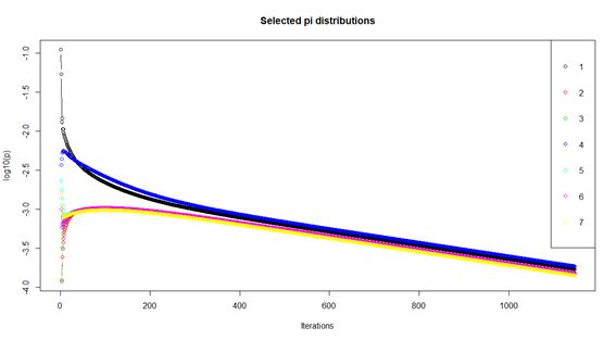
3.4 Evolution of distributions until equilibrium. . . . . . . . . . . . . 36
3.5 The Th-cell differentiation and the HIV-infection processes tend
towards an increasing entropy, S1 ≤ S2 ≤ ... ≤ Sn when reaching
its equilibrium distribution. . . . . . . . . . . . . . . . . . . . . 36
4 Discussion. 39
4.1 Th-cell fully differentiated state as an equilibrium distribution, π n . 39
4.2 Th-differentiation, and HIV-infection processes as martingales. . 40
4.3 Th-cell differentiation, and HIV-infection equilibrium distribu-
tions as discrete generalized beta ones. . . . . . . . . . . . . . . . 41
5 Conclusions & Limitations. 41
6 Acknowledgements. 42
1 Introduction
Human Immunodeficiency Virus infection and the spectrum of related disorders
are relatively new to the human being, with the first known displays of this
disease occurring in the decade of the 1980s, however, efforts were made to
determine a more precise temporal origin of the virus, with some estimates
computing that it appeared as early as in the 1920s decade [311]; while it became
clearer a bit later that HIV may have originated by means of zoonoses from
primates in Africa [252]. During the first-ever recorded outbreak in the 1980s,
the epidemic was said to be limited to the population of the 4 Hs: homosexuals,
haemophiliacs, haitians, heroin addicts [88] nevertheless, was this soon proved to
be a far too narrow assumption, and that the actual boundaries of the infection
were not these, for the set of susceptible population was indeed a larger one.
HIV was later discovered to exhibit two basic genotypes: HIV-1 and HIV-2 with
differences described both in the clinical outcome and course of the disease [236].
HIV is known to exhibit affinity for the receptors in T cells CD4, CCR5, CXCR4,
as well as other cofactors necessary for entry into the cell [302], then using these
cells for replication, inducing later apoptosis in these ones, this apoptosis in
the CD4 (+) T cells (and as well in CD8(+) T cells) is responsible for the
immunodeficiency seen in the HIV(+) patients, in which, when untreated, may
3result in the development of opportunistic infections, including infection by
human herpesvirus type 8, being able this virus to promote the establishment
of a malignancy, say Kaposi’s sarcoma.
1.1 Biology of HIV-1.
Both HIV-1 and HIV-2 are species belonging to the Lentivirus genus which
contains other notable viruses, such as Simian immunodeficiency virus, Feline
immunodeficiency virus and others; this genus belongs itself to the subfamily
Orthoretrovirinae, part of the Retroviridae family which also contains the sub-
family Spumaretrovirinae in which we may find those endogenous retroviruses
which have incorporated into the host’s genome and may take part in the genetic
diversification. The Retroviridae family is part of those reverse transcribing
RNA and DNA viruses [262]. The viral particle is about 100 nm in diameter,
with an outer membrane as envelope, this envelope contains 72 knobs, each knob
comprised by trimers of Env proteins, the Env protein possesses in its structure
the gp120 and the gp41 (gp160) proteins in its stead, where the gp41 anchors to
the lipid membrane and gp120 binds to gp41 finally protruding from the mem-
brane. In its envelope, the viral particle also contains MHC pertaining to the
host. Within the particle, we find another membrane, comprised by the matrix
(p17) and the protease (p10) proteins; attached to the matrix protein membrane
we find the lateral bodies, which are polarized bodies inside the particle. Finally,
the capsid lies in the core of the viral particle, the shell being composed of the
p24 protein (capsid protein) and attached to the matrix protein by means of the
p6 protein (link protein). In the innards of the capsid lies the genome, where
the reverse transcriptase complex is found attached to the genome, along with
the nucleic acid binding protein (p7) [1]. The viral particle’s bilayer is enriched
in specific lipids: aminophospholipids, dyhydrosphingomyelin, plasmenyl phos-
phoethanolamine, phosphatidylserine, phosphatidylethanolamine and phospho-
inositides; being the lipid composition bearers of importance in HIV fusion and
infectivity (chol-chelating compounds inhibit these actions), interaction with
T-cell immunoglobulin and mucin domain proteins block release of HIV from
infected cells, interaction with bavituximab (which targets phosphatidylserine)
suppresses productive HIV infection [111]. The clues leading to the thought of
HIV being a retrovirus came when in the 1980s outbreak, HIV-2 was shown to
also cause AIDS in patients, HIV-2 was related to HIV-1 but also to a simian
virus which caused immunodeficiency in macaques. It has been determined that
the SIV from the Sooty mangabey originated the HIV-2 (groups A-H), while the
SIV from the chimpanzee originated both the HIV-1 (groups M, N and probably
O) as well as the SIV from Western gorillas, which in turn gave place to HIV-1
(groups P and probably O) [252]; it has been proposed as well that HIV origi-
nated from SIV by means of several genetic mutation transitions and that the
use of unsterile injections may have promoted the increase in transmission [161],
other mechanisms have been proposed, such as: population growth, changing
sexual practices, migration, increased hunting and deforestation in post-colonial
Africa [196]; however, retroviruses have been computed to appear as far back
4as 460 to 550 million years ago (during the Palaeozoic Era), along with the
radiation of Vertebrata, nevertheless it remains unclear whether Retroviridae
originated from Metaviridae or whether Retroviridae gave place to Metaviridae
[106]. HIV-1 displays three major groups: major (M) with a transmission event
occurring between 1912 and 1941, outlier (O), and nonmajor and nonoutlier (N);
the virus may have commenced with the HIV-1 O group in gorillas, then spread
among humans from the Congo River into Kinshasa, Zaire, with the earliest
documented case of HIV-1 infection (M group) in humans dating from 1959.
The M group is the predominant circulating group, and it has been divided into
subtypes: A1, A2, A3, A4, B, C, D, F1, F2, F, H, J, K; further recognition of
subtypes by means of full-genome sequencing has been made (CRFs and URFs)
[274]. Meanwhile, HIV-2 displays A-H grups, with the A group possessing two
subdivisions: A1 and A2 [1]. The life of those patients infected with HIV gave
a very important turn with the advent of the antiretroviral drugs, and has ap-
proached the amount of years to be lived at age 20 years two thirds that of
the general population [49], other measurements have been made estimating
expected average ages of death in those patients receiving antiretroviral ther-
apy, finding 67.6 years for men and 67.9 years for women [50]. In addition, the
mutation rate of the HIV-1 genome in vivo has been shown to be rather high,
i.e., 4.1 ± 1.7 × 10⁻³ per base per cell, this rate is contributed 2% by the reverse
transcriptase and 98% by the host’s cytidine deaminases of the A3 family [56].
1.2 Insights into molecular pathophysiology: viral pro-
teins.
1.2.1 gp120: organizing the foundations for infection from mem-
brane to nucleus.
gp120 owes its name to its molecular weight ranging from 110 to 120 kDa
[55], being this protein a portion of the gp160 complexes (due to the fact that
gp120 matures from a larger, gp160 peptide), the gp120 associates with the
gp41 molecule assemblying into a heterodimer, which will trimerize to form the
mature Env protein, in its turn bound to the viral bilayer membrane. The
protein is comprised by five variable regions and five conserved regions, fold-
ing into a globular structure with an inner and an outer domain bound by a
bridging sheet. gp120 is glycated mostly by high-mannose glycans, and com-
plex glycans fucosylated and containing multiple antennas, along with a vari-
able amount of sialic acid ; the high-mannose oligosaccharides were found to be
(GlcN Ac)2 (M an)5 and (GlcN Ac)2 (M an)9 [218]; deficiencies in glycosylation
(as they have been induced in bacteria-produced gp120) result in unsuccess-
ful binding of gp120 to CD4 [78]. gp120 binds to CD4 by means of a pocket
found above the bridging sheet [313], this pocket is known to be hydrophobic in
gp120 and when bound to CD4 a phenylalanine residue caps this pocket, this
residue has proved essential for a successful binding, the whole complex (gp120
hydrophobic pocket + phenylalanine residue) is termed the Phe43 cavity [65];
moreover, the affinity with which CD4 interacts with gp120 has been determined
5to be Kd = 4 × 10⁻⁹ M [275] while the entropy of this process has been seen
to be ∆S = 220 ± 13kJmol−1 and an enthalpy of ∆H = −259 ± 13kJmol−1
(at 310 K) (in which we find that this binding of CD4 to gp120 is an irre-
versible (due to ∆S > 0) and spontaneous (because ∆G < 0, considering ∆G
as the free energy) process [157]) with an intermolecular hydrogen-bond net-
work of 166 atoms (for CD4) and 130 atoms (for gp120) [109]. The binding
of gp120 to CD4 induces conformational changes in gp120, and promotes the
creation of a high-affinity binding site for CCR5, as well as the exposure of
gp41 in order to induce membrane fusion; furthermore, has it been shown that
the binding of the soluble form of CD4 (sCD4) to gp120 promotes the dissoci-
ation of gp120 from gp41, and some variable loops (V1, V2, V3) change their
conformation or become more more exposed; the exposure of these loops (V1
and V2) could be potentially recognized by the monoclonal antibodies 17b and
48d (this nomenclature is due to the epitopes exposed when the conformational
changes are induced) [264], where the monoclonal antibody 17b is an anti-HIV-1
gp120 monoclonal antibody which is obtained from EBV transformation of B
cells (let us recall that these transformed B cells are those infected by EBV in
which EBNA1, EBER1 and EBER2 are expressed, inducing the translocation of
MYC into the immunoglobulin loci, activating permanently this transcription
factor [134]) of an asymptomatic HIV-1 infected individual [226]; however, due
to steric hindrance and conformational masking, these epitopes remain obscured
or unaccesible to the antibodies directed against gp120, therefore, by means of a
chimeric protein comprised of a soluble CD4 bound to the monoclonal antibody
directed towards 17b, the necessary conformational change occurs and the epi-
topes are exposed, method through which, the monoclonal antibody may then
bind to the epitope [136]; nevertheless, there exist also entropic barriers in the
gp120 core which may decrease the potential binding of antibodies [217]. The
binding of CD4 to the heterohexamer ((gp120)3 (gp41)3 ) happens in a stoichiom-
etry of (CD4)3 (gp120)3 (gp41)3 this because two CD4 molecules may be as close
as 19.8nm in the cell membrane context, that is, the root of the CD4 molecules
for these molecules protrude from the cell membrane and be closer in the extra-
cellular space, the maximum distance at which they can be approached one from
the other has been measured to be 3.5nm in the extracellular space and without
steric interference [135]. After the binding of CD4 to gp120, we have described
that there exist some conformational changes which promote the interaction of
CCR5 with gp120 (used in early infection) and with CXCR4 (in late infection),
it has been displayed that in the absence or changes in sequence of the loops V1
and V2, there could be a loss in the ability of gp120 of binding to CD4, then be-
ing able to infect CD4(-) cells with CCR5(+) or CXCR4(+); the conformational
changes of gp120 by means of CD4 diminish the entropy of the protein [217];
this binding of gp120 to the chemokines happens through the V3 loop which is
predominantly composed of 35 residues, connected by a disulfide bridge between
residues 1 and 35, being those residues essential for the binding with the core-
ceptors those from 13 to 21, additionally, gp120 binds with near equal energetic
properties to CCR5 and CXCR4 [269]; the binding of CD4 to gp120 induces a
diminishment in the distance of the trimer to the target cell membrane by 2nm
6[135]. We previously commented on the observed fact that the HIV-1 in its early
infection phase exhibits tropism towards CCR5, while during late infection, this
tropism shifts towards CXCR4 [223] and there have been some reasons of this
shift implicating gp120: loss of N-linked glycosylation site in V2 in late stages,
increase in positive-charged residues in V1/V2 and V4/V5, addition of N-linked
glycosylation site near the CD4-binding site (N362); however the implications of
this change in tropism are viral-fitness-dependent, that is, whether this switch
is convenient for the virus to occur or not, in which, given the case, the switch
shall not occur [174]; this may be challenged (or sustained) by the fact that
CCR5 is expressed in memory T cells, while CXCR4 is in change expressed in
naı̈ve T cells, in which the memory T cells have shown to possess a higher divi-
sion rate than naı̈ve T cells, which promotes an advantage in the fitness of the
virus during early infection, nevertheless when time passes and CD4(+) T cell
depletion occurs, both memory and naı̈ve T cell numbers are consequentially
diminished, but the fraction of naı̈ve T cells in division has been seen to divide
more rapidly in the late stage of disease than memory T cells, those augmenting
the selection of those viruses with CXCR4 tropism or the switch from CCR5
to CXCR4 in late stages of disease [223]. It has been shown that gp120 folding
occurs at the rough endoplasmic reticulum (and not in the Golgi apparatus)
and this folding is essential to the binding of gp120 to the CD4 molecule; the
folding time of this molecule has been shown to be more lengthy than usual,
this due perhaps to the formation of disulfide bonds within gp120, probably
aided by disulfide isomerase (disulfide bond-formation are dependent on several
enzymes, those thiol-disulphide oxidoreductases, such as disulphide isomerase
(this one is yet to know whether it catalyzes de novo formation of disulfide bonds
or shuffles pre-existing bonds), as well as other proteins, such as ERO1 which
is a glycosylated ER protein which induces oxidizing equivalents into the ER
lumen [249]); gp120 is constrained to proper folding for its exit from the rough
endoplasmic reticulum [78]. gp120 not only exerts fusion-related functions, but
also contributes to the pathogenesis of HIV by activating complement, inducing
polyclonal B cell activation, binding to immune complexes, participates in in-
fluencing T cell function (promoting inefficiency in T cell response), augments
production of TGFB [313]. The gp120-dependent activation of complement oc-
curs via a dettachment of gp120 from the viral envelope either spontaneously
or after gp120 binding to the CD4 molecule, then gp120 may circulate in the
patient’s blood and may attach in a soluble form to CD4(+) T cells, promotion
of opsonization and elimination of CD4(+)gp120(+) T cells by means of the
reticuloendothelial system; the complement is activated in its classical pathway,
and gp120 has been seen to bind to C4, C3, C5, C9 complement proteins [266];
thus ending with the membrane atttack complex (MAC) formation in the gp120-
dependent complement activation directed towards CD4(+) T cells, contributing
to the depletion of these cells; this membrane attack complex is assembled by
means of the cleavage of C5 to C5b and C5a, C5b recruits C6 (C5b6) through
C9 and forms the MAC pore [319]; abnormal complement activation has been
observed in HIV patients, associating this infection with certain autoimmune
disorders, such as: immune thrombocytopenic purpura, inflammatory myosi-
7tis, Guillain-Barré syndrome, sarcoidosis, myasthenia gravis, Graves’ disease,
Hashimoto thyroiditis, autoimmune hemolytic anemia, autoimmune hepatitis,
rheumatoid arthritis, systemic lupus erythematosus [287]. Abnormalities of the
humoral response have been observed in HIV patients (which relates to ab-
normal B cell function), such as: polyclonal hypergammaglobulinemia, B-cell
hyperplasia, circulating immune complexes, spontaneous Ig secretion, prolifera-
tion with elevated autoantibodies; that has been shown to be induced by gp120
due to an increase in TNFA, which increases B cell proliferation, this increase
in TNFA not only stimulates cellular proliferation, but also IgM and IgG se-
cretion; furthermore, it has been seen that B cells display binding to the gp120
protein (experimentally, 2.9% to 4.3% of the B cell CD20(+) population binds
to gp120, therefore we may think of this ratio of binding as a probability p
for B cell to bind to gp120 in a range of p ∈ [0.029, 0.043]); in B cells, gp120
also promotes cAMP generation [199] which is known to suppress the activity
of NFKB by means of BCR and TLR4 signaling blocking, possibly through a
protein kinase A [169], NFKB is also a lymphopoietic regulator in B cells, by
maintaining TNFA production at appropriate levels (then we see an increase
in TNFA activity, and suppression of NFKB, which may contribute to a malig-
nancy development in HIV patients, even in those patients with antiretroviral
treatment, there exists the possibility of developing, for instance, Hodgkin’s
lymphoma [119]), and plays also a role in the expression of Igλ in immature
B cells; mature B cell survival and population restoration is dependent on the
NFKB canonical pathway [241]. gp120 also exerts actions down- and upregu-
lating cell cycle- or transcriptional regulation-related genes in T cells (as seen
when T cells are treated with the V3 loop of gp120), some of these upregu-
lated genes are: NOC2L (inhibitor of a histone acetyltransferase independent of
HDAC, which is seen upregulated) [173], SEPT9 (member of a family of GTP-
binding proteins, which exhibit roles in cytokinesis, cytoskeleton, cell cycle con-
trol; whose hypermethylation is related to several cancers [254]), IFI6, SPIN,
HNRNPM; while some other downregulated genes are: ABCG1, PGPEP1, PT-
PLA, SPATA21; overall, the V3 loop of gp120 affects the following sets of genes
within T cells: cell cycle (62 genes), cellular development (function, mainte-
nance, compromise, morphology) (54 genes), aminoacid or lipid metabolism (36
genes), gene expression (65 genes), DNA metabolism (replication, recombina-
tion, repair) (27 genes), cellular assembly and organization (30 genes), cellular
movement (25 genes), hematopoiesis (25 genes), immune response (32 genes),
cellular death (growth and proliferation) (100 genes), infection mechanism (34
genes); therefore we witness a hijacking of the genetic machinery in T cells which
may compromise by all means their further function and proliferation [173]. Var-
iois gp120 inhibitors have been described, such as: BMS-378806, BMS-488043,
BMS-626529, BMS-663068, NBD-556, JRC-II-191, and 18A [150], BMS-378806
does not display action against HIV-2, SIV, and other viruses, whose metabolism
is cythochrome-dependent [310] and thus major drug-drug interactions are ex-
pected [294]. BMS-488043 is an analog of the BMS-378006 compound and has
been seen to display in vivo efficacy against HIV-1 in a monotherapy regime
[150]. BMS-626529 or temsavir is administered as a prodrug (BMS-663068),
8a methyl phosphate prodrug (fostemsavir) which is hydrolized by an esterase,
whose metabolism is equally contributed by the cytochromes [328], the recorded
adverse effects include: headache, rash, and micturition urgency [180]; addition-
ally, this prodrug has shown a better performance in the inhibition of dually
tropic viruses [192]; fostemsavir has very recently been suggested to be used as
a drug in multidrug-resistant HIV-1 infection [130].
1.2.2 gp41: fusion, metabolism, and T-cell function suppression.
This protein mediates fusion with the host’s cell membrane [39], portion of the
env protein pertaining to HIV; this protein displays C-terminal helices which
back around N-terminal helices, forming a six-helix bundle [166], this structure
organizes into an extracellular ectodomain, anchoring gp120 to the cell surface,
a transmembrane domain (promoting further anchoring to the lipid bilayer),
and a cytoplasmic tail, this tail contains a highly conserved endocytosis motif,
and three alpha helical motifs known as lentiviral lytic peptides [80]; where the
transmembrane region of gp41 is followed by a region of high hydrophilicity (the
cytoplasmic domain), containing a highly immunogenic region and a C-terminal
region with two amphipathic segments (the lentiviral lytic peptides , LLPs), as
well as a leucine zipper motif between LLP2 and LLP1; the cytoplasmic domain
of gp41 has been suggested to confer conformational stability to the Env pro-
tein, but it has been determined that the cytoplasmic domain interacts with host
proteins: AP1, AP2, CAM, CTNNA, luman, MA, TP115, perilipin-3, plasma
membrane, PRA1, prohibitin 1/2, TAK1. Furthermore, the cytoplasmic domain
has been proved essential for both replication and incorporation into several cell
lines, such as: monocyte-derived macrophages, peripheral-blood mononuclear
cells, B cells, epithelial carcinoma-derived cells; however, the replication and
incorporation processes have proved independence of the cytoplasmic domain of
gp41 in other cell lines, mostly CD4(+) T cells [208]. Furthermore, gp41 fusion
protein works as an inhibitor of T cell activation by different mechanisms. The
immunosupressive (ISU) sequence impairs T cell activation through the interac-
tion with the T cell receptor (TCR) complex and the direct inhibition of protein
kinase C mediated phosphorylation; gp41 fusion peptide inhibits antigen-specific
T cell activation by binding to the TCRα transmembrane domain; gp41 trans-
membrane domain can enhance the overall immunosupressive effect through an
interaction with the fusion peptide; and the recent described immunosupressive
loop-associated determinant (ISLAD) is another inhibitor of antigen-specific T
cell proliferation and proinflammatory cytokine release by interacting with the
TCRα [12].
Peptides derived from gp41 N-terminal heptad repeat (NHR) and C-terminal
heptad repeat (CHR) sequences can inhibit HIV-1 infection by interaction with
their counterparts in gp41. Some of these peptide fusion inhibitors are: DP178,
later named T20 (generic name: enfuvirtide) was the first fusion inhibitor ap-
proved by the U.S. FDA., but the high cost and inconvenience of twice daily
injection, prevents it from being considered as a regular drug [34]; FB006M
(generic name: albuvirtide) was approved in 2018 by the Chinese FDA.; SFT
9(generic name: sifuvirtide) is a novel and potent gp41 inhibitor that has shown
promising results; other gp41 fusion inhibitors under development and study
are: T1249, 2F5, 4E10, C52L, VIR-576, among others [210].
1.2.3 p7 (nucleoprotein, N C ∈ gag): viral genome package and facil-
itator.
This protein derives from the Gag precursor which is cleaved into p17, p24,
and p7 proteins [164]. Nucleocapsid protein 7 (NCp7) is the major internal
component of the HIV virion core, it has been seen to be highly conserved
(however not so in spumaretroviruses), displays RNA-binding properties, con-
taining two zinc fingers (ZF) [62], reminding us that these domains are main-
tained by a zinc ion, coordinating a cysteine and a histidine molecule, these
motifs have two β-sheets and one α-helix; it has been seen that these proteins
bind to DNA but may as well bind to RNA [38]. p7 possesses functions in-
volved in the selection and packaging of the viral genome, as well as others
which play a role in viral replication [164]; NCp7 interacts with the viral RNA
and is required for its dimerization, encapsidation, and initiation of its reverse
transcription, where NCp7 enhances the reverse transcriptase processivity and
RNase H activity [143]; p7 additionally binds to proviral DNA (by means of
its basic residues), and protects it from nuclease digestion [140]. Interestingly,
zinc ejection or mutations affecting the zinc finger folding and conformation of
the nucleocapsid hydrophobic plateau, lead to non-infectious viral particles [94].
The importance of its conserved structure is the low probability of mutations
found in treatment-resistant strains [127]. Thus, NCp7 represents a promising
therapeutic target for an effective next-generation antiretroviral therapy. NCp7
inhibitors are divided into covalent and non-covalent inhibitors. Covalent in-
hibitors, also referred to as irreversible inhibitors or zinc ejectors, which can
recognise NCp7 among cellular proteins containing ZFs, some examples of them
are 3-Nitrosobenzamides (NOBAs); disulfide-substituted benzamides (DIBAs);
thioesters and pyridinioalkanoyl derivatized thioesters (PATEs); benzisothia-
zolone (BITAs); azodicarbonamide (ADA); thiocarbamates (TICAs); S-acyl 2-
mercaptobenzamides (SAMTs); transition metal complexes; diselenobisbenza-
mides (DISeBAs); and more recently, thioether prodrugs. All of these covalent
inhibitors are structurally characterised by a weak electrophilic group that is
attached by the distal ZF domain, after this covalent complex has been formed,
the zinc is ejected, causing the loss of the tertiary protein structure and con-
sequently, all of its functions [282, 238]. Non-covalent inhibitors and nucleic
acid (NA) binders are another therapeutic target against NCp7. These com-
pounds have weaker antiviral potency when compared with covalent binders,
and they have not been approved for clinical trials. Several interactions in the
NA/NCp7 complexes are involved with the W37 hydrophobic plateau residue,
offering a chance to develop competitive inhibitors. Some of these are pseudod-
inucleotides, HTS-derived small molecules, thiadiazoles, and thiazolidinones.
All of these molecules are characterized by a π-rich area capable of interacting
with W37, seeking to avoid the interaction of NCp7 with NAs. On the other
10hand, NA-binding NCp7 inhibitors include stem loop structure-binders and an-
thraquinones, which block the NCp7-RNA-DNA complex formation, but most
of them are unable to disrupt a preformed complex [117].
1.2.4 p6 (p6 ∈ gag): incorporating vpr into salient viral particles.
This protein, also a byproduct of the Gag precursor cleavage, promotes virus
particle budding, and the incorporation of the vpr protein into the viral particles
[122]; inclusively, p6 is a factor involved in the capsid maturation and virus core
formation processes, this by means of its phosphoprotein features; these func-
tions are performed due to the close Euclidean distance which is encountered
between p6 and the plasma membrane of the cells, this protein may be adsorbed
onto the inner surface of the plasma membrane and promote, for instance, the
incorporation of vpr into the viral particles. This protein possesses two α-helical
domains which are connected by a flexible region, this structure is more pro-
nounced in hydrophobic conditions; its C-terminal region contains vpr-binding
residues; the Ser40 residue has been seen to be a potential protein kinase C
phosphorylation site [194]. TSG101 (tumor susceptibility gene 101) is a key cel-
lular protein as part of the endosomal sorting complexes required for transport
(ESCRT), which is recruited to viral assembly sites via p6, where the Pro-Thr-
Ala-Pro (PTAP) motif in p6 acts as a docking site for TSG101. This process
is critical for HIV release. The duplication of this PTAP motif has shown an
enhanced replication advantage of HIV-1 subtype C by engaging TSG101 with
a higher affinity [250]. Furthermore, the deletion of the YPxn L motif, which
binds to ESCRT component ALG-2-interacting protein X (ALIX), is associated
with a decrease in virus release from infected cells (as seen in HIV-1 subtype
C), and conversely, PYxE motif insertion can reconstitute the p6 binding to
ALIX and consequently, viral budding mediated through the ESCRT pathway
[68]. Depletion of the ESCRT components have shown a powerful block to HIV
particle release [259], for this reason, therapeutic targets are principally focused
on disrupting the p6-TSG101 interface, such as peptoid hydrazones, cyclic pep-
tide 11, F15 (esomeprazole) and N16 (tenatoprazole), these last two are able to
join to the ubiquitin E2 variant domain of TSG101, highlighting the possibility
of interfering with previously unknown therapeutic targets and expanding the
future perspectives of TSG101 inhibitors [66].
1.2.5 p24 (capsid protein, CA ∈ gag): protector of the viral genome.
Capsid protein (CA) is member of the subset of proteins derived from the Gag
polyprotein cleavage, a [24, 25]kDa protein which may be detected before sero-
conversion, this protein may assemble into a protective shell around the viral
RNA [99] which is a spontaneous process [35], this has been described as a
”fullerene cone”, hexamers of p24 link into an hexagonal surface lattice, with 12
capsid-protein pentamers, finally possessing around 1500 p24 monomers. This
protein is comprised by seven α-helices, a β-hairpin (N-terminal region), a C-
terminal region with four α-helices, and a flexible linker [321], when assembled
11into the cone, the N-terminal domain is located on the outer surface of the cone
and the C-terminal domain is oriented towards its interior; inside this cone, the
RNA genome, and the POL proteins are located (that is the integrase, protease,
reverse transcriptase and others); the cone may act in order to shield the genetic
content from a host response [35], such as those induced by STING, DDX14 or
IFI16 acting as foreign DNA sensors [289]. Several models have been proposed
in regards to the viral uncoating, that is, the dissociation of the capsid cone
into monomers or simpler polymers: immediate uncoating, cytoplasmic uncoat-
ing and nuclear pore complex uncoating; there exist studies which support each
of these models, in the first case, the uncoating occurs rapidly after the entry
into the host’s cell has been performed, however, this model has lost its predic-
tive power due to the fact that the core provides protection against the host’s
foreign-DNA sensors, furthermore, the capsid possesses a pocket to which the
reverse transcriptase complex binds to and allows it to be imported into the
nucleus. The second model, the one of cytoplasmic uncoating proposes the dis-
assembly of the capsid after a certain time interval has been spanned within
the host’s cell, this model may be nevertheless challenged as well by the factors
which we have already commented (foreign DNA host’s response), moreover
some additional cellular factors may protect the viral’s genome from the host’s
response, it may occur by means of the HMGA1 protein (high mobility group
AT-hook, also called HMGIY), which has been recollected from the preintegra-
tion complexes, that is the reverse transcriptase complex before they integrate
into the host’s genome [75]. The third model refers to the nuclear pore complex
(NPC) uncoating, in which when the intact capsid reaches the nuclear pores,
and disassembles in situ, while the reverse transcriptase complex is imported
into the nucleus; this model displays another range of problems which are not
compatible entirely with the experimental data, ergo a more suitable model
should be provided in following years. Both viral and host’s factors take part in
the uncoating process: PPIA (or CYPA, a peptidylpropyl isomerase A, with a
native function of accelerating the folding of proteins and isomerize the proline
imidic bonds [188]), dynein (cytoplasmic trafficking), CPSF6 (nuclear import,
which is a cleavage and polyadenylation specific factor, interacts with RNA
[186]), TNPO3 (nuclear import, this protein is member of the importin-β fam-
ily of proteins, has been seen to bind to the viral integrase, and as well interacts
with the capsid protein), NUP358, NUP153 (nuclear import) [35]. Maturation
inhibitors are a novel class of antiretroviral drugs targeting the cleavage site
between the C-terminal portion of CA and the spacer peptide 1 (SP1), this
cleavage site usually triggers a conformational switch that destabilizes the im-
mature Gag and the mature core formation. Maturation inhibitors cause an
accumulation of CA-SP1 precursor, which eventually leads to the loss of viral
infectivity [66]. The first reported maturation inhibitor was Bevirimat (BVM),
which causes an abnormal virion morphology and inhibition of viral replication,
but failed in the phase IIb trial due to resistance mutations in CA-SP1. A sec-
ond compound named PF-46396 was identified, it shows structural differences
compared with BVM, but it induced resistance mutations at different locations,
this lead to the identification of second-generation maturation inhibitors, such
12as GSK3532795, which successfully overcame the inconvenience of drug resis-
tance, but showed a high rate of adverse gastrointestinal events and frequency
of treatment-emergent nucleoside reverse transcriptase inhibitor (NRTI) resis-
tance, reason why its evaluation was interrupted [172]. However, the promising
results obtained support the continued development of drugs against this thera-
peutic target. Small molecules and peptide-based antivirals designed to disrupt
CA-CA interactions in the immature Gag lattice, the mature core, or both have
been studied during the last years. CAP-1 was the first small molecule developed
to target the CA protein, it produced abnormal core morphologies, and conse-
quently noninfectious particles. Besides small molecules compounds, 12-mer
peptide CA inhibitor (CAI), binds in a hydrophobic CA dimerization interface,
but it can not penetrate cell membranes, limiting its clinical use. Some more
stable α-helical peptides, such as NYAD-1 and NYAD-13, showed a stronger
affinity for the binding site than CAI. Other classes of CA inhibitors are ben-
zodiazepienes and benzimidazoles. PF74 and the pyrrolopyrazolones BI-1 and
BI-2, seem to compete with CPSF6 and Nup153 for CA binding, disrupting
nuclear import. The main problem of the compounds described so far, is the
low clinical relevance due to their pharmacological characteristics. Recently, a
new type of CA inhibitors has been described, this group includes GS-CA1 and
its derivative, GS-6207, are promising drugs that have showed higher potency
than PF74 [66].
1.2.6 p17 (matrix protein, M A ∈ gag): multifunctional protein.
The HIV-1 p17 protein (matrix protein, MA) is associated with the inner sur-
face of the viral envelope, and may primarily function as an anchor of the gp41
protein on the virion surface. When in solution it is mainly encountered in
a monomeric form, while in a solid state it trimerizes [163]; it displays five
α-helices, and a three-strand β-sheet, whereby the C-terminal domain, exposes
carboxyl-terminal residues which aid in the early states of HIV infection, and ba-
sic residues promote membrane binding and nuclear localization (the residues
which take part in this process are located in a cationic loop connecting β-
strands one and two). This protein additionally functions in RNA targeting to
the plasma membrane, incorporation of the envelope into virions and particle
assembly, and aids in the transport of the reverse transcriptase complex through
the nuclear pores [162], it also acts as a viral cytokine, by binding to a cellular
receptor, namely p17R [83], this receptor has been seen to be expressed in Raji
B cells, and activates AP-1, as well as ERK1/2 and downregulates AKT, this
by means of maintaining PTEN in its active state through the serine/threonin
kinase ROCK [45]; the identity of this p17R protein is a bit obscured in the
literature, nonetheless through a non-exhaustive search in regards to Raji cells’
receptors we came up with a list of potential proteins which may play such role,
bearing in mind as well that the H9 cell line lacks this p17R protein in its surface,
if we consider that the surface proteins of the Raji cells are members of the set
R = {p1 , ..., pn } and those of the H9 cell line pertain to the set H = {r1 , ..., rm },
therefore ∃pi ∨ ri : pi ∈ R, pi ∈ / H [231]; those proteins in the set R ∩ H are:
13PVRL1, LILRB1, DRD4, CRLF3, and ADGRD2; such that these receptors
may not be the p17R protein; now, those proteins which are exclusively ex-
pressed in Raji cells and take part in PI3K/AKT/PTEN/AP1/GPC (G-protein
coupling) are: NTSR2, TACR3, FOLR2, MC5R, OR2J3, TNFRS13B, PTAFR,
CCR8, GPR173; this sets are shown in Figure 1; the activation of this pathway
(by means of this p17R) may lead to a promotion in proliferation and release
of proinflammatory cytokines from T-cells [83]; but if p17R is also expressed
in B cells, it ought indeed to promote B cell growth and tumorigenesis [45].
MA protein has a fundamental role in virion assembly, because of its highly
conserved PI[4,5]P2 /nucleic acid binding site, it has become an attractive site
for the development of new antiretroviral drugs, nevertheless, other drugs have
been described targeting the nuclear localization signal of MA or the MA-RNA
interaction. Thiadiazolane based compounds where first described, they target
the MA-RNA interaction, but showed significant levels of toxicity. In contrast,
PI[4,5]P2 binding site inhibitors were not associated with cytotoxic effects, but
work is actively ongoing in optimizing the affinity/potency of this type of chemo-
types. New targeting sites are highly desirable, one of them could be the in-
volvement of MA in Env incorporation, where MA trimerization is important
for the recognition of Env cytoplasmic tail (gp41-CT) and virus assembly [66].
1.2.7 p10 (protease, P R ∈ P OL): catalyzer of viral and host’s pro-
teins breakdown.
This protease is encoded by the pol gene being an aspartic protease, and as
in other retroviruses, displays its function when in its homodimeric form; each
monomer is an aspartic peptidase with four elements: two hairpin loops, wide
catalytic aspartic acid loop, and an α helix [70], the dimeric interface comprises
eight N- and C-terminal residues of each chain, some of these are exposed to
the solvent, and some others (the hydrophobic ones) are oriented towards the
interior of the enzyme [320]. This protein displays two states (when in the ho-
modimer form), either open or closed, depending on the presence of a ligand
(open when in the free state, and closed when in the bound state) [308] where
the open form is more stable than the closed one (considering the free energy
of the process ∆G) [320]. The HIV protease cleaves the polyproteins Gag and
Gag-Pol creating protein subunits [167]; all the same the viral protease displays
proteolytic abilities of the host’s proteins, such as those belonging to: the cy-
toskeleton: vimentin, desmin, GFAP [256], actin, troponin [277], laminin [182];
immune system: proIL1B; and transmembrane proteins: APP [277]; cytosolic
proteins: BCL2, CASP8 [182]. Vimentin plays a role in the diapedesis pro-
cess of T cells, whereby this protein, T cells migrate across the endothelium,
and in genotypes vim−/− , there is a diminished capacity of T cells to home to
mesenteric lymph nodes and spleen, this may be mediated by changes in ex-
pression or distribution of ICAM1 and VCAM1 [183]; while actin polymerizes
or depolymerizes when the T cell becomes active, this T cell activation leads
towards the formation of a distal-pole complex which is an actin-rich structure;
disruptions in the cytoskeleton might as well induce changes in the organization
14Figure 1: Venn diagram of the sets representing the receptors expressed in Raji
cells (Raji) and H9 cells (H9), where we regard at their cardinalities, that is,
|R| = 28, |H| = 68, and R ∩ H = 6; in this case the p17R protein lies in the
R − H set.
15of the supramolecular activation clusters in T cells (SMACs) [21] which takes
a central part in the immunological synapse, being the SMAC a nanocluster
comprised of an actin mesh, transmembrane proteins (members of the TCR
complex), and cholesterol-enriched nanodomains [89]; these disruption in the
cytoskeletal proteins might be added to the one induced by the gp120 protein,
as this protein increases ICAM1 expression [221, 273], inducing the formation of
a type of SMAC but in this case gp120-dependent (and likely protease-aided),
in which gp120 clusters in the centre, and LFA1 and ICAM1 concentrate in
the periphery, this type of SMAC is termed the virological synapse, throughout
which the virus may spread between T cells without the need of being exter-
nalized from the cell (no need of viral budding) [159]; this SMAC recruits the
host’s proteins which we may as well find in the immunological synapse, such
as: TCRZ, ZAP70, LAT, SLP76, ITK, PLCG, and only a weak recruitment
of: CD3E, and ABTCR; however when this protein nanocluster was assembled,
there was an abnormal signaling (distinct to the one induced by the immuno-
logical synapse), where no PKC recruitment, no calcium mobilization nor CD69
upregulation were observed [284]. By means of the cleavage of both BCL2 and
CASP8 can the protease induce cell death [182, 142]. HIV-1 protease inhibitors
(PI) play a key role in the antiretroviral treatment. First generation of PIs were
based on hydroxyethylene and hydroxyethylamine isosteresstarted. Saquinavir
was the first PI approved by the FDA in 1995, since then many other PIs have
been developed. Ritonavir was found to be a potent inhibitor of cytochrome
P450 3A, a major metabolic enzyme for PIs [133], due to this finding, ritonavir
is more frequently used as a PI pharmacokinetic booster than as a PI itself.
Other first generation antiretrovirals are indinavir, nelfinavir, and amprenavir.
Due to the major problems of first-generation PIs (high metabolic clearance, low
half-life, poor oral bioavailability, gastrointestinal distress, and the emergence of
drug-resistance strains), second-generation PIs were developed. The balance of
hydrophobicity and hydrophilicity allowed for a longer half-life. Some of these
drugs are lopinavir, atazanavir, tipranavir (reserved as a salvage therapy), and
darunavir (high potency against multidrug-resistance strains) [92]. Despite the
progress in PIs therapy, some drug-resistance variants have emerged, they are
classified in two groups: primary mutations, which involve changes in residues
directly involved in substrate binding and manifest themselves in the active site
of the enzyme; while secondary mutations are located away from the active site
and are usually compensatory mutations to mitigate the deleterious effects of
primary mutations [171]. Even, secondary mutations may not manifest in the
protease itself but instead in the protein cleavage site on the Gag-Pol and Gag
substrates [202]. One of the main strategies to avoid drug resistance, is the
design of PIs by promoting hydrogen bonding interactions with the backbone
atoms in the HIV-1 protease active site, since mutations that cause drug re-
sistance cannot significantly alter protease active site backbone conformation,
as an example we have darunavir, that showed extensive binding interactions
with backbone atoms and maintained a potent antiviral activity against panels
of clinically relevant multidrug-resistant HIV-1 variants [92, 93]. Recently, a
new pharmacokinetic enhancer, cobicistat, which does not have anti-HIV activ-
16ity, has been developed, offering the advantage over ritonavir, where cobicistat
will not contribute toward the emergence of drug-resistant HIV-1 variants [305].
Many other new classes of PIs with innovative ligands are under preclinical de-
velopment for the next generation, molecular design efforts have focused on the
synthesis of P2-ligands promoting enhanced backbone binding and non-peptide
PIs containing different structural scaffolds distinct from hydroxyethylsulfon-
amide isosteres [92].
1.2.8 p51 (reverse transcriptase, RT ∈ P OL): loader of nucleic acids
and structural support.
The reverse transcriptase’s main function is to convert the viral RNA which has
just entered the host’s cell after membrane fusion into double-stranded DNA,
this occurs in the host’s cell cytoplasm, this DNA will later be translocated to-
wards the nucleus in order for it to be finally integrated into the host’s genome.
It is a heterodimer comprised of p66 (or p15 or RNase H) and p51, both of
which derive from a common Gag-Pol polyprotein, cleaved by the viral protease
(p10, PR); these subunits (p66 and p51) share a common amino terminus; the
details of p15 will be addressed in the following section 1.2.9. p51 possesses
4 subdomains (which are homonymous to those of p15 or p66): fingers, palm,
thumb, and connection [240]. The heterodimerization process is dependent on
factors such as: mutations (e.g., the L289K mutation, the L289 residue in the
p51 subunit makes various hydrophobic contacts with p66 but the mutation
does not yield towards an ill-heterodimerization or no heterodimerization at all;
contrarywise, the same mutation but occurring at the protein p66 yields towards
a lack of dimerization), nucleotide substrates, temperature, magnesium, and so-
lution conditions [324]. It has been noted that the catalytic site of the reverse
transcriptase lies in the p15 (p66) domain, however p51 may take part in: struc-
tural support and facilitating the loading of nucleic acids onto p66, which would
explain why the N348I mutation to p51 may confer resistance to reverse tran-
scriptase inhibitors (both nucleoside and non-nucleoside), as well, p51 has been
seen to contribute to the architecture of the RNase H primer grip/phosphate
binding pocket, finally, deletions to the C-terminal sequence of p51 may alter
changes in the RNase H activity [44]. The p51 subunit in itself may undergo
homodimerization under certain conditions, this was observed by small-angle X-
ray scattering (SAXS), this phenomenon occurs dependent on the concentration
of the p51 monomer, and may coexist with the homodimer p51/p51 in equilib-
rium, furthermore it has been noted that p51 may exist in two forms: p51E and
p51C, the latter is the form in which it may be encountered when bound to the
p66 in the heterodimer, and the former is an extended form in which it may
resemble the structure of p66, the homodimerization may occur with one p51E
species and one p51C species assemblying into p51E/p51C, this could happen
due to the stabilizing effect of p51C on the structure of p51E. p51 interacts
with non-nucleoside reverse transcriptase inhibitors (NNRTIs) forming two dif-
ferent species: p51E-NNRTI and p51E/p51C-NNRTI, this due to the fact that
p51E is in possession of a p66-like structure [324]; the monomeric form of p51
17is favored by low concentration, low salt, and low temperature conditions [325].
Reverse transcriptase inhibitors are divided into nucleoside/nucleotide and non-
nucleoside reverse transcriptase inhibitors, NRTIs and NNRTIs, respectively.
NRTIs inhibit viral replication through competition with purines and pyrim-
idines, avoiding adding new nucleotides and consequently, finishing viral DNA
replication. In contrast, NNRTIs bind to an hydrophobic pocket in p66, this
binding leads to a stereo-chemical change in the protein, preventing the addition
of new nucleosides and blocks the cDNA elongation. Typically, NRTIs consti-
tute the backbone of the antiretroviral therapy, among them we can find teno-
fovir disoproxil fumarate (TDF) and tenofovir alafenamide (TAF), which are
adenosine-derivated; emtricitabine (FTC) and lamivudine as cytosine analogs;
abacavir as guanosine analog; zidovudine and stavudine as thymidine analogs;
and didanosine as an inosine derived. An advantage of the current NRTIs is the
low clinically significant drug-drug interactions but they have the disadvantage
of significant side effect profiles and problems with resistant HIV-1 variants [216].
All of them can be affected by selected resistance mutations, either by mutations
in the N-terminal polymerase domain of the enzyme, where the most common
are K65R, L74V, Q151M, and M184V; or by thymidine analog mutations, where
are included M41L, D67N, K70R, L210W, T215Y/F, and K219Q/E [216, 155,
11]. Among NNRTIs, we can find the first generation (efavirenz and nevirap-
ine) and second generation (rilpivirine and etravirine). Resistance mutations
against NNRTIs are based on the inhibited drug interaction with the NNRTI
binding pocket (as seen with K103N and K101E), disruption between the drug
and NNRTI binding domain residues (Y181C and Y188L), or changes in the
conformation or size of the NNRTI binding pocket (Y188L and G190E)[4, 61,
60, 220]. Unlike NRTIs, NNRTIs have a higher plasma half-life, so they can act
as perpetrators of drug interactions additionally to their significant side-effect
profile [216]. New NRTIs and NNRTIs are focused on overcoming resistances
and reduce side effects [215]. Islatravir (MK-8591) is a nucleoside nonobligate
chain terminator that inhibits the enzyme by preventing its translocation, and
therefore it has been categorized as a nucleoside reverse transcriptase transloca-
tion inhibitor (NRTTI), demostrating a robust antiviral activity in vivo against
HIV-1 [156]. Doravirine (MK-1439), a novel NNRTI, has showed its potency
at low doses against common resistant strains and has less potential drug in-
teractions than its counterparts because it does not have appreciable inductive
or inhibitory effects on CYP enzymes [23]. Elsulfavirine is the prodrug of the
active compound VM-1500A, it has shown a high genetic barrier to the develop-
ment of resistant drug mutations [318]. A study showed that elsulfavirine was
not inferior to efavirenz combined with TDF/FTC in virologic control, and in
addition, elsulfavirine was better tolerated [176].
1.2.9 p15 (RNase H, (p15∨p66 ∈ pol): converter of RNA into dsDNA.
We have previously commented that the reverse transcriptase protein is com-
prised of two subunits: p51 and p66 (or p15), where the enzymatic activity of
the protein is performed by the p15 subunit, this enzymatic activity is performed
18by both the polymerase and by the RNase H, the polymerase domain contains 4
subdomains: fingers, palm, thumb, and connection [240]. The RNase H belongs
to a nucleotidyl-transferase superfamily, this family includes enzymes such as:
transposase, retroviral integrase, Holliday junction resolvase, and RISC nucle-
ase Argonaute. In retroviruses, this RNase H converts an ssRNA into dsDNA,
removes the template RNA after the DNA synthesis and produces a polypurine
primer for the second-DNA-strand synthesis. In reactions catalyzed by the
RNase H, the nucleophile is derived from a water molecule or a 3’-OH from the
nucleic acid; the active site of these enzymes is dependent on magnesium or
manganese to bind to the substrate and catalyze the reactions, these reactions
occur in a one-step bimolecular nucleophilic substitution; two metallic ions are
required for the reactions, in which one metal ion activates the hydroxyl nu-
cleophile and the other one stabilizes the pentacovalent intermediate. Various
RNases display a conserved ∼ 100 residue core structure: five stranded β sheet
and three α helices (A,B,D). Specificity for the RNA strand is determined by
contacts between the RNase H and five consecutive 2’-OH groups, and when the
RNA complexes (around 6 bp) with the enzyme, around 100 nm of the enzymatic
surface becomes buried, a groove which protrudes near the site of the buried sur-
face is the active site, where catalytic carboxylates (E109 and D132) make hydro-
gen bonds with 2’-OH groups. The DNA binding site is located in the alpha he-
lices (polymerase), which fold into a groove, a phosphate binding pocket accom-
panies this binding site (comprised of the amino acids T104, N106, S147, T148);
the pocket when not in enzymatic activity is occupied by a sulfate ion, which is
replaced by the DNA’s phosphate [193]. It has been noted that the manganese
may specifically promote a specific hydrolytic activity (hydrolysis of dsRNA)
to the enzyme, rather than the more general endonuclease and directional pro-
cessing activities, divalent metal binding may be mediated by the p66 residue
E478 [46]. The initial RNAse H inhibitors were described almost 30 years ago,
however, the first inhibitor with a relevant effect was N -(4-tert-butylbenzoyl)-2-
hydroxy-1-naphthaldehyde hydrazine (BBNH), although it was also an inhibitor
of the RT DNA polymerase [26]. After, a derivative, (E )-3,4-dihydroxy-N ’-((2-
methoxynaphthalen-1-yl)methylene)benzohydrazide (DHBNH), which has its
binding site between the RT polymerase active site and the polymerase primer
grip, alters the trajectory of the template-primer, so the RNAse H cannot cleave
the RNA strand in RNA/DNA complexes [108]; metalchelating RNase H active-
site inhibitors, such as 1-hydroxy-pyridopyrimidinone, pyridopyrimidinone, di-
hydroxycoumarin, diketo acids, β-keto acids, 3-hydroxypyrimidine-2,4-diones
and hydroxypyridonecarboxylic acids, might sequester Mg+2 ions required for
RNAse H activity [279, 293]. An alternative to metalchelating inhibitors are al-
losteric inhibitors that interfere with the RNA-DNA binding and induce changes
in the RNAse H active site, examples of these are the cycloheptathiophene-3-
carboxamide (cHTC) derivatives [293]. Several other inhibitors have been pro-
posed and developed such as the dual inhibitors RDS1643 (co-targeting HIV-
1 IN and RNAse H), EMAC2005 (co-targeting HIV-1 RNA-dependent DNA
polymerase and RNAse H), and RMNC6 (co-targeting RT and RNAse H) [293].
However, despite extensive research and improvement in RNAse H inhibitors,
19You can also read