Lactiplantibacillus plantarum AR113 Exhibit Accelerated Liver Regeneration by Regulating Gut Microbiota and Plasma Glycerophospholipid
←
→
Page content transcription
If your browser does not render page correctly, please read the page content below
ORIGINAL RESEARCH
                                                                                                                                         published: 13 January 2022
                                                                                                                                   doi: 10.3389/fmicb.2021.800470

                                             Lactiplantibacillus plantarum AR113
                                             Exhibit Accelerated Liver
                                             Regeneration by Regulating Gut
                                             Microbiota and Plasma
                                             Glycerophospholipid
                          Edited by:         Chunliang Xie 1† , Zhoumei Zhang 1† , Manyi Yang 3 , Cha Cao 1 , Yingjun Zhou 1 , Zuohua Zhu 1 ,
                       Xiaodong Xia,         Wenbing Gong 1 , Chao Xu 1 , Li Yan 1 , Zhenxiu Hu 1 , Lianzhong Ai 2* and Yuande Peng 1*
  Dalian Polytechnic University, China
                                             1
                                              Institute of Bast Fiber Crops, Chinese Academy of Agricultural Sciences, Changsha, China, 2 Shanghai Engineering
                       Reviewed by:
                                             Research Center of Food Microbiology, School of Medical Instrument and Food Engineering, University of Shanghai for
                        Fouzia Sadiq,
                                             Science and Technology, Shanghai, China, 3 Department of Hepatobiliary and Pancreatic Surgery, NHC Key Laboratory of
     Shifa Tameer-e-Millat University,
                                             Nanobiological Technology, Xiangya Hospital, Central South University, Changsha, China
                              Pakistan
                          Zhaolai Dai,
   China Agricultural University, China      Emerging evidence indicates that probiotics have been proved to influence liver injury
                      *Correspondence:       and regeneration. In the present study, the effects of Lactiplantibacillus plantarum
                             Lianzhong Ai
               ailianzhong@hotmail.com       AR113 on the liver regeneration were investigated in 70% partial hepatectomy
                            Yuande Peng      (PHx) rats. Sprague-Dawley (SD) rats were gavaged with L. plantarum AR113
                    ibfcpyd313@126.com
                                             suspensions (1 × 10¹⁰ CFU/mL) both before and after partial hepatectomy. The
     † These  authors have contributed
    equally to this work and share first
                                             results showed that L. plantarum AR113 administration 2 weeks before partial
                            authorship       hepatectomy can accelerate liver regeneration by increased hepatocyte proliferation
                                             and tumor necrosis factor-α (TNF-α), hepatocyte growth factor (HGF), and transforming
                   Specialty section:
         This article was submitted to
                                             growth factor-β (TGF-β) expression. Probiotic administration enriched Lactobacillus
                    Food Microbiology,       and Bacteroides and depleted Flavonifractor and Acetatifactor in the gut microbiome.
               a section of the journal
                                             Meanwhile, L. plantarum AR113 showed decline of phosphatidylethanolamine (PE),
             Frontiers in Microbiology
                                             phosphatidylcholine (PC), phosphatidyl serine (PS), and lysophosphatidyl choline
        Received: 23 October 2021
      Accepted: 29 November 2021             (LysoPC) levels in the serum of the rats after the L. plantarum AR113 administration.
       Published: 13 January 2022            Moreover, L. plantarum AR113 treated rats exhibited higher concentrations of L-leucine,
                              Citation:      L-isoleucine, mevalonic acid, and lower 7-oxo-8-amino-nonanoic acid in plasma than
     Xie C, Zhang Z, Yang M, Cao C,
 Zhou Y, Zhu Z, Gong W, Xu C, Yan L,
                                             that in PHx. Spearman correlation analysis revealed a significant correlation between
         Hu Z, Ai L and Peng Y (2022)        changes in gut microbiota composition and glycerophospholipid. These results indicate
  Lactiplantibacillus plantarum AR113
                                             that L. plantarum AR113 is promising for accelerating liver regeneration and provide
              Exhibit Accelerated Liver
     Regeneration by Regulating Gut          new insights regarding the correlations among the microbiome, the metabolome, and
               Microbiota and Plasma         liver regeneration.
                  Glycerophospholipid.
         Front. Microbiol. 12:800470.        Keywords: Lactiplantibacillus plantarum, partial hepatectomy, liver regeneration, gut microbiota, plasma
    doi: 10.3389/fmicb.2021.800470           metabolites, glycerophospholipid

Frontiers in Microbiology | www.frontiersin.org                                   1                                       January 2022 | Volume 12 | Article 800470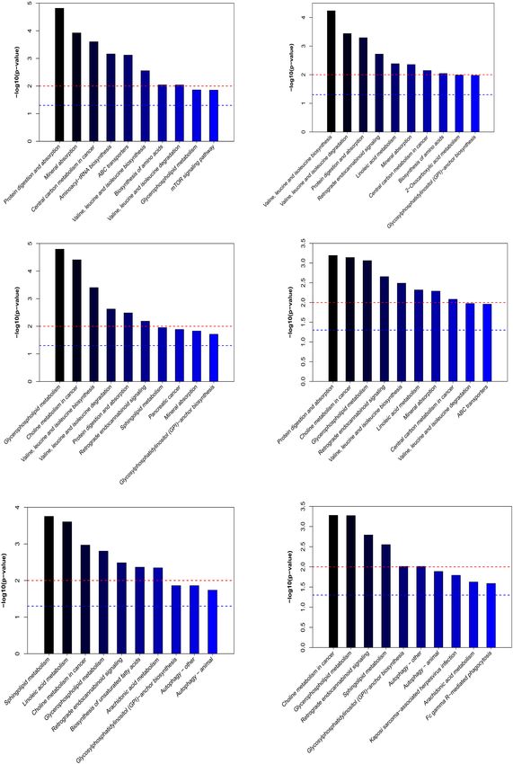
Xie et al.                                                                                    Lactiplantibacillus plantarum AR113 Exhibit Accelerated Liver Regeneration

INTRODUCTION                                                                             bacterial or endotoxin translocation (Cornide-Petronio et al.,
                                                                                         2020). In addition, gut microbiota affects intestinal signaling
Liver diseases are a major medical problem for health care                               and enterohepatic circulation of bile acids (BAs) which have
systems worldwide (Chowdhury et al., 2021). Partial hepatectomy                          been identified as key metabolic signals during liver regeneration
and liver transplantation are currently the only curable methods                         (Liu et al., 2015).
for patients with hepatocellular carcinoma and cirrhosis (Yagi                               Probiotics supplementation is associated with modulation
et al., 2020). However, complications like biliary leakage with                          of the gut microbiota to reduce the inflammation cascade
consecutive bacterial peritonitis have a severe negative impact on                       and enhance the immune system associated with liver surgery
the post-operative course (Tanemura et al., 2018). Therefore, the                        (Nishida et al., 2018). Pediococcus pentoseceus, Lactococcus
liver’s remarkable capacity to regenerate after surgery determines                       raffinolactis, and Lactobacillus paracasei 19 inhibited bacterial
the long-term prognosis and quality of life of patients.                                 translocations after liver resection in rats, and induced hepatocyte
    Liver regeneration is an orchestrated biological process                             mitosis which was delayed by colonic anastomosis (Seehofer
that includes sequential changes in gene expression, growth                              et al., 2004). Treatment with the Linex containing Lactobacillus
factor production, and tissue remodeling (Michalopoulos and                              and Bifidobacterium alleviated hepatic injury and restored
Bhushan, 2021). Following liver resection, hepatocytes, which                            liver function in chronic liver disease patients (Rodes et al.,
are not terminally differentiated, exhibit substantial proliferative                     2014). Although these selected strains have been shown
capacity. Many cytokines, notably HGF, epidermal growth factor,                          to prevent bacterial infections following abdominal surgery,
transforming growth factor-α (TGF-α), interleukin-6 (IL-6), and                          thus far, the experience with selected probiotics in patients
TNF-α, which are involved in liver regeneration, have been                               after PHx is limited.
identified and extensively reviewed (Hoffmann et al., 2020).                                 The present study aimed to evaluate the effects of L. plantarum
However, liver regeneration research has typically focused on                            AR113 in liver regeneration. The results showed that
signaling pathways intrinsic to the liver, overlooking those                             L. plantarum AR113 intervention 2 weeks prior to partial
derived from the gut.                                                                    hepatectomy significantly promoted liver regeneration and
    Due to the intestinal-liver axis interaction, there are natural                      reduced mortality in animals. In addition, comprehensive
and close links between the gut and the liver in terms of                                analyses of cytokines, gut microbiome, and serum metabolites
anatomical structure and physiological function. Gut microbiota                          composition were performed to explore the mechanism
play an important role in different liver diseases (such as non-                         underlying the beneficial effects of L. plantarum AR113 during
alcoholic fatty liver disease, cirrhosis, hepatocellular carcinoma,                      the late phases of liver regeneration. In general, our data suggest
alcoholic liver disease, etc.) (Trebicka et al., 2021). For example,                     that L. plantarum AR113 administration before PHx may be a
in decompensated liver cirrhosis, gut microbiota composition is                          promising strategy to accelerate liver regeneration.
changed due to factors such as liver function decline, decreased
bile secretion, and hepatic portal hypertension (Crismale
and Friedman, 2020). On the contrary, intestinal mucosal                                 MATERIALS AND METHODS
permeability is increased, bacterial overgrowth and translocation
of intestinal bacteria leads to endogenous infection, which is a                         Bacterial Cultures and Growth
common complication of end-stage cirrhosis (Li et al., 2018). It                         Conditions
has been noted that liver regeneration is closely associated with                        Lactiplantibacillus plantarum AR113 was obtained from the
alterations in gut microbiota. In the absence of gut microbiota,                         Shanghai Engineering Research Center of Food Microbiology,
the normal regeneration function of the liver was significantly                          University of Shanghai for Science and Technology (Shanghai,
inhibited (Adolph et al., 2018). Gut microbiota may indirectly                           China), which was kept at the China General Microbiological
interfere with liver regeneration after partial hepatectomy by                           Culture Collection Center, preservation number, CGMCC No.
inducing systemic or local inflammatory responses through                                13909). L. plantarum AR113 was stored in 30% glycerol tubes at
                                                                                         −80◦ C. The bacteria were first streaked on Man-Rogosa-Sharpe
Abbreviations: L. plantarum, Lactiplantibacillus plantarum; PHx, 70% partial             (MRS) agar plates and cultured in an anaerobic station at 37◦ C.
hepatectomy; SD, Sprague-Dawley; TNF-α, tumor necrosis factor-α; HGF,
hepatocyte growth factor; TGF-β, transforming growth factor-β; PE, phosphatidyl          After 3 days of culture, single colonies of bacteria were activated
ethanolamine; PC, phosphatidyl choline; PS, phosphatidyl serine; LysoPC,                 in MRS liquid medium for 2 generations and cultured at 37◦ C for
lysophosphatidyl choline; TGF-α, transforming growth factor-α; IL-6, interleukin-        16 h. The bacteria were centrifuged (8,000 × g, incubated at 4◦ C
6; BAs, bile acids; MRS, Man-Rogosa-Sharpe; PBS, phosphate buffer saline;
H&E, hematoxylin-eosin; TBil-V, total bilirubin; ALT, alanine aminotransferase;
                                                                                         for 10 min) and resuspended with sterile phosphate buffer saline
AST, aspartate aminotransferase; ALB II, albumin II; Glo II, globulin II; TP,            (PBS, pH 7.4) until the final concentration was 1∗1010 CFU/mL.
total protein; PCoA, principal coordinates analysis; LEfSe, linear discriminant
analysis effect size; LDA, linear discriminant analysis; PCA, principle component        Animals and Partial Hepatectomy
analysis; OPLS-DA, orthogonal partial least-squares-discriminant analysis;
VIP, variable importance in the projection; KEGG, Kyoto Encyclopedia                     The animal protocol was reviewed and approved by the Animal
of Genes and Genomes; ANOVA, analysis of variance; Leu, leucine; Ile,                    Care Committee of Institute of Bast Fiber Crops, Chinese
isoleucine; Val, valine; PIs, glycerophosphoinositol; PCs, glycerophosphocholines;       Academy of Agricultural Sciences (no. 2020-016). SD rats
PSs,      glycerophosphoserines;       PGs,     glycerophosphoglycerols;     GPE,
                                                                                         were housed in steel microisolator cages at 22◦ C with a 12-
glycerophosphoethanolamine; GPC, glycerophosphocholine; GPCRs, G protein-
coupled receptors; GPI, glycosylphosphatidylinositol; ARA, arachidonic acid; TG,         h light/dark cycle. Food and water were provided ad libitum
triacylglycerol.                                                                         throughout study. A total of 120 male rats were randomly

Frontiers in Microbiology | www.frontiersin.org                                      2                                        January 2022 | Volume 12 | Article 800470
Xie et al.                                                                        Lactiplantibacillus plantarum AR113 Exhibit Accelerated Liver Regeneration

divided into five groups: (A) control, (B) sham hepatectomy,                 protein (TP), TNF-α, TGF-β, and HGF were determined
the sham hepatectomy consisted of laparotomy and mobilization                by using commercial kits as described in the references
of the liver, (C) PHx, 70% liver resection procedures were                   (Cassano and Dufour, 2019).
performed, (D) AR113+PHx, L. plantarum AR113 was given
by gastric gavage, which was started 14 days before partial                  Analysis of the Gut Microbiota
hepatectomy, and continued until 3 or 7 days after the PHx. (E)              Fecal DNA was manually extracted using QIAamp DNA
PHx+AR113, L. plantarum AR113 was given by gastric gavage,                   Stool Mini Kit (Qiagen, Germany). The extracted DNA
which was started at partial hepatectomy, and continued until 3              from each sample was used as the template to amplify the
or 7 days after the operation. For rats in groups (C–E), 70% liver           V3 and V4 hypervariable regions of ribosomal 16S rRNA
resection procedures were performed according to the method                  genes on a 454-Junior Genome Sequencer (Roche 454 Life
published by Higgins and Anderson (Higgins and Anderson,                     Sciences, Branford, CT, United States) as described. The
1931). L. plantarum AR113 (suspended in physiological saline)                16S rRNA genes were amplified by the universal primers
was given to rat by oral gavage at a dose of 1010 CFU/mL. Rats in            F (50 -ACTCCTACGGGAGGCAGCAG-30 ) and R (50 -
Groups A and B were gavaged the same volume of physiological                 GGACTACHVGGGTWT-CTAAT-30 ). The PCR products were
saline. Rats were killed 3 or 7 days after 2/3 PHx surgery covering          purified with AMPure XP beads (Agencourt, Beckman Coulter,
the time when hepatocytes are actively proliferating. At the end             Brea, CA, United States) and were sequenced on an Illumina
of the experiment, animals were sacrificed, and liver, blood, and            MiSeq platform (Illumina, San Diego, CA, United States). The
fecal samples were collected.                                                clean data were clustered into operational taxonomic units
                                                                             (OTUs) with a 97% threshold by Vsearch software (v2.3.4,
Hepatic Regeneration Rate                                                    Vsearch). OTUs were annotated with RDP classifier as described
Measurement                                                                  to the Ribosomal Database Project (RDP, database v.11.3). The
The liver regeneration rate was calculated as remnant liver                  Chao1 index, Shannon index, and principal coordinates analysis
weight/estimated whole liver weight, which was calculated as                 (PCoA) were calculated by QIIME software (version 1.8.0).
follows:                                                                     Linear discriminant analysis effect size (LEfSe) analysis was
                                                                             performed on the Galaxy web platform to identify discriminant
                                                                             taxa among groups. Linear discriminant analysis (LDA) score
             Liver Regeneration Rate                                         was used to estimate the effect size of different taxon. Results with
                                                                             LDA score greater than 3.5 were defined as discriminative taxa.
               = [Wc − (Wa − Wb)]/[Wa − Wb] × 100
                    Wa = Wb/70%                                              Plasma Metabolites Analysis
                                                                             Each 200 µL serum was added to 600 µL pre-cooled
    Where Wa is the initial weight of rat liver at the start of PHx,         methanol: acetonitrile (2:1 = v:v) and vortexed for 1 min.
and Wb and Wc are the actual weights of the surgically excised               After centrifugation at 2 × 10⁴ g for 20 min, supernatant
liver tissue and the residual liver tissue at the time points of 3 and       was transferred to a new tube and freeze-dried. The dried
7 days after reperfusion.                                                    samples were re-constituted with 10% aqueous methanol, filtered
                                                                             through 0.22 µm polyvinylidene fluoride membrane, and used
Liver Histology                                                              for subsequent LC-MS/MS analysis.
After rats were sacrificed, a portion of each excised liver                     To identify the metabolites from plasma, samples were
was formalin-fixed and sliced into 5 µm thick sections. The                  analyzed by LC-MS/MS analysis, which was described in our
sections were then stained with Hematoxylin-Eosin (H&E) for                  previous study (Heinrich et al., 1988). The extracts were analyzed
morphological examination. Three H&E-stained levels/sections                 by ACQUITY UHPLC system (Waters) coupled to a Xevo
were examined per specimen. Images were then taken at                        G2-XS Q-TOF mass spectrometer, operating in both positive
20× magnification.                                                           and negative ionization mode. The sample was loaded onto an
    Immunohistochemistry was carried out for Ki-67 to estimate               ACQUITY UPLC BEH C18 (100 mm × 2.1 mm, 1.7 µm) column
liver proliferation. The sections were incubated with rabbit anti            held at 45◦ C. The mobile phase consisted of 0.1% (v/v) formic
mouse Ki-67 (1:100 Abcam, Cambridge, United Kingdom) as                      acid (solution A) and acetonitrile contained 0.1% formic acid
primary antibodies overnight at 4◦ C. After washing, the sections            (solution B), with a flow rate of 0.4 mL/min. The elution profile
were incubated with second antibodies HRP polymer detection                  was set as following: 0 min, 1% B; 1 min, 5% B; 2 min, 30% B; 3.5
kit. Digital images were taken around the central vein by using an           min, 60% B; 7.5 min, 90% B; 9.5 min, 100% B; 12.5 min, 100% B;
AxioM1 light microscope (Carl Zeiss, Germany). The number of                 and 12.7 min, 1% B; 16 min, 1%, flow rate, 0.40 mL/min.
Ki-67-positive hepatocytes was manually counted in 20 random                    The ion source condition settings were as follows: desolvation
visual fields at 200X magnification.                                         temperature set at 350◦ C; capillary voltage set at 30 V; mass
                                                                             spectrometry data range was from 100 to 1,200 m/z. The raw
Liver Biochemistry                                                           data from the LC-MS/MS were analyzed using the progenesis
The concentrations of liver plasma total bilirubin-V (TBil-                  QI software (Waters Corporation, Milford, United States). The
V), alanine aminotransferase (ALT), aspartate aminotransferase               internal standard was used for data QC to test reproducibility
(AST), IL-6, albumin II (ALB II), globulin II (Glo II), total                of analysis methods. Principle component analysis (PCA) and

Frontiers in Microbiology | www.frontiersin.org                          3                                        January 2022 | Volume 12 | Article 800470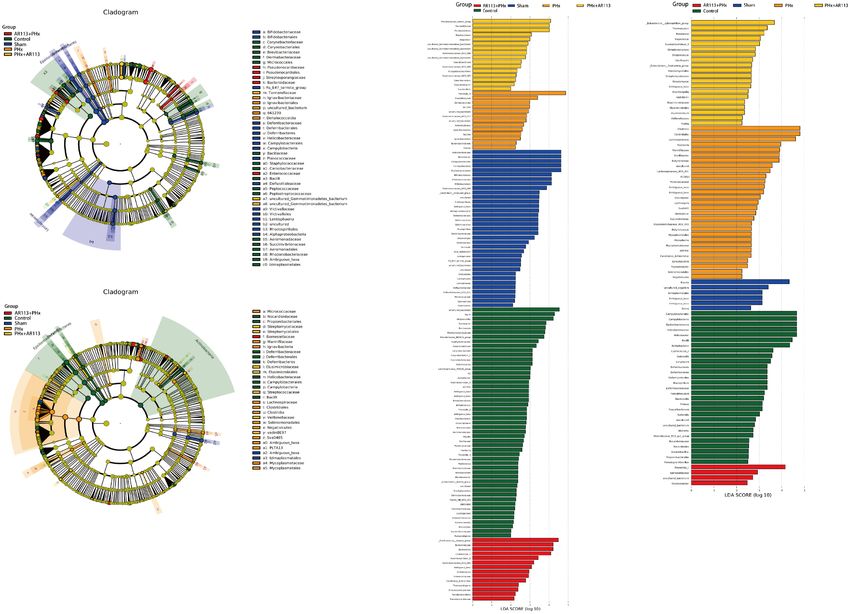
Xie et al.                                                                                   Lactiplantibacillus plantarum AR113 Exhibit Accelerated Liver Regeneration

orthogonal partial least-squares-discriminant analysis (OPLS-
DA) were performed using SIMCA-P+12.0.1.0 chemometrics
software to visualize the metabolites alterations among the
samples. The statistical criteria for preliminary selection of
characteristic metabolites were threshold of variable importance
in the projection (VIP) from the OPLS-DA greater than 1.0 and
q-value < 0.05 in a t-test. Enriched metabolic pathways were
performed using MetaboAnalyst1 based on the pathway library
from Kyoto Encyclopedia of Genes and Genomes (KEGG).

Statistical Analysis
Student’s t-test was used for comparisons of metabolite levels
using the statistical computer package GraphPad Prism version
6 (GraphPad Software Inc., San Diego, CA, United States).
Results in the present study were shown as means ± SEM.
Statistical comparisons were made using two-way analysis of
variance (ANOVA) with Tukey’s post hoc test. P-values < 0.05
were considered as statistical significance. Columns with different
letters differ significantly.

RESULTS
L. plantarum AR113 Administration
Increased Hepatocyte Proliferation and                                                  FIGURE 1 | H&E staining 3 and 7 days after PHx of the liver tissue sections of
                                                                                        Control, Sham, PHx, AR113+PHx, and PHx+AR113 groups. Scale
Accelerated Liver Regeneration of 70%                                                   bar = 50 µm for H&E.
Partial Hepatectomy Rats
In order to investigate the effect of L. plantarum AR113
                                                                                       mortality during PHx. In addition, we also found that hepatic
administration on liver regeneration, hepatocyte proliferation
                                                                                       regeneration rate in L. plantarum AR113 pretreatment rats was
and hepatic regeneration rate were analyzed. The proliferation
                                                                                       significantly accelerated compared to PHx rats at 7 days after
of hepatocytes at different groups after two-thirds PHx was
                                                                                       PHx, but there was no significant difference in the hepatic
evaluated in rats by Ki67 staining. Remarkably, pretreatment
                                                                                       regeneration rate between these two groups at 3 days after PHx,
with L. plantarum AR113 before PHx significantly increased the
                                                                                       suggesting that it takes a long time for probiotics to promote
cell proliferation rate compared with the PHx group at 3 days
                                                                                       liver regeneration.
after PHx (Supplementary Table 1). Because the proliferation
of liver cells occurs within 3 days after surgery, there was no
significant difference in cell proliferation rates between the PHx
                                                                                       Effect of L. plantarum AR113
group and L. plantarum AR113 pretreatment group at 7 days after                        Administration on Liver Histological
PHx (Supplementary Table 1). Therefore, in vivo proliferation                          Changes
analyses demonstrated that proliferation of hepatocytes was                            The liver H&E staining results of the rats in all groups are
enhanced in the presence of L. plantarum AR113 administration.                         presented in Figure 1. In the Control and Sham groups, the liver
   Rats were sacrificed 3 and 7 days after PHx and their livers                        showed a normal structure with well-preserved cell morphology
were collected and analyzed. Intriguingly, Table 1 shows that                          and a prominent nucleus. After PHx, the liver structure showed
L. plantarum AR113 pretreatment can significantly reduce rat                           crypt structure atrophy, mucosal epithelium impairment, and
                                                                                       decreased goblet cells. However, the histological changes were
1
    www.Metaboanalyst.ca                                                               reversed by L. plantarum AR113 administrations, evidenced
                                                                                       by a loss of swollen hepatocytes, cytoplasmic vacuolization,
TABLE 1 | The hepatic regeneration rate and rat mortality after PHx.                   and fat vacuoles.
                        Hepatic regeneration rate              Rat mortality (%)
                                                                                       Effect of L. plantarum AR113
                  3 days after PHx       7 days after PHx                              Administration on Liver Function,
PHx                 45.5% ± 0.57a         58.2% ± 0.83b                40%             Cytokines, and Growth Factors
AR113+PHx          46.49% ± 1.09a         67.72% ± 1.37a               25%             In order to investigate the effect of L. plantarum AR113
PHx+AR113          40.32% ± 0.65b         62.51% ± 0.98a               35%             administration on liver function, the contention of ALT, AST, TP,
Different letters indicate significant differences, P < 0.05 (ANOVA followed by        ALB II, TBil-V, and Glo II were detected. Compared with Control
Tukey’s HSD test).                                                                     and Sham groups, the ALT, AST, and TBil-V levels in PHx rats

Frontiers in Microbiology | www.frontiersin.org                                    4                                         January 2022 | Volume 12 | Article 800470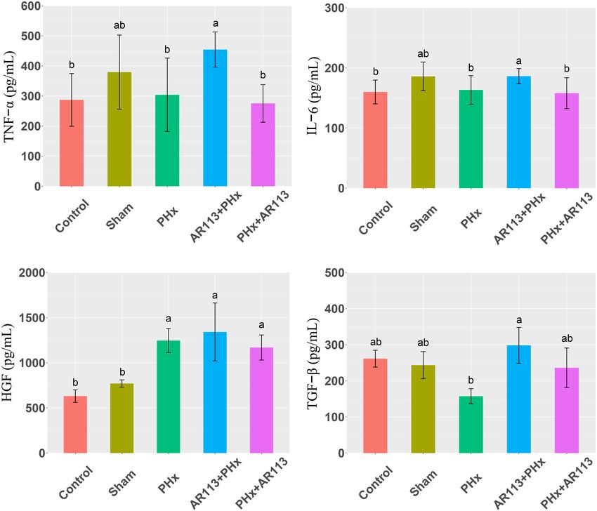
Xie et al.                                                                                  Lactiplantibacillus plantarum AR113 Exhibit Accelerated Liver Regeneration

were significantly increased and ALB II, Glo II, and TP were                           of Control, Sham, PHx, AR113+PHx, and PHx+AR113 groups
significantly decreased. Serum ALT, AST, and TBil-V levels were                        at Day 3 and Day 7 after PHx was conducted. Sequencing of
rapidly elevated at Day 3 after PHx (Supplementary Figure 1),                          16S bacterial RNA retrieved an overall number of 38,155∼41,957
and declined at Day 7 after PHx (Supplementary Figure 2). In                           reads, 26,774∼37,361 after filtering, which were clustered in 1,329
contrast, serum ALB II, Glo II, and TP were decreased sharply at                       operational taxonomic units (OTUs).
Day 3 after PHx (Supplementary Figure 1), and could increase                              Alpha diversity (Shannon and Simpson index) analysis
at Day 7 after PHx (Supplementary Figure 2). L. plantarum                              showed that at Day 3 after PHx, the Shannon index of the
AR113 administration had no significant effect on liver function                       Control group was significantly higher than that of the Sham,
(Supplementary Figure 2).                                                              PHx, and AR113+PHx groups (Supplementary Figure 3), but
    Cytokines and growth factors have prominent roles in liver                         had no significant difference as compared with PHx+AR113.
regeneration. Serum cytokine and growth factors at 3 days after                        However, there was no significant difference in alpha diversity
PHx levels are shown in Figure 2. Compared with the PHx group                          between the Sham, PHx, AR113+PHx, and PHx+AR113 groups.
at 3 days after PHx, TNF-α and HGF were significantly increased                        At Day 7 after PHx, there were no significant differences in alpha
by AR113 pretreatment, while TGF-β and IL-6 did not change                             diversity between groups, suggesting that the reduced diversity
significantly. At 7 days after PHx, TNF-α, IL-6 and TGF-β were                         of microbial communities associated with PHx had returned to
significantly increased by AR113 pretreatment compared with                            normal levels (Supplementary Figure 4).
PHx group (Figure 3). Interestingly, there was no significant                             Remarkable changes in the microbiota community structure
difference in the expression levels of TNF-α, HGF and TGF-β                            were induced by PHx intervention. The microbes in the Control
between the PHx group and the PHx+AR113 group.                                         and Sham groups were more closely clustered relative to
                                                                                       PHx and AR113+PHx groups, which is an indication that
                                                                                       PHx surgery induced similar microbial composition changes.
Effect of L. plantarum AR113                                                           Distinct changes in microbiota composition have revealed
Administration on Microbial                                                            a clear separation between no PHx groups (Control and
Communities                                                                            Sham) and PHx groups (PHx, AR113+PHx, and PHx+AR113)
To elucidate the effects of L. plantarum AR113 administration on                       after PHx at Days 3 and 7 (Supplementary Figure 5).
microbial communities, analysis of the 16S rRNA gene sequences                         L. plantarum AR113 treatment induced significant changes

  FIGURE 2 | Cytokines and growth factors at 3 days after PHx among the Control, Sham, PHx, AR113+PHx, and PHx+AR113 groups. (A) Changes in TNF-α levels;
  (B) Changes in IL-6 levels; (C) Changes in HGF levels; (D) Changes in HGF-β levels. Different letters indicate significant differences, P < 0.05 (ANOVA followed by
  Tukey’s HSD test).

Frontiers in Microbiology | www.frontiersin.org                                    5                                         January 2022 | Volume 12 | Article 800470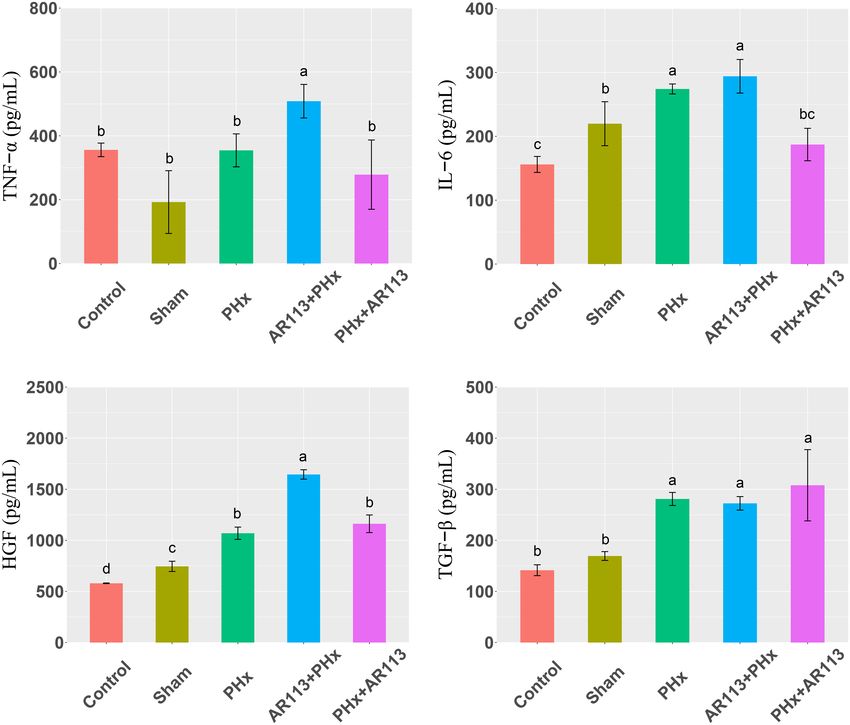
Xie et al.                                                                                  Lactiplantibacillus plantarum AR113 Exhibit Accelerated Liver Regeneration

  FIGURE 3 | Cytokines and growth factors at 7 days after PHx among the Control, Sham, PHx, AR113+PHx, and PHx+AR113 groups. (A) Changes in TNF-α levels;
  (B) Changes in IL-6 levels; (C) Changes in HGF levels; (D) Changes in HGF-β levels. Different letters indicate significant differences, P < 0.05 (ANOVA followed by
  Tukey’s HSD test).

in the gut microbial community. At the phylum level,                                   in the Sham group was significantly higher than that in the
Firmicutes and Bacteroidetes are two major phyla of the                                other three groups.
domain bacteria in gut microbiota (Figure 4). The abundances                              We used the LEfSe analysis to identify the specific bacteria
of Firmicutes and Bacteroidetes were increased in PHx                                  phylotypes that were differentially altered among the five groups,
groups (PHx, AR113+PHx, and PHx+AR113 groups). The                                     the LEfSe algorithm with a logarithmic LDA score cutoff ≥3.0
Firmicutes-to-Bacteroidetes ratio was calculated, and the results                      was then performed (Figure 7 and Supplementary Tables 2, 3).
showed that the F/B ratio was elevated in the PHx and                                  Three days after PHx, the most differentially abundant gut
PHx+AR113 as compared with the AR113+PHx groups                                        microbiota in group PHx were Prevotella_9, Faecalibaculum,
(Figure 5). Proteobacteria abundance in AR113+ PHx and Sham                            Dehalococcoidia, and 661239. The gut microbiota enriched in the
groups was not significantly different but decreased in both PHx                       AR113 administration group (AR113+PHx and PHx+AR113)
and PHx+AR113 groups.                                                                  were Ruminococcus_torques_group, Bacteroidaceae, Bacteroides,
   At the genus level, the abundance of Lactobacillus and                              Coprococcus_2,      Ruminiclostridium_9,       Ruminococcaceae_
Bacteroides from L. plantarum AR113 administration groups                              UCG_004, Ambiguous_taxa, Prevotellaceae_Ga6A1_group,
was higher than that of PHx and Sham groups (Figure 6). The                            Tannerellaceae, Parabacteroides, Mogibacterium, and Atopobium.
abundance of Lachnospiraceae_NK4A136_group was increased                               Seven days after PHx, the most differentially abundant gut
after PHx, which may be related to liver resection. In each genus                      microbiota in group PHx were Clostridia, Clostridiales,
of Bacteroidetes, the abundance of Prevotellaceae_Ga6A1_group                          Lachnospiraceae, Roseburia, Marinifilaceae, Oscillibacter,
has the highest abundance in the AR113+PHx group. In PHx                               Butyricimonas, uncultured, Lachnospiraceae_UGG_001, PLTA13,
groups (PHx, AR113+PHx, and PHx+AR113), the abundance of                               Micrococcaceae, Ambiguous_taxa, and Odoribacter. The gut
the Prevotella_9 genus was significantly higher than in the Sham                       microbiota enriched in the L. plantarum AR113 administration
group, while the abundance of Helicobacter of Proteobacteria                           group were Prevotella_1, Eubacterium_xylanophilum_group,

Frontiers in Microbiology | www.frontiersin.org                                    6                                         January 2022 | Volume 12 | Article 800470
Xie et al.                                                                            Lactiplantibacillus plantarum AR113 Exhibit Accelerated Liver Regeneration

  FIGURE 4 | Distribution of relative abundance at the phylum level among Control, Sham, PHx3, AR113+PHx3, and PHx3+ AR113 groups.

  FIGURE 5 | The Firmicutes-to-Bacteroidetes ratio (F/B) of Control, Sham, PHx, AR113+PHx, and PHx+AR113 groups.

Thermotunica, Roseiarcus, Vagococcus, and Ruminiclostridium_5.                   groups at Day 3 and Day 7, respectively. Ultimately, 17
These results suggest that administration of probiotics can                      metabolites differentially expressed both in Day 3 and Day
significantly alter the composition of gut microbiota after PHx.                 7 were selected (Table 2). Of these metabolites, L-isoleucine,
                                                                                 L-leucine, 3-O-Methylniveusin A, piperidine, PA(22:0/a-25:0),
                                                                                 PI(20:3(5Z,8Z,11Z)/20:3(8Z,11Z,14Z), and mevalonic acid
Effect of L. plantarum AR113                                                     showed an increase in the AR113+PHx group compared
Administration on Overall Plasma                                                 with the PHx group. 1-Arachidonoylglycerophosphoinositol,
Metabolite Content                                                               palmitic      amide,      1-(6-[3]-ladderane-hexanoyl)-2-(8-[3]-
The metabolic profiles were acquired using LC–MS/MS,                             ladderane-octanyl)-sn-glycerophosphoethanolamine,
and 4297 metabolites were identified. Then, multivariate                         7-oxo-8-amino-nonanoic acid, and 5S-HETE di-endoperoxide
analysis was conducted after data normalization. OPLS-DA                         showed a decrease in the PHx group.
model between the PHx and AR113+PHx groups at Day                                   We also compared the plasma metabolite changes between
3 and Day 7 was established and differentially abundant                          the PHx and PHx+AR113 groups. There were 135 and
metabolites were derived from this model with a VIP > 1 and                      115 differentially expressed metabolites identified between
a P-value < 0.05. There were 68 and 74 differentially expressed                  the PHx and PHx+AR113 groups at Day 3 and Day 7,
metabolites identified between the PHx and AR113+PHx                             respectively. There were 26 metabolites differentially expressed

Frontiers in Microbiology | www.frontiersin.org                              7                                        January 2022 | Volume 12 | Article 800470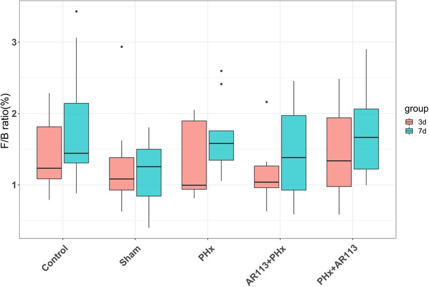
Xie et al. Lactiplantibacillus plantarum AR113 Exhibit Accelerated Liver Regeneration
FIGURE 6 | Distribution of relative abundance at the genus level among Control, Sham, PHx3, AR113+PHx3, and PHx3+ AR113 groups.
both in Day 3 and Day 7 (Table 3). The probiotic group leucine degradation, and mineral absorption pathways. Besides,
showed greater reductions in 4-phosphopantothenoylcysteine, Val, Leu, and Ile biosynthesis, Val, Leu, and Ile degradation,
chrycorin, PI(20:3(5Z,8Z,11Z)/20:3(8Z,11Z,14Z)), PI(20:4(5Z, protein digestion and absorption, mineral absorption, central
8Z,11Z,14Z)/18:0), PC(22:4(7Z,10Z,13Z,16Z)/22:6(4Z,7Z,10Z, carbon metabolism in cancer, and biosynthesis of amino acids
13Z,16Z,19Z)), PC(22:6(4Z,7Z,10Z,13Z,16Z,19Z)/22:4(7Z,10Z, were differentially expressed between PHx and PHx+AR113
13Z,16Z)), 1-Oleoylglycerophosphoserine, and 1-(2-methoxy- groups. These results indicated that L. plantarum AR113
6Z-heptadecenyl)-sn-glycero-3-phosphoserine. In contrast, administration accelerated liver regeneration accompanied by
PE(P-16:0/0:0), sphinganine 1-phosphate, mevalonic acid, a series of changes in metabolism, especially Leu and Ile
PE(18:1(9Z)/0:0), LysoPE(18:2(9Z,12Z)/0:0), LysoPE(20:4(5Z, biosynthesis, Val, Leu, and Ile degradation, and mineral
8Z,11Z,14Z)/0:0), LysoPE(0:0/20:4(5Z,8Z,11Z,14Z)), L-leucine, absorption pathways.
and L-isoleucine were upregulated in the PHx+AR113 group In addition, the differentially expressed metabolites pathways
compared with the PHx group. between pre-probiotic (AR113+PHx) and post-probiotic
(PHx+AR113) treatment groups were also discussed. It was
Effect of L. plantarum AR113 found that different ways of probiotics administration can affect
Administration on Metabolic Pathways choline metabolism in cancer, glycerophospholipid metabolism,
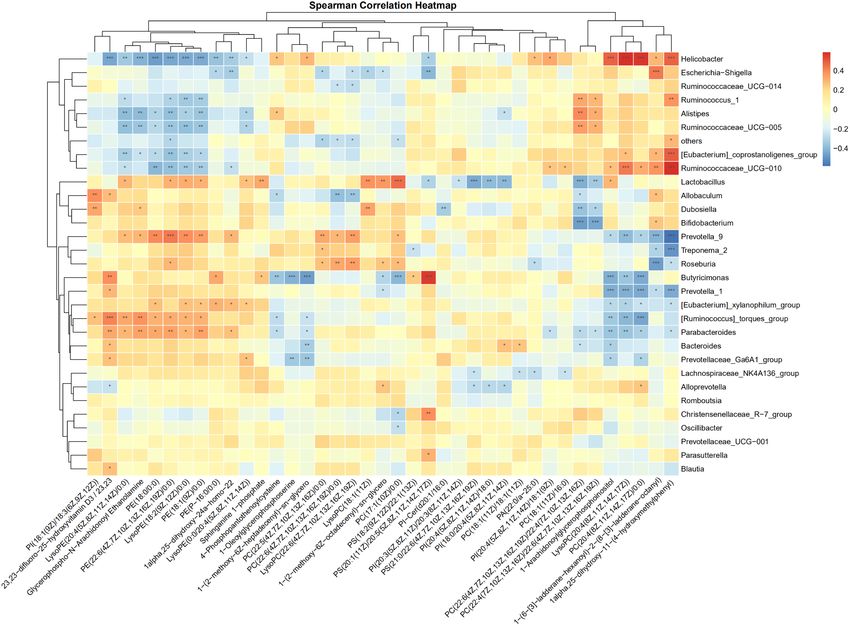
The differential expressed metabolites between PHx and retrograde endocannabinoid signaling, sphingolipid metabolism,
AR113+PHx groups or PHx and PHx+AR113 groups were glycosylphosphatidylinositol (GPI)-anchor biosynthesis,
mapped to the KEGG database2 for metabolic pathway autophagy, and arachidonic acid (ARA) metabolism pathways.
construction (Figure 8). Intriguingly, the differentially expressed
metabolites between PHx and AR113+PHx groups were mainly
associated with protein digestion and absorption, choline
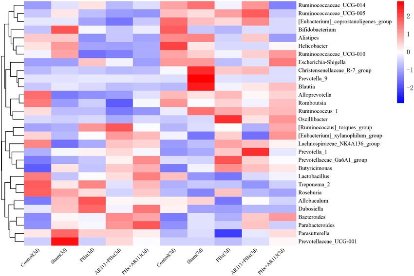
The Correlation Between Gut
metabolism in cancer, glycerophospholipid metabolism, leucine Microbiome and Plasma Metabolome
(Leu) and isoleucine (Ile) biosynthesis, valine (Val), Leu and The correlations of the discriminative gut microbiome
and differential plasma metabolites from Control, Sham,
2
http://www.genome.jp/kegg/ PHx, AR113+PHx, and PHx+AR113 were determined
Frontiers in Microbiology | www.frontiersin.org 8 January 2022 | Volume 12 | Article 800470
Xie et al.                                                                                  Lactiplantibacillus plantarum AR113 Exhibit Accelerated Liver Regeneration

  FIGURE 7 | LEfSe analysis among the Control, Sham, PHx, AR113+PHx, and PHx+AR113 groups. Cladogram displays the taxonomic tree of differentially abundant
  taxa. Histograms represent the LDA scores of bacteria with significant differential abundance between the compared groups, identified by different colors. Features
  with LDA score ≥ 3. (A,C), 3 days after PHx. (B,D), 7 days after PHx.

using Spearman’s rank correlation analysis (Figure 9).                                13Z,16Z)),      LysoPC(22:6(4Z,7Z,10Z,13Z,16Z,19Z)),       and
Taking the correlation analysis of glycerolipids and gut                              LysoPC(18:1(11Z)) were positively correlated with Prevotella_9,
microbiome as an example, the results showed that the                                 Treponema_2, Roseburia, Dubosiella, and Lactobacillus
relative abundances of Helicobacter, Ruminococcus, and                                and     negatively     correlated   with   Escherichia–Shigella,
Ruminococcaceae were negatively correlated with serum levels                          Ruminococcaceae_UCG–014, others, and Allobaculum. Together,
of LysoPE and PE in serum. In contrast, the abundance                                 these data suggested that some species of gut microbial species
of Lactobacillus, Allobaculum, Dubosiella, Prevotella,                                may modulate the levels of glycerophospholipid that are
Roseburia, Butyricimonas, Parabacteroides, and Bacteroides was                        correlated with liver regeneration.
positively correlated with the circulating levels of LysoPE
and PE. In addition, we found PC(18:1(11Z)/18:1(11Z)),
PC(22:6(4Z,7Z,10Z,13Z,16Z,19Z)/22:4(7Z,10Z,13Z,16Z),                                  DISCUSSION
PC(22:4(7Z,10Z,13Z,16Z)/22:6(4Z,7Z,10Z,13Z,16Z,19Z)),
LysoPC(20:4(8Z,11Z,14Z,17Z)), and PC(20:4(8Z,11Z,14Z,17Z)/                            The crosstalk between the gut and liver make probiotics play
0:0) exhibited positive correlations with Helicobacter,                               an important role in the progression of liver diseases (Shavandi
Ruminococcus,      Alistipes,    and     Ruminococcaceae   and                        et al., 2020). Many studies have demonstrated the beneficial
negative correlations with Lactobacillus, Allobaculum,                                effects of probiotics on the modulation of alcohol-induced liver
Dubosiella, Bifidobacterium, Prevotella_9, Butyricimonas,                             injury, D-galactosamine-induced rat liver injury, and chronic
Prevotella_1,                 [Eubacterium]_xylanophilum_group,                       liver disease patients (Nardone et al., 2010). However, to the best
[Ruminococcus]_torques_group, Parabacteroides, Bacteroides,                           of our knowledge, few studies have focused on its effect on liver
and Prevotellaceae_Ga6A1_group. Notably, PC(22:5(4Z,7Z,10Z,                           regeneration. In our research, pretreatment with L. plantarum
13Z,16Z)/0:0), PC(22:6-(4Z,7Z,10Z,13Z,16Z,19Z)/22:4(7Z,10Z,                           AR113 was found to have increased hepatocyte proliferation and

Frontiers in Microbiology | www.frontiersin.org                                   9                                         January 2022 | Volume 12 | Article 800470
Xie et al.                                                                                Lactiplantibacillus plantarum AR113 Exhibit Accelerated Liver Regeneration

TABLE 2 | Summary of the differentially expressed metabolites between PHx and AR113+PHx groups.

m/z                          Ion mode               Metabolites                                                VIP                  P-value                    FC

132.10171                       Pos                 L-Isoleucine                                              3.10                   3.0952                   0.77
203.08349                       Neg                 Phenylalanyl-Glycine                                      3.95                   0.0218                   0.80
130.08765                       Neg                 L-Leucine                                                 1.99                   0.0017                   0.73
881.19349                       Pos                 [Gallocatechin(4alpha->8)]2catechin                       1.50                   0.0009                   0.44
881.44495                       Pos                 1-[2,4-dihydroxy-5-(3-methylbut-2-en-                     1.70                   0.0006                   0.46
                                                    1-yl)phenyl]-2-hydroxy-3-[4-hydroxy-3-
                                                    methoxy-5-(3-methylbut-2-en-1-
                                                    yl)phenyl]propan-1-one
403.23215                       Pos                 5S-HETE di-endoperoxide                                   2.30                   0.0000                   32.63
186.11375                       Neg                 7-oxo-8-amino-nonanoic acid                               2.57                   0.0004                   2.81
129.05583                       Neg                 Mevalonic acid                                            1.35                   0.0255                   0.69
256.26275                       Pos                 Palmitic amide                                            4.28                   0.0264                   1.69
881.6963                        Pos                 PA(22:0/a-25:0)                                           1.61                   0.0005                   0.46
763.53652                       Pos                 1-(6-[3]-ladderane-hexanoyl)-2-(8-[3]-                    1.75                   0.0001                   1.86
                                                    ladderane-octanyl)-sn-
                                                    glycerophosphoethanolamine
619.28723                       Neg                 1-Arachidonoylglycerophosphoinositol                      1.94                   0.0005                   1.53
909.54695                       Neg                 PI(20:3(5Z,8Z,11Z)/20:3(8Z,11Z,14Z))                      1.98                   0.0185                   0.68
254.17573                       Neg                 Labienoxime                                               1.29                   0.0000                   0.34
407.17245                       Neg                 3-O-Methylniveusin A                                      2.58                   0.0176                   0.70
146.05992                       Pos                 4-formyl Indole                                           1.39                   0.0074                   0.82
86.096214                       Pos                 Piperidine                                                2.73                   0.0054                   0.77

Pos, positive ion mode; Neg, negative ion mode.

accelerated liver regeneration of PHx rats. The beneficial effects of              early stage of liver injury (Shi and Line, 2020). Present study
L. plantarum AR113 were associated with increased hepatocyte                       showed that L. plantarum AR113 given two weeks before PHx
proliferation, improved liver function, and modulation of gut                      significantly increased the expression of TNF-α after PHx. but
microbiome and plasma metabolome.                                                  giving probiotics after PHx did not achieve the same effect. In
   It has been reported that although liver regeneration                           addition, we found that TGF-β was increased in AR113+ PHx
results ultimately in restoration of liver mass and function,                      group at day 7 after PHx. TGF-β is produced principally in the
partial hepatectomy is primarily a compensatory hyperplasia                        hepatic stellate cells, and is a representative mitoinhibitory factor,
(Michalopoulos and Bhushan, 2021). To evaluate the degree                          which presumably induces the termination of liver regeneration
of liver injury after 70% partial hepatectomy, liver function                      (Masuda et al., 2020). Our study showed that whether probiotics
was examined. Previous research showed that serum ALT                              are given before or after PHx will affect the expression of cytokine
and AST activity increased rapidly and significantly after 70%                     expression level.
partial hepatectomy on Day 1 and returned almost to pre-                               Accumulating evidence has indicated that the gut microbiome
operative levels after 2–3 days in control rats (Yu et al., 2018).                 is involved in the pathogenesis of liver diseases by influencing the
Not surprisingly, our study found that L. plantarum AR113                          host’s immunity and metabolism (Lederer et al., 2017). Firmicutes
administration does not reduce serum ALT and AST activity and                      and Bacteroidetes are the two most dominant bacterial phyla
ALB II after PHx at Day 3 and Day 7. This is because local                         affecting host energy extraction efficiency and linked with
inflammatory reactions may occur in response to damage around                      excess adiposity (Behari et al., 2021). In the present study,
the ligated area of the liver, resulting in transient increases in                 Lactobacillus, Lachnospiraceae_NK4A136, Ruminococcus_1, and
serum ALT and AST activity.                                                        [Ruminococcus]_torques which were affected by L. plantarum
   Cytokines such as TNF-α, IL-6, HGF, TGF-β, and TNF-α                            AR113 administration belong to Firmicutes. Prevotella_9,
and the activation of NF-κB by cytokines were shown to be                          Bacteroides, Alloprevotella, Prevotellaceae_Ga6A1_group, and
required for the initiation of liver regeneration. Several lines                   Butyricimonas belong to Bacteroidetes. Some studies have
of evidence suggest that TNF-α and IL-6 are among the most                         used the ratio of the two dominant phyla (Firmicutes and
crucial components of the early signaling pathways leading to                      Bacteroidetes) as a marker for microbial dysbiosis (Jasirwan
regeneration (Fathi et al., 2021). In the liver, IL-6 is secreted by               et al., 2021). Changes in this ratio have also been found in
Kupffer cells, and this secretion is stimulated by TNF-α (Jin et al.,              several metabolic disorders. Our study showed that the F/B
2021). HGF is a 97-kd protein that was originally isolated from                    ratio in the AR113+PHx group was the lowest among all the
the peripheral blood of animals after PHx, which is known to                       groups, suggesting that L. plantarum AR113 given 2 weeks
be essential to initiate the process of liver regeneration (Gao and                before PHx could change the microbiome composition. The
Peng, 2021); it could rapidly be elevated by 10- to 20-fold at the                 depletion of genera Alloprevotella and Prevotella can contribute

Frontiers in Microbiology | www.frontiersin.org                               10                                          January 2022 | Volume 12 | Article 800470
Xie et al. Lactiplantibacillus plantarum AR113 Exhibit Accelerated Liver Regeneration
TABLE 3 | Summary of the differentially expressed metabolites between PHx and PHx+ AR113 groups.
m/z Ion mode Metabolites VIP P-value FC
132.10171 Pos L-Isoleucine 3.38 0.0014 0.75
130.08765 Neg L-Leucine 2.28 0.0083 0.70
552.27106 Neg Vignatic acid A 3.58 0.0000 3.18
355.06781 Pos 6-[5-(2-carboxyeth-1-en-1-yl)-2,3-dihydroxyphenoxy]- 3.87 0.0000 1.79
3,4,5-trihydroxyoxane-2-carboxylic
acid
403.23215 Pos 5S-HETE di-endoperoxide 2.28 0.0000 21.29
186.11375 Neg 7-oxo-8-amino-nonanoic acid 2.85 0.0003 3.31
129.05583 Neg Mevalonic acid 1.87 0.0035 0.56
506.32298 Neg PC(17:1(10Z)/0:0) 1.33 0.0280 0.89
904.58372 Pos PC(22:6(4Z,7Z,10Z,13Z,16Z,19Z)/22:4(7Z,10Z,13Z,16Z)) 3.28 0.0004 1.99
904.5835 Pos PC(22:4(7Z,10Z,13Z,16Z)/22:6(4Z,7Z,10Z,13Z,16Z,19Z)) 1.83 0.0000 1.94
478.29196 Pos LysoPE(18:2(9Z,12Z)/0:0) 2.93 0.0433 0.67
500.277 Neg LysoPE(20:4(5Z,8Z,11Z,14Z)/0:0) 3.30 0.0276 0.69
438.29619 Pos PE(P-16:0/0:0) 2.83 0.0200 0.51
500.27645 Neg LysoPE(0:0/20:4(5Z,8Z,11Z,14Z)) 1.48 0.0211 0.71
763.53652 Pos 1-(6-[3]-ladderane-hexanoyl)-2-(8-[3]-ladderane-octanyl)- 1.20 0.0096 1.47
sn-glycerophosphoethanolamine
885.54815 Neg PI(20:4(5Z,8Z,11Z,14Z)/18:0) 7.30 0.0163 1.63
909.54695 Neg PI(20:3(5Z,8Z,11Z)/20:3(8Z,11Z,14Z)) 1.29 0.0389 1.46
504.27176 Neg 1-Oleoylglycerophosphoserine 4.20 0.0000 2.42
506.28682 Neg 1-(2-methoxy-6Z-heptadecenyl)-sn-glycero-3- 3.08 0.0000 2.73
phosphoserine
401.07701 Neg 4-Phosphopantothenoylcysteine 2.24 0.0463 1.39
380.25588 Neg Sphinganine 1-phosphate 2.53 0.0277 0.55
466.33424 Pos 1alpha,25-dihydroxy-24a-homo-22-thiavitamin 2.23 0.0200 0.61
D3/1alpha,25-dihydroxy-24a-homo-22-thiacholecalciferol
480.30672 Pos PE(18:1(9Z)/0:0) 3.11 0.0143 0.61
72.080461 Pos Pyrrolidine 2.24 0.0008 0.59
86.096214 Pos Piperidine 3.04 0.0006 0.74
201.03839 Neg Chrycorin 3.05 0.0007 1.42
Pos, positive ion mode; Neg, negative ion mode.
to non-alcoholic fatty liver disease and microbiome dysbiosis (Hernandez-Conde et al., 2021). We found L-leucine and
(Safari and Gerard, 2019). In addition, genus Prevotella is L-isoleucine were up regulated in the L. plantarum AR113
considered beneficial for promoting hepatic glycogen storage administration group compared with the PHx group. Leu was
and improving glucose metabolism (Monga Kravetz et al., 2020). reported to have a proliferative effect on hepatocyte, suggesting
We found the expression level of Prevotella was decreased that L. plantarum AR113 may promote liver regeneration by
after AR113 administration while Alloprevotella was increased. up-regulating serum Leu. Thus far, the effects of Ile on liver
Meanwhile, the overgrowth of Ruminococcus may lead to regeneration were limited.
metabolic dysfunction and aggravation of liver injury. The Except for branched chain amino acids, our study also
genus Ruminococcus is considered a gut microbiota signature found that L. plantarum AR113 administration had an effect
of non-alcoholic fatty liver disease and was positively correlated on glycerophospholipid metabolism that was evidenced by
with the levels of ALT, AST, TBil-V, and TBA (Demir et al., altered serum levels of glycerophosphoinositol (PIs), PCs,
2020). Our study found the expression level of Prevotella was LysoPC, and PEs which are the fundamental components of
decreased after AR113 administration. Besides, the colonization lipid bilayers of cell membranes. Phospholipids, including PEs,
of L. plantarum AR113 enriched the normal gut microbiota, PCs, glycerophosphoserines (PSs), glycerophosphoglycerols
especially Lactobacillus, which can ferment nutrients into lactic (PGs), and CLs, are the prominent membrane lipids (Xie
acid and benefit health. The decrease in potential pathogens and et al., 2016). PIs were ubiquitous components of eukaryotic
restoration of the normal gut microbiota by L. plantarum AR113 cells that participate in cell proliferation and survival. It was
might improve host metabolism and accelerate liver regeneration. reported that liver regeneration was characterized by increases
Branched chain amino acids, including Leu, Ile, and in PE, and decreases in glycerophosphoethanolamine (GPE)
Val, play critical roles in regulating metabolism of glucose, and glycerophosphocholine (GPC) (Zakian et al., 2005).
lipid, protein synthesis, intestinal health, and immunity Indeed, PHx caused a transient and reversible accumulation
Frontiers in Microbiology | www.frontiersin.org 11 January 2022 | Volume 12 | Article 800470Xie et al. Lactiplantibacillus plantarum AR113 Exhibit Accelerated Liver Regeneration FIGURE 8 | The differentially expressed metabolic pathways among the PHx, AR113+PHx, PHx+AR113 groups of 3 or 7 days after PHx. (A) PHx and AR113+PHx groups of 3 days after PHx; (B) PHx and AR113+PHx groups of 7 days after PHx; (C) PHx and PHx+AR113 groups of 3 days after PHx; (D) PHx and PHx+AR113 groups of 7 days after PHx; (E) AR113+PHx and PHx+AR113 groups of 3 days after PHx; (F) AR113+PHx and PHx+AR113 groups of 7 days after PHx. Frontiers in Microbiology | www.frontiersin.org 12 January 2022 | Volume 12 | Article 800470
Xie et al.                                                                                   Lactiplantibacillus plantarum AR113 Exhibit Accelerated Liver Regeneration

  FIGURE 9 | Spearman correlation analysis heatmaps of gut microbiome and glycerolipids. *, **, *** represents a significant difference, *p < 0.05, **p < 0.01,
  ***p < 0.001 as determined by t-test.

of phospholipids in the liver at the early phase of liver                               given postoperatively. Except for the glycerophospholipid
regeneration. In our study, the probiotic treated group showed                          metabolism, retrograde endocannabinoid signaling, sphingolipid
greater reductions in PI(20:3(5Z,8Z,11Z)/20:3(8Z,11Z,14Z)),                             metabolism, and GPI-anchor biosynthesis pathways, we also
PI(20:4(5Z,8Z,11Z,14Z)/18:0), PC(22:4(7Z,10Z,13Z,16Z)/22:6                              found autophagy and ARA metabolism pathways were affected
(4Z,7Z,10Z,13Z,16Z,19Z)), PC(22:6(4Z,7Z,10Z,13Z,16Z,19Z)/                               by different ways of probiotics administration. ARA is one
22:4(7Z,10Z,13Z,16Z)), 1-Oleoylglycerophosphoserine, and 1-                             of the most abundant polyunsaturated fatty acids present in
(2-methoxy-6Z-heptadecenyl)-sn-glycero-3-phosphoserine,                                 human tissue and represents one of the pivotal signaling
but      had      increased     concentrations     of      PE(P-                        molecules involved in the initiation and propagation of
16:0/0:0),     sphinganine    1-phosphate,     PE(18:1(9Z)/0:0),                        diverse signaling cascades regulating inflammation, pain, and
LysoPE(18:2(9Z,12Z)/0:0),     LysoPE(20:4(5Z,8Z,11Z,14Z)/0:0),                          homeostatic function (Turolo et al., 2021). In humans and
and LysoPE(0:0/20:4(5Z,8Z,11Z,14Z)). PE is an important lipid                           mammals, ARA has been widely observed to reduce the
marker of inflammation in glycerophospholipid metabolism.                               triacylglycerol (TG) accumulation in liver, plasma, and adipose
An increased concentrations of PE has been detected in                                  tissue (Pei et al., 2020). At present, the relationship between
systemic circulation of nonalcoholic steatohepatitis patients                           ARA and liver regeneration remains unclear. In contrast to
(Puri et al., 2009). LysoPE has also been shown to play a role                          ARA metabolism pathways, autophagy was reported to be
in intercellular signaling and in the activation of signaling                           essential for liver regeneration. Autophagy is a homeostatic
enzymes (Yanagida and Valentine, 2020), and has been suggested                          mechanism that regulates turnover of long-lived or damaged
to act through putative G protein-coupled receptors (GPCRs)                             proteins and organelles and supplies amino acids taken from
(Kaluarachchi et al., 2018).                                                            degradation products of the autolysosome (Xu et al., 2020). To
   Based on these results, our study found that probiotics                              our knowledge, our study first reported that different ways of
intervention can affect the metabolic changes of rats after PHx.                        probiotic administration modulate liver regeneration by affecting
More importantly, we also found that L. plantarum AR113                                 autophagy pathways.
administration preoperatively had different effects on liver                                Our data showed that LysoPE and PE showed a positive
regeneration rates and metabolites compared with probiotics                             correlation with Lactobacillus, Allobaculum, Dubosiella,

Frontiers in Microbiology | www.frontiersin.org                                    13                                         January 2022 | Volume 12 | Article 800470
Xie et al.                                                                                          Lactiplantibacillus plantarum AR113 Exhibit Accelerated Liver Regeneration

Prevotella, Roseburia, Butyricimonas, Parabacteroides, and                                   ETHICS STATEMENT
Bacteroides, but an inverse correlation with Helicobacter,
Ruminococcus, and Ruminococcaceae. The opposite correlation                                  The animal study was reviewed and approved by the Hunan SJA
pattern suggests that their counterbalancing role in modulating                              Laboratory Animal Co., Ltd.
lipid homeostasis is beneficial for liver regeneration. Thus,
the constantly changing gut flora acted as an entire system
and exerted various functions on host–microbial nutritional
utilization throughout the course of the liver regeneration to
                                                                                             AUTHOR CONTRIBUTIONS
meet diverse cell proliferation and energy demands during the                                ChuX and ZhoZ conceptualized the study and wrote and
different biological processes (Macchi and Sadler, 2020).                                    prepared the original draft. MY was in charge of the project
                                                                                             administration. CC conceptualized the study. YZ and ZuoZ
CONCLUSION                                                                                   designed the experiments. WG and ChaX supervised the study.
                                                                                             LY and ZH wrote the review. LA and YP conceived and
The present study showed that L. plantarum AR113                                             supervised the work. All authors contributed to the article and
administration increased hepatocyte proliferation and                                        approved the submitted version.
accelerated liver regeneration of PHx rats. Two weeks of
L. plantarum AR113 before PHx induced decreased F/B ratio.
The colonization of L. plantarum AR113 enriched the normal gut                               FUNDING
microbiota, especially Lactobacillus. The decrease in potential
pathogens and restoration of the normal gut microbiota by                                    This work was supported by the grant from the Central
L. plantarum AR113 might improve host metabolism and                                         Public-Interest Scientific Institution Basal Research Fund (no.
accelerated liver regeneration. One of the most profound changes                             Y2019XK15), Training Program for Excellent Young Innovators
was L. plantarum AR113 administration of glycerophospholipid                                 of Changsha (kq 2009089), China Agriculture Research System
metabolism that was evidenced by decreased serum levels of PI                                for Bast and Leaf Fiber Crops (no. CARS-16), and Project
and PCs, and increased LysoPC and PEs. Further investigations                                of Scientific Elitists in National Agricultural Research and
will focus on the key metabolites and ultimately clarify the                                 Agricultural Science and Technology Innovation Program of
molecular basis for these microbe–host interactions during                                   China (CAAS-ASTIP-2021-IBFC).
liver regeneration.

DATA AVAILABILITY STATEMENT                                                                  SUPPLEMENTARY MATERIAL
The original contributions presented in the study are included                               The Supplementary Material for this article can be found
in the article/Supplementary Material, further inquiries can be                              online at: https://www.frontiersin.org/articles/10.3389/fmicb.
directed to the corresponding author/s.                                                      2021.800470/full#supplementary-material

REFERENCES                                                                                       microbiota: ready for prime time? J. Gastroenterol. Hepatol. 35, 1969–1977.
                                                                                                 doi: 10.1111/jgh.15071
Adolph, T. E., Grander, C., Moschen, A. R., and Tilg, H. (2018). Liver-microbiome            Fathi, F., Sanei, B., Ganjalikhani Hakemi, M., Saidi, R. F., and Rezaei, A. (2021).
   axis in health and disease. Trends Immunol. 39, 712–723. doi: 10.1016/j.it.2018.              Liver resection promotes (regulates) proinflammatory cytokines in patients
   05.002                                                                                        with hepatocellular carcinoma. Can. J. Gastroenterol. Hepatol. 2021:5593655.
Behari, J., Graham, L., Wang, R., Schirda, C., Borhani, A. A., Methe, B. A., et al.              doi: 10.1155/2021/5593655
   (2021). Dynamics of hepatic steatosis resolution and changes in gut microbiome            Gao, C., and Peng, J. (2021). All routes lead to rome: multifaceted origin of
   with weight loss in nonalcoholic fatty liver disease. Obes Sci .Pract. 7, 217–225.            hepatocytes during liver regeneration. Cell Regen 10:2. doi: 10.1186/s13619-
   doi: 10.1002/osp4.476                                                                         020-00063-3
Cassano, M., and Dufour, J. F. (2019). Inflammation and microbiota fingerprint:              Heinrich, V. R., Kneist, S., and Kunzel, W. (1988). [Limit of the reparative capacity
   delphi’s oracle for nonalcoholic fatty liver disease-related hepatocellular                   of deciduous dental pulp]. Zahn Mund Kieferheilkd Zentralbl 76, 14–21.
   carcinoma? Hepatology 69, 12–15. doi: 10.1002/hep.30267                                   Hernandez-Conde, M., Llop, E., Gomez-Pimpollo, L., Fernandez Carrillo, C.,
Chowdhury, M. M. H., Salazar, C. J. J., and Nurunnabi, M. (2021). Recent advances                Rodriguez, L., Van Den Brule, E., et al. (2021). Adding branched-chain
   in bionanomaterials for liver cancer diagnosis and treatment. Biomater Sci. 9,                amino acids to an enhanced standard-of-care treatment improves muscle
   4821–4842. doi: 10.1039/d1bm00167a                                                            mass of cirrhotic patients with sarcopenia: a placebo-controlled trial. Am. J.
Cornide-Petronio, M. E., Alvarez-Mercado, A. I., Jimenez-Castro, M. B., and                      Gastroenterol. 116, 2241–2249. doi: 10.14309/ajg.0000000000001301
   Peralta, C. (2020). Current knowledge about the effect of nutritional status,             Higgins, G. M., and Anderson, R. M. (1931). Experimental pathology of the liver. I:
   supplemented nutrition diet, and gut microbiota on hepatic ischemia-                          restoration of the liver of the white rat following partial surgical removal. Arch.
   reperfusion and regeneration in liver surgery. Nutrients 12:284. doi: 10.3390/                Path. Lab. Med. 12, 186–202.
   nu12020284                                                                                Hoffmann, K., Nagel, A. J., Tanabe, K., Fuchs, J., Dehlke, K., Ghamarnejad, O., et al.
Crismale, J. F., and Friedman, S. L. (2020). Acute liver injury and decompensated                (2020). Markers of liver regeneration-the role of growth factors and cytokines:
   cirrhosis. Med. Clin. North Am. 104, 647–662. doi: 10.1016/j.mcna.2020.02.010                 a systematic review. BMC Surg. 20:31. doi: 10.1186/s12893-019-0664-8
Demir, M., Lang, S., Martin, A., Farowski, F., Wisplinghoff, H., Vehreschild,                Jasirwan, C. O. M., Muradi, A., Hasan, I., Simadibrata, M., and Rinaldi, I. (2021).
   M., et al. (2020). Phenotyping non-alcoholic fatty liver disease by the gut                   Correlation of gut firmicutes/bacteroidetes ratio with fibrosis and steatosis

Frontiers in Microbiology | www.frontiersin.org                                         14                                            January 2022 | Volume 12 | Article 800470
Xie et al. Lactiplantibacillus plantarum AR113 Exhibit Accelerated Liver Regeneration
stratified by body mass index in patients with non-alcoholic fatty liver disease. Shavandi, A., Saeedi, P., Gerard, P., Jalalvandi, E., Cannella, D., and Bekhit, A. E.
Biosci. Microbiota Food Health 40, 50–58. doi: 10.12938/bmfh.2020-046 (2020). The role of microbiota in tissue repair and regeneration. J. Tissue Eng.
Jin, S., Yu, C., and Yu, B. (2021). Changes of serum IL-6, IL-10 and TNF-alpha Regen. Med. 14, 539–555. doi: 10.1002/term.3009
levels in patients with systemic lupus erythematosus and their clinical value. Shi, J. H., and Line, P. D. (2020). Hallmarks of postoperative liver regeneration:
Am. J. Transl. Res. 13, 2867–2874. an updated insight on the regulatory mechanisms. J. Gastroenterol Hepatol 35,
Kaluarachchi, M., Boulange, C. L., Karaman, I., Lindon, J. C., Ebbels, T. M. D., 960–966. doi: 10.1111/jgh.14944
Elliott, P., et al. (2018). A comparison of human serum and plasma metabolites Tanemura, A., Mizuno, S., Hayasaki, A., Fujii, T., Iizawa, Y., Kato, H., et al.
using untargeted (1)H NMR spectroscopy and UPLC-MS. Metabolomics 14, 32. (2018). Biliary complications during and after donor hepatectomy in living
doi: 10.1007/s11306-018-1332-1 donor liver transplantation focusing on characteristics of biliary leakage and
Lederer, A. K., Pisarski, P., Kousoulas, L., Fichtner-Feigl, S., Hess, C., and Huber, treatment for intraoperative bile duct injury. Transplant Proc. 50, 2705–2710.
R. (2017). Postoperative changes of the microbiome: are surgical complications doi: 10.1016/j.transproceed.2018.03.045
related to the gut flora? A systematic review. BMC Surg 17:125. doi: 10.1186/ Trebicka, J., Bork, P., Krag, A., and Arumugam, M. (2021). Utilizing the gut
s12893-017-0325-8 microbiome in decompensated cirrhosis and acute-on-chronic liver failure.
Li, B., Zhang, C., and Zhan, Y. T. (2018). Nonalcoholic fatty liver disease cirrhosis: Nat. Rev. Gastroenterol Hepatol 18, 167–180. doi: 10.1038/s41575-020-00
a review of its epidemiology, risk factors, clinical presentation, diagnosis, 376-3
management, and prognosis. Can J. Gastroenterol. Hepatol. 2018:2784537. doi: Turolo, S., Edefonti, A., Mazzocchi, A., Syren, M. L., Morello, W., Agostoni, C.,
10.1155/2018/2784537 et al. (2021). Role of arachidonic acid and its metabolites in the biological
Liu, H. X., Keane, R., Sheng, L., and Wan, Y. J. (2015). Implications of microbiota and clinical manifestations of idiopathic nephrotic syndrome. Int. J. Mol. Sci.
and bile acid in liver injury and regeneration. J. Hepatol. 63, 1502–1510. doi: 22:5452. doi: 10.3390/ijms22115452
10.1016/j.jhep.2015.08.001 Xie, T., Zhou, X., Wang, S., Lu, Y., Zhu, H., Kang, A., et al. (2016).
Macchi, F., and Sadler, K. C. (2020). Unraveling the epigenetic basis of liver Development and application of a comprehensive lipidomic analysis to
development, regeneration and disease. Trends Genet. 36, 587–597. doi: 10. investigate tripterygium wilfordii-induced liver injury. Anal. Bioanal. Chem.
1016/j.tig.2020.05.002 408, 4341–4355. doi: 10.1007/s00216-016-9533-9
Masuda, A., Nakamura, T., Abe, M., Iwamoto, H., Sakaue, T., Tanaka, T., et al. Xu, F., Hua, C., Tautenhahn, H. M., Dirsch, O., and Dahmen, U. (2020). The role
(2020). Promotion of liver regeneration and antifibrotic effects of the TGFbeta of autophagy for the regeneration of the aging liver. Int. J. Mol. Sci. 21:3606.
receptor kinase inhibitor galunisertib in CCl4treated mice. Int. J. Mol. Med. 46, doi: 10.3390/ijms21103606
427–438. doi: 10.3892/ijmm.2020.4594 Yagi, S., Hirata, M., Miyachi, Y., and Uemoto, S. (2020). Liver regeneration after
Michalopoulos, G. K., and Bhushan, B. (2021). Liver regeneration: biological and hepatectomy and partial liver transplantation. Int .J. Mol. Sci. 21:8414. doi:
pathological mechanisms and implications. Nat. Rev. Gastroenterol. Hepatol. 18, 10.3390/ijms21218414
40–55. doi: 10.1038/s41575-020-0342-4 Yanagida, K., and Valentine, W. J. (2020). Druggable lysophospholipid signaling
Monga Kravetz, A., Testerman, T., Galuppo, B., Graf, J., Pierpont, B., Siebel, pathways. Adv. Exp. Med. Biol. 1274, 137–176. doi: 10.1007/978-3-030-50621-
S., et al. (2020). Effect of gut microbiota and PNPLA3 rs738409 variant on 6_7
nonalcoholic fatty liver disease (NAFLD) in obese youth. J. Clin. Endocrinol. Yu, L. H., Yu, W. L., Zhao, T., Wu, M. C., Fu, X. H., and Zhang, Y. J. (2018). Post-
Metab. 105:dgaa382. doi: 10.1210/clinem/dgaa382 operative delayed elevation of ALT correlates with early death in patients with
Nardone, G., Compare, D., Liguori, E., Di Mauro, V., Rocco, A., Barone, M., HBV-related hepatocellular carcinoma and post-hepatectomy liver failure. HPB
et al. (2010). Protective effects of lactobacillus paracasei F19 in a rat model of (Oxford) 20, 321–326. doi: 10.1016/j.hpb.2017.10.001
oxidative and metabolic hepatic injury. Am. J. Physiol. Gastrointest Liver Physiol. Zakian, K. L., Koutcher, J. A., Malhotra, S., Thaler, H., Jarnagin, W., Schwartz,
299, G669–G676. doi: 10.1152/ajpgi.00188.2010 L., et al. (2005). Liver regeneration in humans is characterized by significant
Nishida, A., Inoue, R., Inatomi, O., Bamba, S., Naito, Y., and Andoh, A. (2018). changes in cellular phosphorus metabolism: assessment using proton-
Gut microbiota in the pathogenesis of inflammatory bowel disease. Clin. J. decoupled 31P-magnetic resonance spectroscopic imaging. Magn. Reson. Med.
Gastroenterol. 11, 1–10. doi: 10.1007/s12328-017-0813-5 54, 264–271. doi: 10.1002/mrm.20560
Pei, K., Gui, T., Kan, D., Feng, H., Jin, Y., Yang, Y., et al. (2020). An overview of lipid
metabolism and nonalcoholic fatty liver disease. Biomed Res. Int. 2020:4020249. Conflict of Interest: The authors declare that the research was conducted in the
doi: 10.1155/2020/4020249 absence of any commercial or financial relationships that could be construed as a
Puri, P., Wiest, M. M., Cheung, O., Mirshahi, F., Sargeant, C., Min, H. K., potential conflict of interest.
et al. (2009). The plasma lipidomic signature of nonalcoholic steatohepatitis.
Hepatology 50, 1827–1838. doi: 10.1002/hep.23229 Publisher’s Note: All claims expressed in this article are solely those of the authors
Rodes, L., Saha, S., Tomaro-Duchesneau, C., and Prakash, S. (2014). and do not necessarily represent those of their affiliated organizations, or those of
Microencapsulated bifidobacterium longum subsp. infantis ATCC the publisher, the editors and the reviewers. Any product that may be evaluated in
15697 favorably modulates gut microbiota and reduces circulating this article, or claim that may be made by its manufacturer, is not guaranteed or
endotoxins in F344 rats. Biomed Res. Int. 2014:602832. doi: 10.1155/20 endorsed by the publisher.
14/602832
Safari, Z., and Gerard, P. (2019). The links between the gut microbiome and Copyright © 2022 Xie, Zhang, Yang, Cao, Zhou, Zhu, Gong, Xu, Yan, Hu, Ai and
non-alcoholic fatty liver disease (NAFLD). Cell Mol. Life Sci. 76, 1541–1558. Peng. This is an open-access article distributed under the terms of the Creative
doi: 10.1007/s00018-019-03011-w Commons Attribution License (CC BY). The use, distribution or reproduction in
Seehofer, D., Rayes, N., Schiller, R., Stockmann, M., Muller, A. R., Schirmeier, A., other forums is permitted, provided the original author(s) and the copyright owner(s)
et al. (2004). Probiotics partly reverse increased bacterial translocation after are credited and that the original publication in this journal is cited, in accordance
simultaneous liver resection and colonic anastomosis in rats. J. Surg. Res. 117, with accepted academic practice. No use, distribution or reproduction is permitted
262–271. doi: 10.1016/j.jss.2003.11.021 which does not comply with these terms.
Frontiers in Microbiology | www.frontiersin.org 15 January 2022 | Volume 12 | Article 800470You can also read