Linking Geospatial and Laboratory Sciences to Define Mechanisms behind Landscape Level Drivers of Anthrax Outbreaks - MDPI
←
→
Page content transcription
If your browser does not render page correctly, please read the page content below
International Journal of
           Environmental Research
           and Public Health

Article
Linking Geospatial and Laboratory Sciences to Define
Mechanisms behind Landscape Level Drivers of
Anthrax Outbreaks
Michael H. Norris 1,2,3           and Jason K. Blackburn 1,2,3, *
 1    Department of Geography, University of Florida, Gainesville, FL 32611, USA; mhnorris@ufl.edu
 2    Spatial Epidemiology and Ecology Research Lab, University of Florida, Gainesville, FL 32611, USA
 3    Emerging Pathogens Institute, University of Florida, Gainesville, FL 32611, USA
 *    Correspondence: jkblackburn@ufl.edu; Tel.: +1-352-273-9374

 Received: 6 September 2019; Accepted: 1 October 2019; Published: 4 October 2019

 Abstract: Background: A seasonal predictor of anthrax outbreaks is rainfall, which may be
 approximated by NDVI using remote sensing. How rainfall or vegetative green-up influences
 bacterial physiology or microecology to drive anthrax outbreaks is not known. Methods: Rainfall and
 NDVI dependency of anthrax epizootics was demonstrated with global and local phenological
 analysis. Growth analysis of B. anthracis in response to pH and calcium gradients was carried out.
 The influence of pH and calcium levels on expression of toxin and sporulation related proteins in
 broth culture models was characterized using engineered B. anthracis luminescent reporter strains.
 Results: Short-term bacterial growth and longer-term bacterial survival were altered by pH and
 calcium. These conditions also played a major role in pagA and sspB promoter-driven luminescent
 expression in B. anthracis. Conclusions: Rainfall induced cycling of pH and calcium in soils plays a
 plausible role in amplifying spore load and persistence in endemic anthrax zones. Observed evidence
 of B. anthracis favoring soil alkalinity and high soil calcium levels in the environment were linked to
 physiological conditions that promote bacterial growth, survival, toxin secretion and spore formation;
 illustrating the utility of bringing laboratory-based (controlled) microbiology experiments into the
 fold of zoonotic disease ecology.

 Keywords: anthrax; luminescence; spores; infectious disease; zoonosis; NDVI; outbreak;
 Bacillus anthracis; interdisciplinary

1. Introduction
     Environmentally-mediated pathogen transmission occurs when environmental conditions are
suitable for both hosts and pathogens to survive an intersect. While pathogen survival may vary from
short-term (days or weeks; e.g., Brucella spp.) or long-term (years or decades; e.g., Bacillus anthracis),
disease intensity varies over space and with apparent, but poorly understood, predictability or
seasonality. This may occur across diverse landscapes, but the mechanisms for increased disease
intensity is poorly studied for environmentally mediated pathogens.
     Anthrax, a disease recognized since ancient times, is caused by the Gram-positive spore former
Bacillus anthracis. This same pathogen served as the model organism from which Koch’s postulates
originated and bioterrorism immediately comes to mind. Naturally occurring, anthrax is a zoonosis
with frequent epizootic outbreaks and spillover to humans is common and problematic in many
countries [1]. Soil-borne B. anthracis spores are found in specific soil conditions nearly worldwide and
are infectious to animals such as grazing ruminants, often resulting in high mortality. Phylogenetically,
B. anthracis has traditionally been divided into four clades, A, B, C, and D. Group A B. anthracis has

Int. J. Environ. Res. Public Health 2019, 16, 3747; doi:10.3390/ijerph16193747     www.mdpi.com/journal/ijerph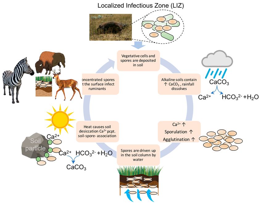
Int. J. Environ. Res. Public Health 2019, 16, 3747                                                 2 of 16

spread to all continents (but Antarctica). Other groups have not dispersed as widely [2]. The highest
degree of genetic diversity is found in southern Africa and was hypothesized as the potential geographic
origin of anthrax evolution because it represented the only location in the world (Kruger National
Park; KNP) where B. anthracis from groups A and B coexist [3]. These strains show geographic
affinities within the park, with group A localizing to the center of the park and group B to the
north. Group A strains were found in soils with a mean soil pH of 6.74 and mean calcium content
of 185.68 me/kg. Group B strains were found in soils with higher mean soil pH and mean calcium
content, pH 7.76 and 274.14 me/kg, respectively. Interestingly, during anthrax epidemics in KNP,
isolates from both groups A and B cause infection. This suggests that sources external to strain,
pH, or calcium content trigger infections/epizootics; Though likely contributors to environmental
persistence of the pathogen, triggers of infections/epizootics peripheral to strain, pH, or calcium
content are suggested by the co-circulation of strains during outbreaks in KNP. Strain differences can
still impact infectiousness, virulence, and microecology of anthrax. The external sources that trigger
outbreaks are as yet underdetermined environmental signals. Spores are reported to have a half-life of
approximately 100 years [4] and environmental elimination is not an easy task as evidenced by the
intensive decontamination efforts on the small Scottish island of Gruinard [5]. Considering the length
and tenacity of the organisms’ environmental persistence, complete eradication of B. anthracis from
endemic areas is seemingly unachievable.
       In the soil environment, the bacterium exists as a dormant (metabolically inactive) spore,
waiting for the right signals to permeate through the exosporium layer and initiate germination.
Vegetative organisms are sensitive to desiccation and heat but can sporulate quickly during conditions
unfavorable for growth. The spore surface, or exosporium, is coated with a glycoprotein that is
involved in spore binding to environmental surfaces, generates spore hydrophobicity and affects spore
germination [17–19]. Spores contact a host through ingestion, inhalation, or cutaneous inoculation then
germinate to the vegetative form and elaborate the A2 B-type anthrax toxins made up of protective
antigen (PAG), which transports both lethal factor (LF) and edema factor (EF) into the cell cytoplasm,
causing animal death by toxemia.

1.1. Seasonal Predictors of Anthrax Epidemics
     Anthrax is often thought of as an important agent of bioterrorism. While certainly such a risk
exists, anthrax most often causes disease in wildlife and livestock due to its persistence in soil around
the world with spillover into proximate human populations. Globally, the disease remains a public and
animal health problem, with recent increases in human disease in the Republic of Georgia illustrating
the threat clearly [20]. Outbreaks still occur with frequency in the US [21], including areas where
vaccination reaches livestock but not wildlife [15]. Outbreaks occur in an episodic fashion with
pronounced seasonality (Figure 1) and some studies have modeled this for systems where there is
no active disease control [22]. Notably, seasonality has been defined study by study, and often with
different metrics, such as cases or outbreaks by month or season. Other studies have measured
outbreaks against precipitation or vegetation indices. Here we compile those data in a single map to
discuss patterns globally. It is estimated that 1.1 billion animals are at risk of anthrax globally and
that 198.2 million B. anthracis Sterne livestock vaccines are administered each year with potential
to impact adjacent human populations totaling 63.5 million [1]. In the high-risk anthrax belt of
Australia, early spring and hot dry summers have been associated with severe livestock anthrax [23].
A close look at the annual normalized vegetation index (NDVI) trajectory, as an evaluation of seasonal
environmental changes, showed that summer green-up (associated with rainfall) often occur in the
weeks or months prior to the major outbreaks in epizootic years possible [13]. In Etosha National
Park (ENP), Namibia, anthrax is also associated with rain events where there is a clear relationship
between monthly precipitation and cases [12,24,25]. In Ghana, a bimodal climatic trend applies to
areas of the most numerous and intense epizootic anthrax outbreaks [11]. Of all anthrax cases in
Zambia, where anthrax is hyper-endemic, ~85% of 2108 human cases over a 10-year period occurred in
Int. J. Environ. Res. Public Health 2019, 16, 3747                                                           3 of 16

the Zambezi floodplain of western Zambia [8]. Livestock cases closely mirror the human cases and
primarily occur during hot-dry months after the rainy season when herding brings animals towards
the dried flood plain. It is also important to note that if the only factor leading to anthrax outbreaks
were early spring green-up and hot summer-associated rainfall then anthrax outbreaks would be
easily predictable. Anthrax outbreaks do not occur annually and are considered episodic [22,26],
possibly with many endemic years between epizootic years. The 2008 anthrax outbreak in Montana
is a prime example. After nearly 50 years of no major outbreaks, a severe outbreak occurred among
bison and elk, killing ~8% of the bison herd [27,28]. Outbreaks in North America are reported at
lower frequency than in Africa, but still ensue near temperature maxima and following periods of
locally high rainfall, as during the localized 1999 Alberta anthrax outbreak which affected seven cattle
farms [10]. The authors have recently (31 July–5 August 2019) returned from an outbreak in West
Texas described by long-time regional veterinarians as the worst outbreak in a lifetime. The 2019 Texas
anthrax outbreak is nearly a perfect fit for the above described predictors. It encompassed at least 2000
km2 of ranchland and claimed numerous animal species (white tailed deer, audad, oryx, barasingha,
antelope, red deer, axis, hartebeest, and gemsbok, among others). West-Texas experienced a wet spring,
followed by a year’s worth of rainfall in June 2019 that preceded dry conditions with temperatures at
or above 38 ◦ C (100 ◦ F). Indeed, in his seminal work on anthrax ecology, Van Ness linked water related
geographic features with anthrax in the incubator hypothesis [29]. Although there are drawbacks to
the incubator hypothesis, it aligns well with the observed cyclic weather events. Additionally, high
soil moisture and organic content are conducive to bacterial survival whether in vegetative or spore
form. Anthrax outbreaks occur across varying geographies and ecologies and can be perpetuated by
place-specific mechanisms.

      Figure 1. Seasonality of anthrax outbreaks is a global phenomenon. Here we illustrate clear patterns
      of outbreak intensity and timing associated with month (orange bounding boxes; Kyrgyzstan [6];
      Northwest Territories, Canada [7]; Zambia [8]), season (red bounding box; Argentina [9]), precipitation
      (blue bounding boxes; Alberta, Canada [10], Ghana [11], Etosha National Park, Namibia [12]), enhanced
      vegetation index (green bounding box; EVI; Montana, Blackburn & Goodin, unpublished data),
      and normalized difference vegetation index (dark green bounding boxes; NDVI; Australia [13] and
      Texas [14]). Red areas in Texas and Australia are the recently defined enzootic zone [15] and re-defined
      anthrax belt [16], respectively.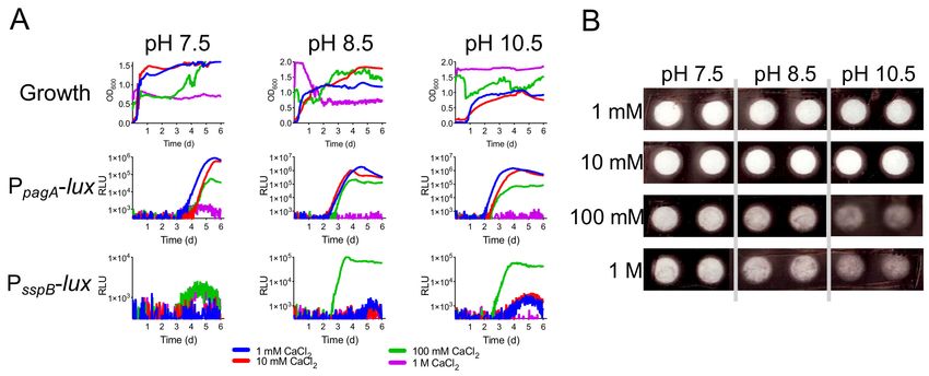
Int. J. Environ. Res. Public Health 2019, 16, 3747                                                 4 of 16

     In Texas, the vegetation is diverse, containing scrub, shrubs, and trees while in the highly endemic
ENP the landscape is relatively homogenous grassland. In ENP, anthrax is most frequently found
in grazing animals, including zebra [24]. It has been shown that B. anthracis is associated with plant
roots and that ruminants, such as zebra, can consume large doses of anthrax during grazing contact
with the soil reservoir of B. anthracis [30]. The 2008 Montana outbreak affected bison and elk, both
grazers, in Montana [27]. In both species, male anthrax mortality rates were much higher than females.
It has been observed that males of both species have a higher frequency of inspecting carcasses than
females translating to higher rates of serological exposure (Blackburn unpublished data). West Texas
is a major anthrax focus in North America, where outbreaks predominantly occur in white-tailed
deer [31] and mixed livestock/wildlife groups. This cycle differs from outbreaks in grasslands such
as ENP and Montana [32]. White-tailed deer are primarily browsers during the anthrax risk period
(summer months). Meaning they primarily feed on leaves, soft shoots, or fruits of high growing,
woody plants, such as shrubs [33], with little to no grass consumption. So how do white-tailed deer get
infected if deer have a low probability of high-dose exposure associated with soil grazing? White-tailed
deer first prefer forbs, which are low growing herbaceous flowering plants (a.k.a. weeds) during
spring and summer, then in late summer and fall switch to acorns, berries and fruits from browse
plants [34]. Summer rain events may increase the forb levels bringing deer into closer contact with low
vegetation. These rain events also cause an explosion in biting flies, as was documented during the
2005 epizootic in West Texas [15]. Following feeding on anthrax-infected animals, tabanid horseflies
may increase the number of cases through mechanical transmission. It has been shown that B. anthracis
can be spread to taller vegetation by necrophagous blowflies [35]. After feeding on anthrax carcasses,
blowflies land on the leaves of surrounding shrubs, deposit anthrax-containing feces and emesis,
thus creating a large three-dimensional infectious zone surrounding the dead animal. The deer then
consume the contaminated leaves during browsing near anthrax carcass sites. The cycle of soil to
animal to plant to animal serves as a case multiplier and contributes to the epizootiology of anthrax in
the white-tailed deer and other exotic animals present across the West-Texas anthrax enzootic area.
Termed localized infectious zones (LIZs), these areas surrounding infected carcasses greatly increase
the probability of healthy animal exposure. Every West-Texas ranch visited by the authors during the
summer of 2019 described a huge explosion in the biting fly population that coincided with the main
outbreak wave. Contribution of the LIZ to anthrax transmission was realized by Pasteur in the 19th
century [36]. Zebra carcasses in ENP are eviscerated quickly (often within 24 h by nighttime scavengers
including hyenas and jackals), but spores persist at the sites for years [25]. The LIZ can be widespread
following carcass destruction (out to several meters from the initial death). Flies do not contribute
to LIZ development in Etosha because rapid consumption by highly efficient vertebrate scavengers
leaves nothing of the carcass for maggot deposition [37]. Horizontal transhumance and migratory
animal herding play a role in anthrax outbreaks by concentrating animal density around LIZs and
increasing probability of LIZ contact [25,38]. Indeed, if ranch lands in West-Texas are mis-managed,
higher than optimal animal densities may drive case numbers higher. The larger a herd that grazes in
a high-load anthrax zone the higher the exposure numbers.
     With every geography, anthrax infected carcasses deposit a large amount of nutrients in the form
of blood and tissue as well as spores. This causes an increase in phosphorous, grass biomass and grass
nitrogen levels that correlate with proximity to the zebra carcass site [30]. Other nutrients have been
measured but without correlates of distance or adequate non-carcass control sites for comparison,
conclusions are uncertain. Vegetative growth and spore load at LIZs have been observed for three years
following zebra fall, after which time vegetation and spore levels presumably decrease to background
levels. However, during the initial two years following an animal carcass death, B. anthracis levels
within 1 m of the site remained at ~104 CFU/g of grass, root, or soil. By year three, spores were not
detectable on above ground plant biomass but root and soil sample CFUs remained high. During the
second year following a carcass fall the increased vegetation appear to increase grazing in the LIZ,
perpetuating a low smoldering of anthrax cases. As we stated above, anthrax epidemics do not occur every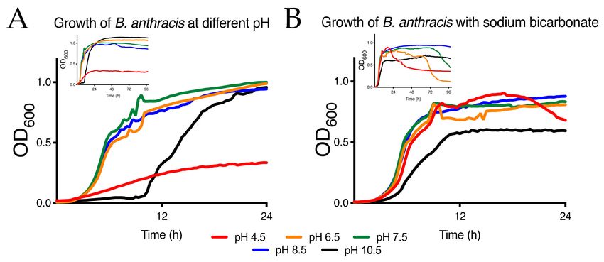
Int. J. Environ. Res. Public Health 2019, 16, 3747                                                         5 of 16

year, so increased amounts of vegetation or spores at the LIZ may contribute to outbreak initiation in combination
with environmental cues and animal behavior. The data do not support LIZs alone as a main driver of
anthrax epizootics, nor does available data directly consider seasonal rainfall as a major contributor to
the process. Because enzootic anthrax often has more than three years between events, it is more likely
that seasonal rainfall cycles increase the infectivity of old LIZs by increasing the probability animals
come to a former LIZ and when they do, a significant infectious dose is received.
      While a wet spring followed by a rain event in a hot dry summer has long been accepted as the
prescription for large anthrax events [26,39], few studies have demonstrated this directly, or measured
it. In one such study, we used satellite derived NDVI remote sensing data as a proxy for rainfall and
discovered the anecdotal evidence do have a firm basis. Earlier vegetation green-up, an earlier start to
spring, is correlated with epizootic outbreak years and increased severity of anthrax outbreaks during
summers in the Enzootic Zone of West Texas [14]. This zone was defined by our group as a region
of frequent large wildlife and livestock outbreaks with evidence of annual cases [15]. For example,
over the course of the decade, both 2001 and 2005 were severe anthrax years, where a well-studied
white-tailed deer herd in Texas experienced epizootic anthrax outbreaks [14]. Both outbreaks had a
statistically significant earlier spring and more intense green-up in the months preceding the large
outbreaks. In contrast, enzootic years, those with few or no detected cases, had significantly less
green-up and rarely had early green-up [14]. In other words, epizootic years had differences in
spring green-up intensity and timing detectable by early April, months ahead of the summer outbreak
risk periods.
      These observations have added to our evolving knowledge of anthrax and demonstrate that this
disease and the way it occurs does not follow the classic textbook descriptions. Further deviation
from the textbook is apparent when considering atypical anthrax caused by Bacillus cereus biovar
anthracis (Bcbva). This new disease was first described in 2004 and is caused by a strain of Bacillus cereus
that had acquired toxin and capsule producing plasmids [40]. Anthrax caused by this organism
does not appear to have seasonality and predominantly infects non-human primates in the forests
of Sub-Saharan Africa, so the epidemiology of wildlife anthrax and anthrax caused by Bcbva is also
vastly different [41–43]. The persistent nature of anthrax in Ta’i National Park represent a problem that
current epidemiological models, including those mentioned in this work, do not address.

1.2. Modeling Episodic Anthrax
     Population dynamics have been used to model single-year anthrax outbreaks [44,45]. Others have
taken into account migration of animals into anthrax areas to infer how populations remain persistently
susceptible to anthrax [46]. These works are useful in understanding single outbreak population
dynamics but become impractical when studying multi-year episodic anthrax outbreaks in areas
that do not receive migratory animals from other endemic areas (like mid-latitude anthrax areas in
Montana and Texas). Recently, our lab devised a mathematical model considering: (1) environmental
cues and (2) host population dynamics to simulate anthrax outbreaks [47]. Without bearing in mind
either, the episodic nature of anthrax outbreaks is not sustained with a given influx of anthrax spores.
When only seasonal dynamics are included the periodicity of anthrax outbreaks can be obtained but
not sustained. When only population dynamics are included, epizootic outbreaks are not observed,
and only enzootic disease is simulated. If both a seasonal driver and population dynamics are included,
episodic epizootic anthrax outbreaks are recreated with in silico simulations. By incorporating population
dynamics and allowing seasonal forcing of infection to be dependent on an external factor we estimated
seasonality to have a large impact on the number of anthrax-related deaths. Thus, environmental
drivers coupled with herd population dynamics are required to sustain the episodic nature of epizootic
anthrax outbreaks. Our model provides general knowledge of environmentally mediated diseases
by explicitly elucidating how intense environmental events determine the tempo and amplitude of
outbreaks of rare diseases.
Int. J. Environ. Res. Public Health 2019, 16, 3747                                                   6 of 16

     Despite the great differences in infectious cycles, vegetation and geography, some variation of
“early wet spring and uncharacteristic heavy rainfall in an otherwise hot-dry summer” appears to hold
true across outbreak environments and animal species in enzootic areas. Another important often
overlooked initiator of outbreak is the contribution of environmental conditions to population changes
of the pathogen and how responses to fluctuating environmental cues affect the ecophysiology of
B. anthracis. To date, laboratory experiments on the life state of B. anthracis have not considered the
landscape-level climatic conditions associated with the observed outbreaks and ecophysiology of the
pathogen. The availability of our vast and growing global database on outbreaks and environmental
conditions across the geography of anthrax [1] allows us the opportunity to define specific laboratory
experiments to test such hypotheses like the “wet spring” or “incubator”. The precise mechanisms
by which rainfall events induce outbreaks are still unknown. B. anthracis spores could be splashed
onto nearby lower vegetation where grazers ingest an infectious dose [30]. Simultaneously, during
these rain events hydrophobic spores deposited from a previous anthrax carcass may rise to the soil
surface as water transitions downwards through the water column or become deposited on plant
surfaces during germination of new vegetation. Higher rainfall could also cause shifts in biomass and
plant species that increase likelihood of animal contact with soil-borne B. anthracis spores. A third
provocative explanation may be that rainfall events induce an increase in B. anthracis soil populations
that then sporulate during dry hot temperatures that follow immediately after.

1.3. Soil pH and Calcium Content
      Soil conditions that are often concurrent with anthrax outbreaks are increased pH and increased
calcium. Anecdotal evidence from outbreak zones around the world suggests elevated pH and
calcium levels correlate with enhanced anthrax activity [26,29]. Analysis of soil chemistries in the
contiguous United States found several minerals, including calcium, were significantly elevated in
counties where zoonotic anthrax cases originated [48]. We hypothesize these two soil characteristics
contribute to anthrax outbreaks by enhancing survival of B. anthracis through increased sporulation
along with mechanical suspension of spores in the soil column. Intact spores bind more efficiently
to high calcium soils [49]. It was hypothesized by the authors that positive Ca2+ ions in the soil
bind to the predominantly negatively charge spore surface, joining the spore to soil particles thus
preventing leaching of calcium from the dipicolinate-rich spore. Leaching of calcium can lead to
germination failure and loss of spore heat resistance, both very important for bacterial survival in the
environment [50,51]. The idea being that the presence of calcium and alkaline pH prevents the spore
from being adrift near the inhospitable surface but bound to calcium rich soils deeper in the soil column.
Rainfall could allow the buoyant spores to move back up to the soil surface, increasing the probability
a grazing host will be inoculated with an infectious dose of spores. Calcium is generally not freely
soluble in soils, existing in the form of calcium carbonate (CaCO3 ) or limestone. High calcium soils are
often alkaline and have calcium predominantly in the form of calcium carbonate. As calcium carbonate
is dissolved by heavy rainfall, free calcium is released and binds to the negatively charged soil particles,
bicarbonate is released; the pH rises [52]. As soils dry, the calcium can precipitate as limestone or
remain free but immobilized on negatively charged soil particles as a site for spore association with
the spore surface serving as a site of calcium nucleation during precipitation [53]. The relationship of
soil pH and calcium could have profound effects on the spore but have not been investigated with
detail. When an animal dies of anthrax, the bodily fluids are teaming with vegetative bacteria. As the
bacteria enter the soil and are exposed to air, they begin to sporulate. The influence of calcium and
pH on vegetative cell survival and sporulation rate is not known but could greatly alter the levels
of spores that are sustained in an environment. Landscapes of the Texas anthrax enzootic zone are
alkaline composites of exposed limestone rock (~40% CaCO3 ) or calciferous loam (~20% CaCO3 ) with
limestone rock underneath.
Int. J. Environ. Res. Public Health 2019, 16, 3747 7 of 16
2. Methods
2.1. Growth and Sporulation of Bacillus Anthracis
Bacillus anthracis Sterne 34F2 (Colorado Serum Company, Denver, CO, USA) was grown in BHI
broth or agar (Difco Laboratories, Inc, Detroit, MI, USA) and either 30 ◦ C or 37 ◦ C depending on the
temperature sensitive nature of the plasmids at use and as described in the manuscript. For sporulation,
B. anthracis Sterne was grown in BHI overnight at 37 ◦ C with shaking at 250 rpm. The overnight
culture was diluted 1:60 in fresh modified G media [54,55] with or without kanamycin at 20 µg/mL then
shaken for 72 h at 30 ◦ C and 250 rpm. Cultures were chilled on ice then harvested by centrifugation
at 5000× g and 4 ◦ C for 15 min in a bench top centrifuge and washed twice with ice cold milli-Q
water. Spores were purified from vegetative cells using gradient centrifugation, then incubated in
100% ethanol for 1 h to remove any residual vegetative cells. The spores were washed twice with ice
cold milli-Q water then stored at 4 ◦ C until used. Completeness of sporulation was ascertained by
microscopy and was typically greater than 99%.
2.2. Bacillus Anthracis Growth Experiments
Growth experiments were carried out in BHI broth (Difco). For pH experiments, the pH of
BHI broth was modified with 1M HCl or 1M NaOH then filter sterilized through a 0.22 µm filter.
B. anthracis Sterne was inoculated into pH 7.0 BHI broth and grown overnight at 37 ◦ C with shaking at
250 rpm. The next day, the culture was adjusted to an OD600 of 1 in BHI and this was used to inoculate
different media at a dilution of 1:100 then aliquoted into clear bottom, black 96 well assay plates and
shaken with double orbital shaking at 30 ◦ C on a Synergy H1M microplate reader (BioTek, Winooski,
VT, USA). The OD600 , and luminescence where applicable, of each well were read every 20–30 min
for the duration of the experiment (Figure 2A,B and Figure 3C,D). Incubation was the same in static
culture except that shaking only occurred for 5 sec before each read. The data shown is the average of
three replicates. The standard deviation is omitted from the figures due to clarity. Spores were used to
initiate growth assays. Spore preparations were enumerated by dilution plating on BHI and 1 × 10⁷
spore forming units (SFU) were inoculated into each well. Sodium bicarbonate was added at 0.4%
(w/v) to growth experiments, as indicated, to investigate the impact of bicarbonate levels on vegetative
growth and survival.
Figure 2. Growth of B. anthracis at different pH. (A) Short-term and long-term (inset) growth of
B. anthracis at different pH. Highly acidic pH is especially detrimental to bacterial survival while
alkaline pH delays growth but does not impact long-term growth. (B) Short-term and long-term
(inset) growth of B. anthracis at different pH in the presence of bicarbonate. The buffering capacity of
bicarbonate is evident by increasing short-term growth rates of B. anthracis. Longer term, bacterial
survival is optimal at pH 8.5 and 10.5 while optical density sharply decreases at neutral and acidic pH
starting at 24 h after growth.Int. J. Environ. Res. Public Health 2019, 16, 3747 8 of 16
Figure 3. Tracking toxin elaboration and sporulation of B. anthracis. (a) A diagrammatic representation
of the plasmid constructs used to track protective antigen (PA) induction from the pagA promoter
(PpagA ) in vegetative B. anthracis (Bav ) or sporulation from the sspB promoter (PsspB ) in cells preparing
to sporulate (Bas ) at 30 ◦ C. (b) A negative luminescent image of cultures grown in liquid broth for
24 h. The strength of expression of PpagA (left) compared to PsspB (right) is apparent in this image.
(c) Static grown cultures of B. anthracis at 30 ◦ C show pagA expression measured in relative luminescent
units (RLUs) is most optimal at pH 8.5 (blue line) and peaks at 3 days. (d) Luminescence (clear boxes)
induced from PsspB during shaking growth (black boxes) at 30◦ C compared to B. anthracis luminescence
without the plasmid (clear circles) and growth (black circles). (e) Validation of our dual-label AmCyan
fluorescence indicator of B. anthracis growth at 37 ◦ C. Culture density measured by OD600 (grey fill)
compared to AmCyan relative fluorescent units (RFU; cyan fill). (f) Normalized short-term luminescent
profiles from PpagA -lux and PsspB -lux expressing B. anthracis illustrates the temporally defined stages of
toxin production and sporulation. (g) PpagA -lux and PsspB -lux expressing B. anthracis compared to OD600
measurements. (h) Long-term luminescence of PpagA -lux and PsspB -lux expressing B. anthracis show that
after an initial defined period of expression, a second less defined period and then increasing levels of
luminescence follow, indicating continued cycles of toxin expression and sporulation in subpopulations
of B. anthracis in long-term cultures.
2.3. Engineering of Bioluminescent B. Anthracis Toxin Elaboration and Sporulation Reporter Strains
Plasmid pMV306hsp+lux was obtained from Addgene [56]. LuxABCDE located on the plasmid
was engineered for optimal expression in Gram-positive bacteria by replacement of the ribosomal
binding site and reorganization of the genes in the operon [57]. The protective antigen promoter, PpagAInt. J. Environ. Res. Public Health 2019, 16, 3747 9 of 16
was synthesized by GENEWIZ (South Plainfield, NJ, USA) and the small sporulation protein B promoter,
PsspB , was amplified by PCR and both were cloned via EcoRI/NotI digestion into pMV306hsp+lux
replacing the hsp promoter with either the Pag promoter or SspB promoter to create pMV306pag+lux
and pMV306sspB+lux, respectively. For maintenance in B. anthracis, the plasmid backbone of
pRP1028 [58], which contains the Gram-positive temperature sensitive replicon repA, kanamycin
resistance marker and origin of transfer, oriT, was amplified by PCR to introduce NheI and PstI sites
then digested with the same enzymes. This fragment was ligated to either PpagA -lux or PsspB -lux
that were cut from pMV306pag+lux and pMV306sspB+lux with the same enzymes. Clones were
verified as bioluminescent. The resultant plasmids were transformed into the mobilizable E. coli strain
RHO3 [59] and mated into B. anthracis Sterne using standard methods. Luminescent plasmid-containing
B. anthracis were selected on BHI agar + 20 µg/mL kanamycin at 30◦ C and plasmid containing colonies
verified by PCR and bioluminescence using a ChemiDoc XRS+ imaging system (Bio-Rad Laboratories,
Hercules, CA, USA).
For production of thermostable dual-labeled pRepU-Kan-AmCyan-(PpagA or PsspB )-lux plasmids,
a PCR fragment from plasmid pRP1099 [58] which encodes the repU B. anthracis replicative plasmid
origin, and AmCyan driven by the strong Gram-positive promote, PFP1, was PCR amplified
with oligos designed using the NEBuilder online tool (nebuilder.neb.com). This fragment was
combined with NotI/PstI digested PpagA-lux or PsspB-lux fragments digested from pMV306pag+lux
or pMV306sspB+lux and assembled using NEBuilder HiFi DNA Assembly master mix according to
the manufacturer’s recommendations (New England BioLabs, Inc., Ipswich, MA, USA). Fluorescence
and luminescence were verified then transferred into B. anthracis Sterne 34F2 as described above.
2.4. B. Anthracis Growth, Toxin Elaboration and Sporulation Induction Assays in the Presence of
Geochemically Relevant Calcium Gradients
Calcium levels in soils of the contiguous United States range from approximately 100 ppm on
the low end to an extreme of almost 320,000 ppm [60]. 100 ppm is equivalent to 2.5 mM while the
extreme 320,000 ppm is equivalent to 8 M. For growth experiments the range at a low of 1 mM and
a maximum of 1 M CaCl2 were used to mimic the geochemically relevant gradient of soil calcium
concentrations found in soils in the United States. pH was modified with calcium concentrations in a
similar manner as in the growth experiments. BHI broth was brought to the desired pH then calcium
chloride in MilliQ water was added to achieve the final concentrations. After addition of spores to
the growth media suspension, images were captured with a BioRad XR Gel Documentation System.
Preparation and growth of the bacteria for these experiments was the same as described above.
3. Results
3.1. Growth of B. Anthracis at Different pH
pH can vary greatly across soils in a given landscape. The connection between soil pH, anthrax
outbreaks and B. anthracis recovery from the soil is widely acknowledged but understanding growth
of B. anthracis and the impact pH can have on the organism using laboratory experiments can lead to
interesting findings. In Figure 2, the effect of pH on growth of B. anthracis from a spore inoculum is
clearly evident. A pH of between 6.5 and 8.5 is ideal for growth of B. anthracis, in agreement with the
notion that spores are found in more alkaline soils. pH of 4.5 supports meager growth of B. anthracis,
reaching stationary at an OD600 of ~0.3. Growth at higher pH causes a significant lag in growth but then
the bacterium reaches the same growth levels when at neutral pH, potentially indicating decreased
germination efficiency (Figure 2A). Longer term growth analysis indicates acidic pH is inhibitory to
growth of B. anthracis (Figure 2A, inset). It is also known that bicarbonate of the human blood pH
buffering system can activate virulence genes and impact germination of spores. Bicarbonate is also
found in alkaline soils. Calcium ions bind bicarbonate in solution and when water evaporates calciumInt. J. Environ. Res. Public Health 2019, 16, 3747 10 of 16
carbonate (limestone) is inevitably formed. By adding bicarbonate to the growth culture, we can
simulate the effect of high moisture levels and alkaline soils on B. anthracis survival.
The short-term (T = 0 to 24 h) growth inhibition of B. anthracis at pH 4.5 is essentially eliminated
and the extended lag phase at pH 10.5 is greatly reduced (Figure 2B). In the longer-term model (96 h),
those gains are not long lived, and bacterial survival is impacted at pH 4.5 to 7.5 (Figure 2B, inset).
Survival in pH 8.5 is the only growth not affected by bicarbonate levels.
3.2. Tracking Toxin Elaboration and Sporulation Using Bioluminescent Reporter Strains
Static growth or perhaps biofilm formation could allow enhanced growth at otherwise inhibitory
pH. To elucidate whether pH can affect induction of anthrax toxin a transcriptional fusion of the
bioluminescent luxABCDE (abr. lux) operon to the protective antigen (PA) encoding gene, pagA,
promoter was utilized (Figure 3A). PA is the common component of the two anthrax toxins. It binds
host cells and translocates either Lethal Factor or Edema Factor into the cell cytoplasm after endocytosis.
Together these factors cause cell signaling dysregulation and eventual cell death. The anthrax toxin
is a major B. anthracis virulence factor so the ability to monitor induction in real-time in various
models provides vital information regarding the life state of the pathogen (Figure 3B). To this end,
we engineered a plasmid for monitoring pagA expression in B. anthracis. The Gram-positive optimized
lux along with a B. anthracis compatible origin of replication was used to monitor pagA expression using
a sensitive bioluminescent plate reader. During static growth in pH 7.5 and 8.5 media pagA was strongly
induced and, in this model, non-existent at all other pH media tested (Figure 3C). Similar patterns of pH
dependent growth were also observed in static B. anthracis cultures. In this model, spiking fluctuations
of optical density indicate biofilm growth from bacteria growing in close association without shaking
(Figure 3C, inset). It is interesting to postulate that association between B. anthracis in soil, bacterial
survival and toxin expression coinciding at elevated pH may be because of a limited soil lifecycle of
B. anthracis. pH is just one of many nutrient parameters that could be investigated with this model.
Comparable experiments can be carried out with sporulation reporter strains to elucidate the
ecological factors contributing to transition from vegetative B. anthracis (BaV ) to spore (BaS ) (Figure 3A,B).
The spore is a fundamental component of the B. anthracis lifecycle and is highly resistant to heat,
desiccation, and many antibiocides. Using the small sporulation B (sspB) protein promoter upstream of
the lux gene as reporter, induction of sporulation in different simulated nutrient environments can be
directly observed (Figure 3D). As nutrients run low, simulated as stationary phase growth, sporulation
proteins are induced, and luminescence rises.
We utilized the B. anthracis Sterne 34F2 strain harboring pTs-Kan-repA-PpagA- lux to follow
toxin induction in static culture growth, similar to those used to produce the human AVA vaccine.
Alternatively, we have created dual labelled strains that allow fluorescent tracking of B. anthracis by
tagging with AmCyan fluorescent protein in combination with pagA or sspB luminescence induction
that is not on a temperature sensitive replicon. These plasmids, pRepU-AmCyan-PpagA- lux and
pRepU-AmCyan-PsspB -lux, were engineered to allow visualization of bacteria within host-cells or
thin soil matrices by using AmCyan fluorescent labeling in conjunction with toxin or sporulation
luminescence expression tracking. The capacity to track growth of the bacterium with fluorescence
was verified by growth curve and a high correlation of fluorescent intensity and optical density
of the culture was found (Figure 3E). It can be seen in Figure 3F that short-term expression from
the pagA and sspB promoter occurs sequentially and peak pagA expression coincides with exit from
exponential growth. The strength (signal is ~ 3 times higher than sspB) and timing (expression from
PsspB ensues ~6 h later) of expression from the pag promoter is also apparent (Figure 3F,G, respectively).
Long-term expression analysis shows that after initial spikes of toxin expression and sporulation
expression, both signals increase again, meaning there are subpopulations in the long-term cultures
that are out of phase in terms of toxin expression and sporulation (Figure 3H). Microscopy confirmed
the presence of vegetative cells and spores in a biofilm like matrix after 6 days (data not shown).Int. J. Environ. Res. Public Health 2019, 16, 3747 11 of 16
Utilizing our luminescent strains, pH influences pagA and sspB expression in cultures grown at
30 ◦ C over a week’s time (Figure 4). Expression of pagA occurred earliest (~48 h) at a pH of 8.5 followed
by pH 7.5. Decreasing pH below 8.5 inhibited expression of pagA. At pH 10.5 expression is not observed
(Figure 4A). A similar scenario is observed with PsspB -lux expression (Figure 4B). Maximum induction
of sspB occurs at pH 8.5 and coincides with maximum pagA expression.
Figure 4. Induction of toxin elaboration and sporulation at different pH are tracked with luminescent
reporter constructs. (A) Heat-map of luminescence production over time at different pH by PpagA -lux
expressing B. anthracis when grown with shaking at 30 ◦ C in BHI broth. (B) Heat-map of luminescence
production over time at different pH by PsspB -lux expressing B. anthracis Green = low expression, Red =
high expression, black = mid-level expression.
3.3. pH and Calcium Combine to Modify the Physiology of B. Anthracis
At pH 7.5 we found that high calcium levels inhibited growth in media while calcium at pH 8.5 and
10.5 restored growth (Figure 5A, top three panels). The highest calcium concntration tested (1 M CaCl2 )
resulted in inhibition of growth. Calcium had minor effects on PpagA induction and expression trends
can be attributed to pH. Studies in KNP found Group A B. anthracis were found in soils with a mean
soil pH of 6.74 and mean calcium content of 185.68 me/kg. Group B strains were found in soils with
higher mean soil pH and mean calcium content, pH 7.76 and 274.14 me/kg, respectively. 185.68 me/kg
of calcium equals 92.5 mM calcium content and 274.14 me/kg equals 137 mM. These numbers are
astoudingly close to our in vitro model where 100 mM CaCl2 elicited maximum long-term sporulation
at slightly alkaline pH (Figure 5A).
Figure 5. Calcium and pH influence lux expression from PpagA and PsspB . (A) Long-term growth
(top three panels) and luminescence of PpagA -lux (middle three panels) and PsspB -lux B. anthracis
(bottom three panels) across a range of alkaline pH and calcium conentrations. (B) Image of precipitation
of B. anthracis after addition to pH (vertical) and calcium (horizontal) gradients in 96-well plates at
dilution of 1:100 at T = 0. RLU, relative luminescence units.Int. J. Environ. Res. Public Health 2019, 16, 3747 12 of 16
In Figure 4 we showed that induction of sporulation was inhibited at pH 10.5. In Figure 5A,
we show that calcium overcomes this inhibition and 100 mM CaCl2 is the optimum concentration
tested. Strong induction of lux from PsspB occurs even at the lower concentrations of calcium and
earlier compared to induction without calcium. When we added bacteria to the 100 mM and 1 M CaCl2
concentrations, bacteria precipitated in the high calcium (Figure 5B). Precipitation occurred in a pH
dependent manner. The more alkalaine the pH, the more precipitate formed.
4. Discussion
The synthesis of information from our spatial studies and other observations of anthrax outbreaks
have shown their cyclical nature on a global scale (Figure 1). This cyclical nature can also be traced to
sporadic anthrax outbreaks. Although annual cycles of enzootic anthrax are apparent, the phenology
of anthrax epizootics is more complex and is the result of a sporadic cycle [47]. We have been able to
differentiate NDVI signatures associated with annual enzootic anthrax and sporadic epizootic anthrax
as summarized in Figure 1. However, at the landscape level there must be other forces that perpetuate
anthrax endemicity and environmental pathogen load. For example, two different Texas ranches
that experience similar annual climatological rainfall events and have strains of similar MLVA type
circulating in enzootic years do not experience epizootics at similar rates. Rainfall events drive pH and
calcium cycling in soil environments high in limestone and organic matter [61]. (Figure 6). It also helps
explain the regional restriction of highly endemic anthrax zones.
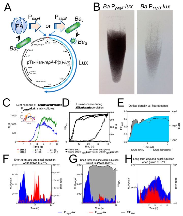
Figure 6. Rainfall, calcium and pH cycling, anthrax microecology and the phenology of anthrax
outbreaks. At the top of the cycle, vegetative and sporulated B. anthracis are deposited in soil form an
anthrax infected animal carcass forming a localized infectious zone (LIZ). Moving clockwise through
the figure; if bacteria are deposited in calcium rich alkaline soils, bacterial survival and sporulation
are promoted. Rainfall dissolves the mineralized calcium further inducing sporulation and causing
bacterial agglutination. Agglutinated spores are likely driven back up through the soil column during
regionally heavy rainfall. When hot dry conditions return, desiccation causes spores to attach via ionic
interactions to soil particles. Concentrated spores at the surface and reduced food sources during
seasonal heat-maxima bring animal hosts into close contact with infectious spore doses. Animals become
infected and the cycle continues.Int. J. Environ. Res. Public Health 2019, 16, 3747 13 of 16
These data and other anecdotal evidence led us to investigate pH and calcium influence on
B. anthracis physiology as a mechanism of anthrax environmental persistence, driven by annual rainfall
cycles observed by NDVI remote sensing. Construction and validation of our luminescent reporter
strains of B. anthracis allowed us to investigate two physiological events vital to B. anthracis survival;
toxin secretion and sporulation. B. anthracis growth, induction of toxin and sporulation are promoted
at alkaline pH. Calcium was slightly detrimental to pagA expression and very high calcium (1 M)
was detrimental to bacterial survival. Toxin is primarily induced during infection in the host, so this
outcome was not surprising. In contrast, sporulation was enhanced by nearly 100-fold at alkaline
pH in the presence of mid-high calcium levels; which correlated with the measured calcium levels in
the highly endemic KNP region. High calcium levels also caused agglutination of B. anthracis spores.
The geographic and laboratory data has allowed synthesis of a model that better explains the landscape
level modification of the ecology of anthrax.
5. Conclusions
We argue identifying global and regional signatures is necessary to fully understand the ecology of
anthrax and protect the public health. All over the world, anthrax outbreaks occur with clear periodicity.
The phenology of seasonal changes culminates with changing animal behavior to drive anthrax
outbreaks following periods of rainfall in a generally hot-dry summer. Figure 1 summarizes data from
multiple sources highlighting global seasonal patterns within seemingly sporadic anthrax outbreaks.
Regionally, anthrax can be limited to certain areas within landscapes that are experiencing similar
weather patterns. Zoonotic outbreaks require the presence of animals but are there environmental
components conducive to “hot zone” formation. We know that many types of animals across
all continents are involved in anthrax outbreaks, and that hot and dry conditions contribute to
outbreaks, whether through animal behavior modification or pathogen ecology alteration. The site of
long-term environmental pathogen persistence is soil, thus regional outbreak restriction and chronically
contaminated anthrax zones could be due to soil conditions and fluctuation of those soil conditions
instigated by rainfall events. pH and calcium have long been linked to increased incidence of anthrax,
so we set out in this work to investigate how regional pH and calcium signatures influence zoonotic
anthrax disease.
Laboratory experiments can help answer questions the medical geographer may have. For example;
what parameters in the soil could increase pathogen burden, persistence, or occurrence? The medical
geographer may have information such as pH of soils in a landscape and GPS coordinates of disease or
other location specific information that can inform the microbiologist or pathogen ecologist. Currently,
medical geographers are analyzing GPS data and ecological factors from the ongoing 2019 West Texas
anthrax outbreak. Back in the lab, microbiologists are analyzing fly, plant, and animal material to
characterize LIZs, decontamination procedures, and outbreak severity. The information can be used as
predictors of risk and impact disease outbreaks of human and animal origin. This work demonstrates
how laboratory experiments can help link landscape level drivers, pathogen reservoir modeling,
and pathogen physiology in pursuit of a holistic understanding of zoonotic disease outbreaks.
Author Contributions: Conceptualization, M.H.N. and J.K.B; methodology, M.H.N. and J.K.B.; software, J.K.B.;
investigation, M.H.N.; resources, M.H.N and J.K.B..; writing—original draft preparation, M.H.N. and J.K.B.;
writing—review and editing, M.H.N and J.K.B.; visualization, M.H.N. and J.K.B.
Funding: This research was partially funded by NIH 1R01GM117617 to J.K.B. and the Emerging Pathogens
Institute at the University of Florida.
Acknowledgments: We would like to thank Herbert P. Schweizer for use of consumables and equipment for
this work.
Conflicts of Interest: The authors declare no conflict of interest.Int. J. Environ. Res. Public Health 2019, 16, 3747 14 of 16
References
1. Carlson, C.J.; Kracalik, I.T.; Ross, N.; Alexander, K.A.; Hugh-Jones, M.E.; Fegan, M.; Elkin, B.T.; Epp, T.;
Shury, T.K.; Zhang, W.; et al. The global distribution of Bacillus anthracis and associated anthrax risk to
humans, livestock and wildlife. Nat. Microbiol. 2019, 4, 1337–1343. [CrossRef] [PubMed]
2. Van Ert, M.N.; Easterday, W.R.; Huynh, L.Y.; Okinaka, R.T.; Hugh-Jones, M.E.; Ravel, J.; Zanecki, S.R.;
Pearson, T.; Simonson, T.S.; U’Ren, J.M.; et al. Global genetic population structure of Bacillus anthracis.
PLoS ONE 2007, 2, e461. [CrossRef] [PubMed]
3. Smith, K.L.; DeVos, V.; Bryden, H.; Price, L.B.; Hugh-Jones, M.E.; Keim, P. Bacillus anthracis diversity in
Kruger National Park. J. Clin. Microbiol. 2000, 38, 3780–3784. [PubMed]
4. Halvorson, H.O. Two generations of spore research: From father to son. Microbiologia (Madrid, Spain) 1997,
13, 131–148.
5. Manchee, R.J.; Broster, M.G.; Stagg, A.J.; Hibbs, S.E. Formaldehyde solution effectively inactivates spores of
Bacillus anthracis on the Scottish island of Gruinard. Appl. Environ. Microbiol. 1994, 60, 4167–4171. [PubMed]
6. Blackburn, J.K.; Matakarimov, S.; Kozhokeeva, S.; Tagaeva, Z.; Bell, L.K.; Kracalik, I.T.; Zhunushov, A.
Modeling the ecological niche of Bacillus anthracis to map anthrax risk in Kyrgyzstan. Am. J. Trop. Med. Hyg.
2017, 96, 550–556. [CrossRef]
7. New, D.; Elkin, B.; Armstrong, T.; Epp, T. Anthrax in the Mackenzie wood bison (Bison bison athabascae)
population: 2012 anthrax outbreak and historical exposure in nonoutbreak years. J. Wildl. Dis. 2017, 53,
769–780. [CrossRef]
8. Munang’andu, H.M.; Banda, F.; Siamudaala, V.M.; Munyeme, M.; Kasanga, C.J.; Hamududu, B. The effect of
seasonal variation on anthrax epidemiology in the upper Zambezi floodplain of western Zambia. J. Vet. Sci.
2012, 13, 293–298. [CrossRef]
9. Noseda, R.; Cordeviola, J.; Bardón, C.; Martínez, A.; Cambessies, G. Bovine anthrax. Prevalence, annual and
seasonal variations in 30 districts of the province of Buenos Aires during the period 1977–1994. Vet. Argent.
1995, 12, 606–611.
10. Parkinson, R.; Rajic, A.; Jenson, C. Investigation of an anthrax outbreak in Alberta in 1999 using a geographic
information system. Can. Vet. J. 2003, 44, 315.
11. Kracalik, I.T.; Kenu, E.; Ayamdooh, E.N.; Allegye-Cudjoe, E.; Polkuu, P.N.; Frimpong, J.A.; Nyarko, K.M.;
Bower, W.A.; Traxler, R.; Blackburn, J.K. Modeling the environmental suitability of anthrax in Ghana and
estimating populations at risk: Implications for vaccination and control. PLoS Negl. Trop. Dis. 2017,
11, e0005885. [CrossRef] [PubMed]
12. Cizauskas, C.A.; Bellan, S.E.; Turner, W.C.; Vance, R.E.; Getz, W.M. Frequent and seasonally variable sublethal
anthrax infections are accompanied by short-lived immunity in an endemic system. J. Anim. Ecol. 2014, 83,
1078–1090. [CrossRef] [PubMed]
13. Barro, A. Integrating Geographical Information Systems and the Ecological Niche Modeling Framework to
Characterize the Spatial Ecology of Anthrax in Australia. Ph.D. Thesis, University of Florida, Gainesville,
FL, USA, 2016.
14. Blackburn, J.K.; Goodin, D.G. Differentiation of springtime vegetation indices associated with summer
anthrax epizootics in West Texas, USA deer. J. Wildl. Dis. 2013, 49, 699–703. [CrossRef] [PubMed]
15. Blackburn, J.; Curtis, A.J.; Hadfield, T.; Hugh-Jones, M.E. Spatial and temporal patterns of anthrax in
white-tailed deer, Odocoileus virginianus, and hematophagous flies in West Texas during the summertime
anthrax risk period. Ann. Assoc. Am. Geogr. 2014, 104, 939–958. [CrossRef]
16. Barro, A.S.; Fegan, M.; Moloney, B.; Porter, K.; Muller, J.; Warner, S.; Blackburn, J.K. Redefining the Australian
anthrax belt: Modeling the ecological niche and predicting the geographic distribution of Bacillus anthracis.
PLoS Negl. Trop. Dis. 2016, 10, e0004689. [CrossRef] [PubMed]
17. Lequette, Y.; Garenaux, E.; Combrouse, T.; Dias Tdel, L.; Ronse, A.; Slomianny, C.; Trivelli, X.; Guerardel, Y.;
Faille, C. Domains of BclA, the major surface glycoprotein of the B. cereus exosporium: Glycosylation patterns
and role in spore surface properties. Biofouling 2011, 27, 751–761. [CrossRef]
18. Lequette, Y.; Garenaux, E.; Tauveron, G.; Dumez, S.; Perchat, S.; Slomianny, C.; Lereclus, D.; Guerardel, Y.;
Faille, C. Role played by exosporium glycoproteins in the surface properties of Bacillus cereus spores and in
their adhesion to stainless steel. Appl. Environ. Microbiol. 2011, 77, 4905–4911. [CrossRef] [PubMed]Int. J. Environ. Res. Public Health 2019, 16, 3747 15 of 16
19. Brahmbhatt, T.N.; Janes, B.K.; Stibitz, E.S.; Darnell, S.C.; Sanz, P.; Rasmussen, S.B.; O’Brien, A.D.
Bacillus anthracis exosporium protein BclA affects spore germination, interaction with extracellular matrix
proteins, and hydrophobicity. Infect. Immun. 2007, 75, 5233–5239. [CrossRef] [PubMed]
20. Kracalik, I.; Malania, L.; Broladze, M.; Navdarashvili, A.; Imnadze, P.; Ryan, S.J.; Blackburn, J.K.
Changing livestock vaccination policy alters the epidemiology of human anthrax, Georgia, 2000–2013.
Vaccine 2017, 35, 6283–6289. [CrossRef]
21. Blackburn, J.K.; McNyset, K.M.; Curtis, A.; Hugh-Jones, M.E. Modeling the geographic distribution of
Bacillus anthracis, the causative agent of anthrax disease, for the contiguous United States using predictive
ecological [corrected] niche modeling. Am. J. Trop. Med. Hyg. 2007, 77, 1103–1110. [CrossRef]
22. Getz, W.M. Biomass transformation webs provide a unified approach to consumer–resource modelling.
Ecol. Lett. 2011, 14, 113–124. [CrossRef] [PubMed]
23. Turner, A.; Galvin, J.; Rubira, R.; Miller, G. Anthrax explodes in an Australian summer. J. App. Microbiol.
1999, 87, 196–199. [CrossRef] [PubMed]
24. Lindeque, P.M.; Turnbull, P.C.B. Ecology and epidemiology of anthrax in the Etosha National Park, Namibia.
Onderstepoort J. Vet. Res. 1994, 61, 71–83. [PubMed]
25. Turner, W.C.; Kausrud, K.L.; Krishnappa, Y.S.; Cromsigt, J.P.; Ganz, H.H.; Mapaure, I.; Cloete, C.C.;
Havarua, Z.; Kusters, M.; Getz, W.M.; et al. Fatal attraction: Vegetation responses to nutrient inputs attract
herbivores to infectious anthrax carcass sites. Proc. Biol. Sci. 2014, 281, 20141785. [CrossRef] [PubMed]
26. Hugh-Jones, M.; Blackburn, J. The ecology of Bacillus anthracis. Mol. Asp. Med. 2009, 30, 356–367. [CrossRef]
[PubMed]
27. Blackburn, J.K.; Asher, V.; Stokke, S.; Hunter, D.L.; Alexander, K.A. Dances with anthrax: Wolves (Canis lupus)
kill anthrax bacteremic plains bison (Bison bison bison) in southwestern Montana. J. Wildl. Dis. 2014, 50,
393–396. [CrossRef]
28. Morris, L.R.; Proffitt, K.M.; Asher, V.; Blackburn, J.K. Elk resource selection and implications for anthrax
management in Montana. J. Wildl. Manag. 2016, 80, 235–244. [CrossRef]
29. Van Ness, G. Ecology of Anthrax. Science 1971, 172, 1303–1307. [CrossRef]
30. Turner, W.C.; Kausrud, K.L.; Beyer, W.; Easterday, W.R.; Barandongo, Z.R.; Blaschke, E.; Cloete, C.C.; Lazak, J.;
Van Ert, M.N.; Ganz, H.H.; et al. Lethal exposure: An integrated approach to pathogen transmission via
environmental reservoirs. Sci. Rep. 2016, 6, 27311. [CrossRef]
31. Kellogg, F.E.; Prestwood, A.K.; Noble, R.E. Anthrax epizootic in white-tailed deer. J. Wildl. Dis. 1970, 6,
226–228. [CrossRef]
32. Blackburn, J.K.; Ganz, H.H.; Ponciano, J.M.; Turner, W.C.; Ryan, S.J.; Kamath, P.; Cizauskas, C.; Kausrud, K.;
Holt, R.D.; Stenseth, N.C. Modeling R0 for Pathogens with Environmental Transmission: Animal Movements,
Pathogen Populations, and Local Infectious Zones. Int. J. Environ. Res. Pub. Health 2019, 16, 954. [CrossRef]
[PubMed]
33. Fulbright, T.E.; Ortega-s, J.A. White-Tailed Deer Habitat: Ecology and Management on Rangelands; Texas A & M
University Press: College Station, TX, USA, 2006; ISBN 1-58544-499-5.
34. Armstrong, W.E.; Harmel, D.E.; Anderegg, M.J.M.S. Traweek Vegetation of the Kerr Wildlife Management Area
and Its Preference by White-Tailed Deer; Texas Parks and Wildlife Department: Austin, TX, USA, 1991.
35. Blackburn, J.K.; Mullins, J.C.; Van Ert, M.; Hadfield, T.; O’Shea, B.; Hugh-Jones, M.E. The necrophagous fly
anthrax transmission pathway: Empirical and genetic evidence from a wildlife epizootic in west Texas 2010.
Vector Borne Zoonotic Dis. 2014, 14, 576–583. [CrossRef] [PubMed]
36. Debré, P. Louis Pasteur; Johns Hopkins University Press: Baltimore, MD, USA, 1998; ISBN 0801858089.
37. Bellan, S.E.; Turnbull, P.C.; Beyer, W.; Getz, W.M. Effects of experimental exclusion of scavengers
from anthrax-infected herbivore carcasses on Bacillus anthracis sporulation, survival and distribution.
Appl. Environ. Microbiol. 2013, 79, 3756–3761. [CrossRef] [PubMed]
38. Havarua, Z.; Turner, W.C.; Mfune, J.K. Seasonal variation in foraging behaviour of plains zebra (Equus quagga)
may alter contact with the anthrax bacterium (Bacillus anthracis). Can. J. Zool. 2014, 92, 331–337. [CrossRef]
39. Hugh-Jones, M.; De Vos, V. Anthrax and wildlife. Revue Sci. Tech. (Int. Off. Epizoot.) 2002, 21, 359. [CrossRef]
[PubMed]
40. Hoffmaster, A.R.; Ravel, J.; Rasko, D.A.; Chapman, G.D.; Chute, M.D.; Marston, C.K.; De, B.K.; Sacchi, C.T.;
Fitzgerald, C.; Mayer, L.W.; et al. Identification of anthrax toxin genes in a Bacillus cereus associated with an
illness resembling inhalation anthrax. Proc. Natl. Acad. Sci. USA 2004, 101, 8449–8454. [CrossRef]You can also read