Low-Energy Shockwave Therapy Improves Ischemic Kidney Microcirculation - Medispec
←
→
Page content transcription
If your browser does not render page correctly, please read the page content below
BASIC RESEARCH           www.jasn.org

Low–Energy Shockwave Therapy Improves Ischemic
Kidney Microcirculation
Xin Zhang,* James D. Krier,* Carolina Amador Carrascal,† James F. Greenleaf,†
Behzad Ebrahimi,* Ahmad F. Hedayat,* Stephen C. Textor,* Amir Lerman,‡ and
Lilach O. Lerman*‡
*Division of Nephrology and Hypertension and Departments of †Physiology and Biomedical Engineering and
‡
 Cardiology, Mayo Clinic, Rochester, Minnesota

ABSTRACT
Microvascular rarefaction distal to renal artery stenosis is linked to renal dysfunction and poor outcomes.
Low–energy shockwave therapy stimulates angiogenesis, but the effect on the kidney microvasculature is
unknown. We hypothesized that low–energy shockwave therapy would restore the microcirculation and
alleviate renal dysfunction in renovascular disease. Normal pigs and pigs subjected to 3 weeks of renal
artery stenosis were treated with six sessions of low–energy shockwave (biweekly for 3 consecutive weeks)
or left untreated. We assessed BP, urinary protein, stenotic renal blood flow, GFR, microvascular structure,
and oxygenation in vivo 4 weeks after completion of treatment, and then, we assessed expression of
angiogenic factors and mechanotransducers (focal adhesion kinase and b1-integrin) ex vivo. A 3-week
low–energy shockwave regimen attenuated renovascular hypertension, normalized stenotic kidney mi-
crovascular density and oxygenation, stabilized function, and alleviated fibrosis in pigs subjected to renal
artery stenosis. These effects associated with elevated renal expression of angiogenic factors and
mechanotransducers, particularly in proximal tubular cells. In additional pigs with prolonged (6 weeks)
renal artery stenosis, shockwave therapy also decreased BP and improved GFR, microvascular density, and
oxygenation in the stenotic kidney. This shockwave regimen did not cause detectable kidney injury in
normal pigs. In conclusion, low–energy shockwave therapy improves stenotic kidney function, likely in part
by mechanotransduction-mediated expression of angiogenic factors in proximal tubular cells, and it may
ameliorate renovascular hypertension. Low–energy shockwave therapy may serve as a novel noninvasive
intervention in the management of renovascular disease.

J Am Soc Nephrol 27: 3715–3724, 2016. doi: 10.1681/ASN.2015060704

Atherosclerotic renal artery stenosis (ARAS) remains          of renal perfusion and vasoconstriction resulting
the leading cause of renovascular hypertension and            from activation of the renin-angiotensin system
is increasing in prevalence because of aging of the           lead to permanent changes in microvascular struc-
population and increased prevalence of atheroscle-            ture (remodeling and regression) associated with
rosis risk factors. As the disease progresses, ARAS           inadequate renal angiogenic signaling involving
results in gradual renal function loss1,2 and cardio-         vascular endothelial growth factor (VEGF). 9,10
vascular events.3
    Restoration of vessel patency by percutaneous
transluminal renal angioplasty and stenting does              Received June 25, 2015. Accepted April 5, 2016.

not often lead to improvement of renal function in            Published online ahead of print. Publication date available at
ARAS compared with optimal medical therapy                    www.jasn.org.
alone,4 likely because correction of an obstruction           Correspondence: Dr. Lilach O. Lerman, Division of Nephrology
in the main renal artery alone cannot reverse the             and Hypertension, Mayo Clinic, 200 First Street SW, Rochester,
                                                              MN 55905. Email: lerman.lilach@mayo.edu
preexisting downstream intrarenal damage.5,6 In
addition to inflammation,7,8 prolonged reduction               Copyright © 2016 by the American Society of Nephrology

J Am Soc Nephrol 27: 3715–3724, 2016                                                                       ISSN : 1046-6673/2712-3715   3715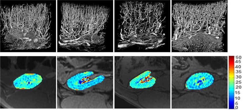
BASIC RESEARCH        www.jasn.org

Ischemia and oxidative stress in ARAS may also compromise             assess its safety. Moreover, the effects of SW on the stenotic
the integrity of the endothelium, leading to endothelial dys-         kidneys were also examined in four additional pigs (prolonged
function.5,11 In addition to glomerular podocytes, tubular            ARAS and SW), in which SW treatment started after 6 weeks
epithelial cells are an important site for VEGF expression,12,13      of RAS, and four other pigs served as controls.
which may fall because of tubular cell damage,14 a common
histologic finding in the ARAS kidney. Furthermore, develop-           SW Improved BP Control and Stabilized Renal
ment of fibrosis restricts expansion of the microcirculation to        Function in ARAS
replace lost vessels, resulting in a vicious cycle of microvascular   Before SW, MAP was similarly elevated in ARAS pigs compared
rarefaction and consequent declines in blood and oxygen sup-          with normal controls (Supplemental Figure 2A). After a 3-week
ply.15 Clearly, novel strategies developed to preserve the micro-     SW regimen, MAP decreased in ARAS and SW pigs but re-
vasculature could be of considerable value to slow functional         mained unchanged in ARAS and normal pigs (Supplemental
decline in kidneys with ARAS.                                         Figure 2A). Four weeks later, MAP of ARAS and SW pigs was
   Low–energy extracorporeal shockwave (SW) therapy, at 10%           lower than that of ARAS pigs, although it remained elevated
energy of the traditional SW used for lithotripsy, evokes             compared with normal (Figure 1A). Plasma renin activity and
neovascularization and improves regional blood flow and                norepinephrine (NE) release were both elevated in the ARAS
function in various ischemic tissues.16–18 The mechanical stim-       stenotic kidney veins but not in ARAS and SW pigs (Table 1),
ulus may be converted into cell signaling by upregulation of          indicating decreased activation of the renin-angiotensin sys-
canonical mechanotransducers, like b1-integrin and its effec-         tem. Scr was similarly elevated in ARAS and SW pigs and
tor, focal adhesion kinase (FAK),19,20 which in turn, activate        ARAS pigs during the 3-week period (Supplemental Figure
VEGF signaling and elicit angiogenesis. Experimental and clin-        2B), but by 16 weeks, it was lower in ARAS and SW pigs (Figure
ical studies in ischemic heart disease have shown improvement         1B). Urinary protein excretion of ARAS pigs was higher than
in myocardial blood flow and cardiac function after SW ther-           normal at 16 weeks, whereas ARAS and SW pigs did not differ
apy.21,22 Given that ischemic kidneys share several patterns of       from normal pigs (Figure 1C). Furthermore, although ARAS
microvascular remodeling with ischemic hearts,23 mechanical           decreased stenotic kidney RBF and GFR, SW improved RBF
forces that improve the myocardial microcirculation and he-           (P.0.10 versus normal) and restored GFR (Figure 1, D and E)
modynamics may also benefit the ARAS kidney. However,                  without affecting the function of the normal kidney.
whether SW can alleviate ARAS–induced ischemic kidney dis-
ease is unknown. Therefore, we hypothesized that low-energy           SW Promoted the Stenotic Kidney Microcirculation and
SW would preserve the stenotic kidney microvasculature and            Stimulated Mechanotransduction and VEGF
stabilize renal function in a unilateral ARAS swine model.            Expression in Proximal Tubular Cells
                                                                      ARAS decreased the density of cortical microvessels and
                                                                      blunted renal oxygenation, but SW improved both (Figure 1,
RESULTS                                                               F–J). Similarly, SW therapy upregulated VEGF expression
                                                                      that was decreased by ARAS, increased angiopoietin-1, and
Animals and SW Treatments                                             downregulated HIF-1a (Figure 2, A–D). SW also increased
Twenty-six pigs were randomized to ARAS (renal artery                 angiopoietin-1 in normal kidneys. Moreover, SW improved the
stenosis [RAS] induced after 6 weeks of an atherogenic diet)          expression of endothelial nitric oxide synthase (eNOS), which
or normal controls treated or untreated with SW. Three weeks          was diminished in ARAS (Supplemental Figure 3, A and D).
after RAS induction, low-energy SW was delivered biweekly for             Expression of the mechanotransducers b1-integrin and
3 consecutive weeks (a total of six sessions) (Supplemental           FAK was unchanged in ARAS pigs but upregulated in ARAS
Figure 1A). Serum creatinine (Scr) and mean arterial pressure         and SW pigs compared with in normal pigs (Figure 2, A, E, and
(MAP) were monitored during the treatment. Four weeks af-             F), indicating stimulation of mechanotransduction signaling.
ter completion of SW therapy, single–kidney renal blood flow           SW also increased FAK expression in normal pigs. Both FAK
(RBF), GFR, and oxygenation (relaxivity index, R2*) were              and VEGF were localized mainly to proximal tubular cells
assessed in vivo. Animals were then euthanized for ex vivo            (Supplemental Figure 4), suggesting them as a major response
studies, including microvascular remodeling as per microvas-          site in transducing SW and translating mechanical forces to
cular density and renal expression of VEGF, angiopoietin-1,           angiogenic signaling.
and hypoxia–inducible factor 1a (HIF-1a); expression of the
mechanotransducers b1-integrin and its downstream effector            SW Alleviated Oxidative Stress and Mediated
FAK; oxidative stress by dihydroethidium (DHE); tubular injury        Tissue Repair
and fibrosis; and tissue repair markers, like stromal–derived          DHE staining revealed increased oxidative stress in the ARAS
factor 1b (SDF-1b), stem cell factor (SCF), octamer–binding           kidney, which was ameliorated by SW (Supplemental Figure 3,
transcription factor 4 (Oct-4), and kidney injury molecule 1          B and E) along with ARAS–induced renal fibrosis (Figure 3, A
(KIM-1). The kidney injury marker neutrophil gelatinase–              and D). SW downregulated TGF-b in normal kidneys, blunted
associated lipocalin (NGAL) was also examined after SW to             its increase in ARAS kidneys (Figure 3, B and E), and alleviated

3716        Journal of the American Society of Nephrology                                            J Am Soc Nephrol 27: 3715–3724, 2016
www.jasn.org BASIC RESEARCH Figure 1. SW improves stenotic kidney function and structure. (A–C) SW lowered MAP and Scr and improved urinary protein excretion in ARAS pigs 4 weeks after completion of the SW regimen. (D and E) SW improved stenotic kidney RBF and GFR in ARAS. (F–J) Representative images of microcomputed tomography (microCT) and blood oxygen level–dependent (BOLD) magnetic resonance imaging from normal, normal and SW, ARAS, and ARAS and SW pigs and quantification of microvascular density and hypoxia (R2*). SW improved microvascular density and kidney oxygenation, which were decreased in ARAS. ♠ARAS, significant effect of ARAS; ♠ARAS 3 SW, significant interaction of ARAS and SW (two-way ANOVA); HZ, Hertz; ♠SW, significant effect of SW. *P,0.05 versus normal; †P,0.05 versus ARAS. tubular injury in ARAS (Supplemental Figure 3, C and F). SW SW pigs compared with ARAS pigs (Table 1), suggesting en- improved renal vein and reduced levels of SDF-1b (P.0.10 ver- hanced tissue repair potency. Moreover, kidney injury–induced sus normal) observed in ARAS, and it increased SCF in ARAS and regeneration markers Oct-4 and KIM-1 were both elevated in J Am Soc Nephrol 27: 3715–3724, 2016 Shockwave Stimulates Renal Repair 3717
BASIC RESEARCH           www.jasn.org

Table 1. Characteristics (mean6SEM) of normal or ARAS pigs treated or untreated with SW)
                                              Normal                           ARAS                    P Value for Two-Way ANOVA
     Characteristics
                                   Untreated                 SW   Untreated           SW            ARAS          SW          ARAS 3 SW
Body weight, kg                    47.261.1           48.562.0    51.564.7         49.362.2         0.25         0.55             0.60
Degree of stenosis, %                 —                  —          7366             7668            —            —                —
LDL, mg/dl                           51627              56621     200647a          194641a         ,0.001        0.96             0.91
Total cholesterol, mg/dl            103634             107619     315661a          301688a          0.004        0.94             0.89
Renal vein NE, ng/ml               0.0160.00          0.0160.00   0.0360.01a       0.0260.00        0.02         0.28             0.38
Renal vein PRA, pg/ml              0.1060.02          0.1060.04   0.2360.04a       0.1160.02        0.002        0.74             0.03
Renal vein SDF-1b, pg/ml          112.269.5          111.0616.7   76.465.8a       112.1612.7        0.04         0.97             0.13
Renal vein SCF-1, pg/ml            21.564.5           21.965.7    17.564.4         54.7618.6b       0.21         0.16             0.01
—, not observed/not performed; PRA, plasma renin activity.
a
 P,0.05 versus normal.
b
  P,0.05 versus ARAS.

the ARAS kidney, and SW downregulated Oct-4 expression, al-             at 12 weeks (Supplemental Figure 6B) compared with in the
though KIM-1 remained unchanged (Figure 3, C, G, and H).                normal and SW group (Supplemental Figure 2B), and it fur-
                                                                        ther increased in ARAS during sham but not in prolonged
SW Did Not Induce Detectable Injury to the Kidney                       ARAS and SW during SW treatment (Supplemental Figure
In two normal animals studied immediately after a single                6B), although at 16 weeks, it remained higher than in the
session of SW, no gross or microscopic hematuria was                    normal group (Supplemental Figure 6C). SW did not change
observed. There was no change in urinary protein excretion              stenotic RBF but significantly improved its GFR (Supplemen-
or either urine or blood NGAL levels (Supplemental Table 1).            tal Figure 6, D and E). At 16 weeks, prolonged ARAS and SW
Renal function, such as GFR, perfusion, and RBF, remained               RBF and GFR did not differ from either ARAS or normal
unaltered (Supplemental Figure 2C), and microscopic inspec-             (Supplemental Figure 6, F and G). In addition, SW also im-
tion of the kidney tissue showed no signs of hemorrhage or              proved cortical microvascular density and renal oxygenation
tubular injury (Supplemental Figure 2, D and E). Hence, SW              in prolonged ARAS (Supplemental Figure 7, A, B, E, and F)
did not induce measurable short–term injury to the kidney.              and alleviated fibrosis (Supplemental Figure 7, C and G), but it
   In the two groups treated with a 3-week SW regimen, vital            did not change tubular injury score (Supplemental Figure 7, D
signs (heart rate and BP) remained stable during each session,          and H). These finding suggest that SW improved structure and
no hematuria or change in urinary protein excretion was                 function of the stenotic kidney in prolonged ARAS, albeit
observed (data not shown), and urine and plasma NGAL levels             slightly less effectively than in ARAS and SW pigs.
were unchanged (Supplemental Figure 2, F and G).
Therefore, a 3-week SW regimen seemed to be safe for the
kidney.
                                                                        DISCUSSION

SW Alleviated Hyperfiltration in the Contralateral                       This study shows that low–energy SW therapy improves the
Kidneys                                                                 poststenotic kidney oxygenation in experimental renovascular
Both RBF and GFR were elevated in the contralateral kidneys of          disease and preserves its function. This was associated with
ARAS, indicating hyperfiltration, but not elevated in those of           upregulation of mechanotransducers and angiogenic factors
ARAS and SW pigs (Supplemental Figure 5, A and B). ARAS                 as well as modulation of vasoactive mediators, resulting in
induced mild contralateral kidney fibrosis (Supplemental Fig-            restoration of the renal microcirculation as well as reduced
ure 5, C and D), which was much lower than in the counter-              oxidative and fibrosis. No signs of renal damage were detected
part stenotic kidneys, and it remained unchanged in ARAS                in SW-treated kidneys. After a more prolonged ARAS, SW also
and SW pigs (Supplemental Figure 5, C and D).                           decreased BP and improved stenotic kidney GFR, albeit
                                                                        slightly less effectively that in ARAS and SW. We also found
SW Improved BP Control and Stabilized Renal Function                    that the contralateral kidney of ARAS developed mild fibrosis
in Prolonged ARAS                                                       and hyperfiltration (increased RBF and GFR) that SW im-
Prolonged ARAS and SW had comparable degrees of stenosis                proved, possibly via improvement of stenotic kidney function
(76%610%) and pretreatment MAP values (Supplemental                     and fall in BP. Collectively, this study suggests a potential role
Figure 6A) to ARAS (both P.0.10). MAP fell in the prolonged             for low–energy SW therapy as a safe, noninvasive, and effective
ARAS and SW group after treatment and became lower than                 treatment of the ischemic kidney distal to ARAS.
ARAS (Supplemental Figure 6A). Similarly, Scr was compara-                  Microvascular remodeling and regression characterize the is-
bly elevated in the prolonged ARAS and SW and ARAS groups               chemic kidney, possibly secondary to protracted vasoconstriction

3718          Journal of the American Society of Nephrology                                             J Am Soc Nephrol 27: 3715–3724, 2016
www.jasn.org BASIC RESEARCH
reparative mechanisms17,18 mediate micro-
circulatory repair. b1-Integrin is a cell sur-
face adhesion receptor with an extracellular
domain linked to the cytoskeleton, which
permits transmission of mechanical forces
generated by SW by modulating the para-
cellular signaling pathway.30 Its effect on the
vasculature is achieved via its chief down-
stream regulator and signaling molecule
FAK,31 which in turn, stimulates VEGF32
and endothelial survival.33 In addition, SW-
induced upregulation of angiopoietin-1,
which promotes microvascular maturation
and stability, can further facilitate FAK acti-
vation34 to enhance angiogenesis. Indeed,
upregulation of angiogenic factors in the
ARAS kidney by SW parallels activation of
mechanotransducers, which possibly accounts
for improved oxygenation (blood oxygen
level–dependent R2*) and downregulated ex-
pression of HIF-1a.
Notably, VEGF expression was not only
coexpressed with mechanotransducers but
also, similarly and selectively localized to
proximal tubules, identifying them as an
important site for VEGF production12,35
after SW treatment. The specific mechanism
responsible for this selective upregulation
of mechanotransducers and angiogenic
factors needs to be further explored. The
population of proximal tubules–derived
regenerating cells expressing Oct-4 and
KIM-1 increases in response to hypoxia or
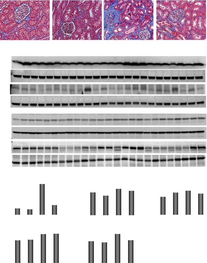
Figure 2. SW enhanced angiogenesis and mechanotransduction. (A–D) Renal ex- injury,36–39 but cellular regeneration might
pression of angiogenic VEGF, angiopoietin-1 (Ang-1), and HIF-1a. SW upregulated be blunted in ARAS because of vasocon-
expression of VEGF, increased angiopoietin-1, and attenuated HIF-1a in ARAS kid- striction and oxidative stress. SW alleviated
neys. (E and F) SW increased both b1-integrin and focal FAK in ARAS kidneys and FAK
vasoconstriction by improving expression
in normal kidneys as well. ♠ARAS, significant effect of ARAS; ♠ARAS 3 SW, significant
of eNOS and reducing oxidative stress,
interaction of ARAS and SW (two-way ANOVA); GAPDH, glyceraldehyde-3-phosphate
dehydrogenase; ♠SW, significant effect of SW (two-way ANOVA). *P,0.05 versus
and it may thereby facilitate regenerative
†
normal; P,0.05 versus ARAS. function and tissue repair as suggested by
normalized expression of Oct-4 in ARAS
and SW.
because of activation of the renin-angiotensin system, shear Interestingly, activation of b1-integrin signaling was only
stress, and increased oxidative stress.24 We have previously observed in ARAS and SW pigs but not in normal and SW pigs,
shown that dyslipidemia alone may increase microvascular suggesting greater responsiveness of the ARAS kidney to SW.
density,25 but its coexistence with renal ischemia exacerbates Because b1-integrin in tubular epithelial cells can redistribute
microvascular loss.26,27 Decreased microvascular density, in to the apical surface during ischemic insults,40 proximal tu-
turn, interferes with the supply and delivery of oxygen and bular cells in ARAS might become more sensitive to SW–
blood, precipitating tissue hypoxia and damage. This study elicited mechanical forces.
shows that SW can alleviate or prevent microvascular loss, Consequent to improved renal structure and oxygenation, SW
which may contribute to preservation of renal function. improved stenotic kidney function. Because stenotic kidney RBF
Low-energy SW generates mechanical forces that induce local- was less markedly affected, the improved GFR may be partly
ized stress on cell membranes that resembles shear stress28 and secondary to alleviated tubular injury and improved tubular-
exerts biologic effects, 29 after which upregulation of angio- glomeruli feedback. After a more prolonged (6-week) ARAS, SW
genic factors, including VEGF and eNOS, and activation of also decreased BP, restored stenotic kidney microcirculation, and
J Am Soc Nephrol 27: 3715–3724, 2016 Shockwave Stimulates Renal Repair 3719BASIC RESEARCH www.jasn.org
longer elevated in ARAS after SW, the neu-
rohumoral pathway might be implicated41
in its BP-lowering effect. Additional studies
are needed to evaluate this link. Moreover,
restored microvasculature and eNOS may
not only improve blood and oxygen deliv-
ery but also, lower BP by antagonizing an-
giotensin II activity, increasing nitric oxide
availability and expression, and alleviating
oxidative stress.
The low–energy SW regimen that we
applied in the kidney exhibited a good
safety profile as reported in the ischemic
heart.42–44 Rather than potentially imposing
tissue damage, like traditional lithotripsy, a
3-week low–energy SW regimen contrarily
decreased proteinuria, attenuated a rise in
Scr observed in ARAS, and increased ste-
notic kidney GFR. This finding in our clin-
ically relevant large animals may increase its
translational potential. In addition, low-
energy SW can promote healing through
direct anti–inflammatory properties in
acute myocardial infarction,45 carotid ar-
tery angioplasty,46 and cutaneous burn in-
jury,47 suggesting that it may be an effective
measure to boost tissue recovery. Interest-
ingly, in an ischemia-reperfusion model,
ultrasound recently suppressed renal in-
flammation by splenic modulation.48 In
our study, SW was selectively applied to
the right kidney, and the spleen was unlikely
affected by SW. Nevertheless, whether
spleen stimulation by SW could facilitate
renal function recovery deserves addi-
Figure 3. SW alleviated fibrosis and tissue injury. (A and D) Representative images and tional studies.
quantification of trichrome staining. (B, E, and F) Renal expression of TGF-b and tissue Our study is limited by the short dura-
inhibitor of metalloproteinases 1 (TIMP-1). (C, G, and H) Renal expression of injury– tion of the disease, but the similarity of renal
induced regenerative markers KIM-1 and Oct-4. SW alleviated ARAS–induced renal structure and function in our swine model
fibrosis and tissue injury. ♠ARAS, significant effect of ARAS; GAPDH, glyceraldehyde- to human kidneys increases the transla-
3-phosphate dehydrogenase; ♠SW, significant effect of SW. *P,0.05 versus normal; tional potential of our results. The temporal
†
P,0.05 versus ARAS. patterns of SW therapy on the microcircu-
lation and its long-term protection of renal
function need to be examined in longitu-
improved its GFR, albeit less effectively that in ARAS and SW; it dinal studies. The effects of low-energy SWon glomerular cells
remained not significantly different from either normal or ARAS and their production of angiogenic factors also warrant
kidneys, possibly because tubular injury was not ameliorated. additional study. We have used the settings implemented on
Furthermore, because RBF did not increase, we cannot exclude our machine and a previously validated regimen.16,43 The op-
the possibility that the improvement in GFR was partly attrib- timal doses, energy levels, and timing of SW treatment in the
utable to afferent vasoconstriction, the mechanism of which ischemic kidney and other kidney diseases need to be defined
would need to be explored in future studies. Overall, the efficacy in additional studies.
of SW is likely at least partly dependent on injury duration. Low–energy extracorporeal SW treatment improved the
This study shows BP-lowering effects of SW in ARAS animals. ARAS kidney microvasculature, alleviated fibrosis, stabilized
Diminished renin release from the treated renal veins might have renal function, and lowered BP. Low–energy SW therapy may
been secondary to improved renal perfusion. Because NE was no be an effective and powerful noninvasive strategy for
3720 Journal of the American Society of Nephrology J Am Soc Nephrol 27: 3715–3724, 2016www.jasn.org BASIC RESEARCH
treatment of chronic ischemic kidney disease and renovascu- time-attenuation curves in each region and obtain measures of renal
lar hypertension. However, its efficacy is likely at least partly function.52 BP was measured in all animals using an arterial catheter
dependent on the duration of preexisting renal injury. during MDCT. Urine was collected via bladder puncture to determine
protein excretion, and blood was collected from the inferior vena cava
for creatinine. Plasma renin activity and NE release were measured in
CONCISE METHODS blood collected from veins draining stenotic/SW-treated kidneys.
Animals and SW Treatments Renal Oxygenation
This study was approved by the Institutional Animal Care and Use Blood oxygen level–dependent magnetic resonance imaging (Signa
Committee. Twenty-six domestic female pigs (50–60 kg) were studied Echo Speed; GE Medical Systems, Milwaukee, WI) scanning was
during 16 weeks of observation. Pigs were randomized to ARAS or performed 2 days before MDCT to assess intrarenal oxygenation
normal without (ARAS and normal groups, n=7 each) or with SW (evaluated as R2*).53,54 For data analysis, ROIs were manually traced
treatment (ARAS and SW, n=7; normal and SW, n=5). Normal pigs in the cortex and medulla on the 7-millisecond echo time images that
were fed isocaloric diets of standard chow, and ARAS pigs were fed give the best anatomic details in each experimental period. For each
with a high-fat diet containing 2% cholesterol (Harlan Teklad, Mad- echo time, the software automatically computed the average of mag-
ison, WI).49 All animals had free access to water. netic resonance signals within each ROI.
RAS was induced after 6 weeks of diet by placing a local irritant Animals were euthanized 3 days after in vivo studies using a lethal
coil in the right main renal artery, leading to gradual development of intravenous dose of sodium pentobarbital (100 mg/kg; Fatal Plus;
unilateral RAS as previously described.50 The degrees of stenosis were Vortech Pharmaceuticals, Fort Washington, PA).55 The kidneys
determined by renal angiography 6 weeks later. Three weeks after were removed using a retroperitoneal incision, and they were immedi-
RAS induction, low–energy SW sessions were initiated and delivered ately dissected and prepared in ice cold normal saline for microcomputed
biweekly for 3 consecutive weeks (a total of six sessions). Because the tomography, frozen in liquid nitrogen (and maintained at 280°C), or
kidney and heart undergo similar processes of microvascular remod- preserved in formalin for tissue studies.
eling secondary to upstream vascular obstructions, we used protocols
that had been successfully applied to the myocardium.43,51 Four Ex Vivo Studies
weeks after completion of this regimen, renal hemodynamics and Microvasculature
oxygenation were assessed in vivo. Animals were then euthanized, After the kidney was flushed, microfil MV122 (an intravascular
and their kidneys were harvested (Supplemental Figure 1A). contrast agent) was perfused into the stenotic kidney through a
Omnispec Vetspec Model (spark voltage =10–24 kV; energy den- cannula ligated in the renal artery. Samples were prepared and
sity =0.09 mJ/mm2 ; frequency =120 pulse/min; Medispec LTD, scanned at 0.5° angular increments at 18-mm resolution, and images
Germantown, MD) was used to deliver SW. An Acuson SC2000 were analyzed as previously described.24,56 The spatial density of
Ultrasound System (Global Siemens Healthcare, Erlangen, Germany) microvessels (defined as diameters ,500 mm) in the inner and outer
was used to guide SW localization on the kidney. Pigs were laid renal cortices was examined.57
prone, the skin of the back was shaved, and ultrasound gel was ap- Renal expression of the angiogenic factors VEGF (1:200; Santa
plied to ensure adequate conduction of the ultrasound wave and SW Cruz Biotechnology, Santa Cruz, CA), angiopoietin-1 (1:200; Santa
(Supplemental Figure 1B). The ultrasound probe was placed at the Cruz Biotechnology), and HIF-1a (1:1000; Abcam, Inc., Cambridge,
lateral aspect of the right/stenotic kidney along its long axis for co- MA) was examined by Western blotting in the kidney. Immuno-
ronal visualization, and the SW applicator was located perpendicu- reactivity of eNOS (1:50; Abcam, Inc.) was measured by immu-
larly above the kidney to distribute energy through the kidney along nofluorescence staining and Western blotting. Expression of the
its short axis (Supplemental Figure 1, B–D). Because the whole kid- mechanotransducers b1-integrin (1:1000; Cell Signaling Technol-
ney is subjected to ischemia distal to the stenosis, the entire kidney ogy, Danvers, MA) and downstream FAK (1:50; Cell Signaling Tech-
was treated with SW with regions evenly selected (Supplemental nology) was assessed by Western blotting and immunofluorescence
Figure 1D), with 200 rapid shots applied to each treatment zone.16 staining, respectively. FAK and VEGF were both costained with the
proximal and distal renal tubular markers Phaseous vulgaris agglu-
In Vivo Studies tinin and peanut agglutinin38 to localize their expression.
BP and Renal Function
Single-kidney function, including RBF and GFR, were assessed in Oxidative Stress, Fibrosis, and Tissue Repair
both the stenotic and contralateral kidneys using multidetector DHE staining was performed to assess renal production of superoxide
computed tomography (MDCT) as described previously.52 Briefly, anion. Fibrosis was evaluated by trichrome staining. Tubular injury
160 consecutive scans were performed after a central venous injec- was scored in hematoxylin and eosin slides on a 1–5 scale (1, ,10%; 2,
tion of iopamidol (0.5 ml kg21 2 s21). Then, MDCT images were 10%–25%; 3, 26%–50%; 4, 51%–75%; and 5, .75% injury) on the
reconstructed and displayed with the Analyze software package basis of tubular dilation, atrophy, cast formation, sloughing tubular
(Biomedical Imaging Resource; Mayo Clinic, Rochester, MN). For endothelial cells, or thickening of basement membrane as previously
data analysis, regions of interest (ROIs) were selected from tomo- described.58 Expression of TGF-b and tissue inhibitor of metallopro-
graphic images from the aorta, renal cortex, and medulla to generate teinases 1 (both 1:200; Santa Cruz Biotechnology) was examined by
J Am Soc Nephrol 27: 3715–3724, 2016 Shockwave Stimulates Renal Repair 3721BASIC RESEARCH www.jasn.org
Western blotting. Renal levels of SDF-1b (MBS735811 ELISA; REFERENCES
MyBioSource) and SCF (MBS2020518 ELISA; MyBioSource), which
mobilize endogenous repair mechanisms,59 were measured in the 1. Safian RD, Textor SC: Renal-artery stenosis. N Engl J Med 344: 431–
stenotic renal vein and inferior vena cava. Expression of regenerating 442, 2001
2. Lerman L, Textor SC: Pathophysiology of ischemic nephropathy. Urol
renal cell markers Oct-4 and KIM-1 that are upregulated in response
Clin North Am 28: 793–803, 2001
to hypoxia and injury36–39 were detected by Western blotting. 3. Edwards MS, Craven TE, Burke GL, Dean RH, Hansen KJ: Renovascular
disease and the risk of adverse coronary events in the elderly: A pro-
Effects of SW in Prolonged ARAS spective, population-based study. Arch Intern Med 165: 207–213,
Of eight additional ARAS pigs, SW was started in four pigs after 6 2005
weeks of RAS (prolonged ARAS and SW); the four other pigs served as 4. Cooper CJ, Murphy TP, Cutlip DE, Jamerson K, Henrich W, Reid
DM, Cohen DJ, Matsumoto AH, Steffes M, Jaff MR, Prince MR, Lewis
controls. BP and Scr were monitored during the 3-week regimen in all
EF, Tuttle KR, Shapiro JI, Rundback JH, Massaro JM, D’Agostino RB
pigs. Stenotic RBF and GFR in prolonged ARAS and SW pigs were Sr., Dworkin LD; CORAL Investigators: Stenting and medical ther-
quantified before and after SW. Microvascular density, oxygenation apy for atherosclerotic renal-artery stenosis. N Engl J Med 370: 13–
levels, fibrosis, and tubular injury score were also assessed. 22, 2014
5. Lerman LO, Textor SC, Grande JP: Mechanisms of tissue injury in renal
artery stenosis: Ischemia and beyond. Prog Cardiovasc Dis 52: 196–
Safety of SW
203, 2009
Short-term safety of low-energy SW was assessed in two normal pigs
6. Textor SC: Issues in renovascular disease and ischemic nephropathy:
receiving a single SW session. Heart rate was continuously monitored Beyond ASTRAL. Curr Opin Nephrol Hypertens 20: 139–145, 2011
using an electrocardiogram monitor throughout the experiment, and 7. Johns EJ: Inflammation: The underlying foe in renovascular hyperten-
BP was recorded every 15 minutes using a cuff placed on the front limb. sion? J Hypertens 27: 1964–1965, 2009
Bladder urine and stenotic renal vein blood were collected before and 2 8. Vaidya VS, Ford GM, Waikar SS, Wang Y, Clement MB, Ramirez V,
hours after SW to examine AKI indices, including microscopic Glaab WE, Troth SP, Sistare FD, Prozialeck WC, Edwards JR, Bobadilla
NA, Mefferd SC, Bonventre JV: A rapid urine test for early detection of
hematuria, urine protein, and urine and plasma NGAL. Single-kidney
kidney injury. Kidney Int 76: 108–114, 2009
function was assessed before and after SW by MDCT for GFR, 9. Eirin A, Zhu XY, Urbieta-Caceres VH, Grande JP, Lerman A, Textor SC,
perfusion, and RBF. Pigs were then euthanized, and the treated kidneys Lerman LO: Persistent kidney dysfunction in swine renal artery stenosis
were immediately removed to inspect histologically for intrarenal correlates with outer cortical microvascular remodeling. Am J Physiol
hemorrhage and tubular injury. Renal Physiol 300: F1394–F1401, 2011
10. Favreau F, Zhu XY, Krier JD, Lin J, Warner L, Textor SC, Lerman LO:
The safety of the 3-week SWregimenwas also tested in normal and SW
Revascularization of swine renal artery stenosis improves renal function
pigs and ARAS and SW pigs. Electrocardiogram and BP were monitored but not the changes in vascular structure. Kidney Int 78: 1110–1118,
during each SW session as described above. AKI markers were measured 2010
before, weekly during, and 4 weeks after completion of SW treatment. 11. Brodsky SV, Yamamoto T, Tada T, Kim B, Chen J, Kajiya F, Goligorsky
MS: Endothelial dysfunction in ischemic acute renal failure: Rescue by
transplanted endothelial cells. Am J Physiol Renal Physiol 282: F1140–
Statistical Analyses
F1149, 2002
Continuous data were expressed as means6SEMs. Two-way ANOVA
12. Schrijvers BF, Flyvbjerg A, De Vriese AS: The role of vascular endo-
was used to analyze the effects of ARAS and SWas separate factors and thelial growth factor (VEGF) in renal pathophysiology. Kidney Int 65:
their interactions followed by Tukey test as appropriate. One-way 2003–2017, 2004
ANOVA was used for comparison among all groups to identify dif- 13. Villegas G, Lange-Sperandio B, Tufro A: Autocrine and paracrine
ferences from prolonged ARAS and SW. Comparisons within groups functions of vascular endothelial growth factor (VEGF) in renal tubular
epithelial cells. Kidney Int 67: 449–457, 2005
were performed using repeated measures ANOVA followed by paired
14. Lindenmeyer MT, Kretzler M, Boucherot A, Berra S, Yasuda Y, Henger
t test. P,0.05 was considered statistically significant. A, Eichinger F, Gaiser S, Schmid H, Rastaldi MP, Schrier RW,
Schlöndorff D, Cohen CD: Interstitial vascular rarefaction and reduced
VEGF-A expression in human diabetic nephropathy. J Am Soc Nephrol
18: 1765–1776, 2007
ACKNOWLEDGMENTS
15. Chade AR, Bentley MD, Zhu X, Rodriguez-Porcel M, Niemeyer S,
Amores-Arriaga B, Napoli C, Ritman EL, Lerman A, Lerman LO: Anti-
We thank Medispec LTD for generously allowing the use of the oxidant intervention prevents renal neovascularization in hypercho-
shockwave machine. lesterolemic pigs. J Am Soc Nephrol 15: 1816–1825, 2004
This research was partly supported by National Institutes of Health 16. Fukumoto Y, Ito A, Uwatoku T, Matoba T, Kishi T, Tanaka H, Takeshita
A, Sunagawa K, Shimokawa H: Extracorporeal cardiac shock wave
grants HL121561, HL123160, DK73608, DK104273, and DK102325
therapy ameliorates myocardial ischemia in patients with severe coro-
and American Heart Association grant 13POST16810064. nary artery disease. Coron Artery Dis 17: 63–70, 2006
The vendor (Medispec LTD) was not involved in data collection or 17. Wang CJ, Huang KE, Sun YC, Yang YJ, Ko JY, Weng LH, Wang FS:
analysis. VEGF modulates angiogenesis and osteogenesis in shockwave-
promoted fracture healing in rabbits. J Surg Res 171: 114–119, 2011
18. Aicher A, Heeschen C, Sasaki K, Urbich C, Zeiher AM, Dimmeler S: Low-
energy shock wave for enhancing recruitment of endothelial progenitor
DISCLOSURES cells: A new modality to increase efficacy of cell therapy in chronic hind
None. limb ischemia. Circulation 114: 2823–2830, 2006
3722 Journal of the American Society of Nephrology J Am Soc Nephrol 27: 3715–3724, 2016www.jasn.org BASIC RESEARCH
19. Luu NT, Glen KE, Egginton S, Rainger GE, Nash GB: Integrin- angiogenesis in epithelial-endothelial co-cultures. J Am Soc Nephrol
substrate interactions underlying shear-induced inhibition of the 13: 2027–2036, 2002
inflammatory response of endothelial cells. Thromb Haemost 109: 298– 36. Smeets B, Boor P, Dijkman H, Sharma SV, Jirak P, Mooren F, Berger K,
308, 2013 Bornemann J, Gelman IH, Floege J, van der Vlag J, Wetzels JF, Moeller
20. Zebda N, Dubrovskyi O, Birukov KG: Focal adhesion kinase regulation MJ: Proximal tubular cells contain a phenotypically distinct, scattered
of mechanotransduction and its impact on endothelial cell functions. cell population involved in tubular regeneration. J Pathol 229: 645–
Microvasc Res 83: 71–81, 2012 659, 2013
21. Wang Y, Guo T, Cai HY, Ma TK, Tao SM, Sun S, Chen MQ, Gu Y, Pang 37. Berger K, Bangen JM, Hammerich L, Liedtke C, Floege J, Smeets B,
JH, Xiao JM, Yang XY, Yang C: Cardiac shock wave therapy reduces Moeller MJ: Origin of regenerating tubular cells after acute kidney in-
angina and improves myocardial function in patients with refractory jury. Proc Natl Acad Sci U S A 111: 1533–1538, 2014
coronary artery disease. Clin Cardiol 33: 693–699, 2010 38. Gupta S, Verfaillie C, Chmielewski D, Kren S, Eidman K, Connaire J,
22. Kikuchi Y, Ito K, Ito Y, Shiroto T, Tsuburaya R, Aizawa K, Hao K, Heremans Y, Lund T, Blackstad M, Jiang Y, Luttun A, Rosenberg ME:
Fukumoto Y, Takahashi J, Takeda M, Nakayama M, Yasuda S, Kuriyama Isolation and characterization of kidney-derived stem cells. J Am Soc
S, Tsuji I, Shimokawa H: Double-blind and placebo-controlled study of Nephrol 17: 3028–3040, 2006
the effectiveness and safety of extracorporeal cardiac shock wave 39. Bussolati B, Moggio A, Collino F, Aghemo G, D’Armento G, Grange C,
therapy for severe angina pectoris. Circ J 74: 589–591, 2010 Camussi G: Hypoxia modulates the undifferentiated phenotype of
23. Urbieta Caceres VH, Lin J, Zhu XY, Favreau FD, Gibson ME, Crane JA, human renal inner medullary CD133+ progenitors through Oct4/miR-145
Lerman A, Lerman LO: Early experimental hypertension preserves the balance. Am J Physiol Renal Physiol 302: F116–F128, 2012
myocardial microvasculature but aggravates cardiac injury distal to 40. Zuk A, Bonventre JV, Brown D, Matlin KS: Polarity, integrin, and ex-
chronic coronary artery obstruction. Am J Physiol Heart Circ Physiol tracellular matrix dynamics in the postischemic rat kidney. Am J Physiol
300: H693–H701, 2011 275: C711–C731, 1998
24. Bentley MD, Rodriguez-Porcel M, Lerman A, Sarafov MH, Romero JC, 41. Grisk O, Rettig R: Interactions between the sympathetic nervous system
Pelaez LI, Grande JP, Ritman EL, Lerman LO: Enhanced renal cortical and the kidneys in arterial hypertension. Cardiovasc Res 61: 238–246,
vascularization in experimental hypercholesterolemia. Kidney Int 61: 2004
1056–1063, 2002 42. Nishida T, Shimokawa H, Oi K, Tatewaki H, Uwatoku T, Abe K,
25. Li ZL, Woollard JR, Ebrahimi B, Crane JA, Jordan KL, Lerman A, Wang Matsumoto Y, Kajihara N, Eto M, Matsuda T, Yasui H, Takeshita A,
SM, Lerman LO: Transition from obesity to metabolic syndrome is as- Sunagawa K: Extracorporeal cardiac shock wave therapy markedly
sociated with altered myocardial autophagy and apoptosis. Arte- ameliorates ischemia-induced myocardial dysfunction in pigs in vivo.
rioscler Thromb Vasc Biol 32: 1132–1141, 2012 Circulation 110: 3055–3061, 2004
26. Zhang X, Li ZL, Woollard JR, Eirin A, Ebrahimi B, Crane JA, Zhu XY, 43. Cassar A, Prasad M, Rodriguez-Porcel M, Reeder GS, Karia D, DeMaria
Pawar AS, Krier JD, Jordan KL, Tang H, Textor SC, Lerman A, Lerman AN, Lerman A: Safety and efficacy of extracorporeal shock wave myo-
LO: Obesity-metabolic derangement preserves hemodynamics but cardial revascularization therapy for refractory angina pectoris. Mayo
promotes intrarenal adiposity and macrophage infiltration in swine Clin Proc 89: 346–354, 2014
renovascular disease. Am J Physiol Renal Physiol 305: F265–F276, 44. Prasad M, Wan Ahmad WA, Sukmawan R, Magsombol EB, Cassar A,
2013 Vinshtok Y, Ismail MD, Mahmood Zuhdi AS, Locnen SA, Jimenez R,
27. Sun D, Eirin A, Ebrahimi B, Textor SC, Lerman A, Lerman LO: Early Callleja H, Lerman A: Extracorporeal shockwave myocardial therapy
atherosclerosis aggravates renal microvascular loss and fibrosis in swine is efficacious in improving symptoms in patients with refractory an-
renal artery stenosis. J Am Soc Hypertens 10: 325–335, 2016 gina pectoris–a multicenter study. Coron Artery Dis 26: 194–200,
28. Maisonhaute E, Prado C, White PC, Compton RG: Surface acoustic 2015
cavitation understood via nanosecond electrochemistry. Part III: Shear 45. Abe Y, Ito K, Hao K, Shindo T, Ogata T, Kagaya Y, Kurosawa R,
stress in ultrasonic cleaning. Ultrason Sonochem 9: 297–303, 2002 Nishimiya K, Satoh K, Miyata S, Kawakami K, Shimokawa H: Extracor-
29. Sun CK, Shao PL, Wang CJ, Yip HK: Study of vascular injuries using poreal low-energy shock-wave therapy exerts anti-inflammatory effects
endothelial denudation model and the therapeutic application of shock in a rat model of acute myocardial infarction. Circ J 78: 2915–2925,
wave: A review. Am J Transl Res 3: 259–268, 2011 2014
30. Elias BC, Mathew S, Srichai MB, Palamuttam R, Bulus N, Mernaugh G, 46. Shao PL, Chiu CC, Yuen CM, Chua S, Chang LT, Sheu JJ, Sun CK, Wu
Singh AB, Sanders CR, Harris RC, Pozzi A, Zent R: The integrin b1 CJ, Wang CJ, Yip HK: Shock wave therapy effectively attenuates in-
subunit regulates paracellular permeability of kidney proximal tubule flammation in rat carotid artery following endothelial denudation by
cells. J Biol Chem 289: 8532–8544, 2014 balloon catheter. Cardiology 115: 130–144, 2010
31. Eke I, Deuse Y, Hehlgans S, Gurtner K, Krause M, Baumann M, 47. Davis TA, Stojadinovic A, Anam K, Amare M, Naik S, Peoples GE,
Shevchenko A, Sandfort V, Cordes N: b₁Integrin/FAK/cortactin sig- Tadaki D, Elster EA: Extracorporeal shock wave therapy suppresses the
naling is essential for human head and neck cancer resistance to ra- early proinflammatory immune response to a severe cutaneous burn
diotherapy. J Clin Invest 122: 1529–1540, 2012 injury. Int Wound J 6: 11–21, 2009
32. Chen XL, Nam JO, Jean C, Lawson C, Walsh CT, Goka E, Lim ST, Tomar 48. Gigliotti JC, Huang L, Ye H, Bajwa A, Chattrabhuti K, Lee S, Klibanov
A, Tancioni I, Uryu S, Guan JL, Acevedo LM, Weis SM, Cheresh DA, AL, Kalantari K, Rosin DL, Okusa MD: Ultrasound prevents renal is-
Schlaepfer DD: VEGF-induced vascular permeability is mediated by chemia-reperfusion injury by stimulating the splenic cholinergic anti-
FAK. Dev Cell 22: 146–157, 2012 inflammatory pathway. J Am Soc Nephrol 24: 1451–1460, 2013
33. Braren R, Hu H, Kim YH, Beggs HE, Reichardt LF, Wang R: Endothelial 49. Stulak JM, Lerman A, Porcel MR, Caccitolo JA, Romero JC, Schaff HV,
FAK is essential for vascular network stability, cell survival, and la- Napoli C, Lerman LO: Renal vascular function in hypercholesterolemia
mellipodial formation. J Cell Biol 172: 151–162, 2006 is preserved by chronic antioxidant supplementation. J Am Soc
34. Cascone I, Napione L, Maniero F, Serini G, Bussolino F: Stable in- Nephrol 12: 1882–1891, 2001
teraction between alpha5beta1 integrin and Tie2 tyrosine kinase re- 50. Lerman LO, Schwartz RS, Grande JP, Sheedy PD, Romero JC: Non-
ceptor regulates endothelial cell response to Ang-1. J Cell Biol 170: invasive evaluation of a novel swine model of renal artery stenosis. J Am
993–1004, 2005 Soc Nephrol 10: 1455–1465, 1999
35. Kim BS, Chen J, Weinstein T, Noiri E, Goligorsky MS: VEGF expres- 51. Zuozienė G, Laucevicius A, Leibowitz D: Extracorporeal shockwave
sion in hypoxia and hyperglycemia: Reciprocal effect on branching myocardial revascularization improves clinical symptoms and left
J Am Soc Nephrol 27: 3715–3724, 2016 Shockwave Stimulates Renal Repair 3723BASIC RESEARCH www.jasn.org
ventricular function in patients with refractory angina. Coron Artery Dis attenuates myocardial neovascularization in hypercholesterolemia.
23: 62–67, 2012 Circulation 109: 2109–2115, 2004
52. Daghini E, Primak AN, Chade AR, Krier JD, Zhu XY, Ritman EL, 57. Zhang X, Eirin A, Li Z-L, Crane JA, Krier JD, Ebrahimi B, Pawar AS, Zhu X-Y,
McCollough CH, Lerman LO: Assessment of renal hemodynamics and Tang H, Jordan KL, Lerman A, Textor SC, Lerman LO: Angiotensin re-
function in pigs with 64-section multidetector CT: Comparison with ceptor blockade has protective effects on the poststenotic porcine
electron-beam CT. Radiology 243: 405–412, 2007 kidney. Kidney Int 84: 767–775, 2013
53. Warner L, Glockner JF, Woollard J, Textor SC, Romero JC, Lerman LO: 58. Eirin A, Li Z, Zhang X, Krier JD, Woollard JR, Zhu XY, Tang H, Herrmann
SM, Lerman A, Textor SC, Lerman LO: A mitochondrial permeabil-
Determinations of renal cortical and medullary oxygenation using
ity transition pore inhibitor improves renal outcomes after revascu-
blood oxygen level-dependent magnetic resonance imaging and se-
larization in experimental atherosclerotic renal artery stenosis.
lective diuretics. Invest Radiol 46: 41–47, 2011
Hypertension 60: 1242–1249, 2012
54. Ebrahimi B, Gloviczki M, Woollard JR, Crane JA, Textor SC, Lerman LO:
59. Yamaguchi J, Kusano KF, Masuo O, Kawamoto A, Silver M, Murasawa S,
Compartmental analysis of renal BOLD MRI data: Introduction and
Bosch-Marce M, Masuda H, Losordo DW, Isner JM, Asahara T: Stromal
validation. Invest Radiol 47: 175–182, 2012 cell-derived factor-1 effects on ex vivo expanded endothelial pro-
55. Zhang X, Li ZL, Crane JA, Jordan KL, Pawar AS, Textor SC, Lerman A, genitor cell recruitment for ischemic neovascularization. Circulation
Lerman LO: Valsartan regulates myocardial autophagy and mitochon- 107: 1322–1328, 2003
drial turnover in experimental hypertension. Hypertension 64: 87–93,
2014
56. Zhu XY, Rodriguez-Porcel M, Bentley MD, Chade AR, Sica V, Napoli C, This article contains supplemental material online at http://jasn.asnjournals.
Caplice N, Ritman EL, Lerman A, Lerman LO: Antioxidant intervention org/lookup/suppl/doi:10.1681/ASN.2015060704/-/DCSupplemental.
3724 Journal of the American Society of Nephrology J Am Soc Nephrol 27: 3715–3724, 2016You can also read


