Overcoming the bottleneck to widespread testing: A rapid review of nucleic acid testing approaches for COVID-19 detection
←
→
Page content transcription
If your browser does not render page correctly, please read the page content below
Downloaded from rnajournal.cshlp.org on September 17, 2020 - Published by Cold Spring Harbor Laboratory Press

 Overcoming the bottleneck to widespread testing: A rapid review of
 nucleic acid testing approaches for COVID-19 detection

Meagan N. Esbin†, Oscar N. Whitney†, Shasha Chong†,‡, Anna Maurer†, Xavier Darzacq†,
 Robert Tjian*,†,‡

†
 Department of Molecular and Cell Biology, University of California Berkeley, Berkeley, California 94720, United
States

‡
 The Howard Hughes Medical Institute, University of California Berkeley, Berkeley, California 94720, United States

Abstract
 The current COVID-19 pandemic presents a serious public health crisis, and a better
understanding of the scope and spread of the virus would be aided by more widespread testing. Nucleic-
acid based tests currently offer the most sensitive and early detection of COVID-19. However, the “gold
standard” test pioneered by the United States Center for Disease Control & Prevention, takes several
hours to complete and requires extensive human labor, materials such as RNA extraction kits that could
become in short supply and relatively scarce qPCR machines. It is clear that a huge effort needs to be
made to scale up current COVID-19 testing by orders of magnitude. There is thus a pressing need to
evaluate alternative protocols, reagents, and approaches to allow nucleic-acid testing to continue in the
face of these potential shortages. There has been a tremendous explosion in the number of papers
written within the first weeks of the pandemic evaluating potential advances, comparable reagents, and
alternatives to the “gold-standard” CDC RT-PCR test. Here we present a collection of these recent
advances in COVID-19 nucleic acid testing, including both peer-reviewed and preprint articles. Due to
the rapid developments during this crisis, we have included as many publications as possible, but many
of the cited sources have not yet been peer-reviewed, so we urge researchers to further validate results
in their own labs. We hope that this review can urgently consolidate and disseminate information to aid
researchers in designing and implementing optimized COVID-19 testing protocols to increase the
availability, accuracy, and speed of widespread COVID-19 testing.
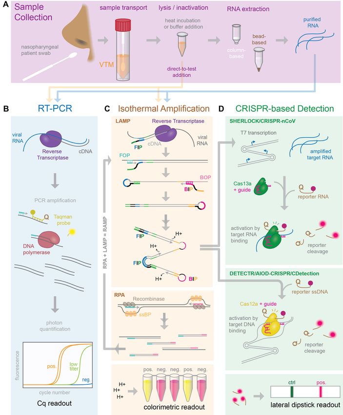
Downloaded from rnajournal.cshlp.org on September 17, 2020 - Published by Cold Spring Harbor Laboratory Press

Figure 1. An overview of COVID-19 nucleic acid testing. Samples collected via nasopharyngeal swab are lysed
and inactivated, and an amplification reaction is performed using either crude swab sample or purified RNA.
Amplification of specific viral sequences by RT-PCR, LAMP, or RPA is detected using fluorescent or colorimetric
dyes, sequence-specific CRISPR-Cas nuclease cleavage of a reporter, or separation of reaction products on a
lateral flow dipstick.

 2
Downloaded from rnajournal.cshlp.org on September 17, 2020 - Published by Cold Spring Harbor Laboratory Press

Overview
 On March 11th, 2020 the World Health Organization deemed COVID-19 a global pandemic
(World Health Organization 2020). As of April 26th, SARS-CoV-2 infections have been confirmed in
almost 3 million people worldwide, yet even this staggering figure is likely to be an underestimate
(Elflein). To have any actionable impact on our control of the pandemic propagation, tests should be
performed repetitively on a large fraction of the population in order to detect outbakes before they
spread. Current estimates of the testing capacity needed to end the pandemic are in the range of tens of
millions of tests per day in the U.S., far above the ~145,000 tests currently conducted nationally (Irfan
2020; Goodnough et al. 2020). A solution to massively scaling up COVID-19 testing by orders of
magnitudes may be aided by an innovative combination of the molecular tools presented here. Current
testing approaches fall into two categories - nucleic-acid or serological. Nucleic-acid tests directly probe
for the RNA of viruses swabbed from a patient’s throat or nasal passage (Figure 1), while serological
tests detect antibodies present in the patient’s serum. During the first days of infection, patient viral
titers are high and a single patient nasopharyngeal swab may harbor close to 1 million SARS-COV-2 viral
particles (Wölfel et al. 2020). However, patient IgG and IgM antibody production, termed
seroconversion, typically occurs 5-10 days after the onset of initial symptoms (Wölfel et al. 2020).
Therefore, nucleic acid tests offer the earliest and most sensitive detection for the presence of SARS-
COV-2 and will be the subject of this review. The RT-PCR test pioneered by the CDC has been deemed
the “gold standard” for clinical diagnosis but takes hours to perform and requires specialized reagents,
equipment, and training (Centers for Disease Control and Prevention 2020). In the first few weeks of the
global SARS-CoV-2 pandemic, required reagents have already been in short supply and researchers and
testing centers have reported issues acquiring almost every necessary reagent from commercial
suppliers - from patient nasopharyngeal swabs to lysis buffer to RNA extraction kits (Baird 2020; Akst
2020). Some testing centers have even been running multiple testing protocols side-by-side to increase
throughput and allow for decreased reliance on a single reagent (Soucheray 2020). A few commercial
test systems exist, but are primarily designed to give single-patient results (Cepheid 2020; Abbott 2020).
A scalable, high-throughput platform will be required to deliver millions of tests per day. Here we
investigate recent advances and approaches to nucleic-acid testing for COVID-19. We highlight some
findings from research groups who have compared commercial reagents or created homemade
solutions in order to decrease cost or reliance on particular commercial reagents. We also outline
several alternative nucleic-acid tests involving isothermal amplification or CRISPR-based detection.
Finally, we examine some recent applications of specialized techniques such as sequencing, digital PCR
and DNA nanoswitches as tools for COVID-19 detection. We have tried to be as exhaustive as possible
throughout this review, but due to the rapid daily developments in testing we may have unwittingly

 Figure 2. An overview of sample processing. Patient nasopharyngeal swabs are collected and transported for
 testing. Viral particles are inactivated and lysed by heat and/or lysis buffer addition. Swab sample is then added
 directly to amplification reactions or RNA is purified from the sample and then amplified.

 3
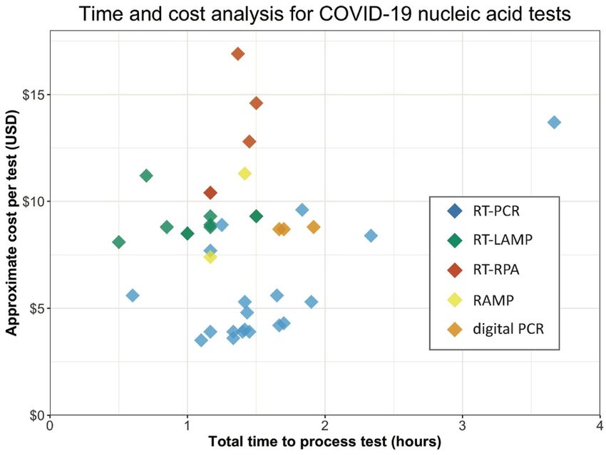
Downloaded from rnajournal.cshlp.org on September 17, 2020 - Published by Cold Spring Harbor Laboratory Press

 excluded some published
 works. Another review by Shen
 et al. published in late
 February may be useful to
 readers (Shen et al. 2020). In
 this review, we greatly expand
 the scope to evaluate and
 compare many more recently
 published articles, address
 advancements in sample lysis,
 direct addition, and novel
 detection methods, and
 include a quantitative
 comparison of these methods
 including workflow time, cost,
 and limit of detection.
 The general workflow
 for RT-PCR tests, such as that
 approved by the CDC, includes
 Figure 3. An analysis of the total workflow time and calculated cost (in U.S.
 3 main steps: sample
 dollars) of published COVID-19 nucleic acid tests. Calculated costs are
 collection and transport, lysis
 estimated from available online pricing for consumables and do not
 include labor or equipment. Protocols which required key reagents to be and RNA purification, and
 synthesized or created in a laboratory are not included but are likely to be amplification (Figure 2).
 even cheaper than commercially priced reagents. All raw data available in Typically, a clinician collects a
 Table S1 and S2. nasopharyngeal swab and
 transfers it to a vial containing
a few milliliters of viral transport medium (VTM), which is transported to a laboratory for testing.
Chemical lysis buffers or heating may be used to lyse and inactivate viral particles. The viral RNA is then
purified from a fraction of the swab sample (typically 1/20th of the swab) using column-based RNA
purification kits or magnetic beads. The eluted purified RNA is then amplified using a 1-step master mix
containing reverse transcriptase and DNA polymerase enzymes with 3 primers targeting specific regions
of the viral genome. Primers targeting a human gene, such as RNaseP are also included as a positive
control for swab collection, RNA extraction and amplification. A spike-in control RNA, such as MS2
bacteriophage genomic RNA, may alternatively be used. Amplified products can be detected using
TaqMan probe fluorescence or DNA-intercalating dyes and a threshold cycle of amplification is set to
distinguish positive from negative results. A test result is typically considered positive if amplification is
observed for two or more viral targets, while it is considered negative if amplification is observed for the
control RNA but for none of the viral targets (Centers for Disease Control and Prevention 2020).
 The standard CDC RT-PCR test takes about 3 hours to perform and costs ~$10 per test (Table
S1). Specialized reagents or equipment can lead to high per-test costs and may limit the number of tests
that can be conducted, in some cases resulting in a lag of several days before a patient receives a
diagnosis. The variety of approaches presented here span a wide range of costs and processing times,
with several published protocols reaching results in less than 1 hour (Figure 3). Some investigators have
found homemade solutions that drastically decrease the required reagent cost allowing for tests to be
performed for just a few dollars (Table S2). Others have proposed completely novel solutions that can
cut the testing time to tens of minutes but may still require costly reagents to perform. While
widespread testing will necessarily require high-throughput approaches, other tests may offer higher
sensitivity for low titer cases or rapid turn-around for point-of-care diagnosis. Recent ingenuity in

 4
Downloaded from rnajournal.cshlp.org on September 17, 2020 - Published by Cold Spring Harbor Laboratory Press

COVID-19 nucleic-acid testing offers a wide range of solutions and further innovation may be required to
maximize testing accuracy while providing a low-cost and fast-turnaround solution.

Sample lysis and direct addition
 Testing for the presence of SARS-CoV-2 viral RNA typically begins with the collection of a patient
swab sample which is stored and transported to a testing facility in Viral Transport Medium (VTM).
These samples are lysed and viral RNA is typically purified using either RNA extraction columns or
magnetic beads (Figure 2) (Centers for Disease Control and Prevention 2020). One advantage of RNA
purification, is that the viral RNA present in the more dilute swab sample can be concentrated and
eluted in a buffer compatible with RT-PCR. However, in order to decrease reliance on commercial lysis
buffers and viral RNA extraction kits and simplify COVID-19 testing, there has been great interest in
finding alternative strategies or eliminating RNA purification altogether by adding patient swab samples
directly to the RT-PCR reaction. Additionally, eliminating RNA purification can dramatically speed up the
overall workflow time per test and may be an ideal solution for streamlining testing times (Figure 4).

 RT-PCR

 RT-LAMP

 RT-RPA

 RAMP
 RT-RAA

 digital PCR

 * sequencing
 0 2 3

 Figure 4. Examination of the total workflow for published COVID-19 testing methods. Each step of the
 workflow is shown with colored bars. Four example commercial RT-PCR kits are included for reference (blue)
 and were directly compared within a single publication. The CDC RT-PCR test is shown in red. *Sequencing
 typically takes 4-12 hours but can vary significantly depending on library preparation and the platform used
 and was not specifically stated in the cited protocols. Raw data available in Table S1.

 5

Downloaded from rnajournal.cshlp.org on September 17, 2020 - Published by Cold Spring Harbor Laboratory Press

 Swab samples must be lysed to release viral RNA into solution for purification and to neutralize
the virus for safe handling. Many protocols use commercial reagents for lysis, including DNA/RNA Shield
(Zymo Research), Buffer RLT (Qiagen), and MagNA Pure External Lysis Buffer (Roche). However, multiple
researchers have recently found that when compared to commercial solutions, homemade solutions
containing 4M (Scallan et al. 2020; Sridhar et al. 2020) or 5M (Aitken et al. 2020) guanidinium
thiocyanate work equally well to lyse samples and recover viral RNA after purification. However, these
solutions contain strong denaturants and are therefore not compatible with addition directly into
amplification reactions. Other laboratories have assessed lysis conditions that are compatible with direct
addition in order to streamline sample preparation and reduce overall test time. Preliminary studies
report that direct-to-test addition of unprocessed swab samples generally allows for SARS-CoV-2
detection but may decrease test sensitivity. Viral RNA stability and compatibility with downstream
reactions will be heavily dependent on the buffer used for swab collection and transport. Arumugam
and Wong have shown that RNA can be detected from non-replicative recombinant virus particles
(SeraCare AccuPlex) in VTM spiked directly into the RT-PCR master mix without an RNA extraction step
(Arumugam and Wong 2020). Merindol et al. compared a few common swab collection buffers for
compatibility with direct PCR addition. Swab samples stored in Hank’s medium or saline solution and
directly added to RT-PCR reactions amplified poorly using either the RealStar SARS-CoV-2 RT-PCR kit
(Altona Diagnostics) or the Allplex 2019-nCoV RT-QPCR kit (Seegene) compared to purified RNA from the
same swabs. Interestingly, however, viral RNA added directly from swabs stored in water or UTM
(Remel) at 4°C showed equivalent RT-PCR amplification to RNA purified from the same swabs. In the
presence of RNase inhibitor, viral RNA could be amplified with similar efficiency from swabs stored in
water at 4°C for up to 5 days (Merindol et al.).
 Many groups are further optimizing direct-to-test addition by heating and/or lysing swab
samples prior to testing. In one study, direct addition of swab samples in viral transport media to the
Luna Universal Probe One Step RT qPCR master mix (New England Biolabs) accurately identified 92% of
155 COVID-19 cases but reached the detection threshold four cycles later (corresponding to a 16-fold
loss in detection of starting material) than a test using RNA extracted from a swab sample using the
QIAamp Viral RNA Mini kit (Qiagen) (Bruce et al. 2020). This procedure could correctly identify cases
across low, medium or high SARS-CoV-2 RNA copy loads (as defined by cycles to detection from tests of
the same samples after RNA purification). Heating the swab sample at 95°C for 10 minutes before
direct-to-test addition improved detection of low copy load samples (Bruce et al. 2020). Another group
reported that directly added samples were detected 3.5 cycles later than RNA isolated using the MagNA
Pure kit (Roche), but heating the sample at 95°C for 5 minutes before direct-to-test addition resulted in
detection only one cycle later, with 97.4% accuracy compared to tests using purified RNA (Fomsgaard
and Rosenstierne 2020). However, Grant et al. found the opposite - heating direct-to-test samples in
VTM at 95°C for 10 minutes delayed detection of viral RNA compared to directly added samples not
heated prior to amplification (Grant et al. 2020). Additionally, they found that direct sample addition in
VTM without heating to the TaqMan Fast Virus 1-step Master Mix (Thermo Fisher) RT-qPCR reaction
allowed detection 3.77 cycles earlier than the same test performed with RNA purified using the EZ1
Qiagen kit. Overall, their test using direct, unheated sample had 98.8% diagnostic accuracy when
compared to cartridge-based RNA purification and RT-qPCR using the Panther Fusion system (Grant et
al. 2020). Intermediate inactivation temperatures seem to perform worse than high heat or no heating
at all. One group reported that swab sample heat treatment at 75°C for 10 minutes prior to direct-to-
test addition delayed detection by 6.1 cycles (Alcoba-Florez et al. 2020). Multiple groups have reported
contradictory findings on the advantages of heating samples before direct addition into RT-PCR mixes.
RNases present in the nasal swab are likely active even at high temperatures and thus RNA degradation
may be particularly sensitive to the temperature and buffer conditions of inactivation.

 6
Downloaded from rnajournal.cshlp.org on September 17, 2020 - Published by Cold Spring Harbor Laboratory Press

 Slightly more complex approaches employ lysis buffers to aid RNA recovery and improve RT-
qPCR test sensitivity. In one report, positive patient swab samples diluted 1:1 into Quick Extract DNA
extraction solution (a buffer containing detergents and proteinase K), heat inactivated and directly
added to the RT-PCR reaction mix were detected at the same amplification cycle as, or even slightly
before, samples processed with the QIAmp Viral RNA Miniprep kit (Qiagen) (Ladha et al.). Another group
reported that swab samples added to Quick Extract DNA extraction solution were detected with equal
sensitivity to column-purified RNA (Sentmanat et al. 2020). Finally, another study reported that direct
addition of swab samples treated 15 minutes with proteinase K yielded sensitivity comparable to the use
of RNA isolated with the automated ELITe InGenius Sp200 system (ELITech Group) (Marzinotto et al.).
 The discrepancy between the sensitivities of direct-to-test addition procedures may be due to
differences in protocols and kits used for the RT-qPCR test and isolation of control RNA, types of lysis
buffers, heating parameters, and varying viral RNA loads in the swab samples of each study. Despite
these discrepancies, it appears that direct-to-test addition of a small volume of swab sample treated
with lysis buffer or Proteinase K allows for robust SARS-CoV-2 RNA detection. Direct-to-test addition of
patient swab samples may prove useful in settings where there is a lack of RNA purification reagents or
time constraints that render laborious RNA isolations infeasible. Further work is required to ascertain
optimal swab sample lysis, heating and storage conditions prior to direct-to test addition, as well as
whether direct-to-test addition could be employed in tests other than RT-qPCR.

RNA purification
 As uncovered by multiple groups, eliminating RNA isolation prior to RT-PCR altogether may be
possible. However, a dedicated RNA isolation step may improve detection sensitivity or be necessary to
remove incompatible sample buffers prior to amplification for some protocols. However, column-based
kits used to purify RNA from the patient swab sample can also occasionally lead to unintentional carry-
over of ethanol or retention of some RNA, which can be kit-specific. In our laboratory, we have found
that the RNeasy Mini Kit (Qiagen) leads to a ~8-fold (3 Ct) loss of synthesized SARS-CoV-2 viral N gene
RNA after column purification. We found similar results with inactivated positive patient swab samples;
Ct values were consistently lower when RNA was purified via isopropanol precipitation or using the
Direct-zol RNA Miniprep Plus kit (Zymo Research) when compared to the RNeasy Mini Kit (Qiagen).
However, we have not compared directly with the CDC recommended Qiagen QIAmp Viral RNA kit (C
Dugast-Darzacq, T Graham, G M Dailey et al., unpublished). Several recent papers have investigated
alternative methods for RNA purification, including unique approaches as well as traditional laboratory
techniques. Zhao et al. present a synthesis protocol for magnetic nanoparticles that can combine sample
lysis and RNA binding in a single step. The poly amino ester with carboxyl groups-coated magnetic
nanoparticles (pcMNPs) are also directly compatible with the RT-PCR reaction, greatly streamlining the
protocol from lysis through RNA purification and the pcMNPs can be synthesized on-site (Zhao et al.
2020). Kalikir et al. find that AmpureXP beads (Beckman Coulter) yield equal sensitivity to the NucliSENS
easyMAG automated extraction platform (bioMérieux) (Kalikiri et al. 2020). Other commonly used
laboratory reagents for RNA purification include TRIzol which includes guandinium thiocyanate and
phenol-chloroform to extract RNA from cellular samples. Won et al. describe a complete workflow for
COVID-19 testing which includes TRIzol extraction and isopropanol precipitation of the RNA from swab
samples. The authors found no difference between TRIzol and the approved Qiagen QIAamp Viral RNA
Mini Kit in RNA extraction efficiency from Lentivirus-infected HEK293 cells, but was not directly
compared using SARS-CoV-2 RNA (Won et al. 2020). In our laboratory, isopropanol precipitation of
synthesized SARS-CoV-2 viral N gene RNA resulted in almost no loss of RNA, with or without the
presence of additional human RNA as a carrier (C Dugast-Darzacq, T Graham, G M Dailey et al.,
unpublished). Standard laboratory RNA purification methods offer an attractive alternative to

 7
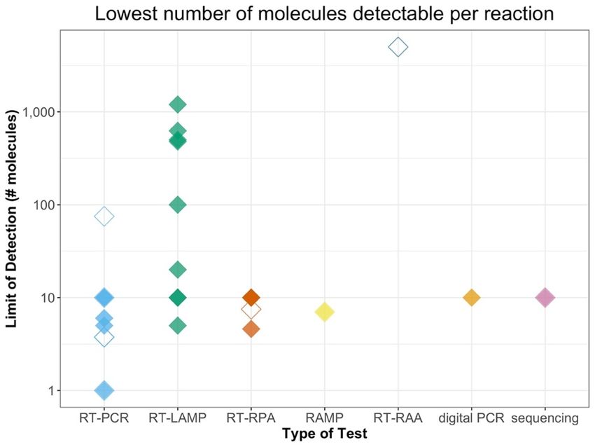
Downloaded from rnajournal.cshlp.org on September 17, 2020 - Published by Cold Spring Harbor Laboratory Press

 commercial kits, as they generally employ inexpensive, abundant
 materials. For clinical testing, however, these solutions may be
 difficult to scale to high-throughput pipelines and may require
 special handling of hazardous materials. It may be useful to
 assess where RNA extraction can be eliminated while
 maintaining the necessary sensitivity and accuracy of testing. If
 eliminating RNA purification is not possible, however, these
 procedures could be useful as cheap, home-made solutions for
 small-scale testing operations.

 RT-PCR
 RT-PCR master mixes use a mixture of reverse transcriptase
 enzymes such as the thermostable MMLV RT and a DNA
 polymerase, like Taq. Primers that anneal specifically to the
 SARS-COV-2 viral genome are included to prime amplification.
 The U.S. CDC protocol utilizes primers that target the viral N
 gene, while the China CDC uses primers matching both the N
 gene and the ORF1ab region and Charité Germany primers target
 the RdRp and E genes (Reviewed in Udugama et al. 2020). For
 detection of amplification, qPCR can be performed using
 intercalating dyes like SybrGreen. Because these dyes are non-
 specific for DNA products, any amplification (specific or not) will
 lead to an increased fluorescent readout. Higher sequence
 specificity in the detection of amplicons can be achieved using
 Taqman probes. These short oligonucleotides contain a 5’
 fluorophore and 3’ quencher and anneal to sequences within the
 DNA template. Taq polymerase degrades the annealed probe by
 its 5’ to 3’ exonuclease activity and cleaves off the fluorophore
 thereby releasing it from being quenched. This fluorescence is
 proportional to the number of amplified product molecules, is
 sequence-specific for the correct amplified product, and can be
 measured in real-time on a qPCR machine (Figure 5).
 RT-PCR has been deemed the “gold standard” for COVID-
 19 diagnosis because it has shown to be very sensitive for
 accurately detecting viral genomes present, down to just one
 molecule of RNA (Figure 6). Multiple commercial master mixes
 exist that enable sensitive 1-step RT-PCR. The original CDC
 protocol approved four commercial master mixes for the RT-PCR
 test from Quantabio, Promega, and ThermoFisher (Centers for
 Disease Control and Prevention 2020). However, published RT-
 PCR protocols have also successfully employed 1-step RT-PCR
 master mixes from a variety of companies including NEB, Applied
 Biosciences, Qiagen, Roche, Takara, and others and a growing list
 of approved alternative commercial reagents can be found at the
Figure 5. Molecular overview of the RT-PCR reaction. Taqman probes are used to visualize increased
fluorescence during each cycle of amplification. Amplification is quantified by Cq readout and a threshold is set
for positive detection of the target amplicon.

 8
Downloaded from rnajournal.cshlp.org on September 17, 2020 - Published by Cold Spring Harbor Laboratory Press

FDA EUA website (Chandler-
Brown et al. 2020; Kalikiri et
al. 2020; Zhao et al. 2020;
Won et al. 2020; Merindol et
al.; Alcoba-Florez et al. 2020;
Marzinotto et al.; Xu et al.
2020; Food and Drug
Administration). Many
commercial master mixes
seem to function well in the
detection of SARS-CoV-2,
although a detailed side-by-
side comparison of the
numerous commercial
reagents is lacking. Brown et
al. compared 4 popular 1-
step RT-PCR kits (Takara One
Step PrimeScript III kit,
Qiagen Quantifast Multiplex
 Figure 6. The limit of detection for published tests equivalent to the fewest
RT-PCR +R Master Mix,
 number of molecules accurately assayed in a single reaction. For some spike-
ThermoFisher TaqPath 1-
 in controls, authors used viral DNA, plasmid DNA, or a pseudovirus instead of
step RT-qPCR Master Mix, viral RNA (shown as open diamonds) which may have a different
and the Thermo Fisher amplification efficiency than SARS-CoV-2 RNA and thus alter their calculated
Taqman Fast Virus 1-step limit of detection. Raw data available in Table S1.
Master Mix) on 74 patient
nose and/or throat swabs (Brown et al. 2020). Comparison of the 4 master mixes showed that 3 out of
the 4 mixes performed optimally with the N2 primers for SARS-CoV-2 detection – the Takara, Qiagen,
and TaqPath. The best, however, seemed to be the Takara master mix which was able to detect just a
single viral genome copy using the N1 primers. Consistent with the Takara mix being the most sensitive,
none of their patient samples that tested negative with the Takara mix tested positive with the Qiagen
kit, whereas the reverse did not hold (Brown et al. 2020).
 Additionally, in order to decrease reliance on a particular company to generate master mix
reagents for testing, which in the course of the pandemic could experience supply chain disruptions or
delays, at least one lab has developed a completely home-made, open-source master mix. Bhadra et al.
have developed master mixes using the evolved reverse transcriptase/DNA polymerase RTX that are
compatible both with either dye-based or TaqMan qPCR. The RTX enzyme can be expressed in E. coli
and purified using Ni-NTA agarose and heparin columns and master mix buffers can be made easily and
cheaply in a laboratory. The authors demonstrated detection of as few as 100 molecules of in vitro
transcribed SARS-CoV-2 N gene RNA, using either RTX enzyme alone in a dye-based reaction or a
mixture of RTX and Taq in a TaqMan reaction. TaqMan reactions with RTX and Taq showed Cq values
comparable to the commercial TaqPath kit (Bhadra et al. 2020). Future studies should assess home-
made master mixes using patient samples, to provide inexpensive, open-source options for testing.
While a variety of commercial and laboratory options exist for RT-PCR master mixes, active enzymes
typically require careful refrigeration for storage and transport. Xu et al. have demonstrated that the
Takara RT-PCR mix maintains its activity after being freeze-dried and stored at room temperature for 28
days (Xu et al. 2020). Further innovation in homemade or room-temperature stable reagents may
improve testing capabilities in remote locations or at the point-of-care.

 9
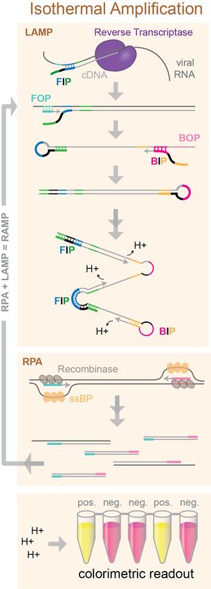
Downloaded from rnajournal.cshlp.org on September 17, 2020 - Published by Cold Spring Harbor Laboratory Press

Isothermal amplification
 A promising alternative to RT-PCR is isothermal amplification, which does not require
thermocycling. Two isothermal techniques used for rapid and
sensitive diagnostics are Loop-mediated Isothermal Amplification
(LAMP) and Recombinase Polymerase Amplification (RPA) (Figure
7). LAMP uses a strand-displacing DNA polymerase together with
four specially designed primers containing regions of
complementarity to six target sequences. The 3’ end of the
forward inner primer (FIP) primes synthesis of an initial DNA
strand, which is subsequently displaced by synthesis primed by the
forward outer primer (FOP). A reverse-complementary sequence in
the 5' end of the FIP anneals with a downstream sequence in the
displaced ssDNA strand, forming a loop. The same process repeats
with the backward inner and outer primers (BIP and BOP) at the
opposite end of the amplicon. Repeated rounds of priming and
strand extension generate a mixture of stem-loop and
“cauliflower” structured products. Because LAMP includes primers
that anneal to six unique target regions, it is highly sequence
specific (Notomi et al. 2000). Release of hydrogen ions upon
incorporation of dNTPs into the nascent DNA chain can be
detected using colorimetric pH indicator dyes (Tanner et al. 2015).
RT-LAMP has been validated for detection of a multitude of RNA
viruses including influenza, Zika, Ebola, and MERS (Reviewed in
Wong et al. 2018).
 A slightly more recent addition to the isothermal
amplification toolkit is RPA. RPA uses a recombinase to catalyze
strand invasion of a primer into dsDNA. Single-stranded binding
proteins are included to stabilize the open duplex structure and a
strand-displacing DNA polymerase extends the primer (Piepenburg
et al. 2006). Some groups have demonstrated very high sensitivity
and specificity of target amplification by combining RPA and LAMP
into a 2-stage amplification protocol, termed RAMP. The outer
LAMP primers can be used for RPA amplification and then
combined with the additional LAMP primers for further
amplification in a single tube or microfluidic device. The combined
RAMP approach exhibits the extremely high specificity of LAMP,
 Figure 7. Molecular overview of
together with enhanced sensitivity from dual amplification, and a isothermal amplification
higher tolerance to inhibitors (Song et al. 2017). Song et al. techniques. LAMP uses specially
demonstrated the huge potential of the RAMP approach for designed nested primers with
diagnostics by multiplexing 16 pathogenic targets including HIV-1 complementary regions that form
and multiple strains of HPV ZIKV (Song et al. 2017). These hairpins to permit priming of
isothermal methods are relatively fast and can be read out subsequent rounds of
colorimetrically, with a lateral-flow stick, or even with amplification. RPA uses
nanoparticle-based biosensors (Zhu et al. 2020) making them easy recombinase-catalyzed strand
to use at home or at remote points of care. invasion to prime amplification.
 Colorimetric pH indicators can be
 Several groups have now developed novel isothermal
 used to detect hydrogen ion
protocols for detection of SARS-CoV-2 RNA. Lu et al. tested their release during dNTP incorporation.

 10
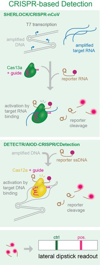
Downloaded from rnajournal.cshlp.org on September 17, 2020 - Published by Cold Spring Harbor Laboratory Press

LAMP-based detection method with spiked-in SARS-CoV-2 RNA and were able to detect a colorimetric
change indicating a positive result after just 40 minutes of amplification, with sensitivity down to 30 viral
RNA copies per reaction (Lu et al. 2020b). They and others have demonstrated that LAMP detection of
SARS-CoV-2 is specific by showing no cross-reactivity to other respiratory pathogens including human
coronavirus strains HCoV-OC43 and HCoV-229E (Lu et al. 2020b; Park et al. 2020). Zhang, et al. have
shown that their LAMP strategy gives results that match the RT-PCR standard test in COVID-19 positive
patient samples, reporting 100% sensitivity and specificity. They also find that the LAMP protocol may
be compatible with cell lysates, potentially eliminating the need for RNA purification from patient
samples (Zhang et al. 2020). Using 130 samples, Yan et al. were able to directly compare RT-PCR with RT-
LAMP. The LAMP assay gave identical clinical diagnoses to the RT-PCR test, with similar sensitivity, and it
was faster and easier to read out (Yan et al. 2020). Others have reported similar success, with LAMP
amplification yielding 90-100% sensitivity and 95-99% specificity in patient samples with improved
accuracy for amplification of multiple gene targets (Mehmood Butt et al.; Yang et al.; Yu et al.; Jiang et
al.). A smaller cohort study found that their RT-LAMP test had a sensitivity of 80%, compared to
consecutive RT-PCR swabs, which could be adequate clinically, they suggest, if repeated testing were
employed (Österdahl et al.). By combining two common isothermal techniques, LAMP and RPA, into a
single-tube RAMP reaction, El-Tholoth and colleagues were able to improve detection 100-fold over RT-
PCR in mimic patient samples, providing an early proof-of-concept for an extremely sensitive method
that can detect down to just a few viral RNA copies, but that to date has not yet been tested on patient
samples (El-Tholoth et al. 2020). From these early demonstrations, under optimized conditions
isothermal amplification techniques can provide equal sensitivity and specificity to the RT-PCR test for
SARS-CoV-2 detection. These methods allow for faster amplification, less specialized equipment, and
easy readout. LAMP methods also benefit from the ability to multiplex targets in a single reaction and
can be combined with other isothermal methods, like RPA in the RAMP technique, to increase test
accuracy even more. These techniques may be particularly useful for rapid, point-of-care diagnoses or
for remote clinical testing without the need for laboratory equipment.

CRISPR-based detection
 A unique group of Cas nucleases, including Cas12 and Cas13 were recently discovered to have
promiscuous DNA or RNA cleavage activities (Gootenberg et al. 2017; Chen et al. 2018; Li et al. 2018),
which have been exploited for nucleic acid detection. Multiple assays combining isothermal
amplification and CRISPR have recently emerged as diagnostic tools for rapid detection of SARS-CoV-2
viral RNA (Figure 8).
 Cas13a is a non-specific RNase that remains inactive until it binds its programmed RNA target. It
has been harnessed for sensitive DNA or RNA detection in a method termed SHERLOCK (Gootenberg et
al. 2017) In SHERLOCK, the target RNA is first amplified by a combination of RT-RPA and T7 transcription.
The amplified product RNA activates Cas13a, which in turn cleaves a reporter RNA, liberating a
fluorescent dye from a quencher. This method consistently detects synthetic SARS-CoV-2 viral RNA in
the range between 10 and 100 copies per L of input, only requires a lateral flow dipstick for visual
readout of the detection result, and can be completed in 40 minutes (Hou et al. 2020) or 57 minutes
(Zhang et al. 2020; Metsky et al. 2020) after the RNA extraction step. SHERLOCK (termed “CRISPR-nCoV”
in Hou et al. 2020) also demonstrated its diagnostic potential by detecting SARS-CoV-2 RNA with 100%
sensitivity in 52 patient samples (Hou et al. 2020).
 Cas12 is another member of the CRISPR-Cas effector family. It is an RNA-guided DNase that
indiscriminately cleaves ssDNA upon binding its target sequence. In a method termed “DETECTR”,
Cas12a ssDNase activation is combined with isothermal amplification to achieve sensitive and specific
DNA detection (Chen et al. 2018). Multiple groups have recently used DETECTR for SARS-CoV-2 RNA

 11
Downloaded from rnajournal.cshlp.org on September 17, 2020 - Published by Cold Spring Harbor Laboratory Press

 detection. Viral RNA is first converted to DNA and isothermally
 amplified. Specific target sequences in the amplified DNA
 activate Cas12a, which in turn cleaves a ssDNA reporter to
 unquench a fluorophore. Using RT-RPA for amplification in
 DETECTR, Lucia et al. detected 10 copies of SARS-CoV-2 RNA per
 L of input within 60 minutes (after RNA sample preparation)
 (Lucia et al. 2020). Ding et al. improved the protocol by
 combining RT-RPA and CRISPR-based detection in a one-pot
 reaction and incubating at a single temperature. This “All-In-
 One Dual CRISPR-Cas12a” (AIOD-CRISPR) assay detected as few
 as 4.6 copies of SARS-CoV-2 RNA per L of input in 40 minutes
 (Ding et al. 2020). Similarly, Guo et al. developed another
 single-tube and constant temperature protocol (“CDetection”),
 in which they used recombinase-aided amplification (RAA)
 instead of RPA for nucleic acid amplification. They showed that
 Cas12b behaves similarly to Cas12a for ssDNA reporter cleavage
 and can achieve a detection limit of 5 copies/ L in 40-60
 minutes (Guo et al. 2020). Moreover, Broughton et al. used
 LAMP instead of RPA in DETECTR and further reduced the
 testing time for SARS-CoV-2 RNA to 30-32 minutes while
 maintaining a low detection limit (10 copies/ L) (Broughton et
 al. 2020).
 In combination with fast isothermal amplification,
 CRISPR-based techniques can harness highly specific nucleases
 to achieve fast read-outs and sensitivity down to a few viral
 RNA copies. CRISPR detection can be coupled to lateral flow
 readouts, which are an attractive option for easy, at-home
 testing scenarios.

 Sequencing for diagnosis
 Sequencing-based detection methods provide the
 benefit of collecting base-pair-level information of patient
 strains, which allows for viral mutation tracing but comes at the
 cost of expensive sequencing platforms and lengthy sample
 Figure 8. Molecular overview of processing times. However, several labs have investigated high-
 CRISPR detection of amplified throughput approaches or portable, fast sequencing to use this
 products. Binding to specific target technology as a diagnostic tool for COVID-19. Nanopore target
 sequences in amplified RNA or DNA sequencing (NTS) is an attractive option for clinical testing
 activates Cas nucleases, which cleave
 because it is fast, highly portable, and sensitive. Wang et al.
 reporter molecules. Reporter cleavage
 have developed an NTS approach targeting 11 viral regions that
 can then be assayed using a lateral
 dipstick. is able to detect as few as 10 viral copies/mL with 1 hour of
 sequencing. By relying on a sequencing-based approach, this
group also demonstrated that viral genome mutations can be identified within the target regions, and
that an additional panel of targets against common respiratory viruses can be included to detect co-
infection (Wang et al. 2020). Additionally, commercial sequencing approaches have also been adapted
for high-throughput SARS-CoV-2 testing. BillionToOne Inc. seeks to employ the extensive national
infrastructure for Sanger sequencing, which they propose could “unlock more than 1,000,000 tests per

 12
Downloaded from rnajournal.cshlp.org on September 17, 2020 - Published by Cold Spring Harbor Laboratory Press

day in the US”. (Chandler-Brown et al. 2020) BillionToOne uses a 1-step RT-PCR mix to amplify viral RNA
directly from swab samples, which are collected in viral transport medium rather than a custom lysis
buffer. Sanger sequencing then proceeds with inclusion of a synthetic, shortened SARS-CoV-2 sequence
as a spike-in control allowing for careful quantitation of viral abundance down to ~10 genomic
equivalents (Chandler-Brown et al. 2020). While traditional sequencing approaches typically require
substantial cost and specialization, repurposed portable or quantitative sequencing approaches may
offer extremely accurate high-throughput diagnostics during the pandemic.

Other NATs (“thinking outside the box”)
 Beyond the approaches described above, ingenious methods are being developed for
widespread, at-home or point-of-care COVID-19 diagnostics. Most isothermal amplification steps
require incubation at elevated temperatures around 60°C. To facilitate isothermal amplification at
remote testing facilities, González-González et al. developed a 3D-printed water circulator that can act
as a heat block for LAMP amplification and have demonstrated the ability to detect as few as 62 viral
RNA molecules after 1 hour of incubation (González-González et al. 2020). To make RT-PCR more
accessible for remote testing, Wee et al. have demonstrated a rapid, extraction-free PCR protocol that
can detect 6 SARS-CoV-2 RNA copies using a portable thermocycler (Wee et al.).
 While samples collected from patients with symptoms or who have been hospitalized seem to
present relatively high viral titers that are likely to be easier to detect (Wölfel et al. 2020), testing of
asymptomatic patients or testing prior to quarantine release may require extremely sensitive tests.
While specialized reagents and equipment are required, digital and digital-droplet PCR may allow for
even more sensitive testing than RT-PCR. Lu et al. report 96.3% accuracy for testing of clinical samples
using digital PCR and were able to detect virus in 4 patient samples that were deemed negative by RT-
PCR (Lu et al. 2020a). Furthermore, digital droplet methods have been shown to be capable of detecting
down to 0.4 viral RNA copies/µL in patient samples (Suo et al. 2020). Because digital PCR allows for more
careful quantitation of viral RNA copy number over the course of the disease, this highly sensitive test
may also be useful to evaluate treatment progress or assess patient release after quarantine.
 Particularly as testing becomes more widespread, testing of the general population and
asymptomatic individuals may lead to a large number of negative samples and a huge increase in the
demand of testing supplies. In an innovative effort to further conserve resources using existing testing
methodologies, some groups have investigated pooling many patient samples to decrease the number
of tests required for larger populations. Proposed pooling approaches can be adaptive, where samples
are first pooled and tested and positive pools are re-tested individually. This is a relatively simple
solution which decreases overall testing resources used but may introduce several disadvantages,
including longer wait times for results since positive samples must be iteratively tested, and a slight loss
in sensitivity from diluting positive patient samples with negative ones. Multiple groups have modeled
patient pooling and proposed algorithms that optimize positive sample detection and testing efficiency
(Sinnott-Armstrong et al. 2020; Noriega and Samore). Some simple approaches like pooling the rows and
columns of a 96-well plate during testing can increase efficiency 4 to 8-fold for low prevalence
populations (Sinnott-Armstrong et al. 2020). Random pooling has also been shown to be useful for
estimating disease prevalence and transmission within a local area (Hogan et al. 2020). More
complicated pooling assignments and non-adaptive approaches have been proposed and may
significantly increase efficiency and allow for single-iteration pooled testing, but may be more difficult to
implement with common clinical workflows and robotic pipetting (Täufer 2020). The solution to
widespread testing is likely to require an adaptive, multi-pronged approach. While pooling of samples
may not be the most appropriate solution for very sensitive testing, pooling may drastically improve our
ability to screen large populations while conserving limited testing resources.

 13
Downloaded from rnajournal.cshlp.org on September 17, 2020 - Published by Cold Spring Harbor Laboratory Press

 Lastly, it is important to consider that enzyme-based tests are not always feasible in resource-
limited scenarios. One potential alternative lies in the use of DNA “nanoswitch” based tests that have
been developed for Zika virus detection. Based on a DNA origami design, these nanoswitch DNA
oligomers bind viral RNA to undergo a conformational change that can be visualized on an agarose gel.
DNA nanoswitches targeting different species of RNA viruses can also be combined in one test, allowing
for the detection of co-infections. Unfortunately, the Zika nanoswitch test requires ~5.2 x 10⁵ Zika RNA
genomes per test for reliable detection and is thus far less sensitive than RT-PCR (Zhou et al. 2020). Due
to being a gel-based method, the throughput of DNA nanoswitch tests is also severely limited. To avoid
low-throughput gel detection, the Godin group developed a nanopore sensor capable of detecting these
conformational changes and could detect as few as ~500 target molecules (Beamish et al. 2019). Further
innovation in DNA nanoswitch detection of SARS-CoV-2 and developments in generating fluorescent or
colorimetric outputs could significantly improve the test’s throughput and facilitate its use for COVID-19
detection in low-resource areas.

Outlook
 In response to the tremendous global toll of COVID-19, researchers have rapidly mobilized to
investigate solutions for testing, diagnosis, and treatment. Pre-print and published articles from the past
several months describe a variety of options for rapid, affordable, sensitive, and high-throughput nucleic
acid testing, which is currently the most reliable approach for early detection of SARS-CoV-2. To address
the dire need for increased testing, researchers across disciplines have quickly compared widely
available commercial products, proposed repurposing existing reagents and infrastructure, and created
novel laboratory solutions to optimize the COVID-19 testing pipeline. Academic researchers at a few
institutions around the world have published detailed blueprints for establishing local pop-up testing
centers, and others are likely to follow (Consortium and Doudna 2020; Sridhar et al. 2020; Aitken et al.
2020). A combination of testing approaches may be the most efficient way to fill the current gaps in
testing. We are hopeful that the explosion of creative and multi-faceted approaches to COVID-19
nucleic-acid testing will continue to seed solutions as society addresses the COVID-19 pandemic.

 14
Downloaded from rnajournal.cshlp.org on September 17, 2020 - Published by Cold Spring Harbor Laboratory Press

Glossary

RT-PCR – reverse transcription polymerase chain reaction; amplification of RNA in a 1-step reaction
containing a reverse transcriptase enzyme, DNA polymerase, and a specific primer complementary to a
target region.
LAMP – loop-mediated isothermal amplification that uses four primers that recognize six
complementary regions within the target. Amplification occurs from a strand-displacing polymerase and
elongation of primers with self-complimentary regions for hairpins that prime further rounds of
amplification forming large “cauliflower” structures of amplified products.
RPA – rapid amplification; recombinase polymerase amplification uses a recombinase-primer complex
which finds matches in the DNA or RNA template and enables strand exchange to form an open
complex. Single-stranded binding proteins stabilize the open duplex a strand-displacing DNA polymerase
amplifies the template.
RAMP – a 2-stage isothermal amplification technique combining a primary RPA reaction using the
outside LAMP primers with a secondary LAMP reaction including self-complimentary internal primers.
Ct or Cq value – in qPCR, the amplification cycle where the fluorescence curve exhibits the greatest
curvature and exceeds the background fluorescence threshold.

Sensitivity (Baratloo et al. 2015) – the ability of a test to detect a true positive =
 !"#$ &'()!)*$
!"#$ &'()!)*$ + ,-.($ /$0-!)*$
 ∗ 100

Specificity (Baratloo et al. 2015) – the ability of a test to detect a true negative =
 !"#$ /$0-!)*$
 ∗ 100
,-.($ &'()!)*$ + !"#$ /$0-!)*$

Accuracy (Baratloo et al. 2015) – the ability of a test to differentiate true positive and negative results
 !"#$ &'()!)*$ + !"#$ /$0-!)*$
correctly from the total tests = !"#$ &'()!)*$+ ,-.($ &'()!)*$ + !"#$ /$0-!)*$+,-.($ /$0-!)*$ ∗ 100

Supplemental Information

Table S1. The approach, accuracy, sensitivity, speed, throughput, and total cost of each published
COVID-19 test that is discussed in the current review. “Time to complete the test” is equal to “Time for
RNA preparation” + “Time for amplification and detection” and is used in plots of Figure 3 and Figure 4.
“Approximate consumable cost per test (USD)” is calculated in Table S2 and is plotted in Figure 3. For
CRISPR methods that list costs for both lateral flow and fluorescence detection, only the fluorescence
cost is plotted to more closely compare with other CRISPR protocols that use fluorescent readout.
“Standardized LoD (minimum detectable viral RNA copies)” is plotted in Figure 6.

Table S2. Costs, amounts, and vendors of required reagents for representative COVID-19 tests. Costs
include consumables but do not include labor or purchase cost of machinery required. Prices are quoted
directly from publicly available vendor websites, current as of April 26, 2020.

 15
Downloaded from rnajournal.cshlp.org on September 17, 2020 - Published by Cold Spring Harbor Laboratory Press

Acknowledgments
We thank all of the members of the Tjian+Darzacq lab for their reading, insights and helpful discussions
on this review. We would expressly like to thank the Tjian+Darzacq COVID-19 response team for their
unpublished findings relevant to COVID-19 testing presented here.

Funding Information
The authors gratefully acknowledge funding from the Howard Hughes Medical Institute (Grant CC34430
to R.T. and M.N.E.) and the National Institutes of Health (NIH training program Grant T32GM098218 to
M.N.E. and NIH training program Grant T32GM007232 to O.N.W.).

Author Information

Corresponding Author
*
 Email: jmlim@berkeley.edu

Contributions
M.N.E. developed the concept; M.N.E., O.N.W., and S.C. contributed to the design and writing of the
review and A.M. prepared the figures. All authors contributed to the reading, collection, and analysis of
sources for this review.

Ethics declaration
The authors declare no competing interests.

References

Abbott. 2020. ID NOWTM COVID-19 . ID NOWTM COVID-19 Prod Inser.
 https://www.alere.com/en/home/product-details/id-now-covid-19.html (Accessed April 23, 2020).
Aitken J, Ambrose K, Barrell S, Beale R, Bineva-Todd G, Biswas D, Byrne R, Caidan S, Cherepanov P,
 Churchward L, et al. 2020. Scalable and Resilient SARS-CoV2 testing in an Academic Centre.
 medRxiv 2020.04.19.20071373.
Akst J. 2020. RNA Extraction Kits for COVID-19 Tests Are in Short Supply in US | The Scientist Magazine®.
 Sci. https://www.the-scientist.com/news-opinion/rna-extraction-kits-for-covid-19-tests-are-in-
 short-supply-in-us-67250 (Accessed April 23, 2020).
Alcoba-Florez J, Gonzalez-Montelongo R, Inigo-Campos A, Artola DG-M de, Gil-Campesino H, Team
 TMTS, Ciuffreda L, Valenzuela-Fernandez A, Flores C. 2020. Fast SARS-CoV-2 detection by RT-qPCR
 in preheated nasopharyngeal swab samples. medRxiv 2020.04.08.20058495.
Arumugam A, Wong SS. 2020. The Potential Use of Unprocessed Sample for RT-qPCR Detection of
 COVID-19 without an RNA Extraction Step. bioRxiv.
Baird R. 2020. Why Widespread Coronavirus Testing Isn’t Coming Anytime Soon | The New Yorker. The
 New Yorker, March 24 https://www.newyorker.com/news/news-desk/why-widespread-
 coronavirus-testing-isnt-coming-anytime-soon (Accessed April 23, 2020).
Baratloo A, Hosseini M, Negida A, El Ashal G. 2015. Part 1: Simple Definition and Calculation of Accuracy,
 Sensitivity and Specificity. Emerg (Tehran, Iran) 3: 48–9.

 16
Downloaded from rnajournal.cshlp.org on September 17, 2020 - Published by Cold Spring Harbor Laboratory Press

 http://www.ncbi.nlm.nih.gov/pubmed/26495380 (Accessed April 23, 2020).
Beamish E, Tabard-Cossa V, Godin M. 2019. Programmable DNA Nanoswitch Sensing with Solid-State
 Nanopores. ACS Sensors 4: 2458–2464.
Bhadra S, Maranhao AC, Ellington AD. 2020. A one-enzyme RT-qPCR assay for SARS-CoV-2, and
 procedures for reagent production. bioRxiv 2020.03.29.013342.
Broughton JP, Deng X, Yu G, Fasching CL, Servellita V, Singh J, Miao X, Streithorst JA, Granados A,
 Sotomayor-Gonzalez A, et al. 2020. CRISPR-Cas12-based detection of SARS-CoV-2. Nat Biotechnol
 1–5. http://www.ncbi.nlm.nih.gov/pubmed/32300245 (Accessed April 23, 2020).
Brown JR, O’sullivan D, Pereira RP, Whale AS, Busby E, Huggett J, Harris K. 2020. Comparison of SARS-
 CoV2 N gene real-time RT-PCR targets and commercially available mastermixes. bioRxiv.
 https://doi.org/10.1101/2020.04.17.047118 (Accessed April 23, 2020).
Bruce EA, Tighe S, Hoffman JJ, Laaguiby P, Gerrard DL, Diehl SA, Leonard DGB, Huston CD, Kirkpatrick BD,
 Crothers JW, et al. 2020. RT-qPCR DETECTION OF SARS-CoV-2 RNA FROM PATIENT
 NASOPHARYNGEAL SWAB USING QIAGEN RNEASY KITS OR DIRECTLY VIA OMISSION OF AN RNA
 EXTRACTION STEP. bioRxiv.
Centers for Disease Control and Prevention. 2020. CDC 2019-Novel Coronavirus (2019-nCoV) Real-Time
 RT-PCR Diagnostic Panel For Emergency Use Only Instructions for Use.
Cepheid. 2020. Xpert ® Xpress SARS-CoV-2 Instructions for Use.
Chandler-Brown D, Bueno AM, Atay O, STsao D. 2020. A Highly Scalable and Rapidly Deployable RNA
 Extraction-Free COVID-19 Assay by Quantitative Sanger Sequencing. bioRxiv.
 https://doi.org/10.1101/2020.04.07.029199 (Accessed April 23, 2020).
Chen JS, Ma E, Harrington LB, Da Costa M, Tian X, Palefsky JM, Doudna JA. 2018. CRISPR-Cas12a target
 binding unleashes indiscriminate single-stranded DNase activity. Science (80- ) 360: 436–439.
Consortium IGIS-C-2 T, Doudna JA. 2020. Blueprint for a Pop-up SARS-CoV-2 Testing Lab. medRxiv
 2020.04.11.20061424.
Ding X, Yin K, Li Z, Liu C. 2020. All-in-One Dual CRISPR-Cas12a (AIOD-CRISPR) Assay: A Case for Rapid,
 Ultrasensitive and Visual Detection of Novel Coronavirus SARS-CoV-2 and HIV virus. bioRxiv
 2020.03.19.998724.
El-Tholoth M, Bau HH, Song J. 2020. A Single and Two-Stage, Closed-Tube, Molecular Test for the 2019
 Novel Coronavirus (COVID-19) at Home, Clinic, and Points of Entry. ChemRxiv.
Elflein J. • Coronavirus cases worldwide 2020 | Statista.
 https://www.statista.com/statistics/1043366/novel-coronavirus-2019ncov-cases-worldwide-by-
 country/ (Accessed April 26, 2020).
Fomsgaard AS, Rosenstierne MW. 2020. An alternative workflow for molecular detection of SARS-CoV-2
 - escape from the NA extraction kit-shortage. medRxiv.
Food and Drug Administration. Emergency Use Authorizations | FDA. https://www.fda.gov/medical-
 devices/emergency-situations-medical-devices/emergency-use-authorizations#covid19ivd
 (Accessed April 23, 2020).
González-González E, Montserrat Lara-Mayorga I, García-Rubio A, Ezio Garciaméndez-Mijares C, Emilio-
 Guerra-Alvarez G, García- G, Aguayo J, Shrike Zhang Y, Omar Martínez-Chapa S, Santiago T, et al.
 2020. Scaling diagnostics in times of COVID-19: Rapid prototyping of 3D-1 printed water circulators
 for Loop-mediated Isothermal Amplification 2 (LAMP) and detection of SARS-CoV-2 virus 3 4.
 medRxiv. https://doi.org/10.1101/2020.04.09.20058651 (Accessed April 23, 2020).
Goodnough A, Thomas K, Kaplan S. 2020. Coronavirus Testing Falls Woefully Short as Trump Seeks to
 Reopen U.S. - The New York Times. New York Times.
 https://www.nytimes.com/2020/04/15/us/coronavirus-testing-trump.html (Accessed April 23,
 2020).
Gootenberg JS, Abudayyeh OO, Lee JW, Essletzbichler P, Dy AJ, Joung J, Verdine V, Donghia N, Daringer

 17
Downloaded from rnajournal.cshlp.org on September 17, 2020 - Published by Cold Spring Harbor Laboratory Press

 NM, Freije CA, et al. 2017. Nucleic acid detection with CRISPR-Cas13a/C2c2. Science (80- ) 356:
 438–442.
Grant PR, Turner MA, Shin GY, Nastouli E, Levett LJ. 2020. Extraction-free COVID-19 (SARS-CoV-2)
 diagnosis by RT-PCR to increase capacity for national testing programmes during a pandemic.
 bioRxiv.
Guo L, Sun X, Wang X, Liang C, Jiang H, Gao Q, Dai M, Qu B, Fang S, Mao Y, et al. 2020. SARS-CoV-2
 detection with CRISPR diagnostics. bioRxiv. https://doi.org/10.1101/2020.04.10.023358 (Accessed
 April 23, 2020).
Hogan CA, Sahoo MK, Pinsky BA. 2020. Sample Pooling as a Strategy to Detect Community Transmission
 of SARS-CoV-2. JAMA. https://jamanetwork.com/journals/jama/fullarticle/2764364 (Accessed April
 23, 2020).
Hou T, Zeng W, Yang M, Chen W, Ren L, Ai J, Wu J, Liao Y, Gou X, Li Y, et al. 2020. Development and
 Evaluation of A CRISPR-based Diagnostic For 2019-novel Coronavirus. medRxiv
 2020.02.22.20025460.
Irfan U. 2020. Coronavirus tests: Why the US needs millions of tests per day - Vox. Vox.
 https://www.vox.com/2020/4/13/21215133/coronavirus-testing-covid-19-tests-screening
 (Accessed April 23, 2020).
Jiang M, Pan W, Aratehfar A, Fang W, Ling L, Fang H, Daneshnia F, Yu J, Liao W, 6# HP, et al.
 Development and validation of a rapid, single-step reverse transcriptase loop-mediated isothermal
 amplification (RT-LAMP) system potentially to be used for reliable and high-throughput screening
 of COVID-19. medRxiv. https://doi.org/10.1101/2020.03.15.20036376 (Accessed April 23, 2020).
Kalikiri MKR, Mohammad ;, Hasan R, Faheem Mirza ;, Xaba ; Thabisile, Patrick Tang ;, Lorenz S. 2020.
 High-throughput extraction of SARS-CoV-2 RNA from nasopha-ryngeal swabs using solid-phase
 reverse immobilization beads. medRxiv. https://doi.org/10.1101/2020.04.08.20055731 (Accessed
 April 23, 2020).
Ladha A, Joung J, Abudayyeh OO, Gootenberg JS, Zhang F. A 5-min RNA preparation method for COVID-
 19 detection with RT-qPCR.
 https://static1.squarespace.com/static/5b7c640be2ccd1703a3da4d3/t/5e8bcd0923fb224b2f3b13
 56/1586220297644/Ladha+et+al+-+RNA+QE+Extraction.pdf (Accessed April 23, 2020).
Li SY, Cheng QX, Li XY, Zhang ZL, Gao S, Cao RB, Zhao GP, Wang J, Wang JM. 2018. CRISPR-Cas12a-
 assisted nucleic acid detection. Cell Discov 4: 1–4.
Lu R, Wang J, Li M, Wang Y, Dong J, Cai W. 2020a. SARS-CoV-2 detection using digital PCR for COVID-19
 diagnosis, treatment monitoring and criteria for discharge. medRxiv 2020.03.24.20042689.
Lu R, Wu X, Wan Z, Li Y, Zuo L, Qin J, Jin X, Zhang C. 2020b. Development of a Novel Reverse
 Transcription Loop-Mediated Isothermal Amplification Method for Rapid Detection of SARS-CoV-2.
 Virol Sin.
Lucia C, Federico P-B, Alejandra GC. 2020. An ultrasensitive, rapid, and portable coronavirus SARS-CoV-2
 sequence detection method based on CRISPR-Cas12. bioRxiv 2020.02.29.971127.
Marzinotto S, Mio C, Cifù A, Verardo R, Pipan C, Schneider C, Curcio F. A streamlined approach to rapidly
 detect SARS-CoV-2 infection, avoiding RNA purification. medRxiv.
 https://doi.org/10.1101/2020.04.06.20054114 (Accessed April 23, 2020).
Mehmood Butt A, Siddique S, An X, Tong Y. Development of a dual-gene loop-mediated isothermal
 amplification (LAMP) detection assay for SARS-CoV-2: A preliminary study. medRxiv.
 https://doi.org/10.1101/2020.04.08.20056986 (Accessed April 23, 2020).
Merindol N, Pépin G, Marchand C, Rheault M, Poirier A, Germain H, Danylo A. Optimization of SARS-
 CoV-2 detection by RT-QPCR without RNA extraction. BioRxiv.
 https://doi.org/10.1101/2020.04.06.028902 (Accessed April 23, 2020).
Metsky HC, Freije CA, Kosoko-Thoroddsen T-SF, Sabeti PC, Myhrvold C. 2020. CRISPR-based surveillance

 18
You can also read


