PLASMODIUM FALCIPARUM PARASITE LINES EXPRESSING DC8 AND GROUP A PFEMP1 BIND TO BRAIN, INTESTINAL, AND KIDNEY ENDOTHELIAL CELLS
←
→
Page content transcription
If your browser does not render page correctly, please read the page content below
ORIGINAL RESEARCH
                                                                                                                                               published: 28 January 2022
                                                                                                                                         doi: 10.3389/fcimb.2022.813011

                                             Plasmodium falciparum Parasite
                                             Lines Expressing DC8 and Group A
                                             PfEMP1 Bind to Brain, Intestinal, and
                                             Kidney Endothelial Cells
                                             Luana S. Ortolan 1, Marion Avril 1, Jun Xue 2, Karl B. Seydel 3,4, Ying Zheng 2
                                             and Joseph D. Smith 1,5*
                                             1Center for Global Infectious Disease Research, Seattle Children’s Research Institute, Seattle, WA, United States,
                                             2Department of Bioengineering, University of Washington, Seattle, WA, United States, 3 Department of Osteopathic Medical
                            Edited by:
                    Andrea L. Conroy,        Specialties, College of Osteopathic Medicine, Michigan State University, East Lansing, MI, United States, 4 Blantyre Malaria
      Indiana University, United States      Project, Kamuzu University of Health Sciences, Blantyre, Malawi, 5 Department of Pediatrics, University of Washington,
                                             Seattle, WA, United States
                      Reviewed by:
              Julio Gallego-Delgado,
       Lehman College, United States         Cytoadhesion of Plasmodium falciparum-infected red blood cells is a virulence determinant
                      Gaoqian Feng,
           Burnet Institute, Australia
                                             associated with microvascular obstruction and organ complications. The gastrointestinal
                  *Correspondence:
                                             tract is a major site of sequestration in fatal cerebral malaria cases and kidney complications
                    Joseph D. Smith          are common in severe malaria, but parasite interactions with these microvascular sites are
        joe.smith@seattlechildrens.org
                                             poorly characterized. To study parasite tropism for different microvascular sites, we
                    Specialty section:
                                             investigated binding of parasite lines to primary human microvascular endothelial cells
          This article was submitted to      from intestine (HIMEC) and peritubular kidney (HKMEC) sites. Of the three major host
                      Parasite and Host,     receptors for P. falciparum, CD36 had low or negligible expression; endothelial protein C
                a section of the journal
               Frontiers in Cellular and     receptor (EPCR) had the broadest constitutive expression; and intercellular adhesion
                 Infection Microbiology      molecule 1 (ICAM-1) was weakly expressed on resting cells and was strongly
       Received: 11 November 2021            upregulated by TNF-a on primary endothelial cells from the brain, intestine, and
        Accepted: 10 January 2022
        Published: 28 January 2022
                                             peritubular kidney sites. By studying parasite lines expressing var genes linked to severe
                                 Citation:
                                             malaria, we provide evidence that both the DC8 and Group A EPCR-binding subsets of the
Ortolan LS, Avril M, Xue J, Seydel KB,       P. falciparum erythrocyte membrane protein 1 (PfEMP1) family encodes binding affinity for
         Zheng Y and Smith JD (2022)         brain, intestinal, and peritubular kidney endothelial cells, and that DC8 parasite adhesion
       Plasmodium falciparum Parasite
  Lines Expressing DC8 and Group A           was partially dependent on EPCR. Collectively, these findings raise the possibility of a brain-
      PfEMP1 Bind to Brain, Intestinal,      gut-kidney binding axis contributing to multi-organ complications in severe malaria.
          and Kidney Endothelial Cells.
Front. Cell. Infect. Microbiol. 12:813011.   Keywords: Plasmodium falciparum, malaria, cytoadhesion, endothelial protein C receptor, kidney endothelial cell,
       doi: 10.3389/fcimb.2022.813011        intestinal endothelial cell

Frontiers in Cellular and Infection Microbiology | www.frontiersin.org               1                                       January 2022 | Volume 12 | Article 813011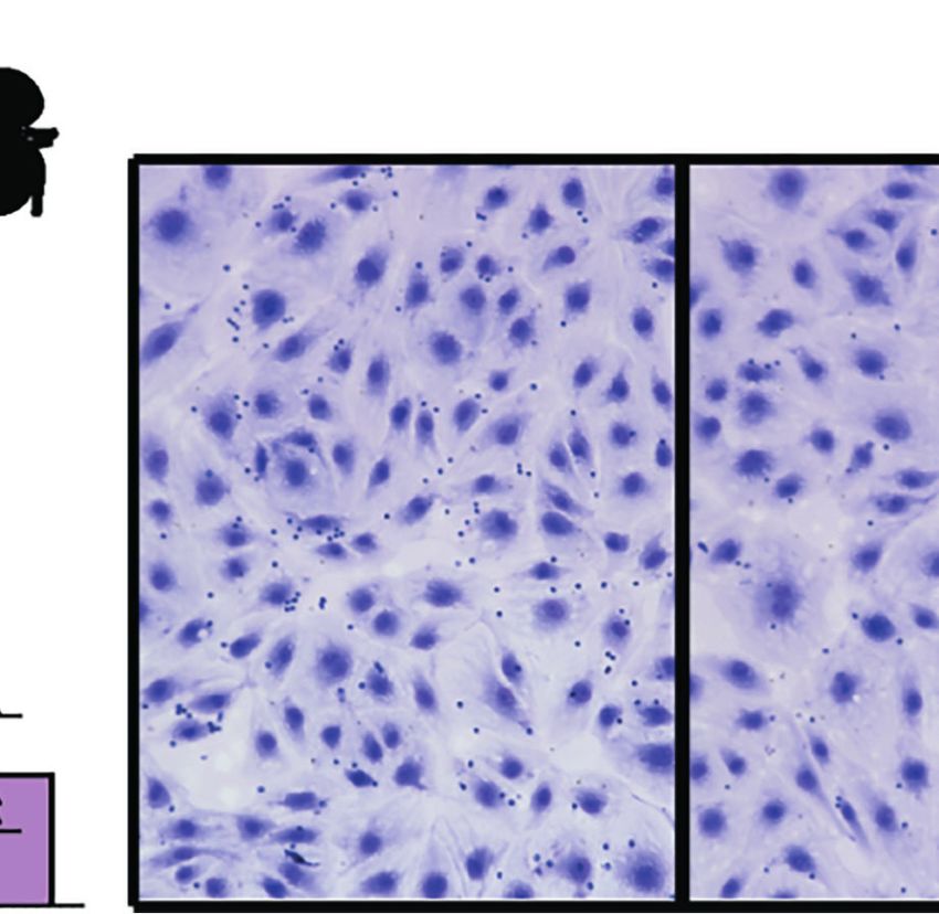
Ortolan et al.                                                                                                  Malaria and Endothelial Cell Interactions

INTRODUCTION                                                                 instance, different subsets of PfEMP1 proteins encode binding
                                                                             activity for CD36 (Robinson et al., 2003; Hsieh et al., 2016),
The distinctive virulence of Plasmodium falciparum is caused in              endothelial protein C receptor (EPCR) (Turner et al., 2013; Lau
part through the cytoadhesion of P. falciparum-infected                      et al., 2015), and intercellular adhesion molecule 1 (ICAM-1)
erythrocytes (IEs) in the microcirculation of different organs.              (Smith et al., 2000; Lennartz et al., 2019). Whereas the EPCR
Extensive sequestration of P. falciparum-IEs obstructs blood flow             binders comprise only a small minority of the var gene repertoire
and can promote localized inflammation and organ dysfunction                  (~10% of genes) (Rask et al., 2010), this subset is transcriptionally
(Miller et al., 2002). The best studied examples are cerebral                elevated in severe malaria infections and both the DC8 and Group
malaria, associated with high sequestered parasite burdens in the            A PfEMP1 variants are linked to severe malaria complications
brain microcirculation (Marchiafava and Bignami, 1892;                       (Lavstsen et al., 2012; Turner et al., 2013; Bernabeu et al., 2016;
MacPherson et al., 1985; Taylor et al., 2004), and placental                 Kessler et al., 2017; Lennartz et al., 2017; Mkumbaye et al., 2017;
malaria, associated with high parasite burdens in the placental              Sahu et al., 2021; Wichers et al., 2021). From in vitro studies,
intervillous blood circulation (Brabin et al., 2004). However,               EPCR-binding variants adhere to human brain endothelial cells
mature forms of P. falciparum-IEs have broad sequestration in                (Turner et al., 2013; Avril et al., 2016; Lennartz et al., 2017;
diverse microvascular beds, including the brain, gastrointestinal            Bernabeu et al., 2019; Storm et al., 2019) and to primary human
tract, subcutaneous adipose tissue of the skin, heart, lung, spleen          heart and lung microvascular endothelial cells (Avril et al., 2013;
and to a lesser extent, the kidney (Spitz, 1946; MacPherson et al.,          Gillrie et al., 2015), suggesting they may have broad affinity for
1985; Milner et al., 2014; Milner et al., 2015), but parasite tropism        diverse microvascular beds.
for most organ sites remains poorly characterized.                               Given that kidney injury and gastrointestinal sequestration
    Whereas the kidney is not a major site of parasite                       are common in severe and fatal malaria cases, we investigated if
sequestration (Spitz, 1946; Milner et al., 2014), kidney injury is           similar parasite binding variants may have affinity for brain,
common in severe malaria. For instance, recent evidence                      intestinal, and kidney microvascular sites. Our analysis
indicates that acute kidney injury (AKI) is a common                         demonstrates that parasites expressing DC8 or Group A
complication in African children with severe malaria and is                  PfEMP1 encode broad binding affinity for primary human
associated with increased morbidity and mortality (Conroy                    brain, intestinal, and kidney endothelial cells and are partially
et al., 2016; Sypniewska et al., 2017; Conroy et al., 2019; Batte            dependent on EPCR for adhesion.
et al., 2021). Moreover, renal impairment occurs in up to 37% of
adult severe malaria cases and increased the risk of death by 4-
fold (Dondorp et al., 2008b). In a large autopsy series, renal
failure was characterized by acute tubular necrosis with                     MATERIALS AND METHODS
accumulation of host monocytes and P. falciparum-IEs in the
kidneys, especially in the peritubular capillary microcirculation            Human Brain, Intestinal, and Peritubular
(Nguansangiam et al., 2007). The gastrointestinal tract has been             Kidney Microvascular Endothelial Cell
considered a relatively non-pathogenic sequestration site.                   Cultures
However, it is a major site of parasite sequestration and there              Primary human brain microvascular endothelial cells (HBMEC)
is increasing evidence that gastrointestinal barrier function may            (Cell System, ACBRI 376) were cultured with endothelial cell
be compromised at higher parasite burdens (Wilairatana et al.,               growth basal medium-2 (EBM-2, Lonza, CC-3156) in
1997). For instance, the gastrointestinal tract was the most                 accordance with manufacturer specifications with 5% fetal
intense site of parasite sequestration in fatal pediatric CM cases           bovine serum (FBS) and supplements provided (Lonza, CC-
(Seydel et al., 2012; Milner et al., 2014) and blocked capillaries in        4147) in tissue culture flasks treated with rat collagen I (Corning,
the rectal mucosa correlate to disease severity and metabolic                354236). Primary human intestinal microvascular endothelial
acidosis in adult SM cases (Dondorp et al., 2008a). Moreover,                cells (HIMEC) (Cell Systems, ACBRI 666) were cultured in
acidic microbial products contribute to metabolic acidosis in                complete classic medium with 10% FBS serum and culture
adult severe malaria (Leopold et al., 2019) and children with                boost (Cell Systems 4Z0-500) supplemented with Bac-off (Cell
severe malaria are at risk for invasive bacterial disease (Scott             Systems) on an extracellular matrix-coated surface (attachment
et al., 2011), suggesting compromise of gut integrity. Thus, while           factor, Cell Systems). HBMEC and HIMEC were certified
kidney and gut pathogenesis are not well understood, autopsy                 positive by the manufacturer for expression of Von Willebrand
studies have led to the hypothesis that organ injury may result              factor, acetylated low-density lipoprotein uptake, and CD31.
from common pathological processes, precipitated by                          HBMEC and HIMEC were used in experiments at passages 5
cytoadherence of P. falciparum-IEs.                                          to 10. Primary human kidney peritubular microvascular
    Cytoadhesion of P. falciparum-IEs is mediated by the P.                  endothelial cells (HKMEC) were isolated and purified from
falciparum erythrocyte membrane protein-1 (PfEMP1) family,                   fetal kidneys as previously reported (Ligresti et al., 2016) and
encoded by a repertoire of about 60 var genes per parasite                   used in experiments at passages 3 to 5. Fetal human kidneys were
genotype (Baruch et al., 1995; Smith et al., 1995; Su et al., 1995).         obtained after informed consent from patients at the University
PfEMP1 proteins encode multiple adhesion domains, called Duffy               of Washington Medical Center in compliance with Institutional
binding-like (DBL) and cysteine-rich interdomain region (CIDR),              Review Board protocol (IRB 447773EA). Cells were cultured and
which confer different binding properties (Smith et al., 2013). For          expanded on matrix-coated surface with 0.2% gelatin (Sigma,

Frontiers in Cellular and Infection Microbiology | www.frontiersin.org   2                                  January 2022 | Volume 12 | Article 813011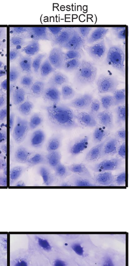
Ortolan et al.                                                                                                 Malaria and Endothelial Cell Interactions

G1890) with EBM2 medium (Lonza, CC-3156) with 10% FBS                        Determination of var Gene Transcripts
and supplemented with 5 mg/ml EGCS (Cell Biologics, 1166), 1%                P. falciparum-IEs at ring stage were collected in TRIZOL LS
antibiotic-antimycotic (Gibco, 15240062), 50 mg/ml of heparin                (ThermoFisher Scientific) and RNA was isolated by chloroform
(Sigma, H3149) and 20 ng/ml of vascular endothelial growth                   and purified with RNeasy Micro Kit (Qiagen), following
factor (VEGF) (R&D Systems, 293-VE-10).                                      manufacturer’s instructions. Contaminating genomic DNA was
                                                                             eliminated with DNase I treatment. cDNA was synthesized by
Characterization of Surface Markers on                                       reverse transcription reaction. For the IT4var19 and HB3var03
Endothelial Cells by Flow Cytometry                                          parasite lines, var transcription was analyzed by real time-PCR
HBMEC, HIMEC and HKMEC monolayers were cultured on                           with SYBR green (Power SYBR Green PCR Master Mix, Thermo
matrix-coated tissue culture flasks until confluence. Cells were               Fisher Scientific) using a set of IT4var primers (Janes et al., 2011)
washed with 1X PBS, lifted with 8 mM EDTA in 1X PBS and                      or HB3var primers (Soerli et al., 2009). Transcription unit levels
then resuspended in 1X PBS (supplemented with 2% FBS). Cell                  were normalized to the housekeeping control gene coding for
suspensions were stained with monoclonal antibodies (mAb) for                STS (seryl-tRNA synthetase). To determine the proportion of
CD31-PE (Biolegend, clone WM59), CD36-FITC (Biolegend,                       3173-S var1 transcripts expressed by the 3173-S parasite line,
clone 5-271), EPCR-APC (Biolegend, clone RCR-401) and                        DBLa tags were PCR-amplified using the varF_dg2 and brlong2
CD54/ICAM-1-PE-Cy-7 (Biolegend, clone HA58) at 1:100                         primers (Lavstsen et al., 2012), cloned into a Zero Blunt TOPO
dilution and with Live/Dead-V450 (Tonbo Biosciences) for 30                  vector, sequenced, and compared to a previous report (Bernabeu
min on ice. For assays with proinflammatory pre-stimulation,                  et al., 2019).
confluent cell monolayers were stimulated with 10 ng/ml TNF-a
(Sigma, T0157) for 20-24 hours at 37°C. Cells were analyzed in a             Binding Assay
LSRII (Becton & Dickson) with 100,000 events/sample. Gates                   For IE binding assays, HBMEC, HIMEC and HKMEC were
were set based on fluorescence minus one (FMO) and IgG                        seeded on collagen coated 8 well slides (BD Biocoat) 3–4 days
isotype controls (Biolegend). Results were expressed relative to             before the assays and allowed to grow to confluency. Mature
IgG isotype controls. Data was analyzed using FlowJo v10                     forms of P. falciparum were purified by magnetic purification
software (TreeStar Inc.).                                                    with LD columns (Miltenyi Biotec) and checked for purity by
                                                                             Giemsa staining. Binding assays and washes were performed
Immunofluorescence                                                            with pre-warmed binding medium (RPMI-1640 medium
HBMEC, HIMEC and HKMEC endothelial cells, were cultured                      containing 0.5% BSA, pH 7.2). For binding assays, 5x10⁶ IE/ml
in an 8 well pretreated chamber slide until confluence. For assays            were resuspended in 200 µl and added to the wells with
with proinflammatory pre-stimulation, confluent cell                           endothelial cells. After 1 hour incubation at 37°C, non-bound
monolayers were stimulated with 10 ng/ml TNF-a (Sigma,                       IE were washed by flipping the slides in a gravity wash for 10
T0157) for 24 hours at 5% CO2 and 37°C. Cells were fixed                      min. For binding quantification, slides were fixed in 1%
with 3.7% paraformaldehyde for 30 min. For antibody labeling,                glutaraldehyde for 30 min at room temperature, then stained
cells were pretreated with background blocking agent                         with 1× Giemsa for 5 min. Binding was quantified in a blinded
(Background Buster, Innovex Biosciences, NB306) for 30 min.                  fashion, by determining the number of IEs adhering per mm2 of
Cells were then washed with 1x PBS and incubated with primary                endothelial cells in six random fields with images taken under
mouse-anti-human-ICAM-1 (1:200 dilution) in PBS                              400× magnification (Keyence BZ-X series microscope). For
(supplemented with 2% BSA) for 1 hour (Biosource                             assays with proinflammatory pre-stimulation, confluent cell
International, ThermoFisher Scientific, clone C14), followed by               monolayers were stimulated with 10 ng/ml TNF-a (Sigma,
the secondary antibody goat anti-mouse Alexa Fluor 488 (1:200                T0157) for 20-24 hours at 37°C prior to the binding assay. For
dilution) (Invitrogen) for 1 hour. Images taken under 400x                   binding inhibition assays, rat anti-human EPCR monoclonal
magnification (Keyence BZ-X series microscope).                               antibody (20 µg/ml, clone RCR-252; Sigma E6280) or rat control
                                                                             IgG (eBiosciences, 20 µg/ml) was added to cells for 30 min of
P. falciparum Culture                                                        incubation at 37°C prior to the addition of IEs.
The P. falciparum parasites lines IT4var19 (Avril et al., 2012),
HB3var03 (Claessens et al., 2012; Avril et al., 2016), and 3173-S            Statistical Analysis
(Bernabeu et al., 2019) were cultured in human red blood cells               Statistical analysis was performed using Prism (version
(O+) and 10% pooled human A+ serum-rich media (RPMI 1640                     8, GraphPad Software Inc.). Flow cytometry data were
medium, GIBCO) and 0.5% Albumax II (Thermo Fischer                           compared using Two-way ANOVA, Sidak’s multiple
Scientific) at 5% hematocrit. The IT4var19 and HB3var03                       comparison test. Binding assays were compared using Two-
parasite lines were grown in a gas mixture of 5% O2, 5% CO2,                 tailed Unpaired T test. Flow cytometry assays shows the mean
and 90% N2 and 3173-S was grown in a gas mixture of 1% O2, 5%                of two technical replicates from three or four different
CO2, and 94% N2. All parasites were routinely synchronized by                experiments. Binding assays with P. falciparum-IEs were
treatment with 5% sorbitol and gelatin flotation to ensure that               performed with two technical replicates and conducted in
IEs maintained their ‘knob-like’ adhesion complexes.                         three to six independent experiments.

Frontiers in Cellular and Infection Microbiology | www.frontiersin.org   3                                 January 2022 | Volume 12 | Article 813011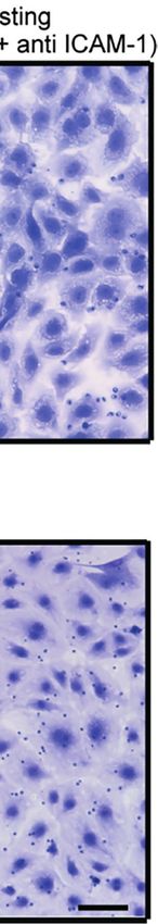
Ortolan et al.                                                                                                                   Malaria and Endothelial Cell Interactions

RESULTS                                                                                expression was substantially increased by TNF-a treatment
                                                                                       reaching nearly 100% surface positive for all three endothelial
Expression of CD36, ICAM-1, and EPCR                                                   cell types (Figure 1 and Supplementary Figure 2). Whereas
on Brain, Intestinal, and Kidney Endothelial                                           resting EPCR surface levels were higher in HBMEC and HIMEC
Cells                                                                                  than HKMEC, constitutive ICAM-1 expression was highest in
To investigate parasite tropism for brain, gut, and kidney                             HKMEC (Figures 1A, B). The distinctive expression of the two
microvascular sites, we first characterized the expression of                           receptors in HKMEC may be because these cells were cultured in
CD36, ICAM-1 and EPCR on CD31 + primary human                                          medium supplemented with high concentrations of VEGF to
microvascular endothelial cells. Malaria autopsy studies have                          enhance their growth (Ligresti et al., 2016) and VEGF induces
indicated that parasite sequestration is higher in the peritubular                     ICAM-1 expression on endothelial cells (Kim et al., 2001).
than the glomerular capillaries (Nguansangiam et al., 2007).                           Overall, there were similarities in receptor profiles between the
Therefore, we compared endothelial cells from brain                                    three endothelial cell types, especially between the intestinal and
(HBMEC), intestine (HIMEC) and kidney peritubular                                      brain endothelial cells.
(HKMEC) sites by flow cytometry. Cells were compared under
resting conditions or following overnight stimulation with
TNF-a (see gating strategies in Supplementary Figure 1).                               A Parasite Line Expressing a DC8-PfEMP1
   Of the three receptors, CD36 had very low expression levels                         Binds to HIMEC and HKMEC and Is
on HBMEC and HIMEC and negligible or absent expression on                              Partially Dependent on EPCR
HKMEC under both resting and TNF-a treatment conditions                                We next evaluated binding of the IT4var19 clonal line to HIMEC
(Figure 1). EPCR had the broadest constitutive expression (100%                        and HKMEC cells. The IT4var19 parasite line expresses a DC8-
of HBMEC and HIMEC cells and ~60% of HKMEC cells) and                                  PfEMP1 variant and was originally selected by repeated panning
was slightly downregulated on HBMEC and HIMEC following                                on an immortalized human brain endothelial cell line followed
TNF-a treatment (Figure 1). By comparison, ICAM-1 had very                             by limited dilution cloning (Avril et al., 2012). From previous
low constitutive expression levels on a minority of cells                              work, IT4var19 is partially dependent on EPCR for binding to
(HBMEC, 10%; HIMEC, 1-2%; HKMEC, 10-57%), but its                                      brain endothelial cells (Turner et al., 2013; Sampath et al., 2015;

                             A

                             B

                             C

 FIGURE 1 | Surface expression of CD36, EPCR, and ICAM-1 on primary human brain, intestinal and kidney endothelial cells. (A) Brain, (B) Intestinal, or (C) Peritubular
 kidney microvascular endothelial cells were stained for CD36, EPCR and ICAM-1 expression on resting or TNF-a-stimulated cells (20-24 hours treatment). Histograms
 show expression levels on live/CD31+ cells. The percentage of each receptor expression was determined by subtracting isotype control antibody from the target
 antibody levels. Positive is defined as being above the vertical dashed line in the histogram. Data in bar graphs are expressed as mean ± SD and were analyzed by
 2-way ANOVA, using Sidak’s multiple comparisons test. *p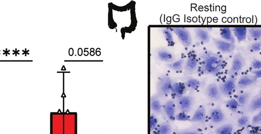
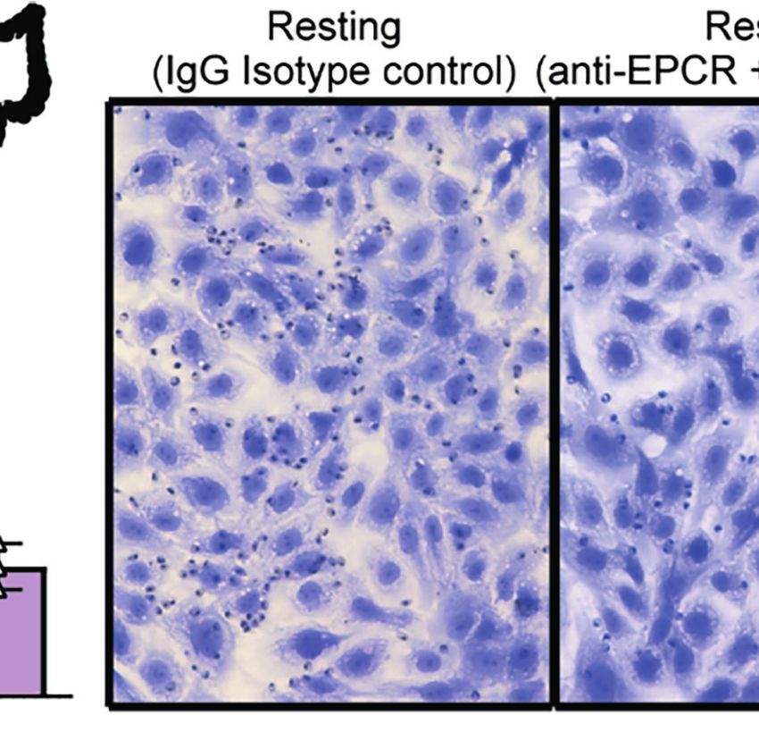
Ortolan et al.                                                                                                                Malaria and Endothelial Cell Interactions

Azasi et al., 2018; Bernabeu et al., 2019). The predominant                          A Parasite Line Expressing a Group A-
expression of the var/PfEMP1 of interest was confirmed by                             PfEMP1 Binds to HIMEC and HKMEC
RT-PCR using var strain-specific primers for the IT4 parasite                         We next investigated a parasite line expressing a Group A PfEMP1
genotype (Supplementary Figure 3). IT4var19-IEs bound at a                           variant. The HB3var03 parasite line was originally selected by
slightly higher level to resting than TNF-a-activated HIMEC,                         repeated panning on an immortalized human brain endothelial
albeit it did not reach statistical significance (Figure 2A).                         cell line (Claessens et al., 2012) and encodes dual binding activity
Binding to both resting and TNF-a-activated cells was                                for EPCR and ICAM-1 (Avril et al., 2016; Lennartz et al., 2017;
substantially inhibited by anti-EPCR monoclonal antibody                             Bernabeu et al., 2019). At the time of this study, the HB3var03
(median inhibition 80% on resting HIMEC, p

Ortolan et al.                                                                                                              Malaria and Endothelial Cell Interactions

                                                                   A

                                                                    B

 FIGURE 3 | Binding of the HB3var03 parasite line to HIMEC and HKMEC. The HB3var03 parasite was previously selected on brain endothelial cells in vitro and
 expresses a Group A-PfEMP1 protein that interacts with EPCR and ICAM-1 (cartoon). (A) Binding of HB3var03-IEs to resting and TNFa-stimulated HIMEC cells in the
 presence or absence of combined anti-EPCR and anti-ICAM-1 antibodies (B) Binding of HB3var03-IEs to resting and TNF-a-stimulated HKMEC cells in the presence or
 absence of combined anti-EPCR and anti-ICAM-1 antibodies. Data are expressed as mean ± SD and were analyzed by unpaired t test. **p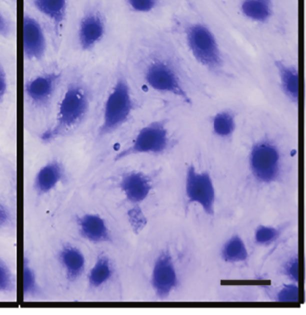
Ortolan et al.                                                                                                                  Malaria and Endothelial Cell Interactions

                             A                                           B

                                                                         C

                                                                         D

 FIGURE 4 | A DC8-PfEMP1 expressing parasite line (3173-S) isolated from a cerebral malaria patient binds to brain, intestinal and kidney endothelial cells. (A) The
 3173-S parasite line predominantly expresses the 3173-S var1 transcript (DC8-PfEMP1) based on sequencing of DBLa sequence tags. Binding of 3173-S was
 compared to primary human microvascular endothelial cells from (B) brain, (C) intestinal, and (D) peritubular kidney endothelial cells. Binding levels were compared
 on resting and TNFa-stimulated cells in the presence and absence of anti-EPCR antibody. Data are expressed as mean ± SD and were analyzed by unpaired t test.
 *p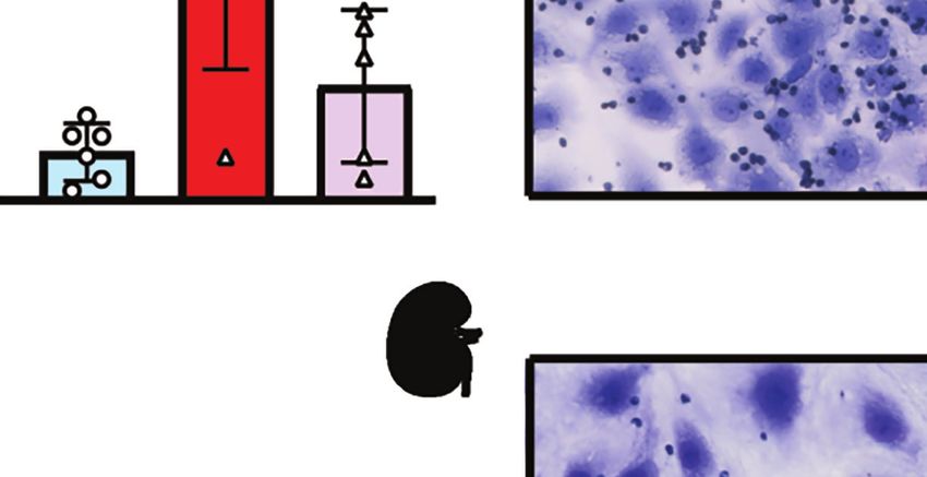
Ortolan et al.                                                                                                                               Malaria and Endothelial Cell Interactions

Sahu et al., 2021; Wichers et al., 2021), little is known about their                           ETHICS STATEMENT
endothelial binding specificity. Here, we provide evidence that
the DC8 and Group A EPCR-binding PfEMP1 subsets have                                            HKMEC were obtained after voluntary pregnancy interruptions
binding activity for brain, gut, and kidney endothelial cells. Our                              performed at the University of Washington Medical Center in
findings do not preclude that other PfEMP1 variants can                                          compliance with Institutional Review Board protocol (IRB447773EA).
sequester in these organs, but they raise the possibility of
brain-gut-kidney binding axis contributing to multi-organ
complications in severe malaria. A limitation of this study is
that binding was studied using cell monolayers and static binding                               AUTHOR CONTRIBUTIONS
assays. Further work is needed to model parasite binding in
                                                                                                LO, MA, and JS conceived and designed the study. KS provided
organ-specific microvasculature models that better mimic the
                                                                                                the parasite line recovered from a pediatric cerebral malaria
distinct 3D architecture and microfluidic dynamics of different
                                                                                                patient. JX and YZ provided HKMEC cells and culture
organs where P. falciparum-IEs sequester. Nevertheless, as
                                                                                                conditions. LO and MA performed experiments and data
EPCR-binding variants can inhibit the pro-homeostatic and
                                                                                                analysis. LO and JS wrote the first draft. All authors
pro-barrier functions of EPCR by blocking its interaction with
                                                                                                contributed to the article and approved the submitted version.
the native ligand (Turner et al., 2013; Gillrie et al., 2015; Lau
et al., 2015; Petersen et al., 2015; Sampath et al., 2015), multiple
organs may be impacted at high parasite burdens.
                                                                                                FUNDING
CONCLUSION
                                                                                                This work was supported by RO1 AI141602 (JS and KS),
This study provides evidence that DC8 and Group A EPCR-                                         U19AI089688 (JS), and UG3/UH3 TR002158 (YZ). The
binding PfEMP1 variants have broad affinity for brain, intestinal,                               content is solely the responsibility of the authors and does not
and kidney microvascular endothelial cells and raise the                                        necessarily represent the official views of the National Institutes
possibility that a parasite brain-gut-kidney binding axis may                                   of Health.
contribute to multi-organ dysfunction in severe malaria.

DATA AVAILABILITY STATEMENT                                                                     SUPPLEMENTARY MATERIAL
The original contributions presented in the study are included in                               The Supplementary Material for this article can be found online
the article/Supplementary Material. Further inquiries can be                                    at: https://www.frontiersin.org/articles/10.3389/fcimb.2022.
directed to the corresponding author.                                                           813011/full#supplementary-material

REFERENCES                                                                                         Prevalence, Pathophysiology, Impact, and Management Challenges. Int. J.
                                                                                                   Nephrol. Renovasc. Dis. 14, 235–253. doi: 10.2147/IJNRD.S239157
Avril, M., Bernabeu, M., Benjamin, M., Brazier, A. J., and Smith, J. D. (2016).                 Bernabeu, M., Danziger, S. A., Avril, M., Vaz, M., Babar, P. H., Brazier, A. J., et al.
   Interaction Between Endothelial Protein C Receptor and Intercellular                            (2016). Severe Adult Malaria is Associated With Specific PfEMP1 Adhesion
   Adhesion Molecule 1 to Mediate Binding of Plasmodium Falciparum-                                Types and High Parasite Biomass. Proc. Natl. Acad. Sci. U.S.A. 113 (23),
   Infected Erythrocytes to Endothelial Cells. MBio 7 (4), e00615–16.                              E3270–E3279. doi: 10.1073/pnas.1524294113
   doi: 10.1128/mBio.00615-16                                                                   Bernabeu, M., Gunnarsson, C., Vishnyakova, M., Howard, C. C., Nagao, R. J.,
Avril, M., Brazier, A. J., Melcher, M., Sampath, S., and Smith, J. D. (2013). DC8                  Avril, M., et al. (2019). Binding Heterogeneity of Plasmodium Falciparum to
   and DC13 Var Genes Associated With Severe Malaria Bind Avidly to Diverse                        Engineered 3d Brain Microvessels Is Mediated by EPCR and ICAM-1. MBio 10
   Endothelial Cells. PloS Pathog. 9 (6), e1003430. doi: 10.1371/journal.                          (3), e00420-19. doi: 10.1128/mBio.00420-19
   ppat.1003430                                                                                 Brabin, B. J., Romagosa, C., Abdelgalil, S., Menendez, C., Verhoeff, F. H.,
Avril, M., Tripathi, A. K., Brazier, A. J., Andisi, C., Janes, J. H., Soma, V. L., et al.          McGready, R., et al. (2004). The Sick Placenta-the Role of Malaria. Placenta
   (2012). A Restricted Subset of Var Genes Mediates Adherence of Plasmodium                       25 (5), 359–378. doi: 10.1016/j.placenta.2003.10.019
   Falciparum-Infected Erythrocytes to Brain Endothelial Cells. Proc. Natl. Acad.               Claessens, A., Adams, Y., Ghumra, A., Lindergard, G., Buchan, C. C., Andisi, C.,
   Sci. U.S.A. 109 (26), E1782–E1790. doi: 10.1073/pnas.1120534109                                 et al. (2012). A Subset of Group A-Like Var Genes Encodes the Malaria
Azasi, Y., Lindergard, G., Ghumra, A., Mu, J., Miller, L. H., and Rowe, J. A. (2018).              Parasite Ligands for Binding to Human Brain Endothelial Cells. Proc. Natl.
   Infected Erythrocytes Expressing DC13 PfEMP1 Differ From Recombinant                            Acad. Sci. U.S.A. 109 (26), E1772–E1781. doi: 10.1073/pnas.1120461109
   Proteins in EPCR-Binding Function. Proc. Natl. Acad. Sci. U.S.A. 115 (5),                    Conroy, A. L., Hawkes, M., Elphinstone, R. E., Morgan, C., Hermann, L., Barker,
   1063–1068. doi: 10.1073/pnas.1712879115                                                         K. R., et al. (2016). Acute Kidney Injury Is Common in Pediatric Severe Malaria
Baruch, D. I., Pasloske, B. L., Singh, H. B., Bi, X., Ma, X. C., Feldman, M., et al.               and Is Associated With Increased Mortality. Open Forum Infect. Dis. 3 (2),
   (1995). Cloning the P. Falciparum Gene Encoding PfEMP1, a Malarial Variant                      ofw046. doi: 10.1093/ofid/ofw046
   Antigen and Adherence Receptor on the Surface of Parasitized Human                           Conroy, A. L., Opoka, R. O., Bangirana, P., Idro, R., Ssenkusu, J. M., Datta, D., et al.
   Erythrocytes. Cell 82 (1), 77–87. doi: 10.1016/0092-8674(95)90054-3                             (2019). Acute Kidney Injury is Associated With Impaired Cognition and
Batte, A., Berrens, Z., Murphy, K., Mufumba, I., Sarangam, M. L., Hawkes, M. T.,                   Chronic Kidney Disease in a Prospective Cohort of Children With Severe
   et al. (2021). Malaria-Associated Acute Kidney Injury in African Children:                      Malaria. BMC Med. 17 (1), 98. doi: 10.1186/s12916-019-1332-7

Frontiers in Cellular and Infection Microbiology | www.frontiersin.org                      8                                          January 2022 | Volume 12 | Article 813011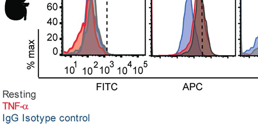
Ortolan et al.                                                                                                                                       Malaria and Endothelial Cell Interactions

Dondorp, A. M., Ince, C., Charunwatthana, P., Hanson, J., van, K. A., Faiz, M. A.,                    Nguansangiam, S., Day, N. P., Hien, T. T., Mai, N. T., Chaisri, U., Riganti, M., et al.
   et al. (2008a). Direct In Vivo Assessment of Microcirculatory Dysfunction in                           (2007). A Quantitative Ultrastructural Study of Renal Pathology in Fatal
   Severe Falciparum Malaria. J. Infect. Dis. 197 (1), 79–84. doi: 10.1086/523762                         Plasmodium Falciparum Malaria. Trop. Med. Int. Health 12 (9), 1037–1050.
Dondorp, A. M., Lee, S. J., Faiz, M. A., Mishra, S., Price, R., Tjitra, E., et al. (2008b). The           doi: 10.1111/j.1365-3156.2007.01881.x
   Relationship Between Age and the Manifestations of and Mortality Associated                        Petersen, J. V., Bouwens, E. M., Tamayo, I., Turner, L., Wang, C. W., Stins, M., et al.
   With Severe Malaria. Clin. Infect. Dis. 47 (2), 151–157. doi: 10.1086/589287                           (2015). Protein C System Defects Inflicted by the Malaria Parasite Protein PfEMP1
Gillrie, M. R., Avril, M., Brazier, A. J., Davis, S. P., Stins, M. F., Smith, J. D., et al.               can be Overcome by a Soluble EPCR Variant. Thromb. Haemost. 114 (5), 1038–1048.
   (2015). Diverse Functional Outcomes of Plasmodium Falciparum Ligation of                               doi: 10.1160/TH15-01-0018
   EPCR: Potential Implications for Malarial Pathogenesis. Cell Microbiol. 17                         Rask, T. S., Hansen, D. A., Theander, T. G., Gorm, P. A., and Lavstsen, T. (2010).
   (12), 1883–1899. doi: 10.1111/cmi.12479                                                                Plasmodium Falciparum Erythrocyte Membrane Protein 1 Diversity in Seven
Hsieh, F. L., Turner, L., Bolla, J. R., Robinson, C. V., Lavstsen, T., and Higgins, M.                    Genomes - Divide and Conquer. PloS Comput. Biol. 6 (9), e1000933.
   K. (2016). The Structural Basis for CD36 Binding by the Malaria Parasite. Nat.                         doi: 10.1371/journal.pcbi.1000933
   Commun. 7, 12837. doi: 10.1038/ncomms12837                                                         Robinson, B. A., Welch, T. L., and Smith, J. D. (2003). Widespread Functional
Janes, J. H., Wang, C. P., Levin-Edens, E., Vigan-Womas, I., Guillotte, M., Melcher,                      Specialization of Plasmodium Falciparum Erythrocyte Membrane Protein 1
   M., et al. (2011). Investigating the Host Binding Signature on the Plasmodium                          Family Members to Bind CD36 Analysed Across a Parasite Genome. Mol.
   Falciparum PfEMP1 Protein Family. PloS Pathog. 7 (5), e1002032.                                        Microbiol. 47 (5), 1265–1278. doi: 10.1046/j.1365-2958.2003.03378.x
   doi: 10.1371/journal.ppat.1002032                                                                  Sahu, P. K., Duffy, F. J., Dankwa, S., Vishnyakova, M., Majhi, M., Pirpamer, L.,
Kessler, A., Dankwa, S., Bernabeu, M., Harawa, V., Danziger, S. A., Duffy, F., et al.                     et al. (2021). Determinants of Brain Swelling in Pediatric and Adult Cerebral
   (2017). Linking EPCR-Binding PfEMP1 to Brain Swelling in Pediatric Cerebral                            Malaria. JCI Insight 6 (18), e145823. doi: 10.1172/jci.insight.145823
   Malaria. Cell Host Microbe 22601-614 (5), e605. doi: 10.1016/j.chom.2017.09.009                    Sampath, S., Brazier, A. J., Avril, M., Bernabeu, M., Vigdorovich, V., Mascarenhas,
Kim, I., Moon, S. O., Kim, S. H., Kim, H. J., Koh, Y. S., and Koh, G. Y. (2001).                          A., et al. (2015). Plasmodium Falciparum Adhesion Domains Linked to Severe
   Vascular Endothelial Growth Factor Expression of Intercellular Adhesion                                Malaria Differ in Blockade of Endothelial Protein C Receptor. Cell Microbiol.
   Molecule 1 (ICAM-1), Vascular Cell Adhesion Molecule 1 (VCAM-1), and                                   17 (12), 1868–1882. doi: 10.1111/cmi.12478
   E-Selectin Through Nuclear Factor-Kappa B Activation in Endothelial Cells.                         Scott, J. A., Berkley, J. A., Mwangi, I., Ochola, L., Uyoga, S., Macharia, A., et al.
   J. Biol. Chem. 276 (10), 7614–7620. doi: 10.1074/jbc.M009705200                                        (2011). Relation Between Falciparum Malaria and Bacteraemia in Kenyan
Lau, C. K., Turner, L., Jespersen, J. S., Lowe, E. D., Petersen, B., Wang, C. W., et al.                  Children: A Population-Based, Case-Control Study and a Longitudinal Study.
   (2015). Structural Conservation Despite Huge Sequence Diversity Allows                                 Lancet 378 (9799), 1316–1323. doi: 10.1016/S0140-6736(11)60888-X
   EPCR Binding by the PfEMP1 Family Implicated in Severe Childhood                                   Seydel, K. B., Fox, L. L., Glover, S. J., Reeves, M. J., Pensulo, P., Muiruri, A., et al.
   Malaria. Cell Host Microbe 17 (1), 118–129. doi: 10.1016/j.chom.2014.11.007                            (2012). Plasma Concentrations of Parasite Histidine-Rich Protein 2 Distinguish
Lavstsen, T., Turner, L., Saguti, F., Magistrado, P., Rask, T. S., Jespersen, J. S., et al.               Between Retinopathy-Positive and Retinopathy-Negative Cerebral Malaria in
   (2012). Plasmodium Falciparum Erythrocyte Membrane Protein 1 Domain                                    Malawian Children. J. Infect. Dis. 206 (3), 309–318. doi: 10.1093/infdis/jis371
   Cassettes 8 and 13 are Associated With Severe Malaria in Children. Proc. Natl.                     Smith, J. D., Chitnis, C. E., Craig, A. G., Roberts, D. J., Hudson-Taylor, D. E.,
   Acad. Sci. U.S.A. 109 (26), E1791–E1800. doi: 10.1073/pnas.1120455109                                  Peterson, D. S., et al. (1995). Switches in Expression of Plasmodium Falciparum
Lennartz, F., Adams, Y., Bengtsson, A., Olsen, R. W., Turner, L., Ndam, N. T., et al.                     Var Genes Correlate With Changes in Antigenic and Cytoadherent
   (2017). Structure-Guided Identification of a Family of Dual Receptor-Binding                            Phenotypes of Infected Erythrocytes. Cell 82 (1), 101–110. doi: 10.1016/
   PfEMP1 That Is Associated With Cerebral Malaria. Cell Host Microbe 21 (3),                             0092-8674(95)90056-X
   403–414. doi: 10.1016/j.chom.2017.02.009                                                           Smith, J. D., Craig, A. G., Kriek, N., Hudson-Taylor, D., Kyes, S., Fagen, T., et al. (2000).
Lennartz, F., Smith, C., Craig, A. G., and Higgins, M. K. (2019). Structural Insights                     Identification of a Plasmodium Falciparum Intercellular Adhesion Molecule-1
   Into Diverse Modes of ICAM-1 Binding by Plasmodium Falciparum-Infected                                 Binding Domain: A Parasite Adhesion Trait Implicated in Cerebral Malaria. Proc.
   Erythrocytes. Proc. Natl. Acad. Sci. U.S.A. 116 (40), 20124–20134. doi: 10.1073/                       Natl. Acad. Sci. U.S.A. 97 (4), 1766–1771. doi: 10.1073/pnas.040545897
   pnas.1911900116                                                                                    Smith, J. D., Rowe, J. A., Higgins, M. K., and Lavstsen, T. (2013). Malaria’s Deadly
Leopold, S. J., Ghose, A., Allman, E. L., Kingston, H. W. F., Hossain, A., Dutta, A.                      Grip: Cytoadhesion of Plasmodium Falciparum-Infected Erythrocytes. Cell
   K., et al. (2019). Identifying the Components of Acidosis in Patients With                             Microbiol. 15 (12), 1976–1983. doi: 10.1111/cmi.12183
   Severe Plasmodium Falciparum Malaria Using Metabolomics. J. Infect. Dis. 219                       Spitz, S. (1946). The Pathology of Acute Falciparum Malaria. Mil Surg. 99 (5),
   (11), 1766–1776. doi: 10.1093/infdis/jiy727                                                            555–572. doi: 10.1093/milmed/99.5.555
Ligresti, G., Nagao, R. J., Xue, J., Choi, Y. J., Xu, J., Ren, S., et al. (2016). A Novel             Storm, J., Jespersen, J. S., Seydel, K. B., Szestak, T., Mbewe, M., Chisala, N. V., et al.
   Three-Dimensional Human Peritubular Microvascular System. J. Am. Soc.                                  (2019). Cerebral Malaria is Associated With Differential Cytoadherence to Brain
   Nephrol. 27 (8), 2370–2381. doi: 10.1681/ASN.2015070747                                                Endothelial Cells. EMBO Mol. Med. 11 (2), e9164. doi: 10.15252/emmm.201809164
MacPherson, G. G., Warrell, M. J., White, N. J., Looareesuwan, S., and Warrell, D.                    Su, X. Z., Heatwole, V. M., Wertheimer, S. P., Guinet, F., Herrfeldt, J. A., Peterson, D.
   A. (1985). Human Cerebral Malaria. A Quantitative Ultrastructural Analysis of                          S., et al. (1995). The Large Diverse Gene Family Var Encodes Proteins Involved
   Parasitized Erythrocyte Sequestration. Am. J. Pathol. 119 (3), 385–401.                                in Cytoadherence and Antigenic Variation of Plasmodium Falciparum-Infected
Marchiafava, E., and Bignami, A. (1892). Two Monographs on Malaria and the                                Erythrocytes. Cell 82 (1), 89–100. doi: 10.1016/0092-8674(95)90055-1
   Parasites of Malaria Fevers (London: The New Sydenham Society).                                    Sypniewska, P., Duda, J. F., Locatelli, I., Althaus, C. R., Althaus, F., and Genton, B.
Miller, L. H., Baruch, D. I., Marsh, K., and Doumbo, O. K. (2002). The Pathogenic                         (2017). Clinical and Laboratory Predictors of Death in African Children With
   Basis of Malaria. Nature 415 (6872), 673–679. doi: 10.1038/415673a                                     Features of Severe Malaria: A Systematic Review and Meta-Analysis. BMC
Milner, D. A.Jr., Lee, J. J., Frantzreb, C., Whitten, R. O., Kamiza, S., Carr, R. A.,                     Med. 15 (1), 147. doi: 10.1186/s12916-017-0906-5
   et al. (2015). Quantitative Assessment of Multiorgan Sequestration of Parasites                    Taylor, T. E., Fu, W. J., Carr, R. A., Whitten, R. O., Mueller, J. S., Fosiko, N. G.,
   in Fatal Pediatric Cerebral Malaria. J. Infect. Dis. 212 (8), 1317–1321.                               et al. (2004). Differentiating the Pathologies of Cerebral Malaria by
   doi: 10.1093/infdis/jiv205                                                                             Postmortem Parasite Counts. Nat. Med. 10 (2), 143–145. doi: 10.1038/nm986
Milner, D. A.Jr., Whitten, R. O., Kamiza, S., Carr, R., Liomba, G., Dzamalala, C.,                    Turner, L., Lavstsen, T., Berger, S. S., Wang, C. W., Petersen, J. E., Avril, M., et al.
   et al. (2014). The Systemic Pathology of Cerebral Malaria in African Children.                         (2013). Severe Malaria Is Associated With Parasite Binding to Endothelial
   Front. Cell Infect. Microbiol. 4. doi: 10.3389/fcimb.2014.00104                                        Protein C Receptor. Nature 498 (7455), 502–505. doi: 10.1038/nature12216
Mkumbaye, S. I., Wang, C. W., Lyimo, E., Jespersen, J. S., Manjurano, A., Mosha,                      Turner, G. D., Morrison, H., Jones, M., Davis, T. M., Looareesuwan, S., Buley, I. D.,
   J., et al. (2017). The Severity of Plasmodium Falciparum Infection Is Associated                       et al. (1994). An Immunohistochemical Study of the Pathology of Fatal
   With Transcript Levels of Var Genes Encoding Endothelial Protein C                                     Malaria. Evidence for Widespread Endothelial Activation and a Potential
   Receptor-Binding P. Falciparum Erythrocyte Membrane Protein 1. Infect.                                 Role for Intercellular Adhesion Molecule-1 in Cerebral Sequestration. Am. J.
   Immun. 85 (4), e00841–16. doi: 10.1128/IAI.00841-16                                                    Pathol. 145 (5), 1057–1069.

Frontiers in Cellular and Infection Microbiology | www.frontiersin.org                            9                                            January 2022 | Volume 12 | Article 813011
Ortolan et al.                                                                                                                        Malaria and Endothelial Cell Interactions

Wichers, J. S., Tonkin-Hill, G., Thye, T., Krumkamp, R., Kreuels, B., Strauss, J.,        Publisher’s Note: All claims expressed in this article are solely those of the authors
  et al. (2021). Common Virulence Gene Expression in Adult First-Time                     and do not necessarily represent those of their affiliated organizations, or those of
  Infected Malaria Patients and Severe Cases. Elife 10, e69040. doi: 10.7554/             the publisher, the editors and the reviewers. Any product that may be evaluated in
  eLife.69040                                                                             this article, or claim that may be made by its manufacturer, is not guaranteed or
Wilairatana, P., Meddings, J. B., Ho, M., Vannaphan, S., and Looareesuwan, S.             endorsed by the publisher.
  (1997). Increased Gastrointestinal Permeability in Patients With Plasmodium
  Falciparum Malaria. Clin. Infect. Dis. 24 (3), 430–435. doi: 10.1093/clinids/           Copyright © 2022 Ortolan, Avril, Xue, Seydel, Zheng and Smith. This is an open-
  24.3.430                                                                                access article distributed under the terms of the Creative Commons Attribution
                                                                                          License (CC BY). The use, distribution or reproduction in other forums is permitted,
                                                                                          provided the original author(s) and the copyright owner(s) are credited and that the
Conflict of Interest: The authors declare that the research was conducted in the           original publication in this journal is cited, in accordance with accepted academic
absence of any commercial or financial relationships that could be construed as a          practice. No use, distribution or reproduction is permitted which does not comply with
potential conflict of interest.                                                            these terms.

Frontiers in Cellular and Infection Microbiology | www.frontiersin.org               10                                         January 2022 | Volume 12 | Article 813011
You can also read