RSC Chemical Biology - RSC Publishing
←
→
Page content transcription
If your browser does not render page correctly, please read the page content below
Volume 1
                                                                           Number 5
                                                                           December 2020
                                                                           Pages 291–456

RSC
Chemical Biology
rsc.li/rsc-chembio

                     ISSN 2633-0679

                     PAPER
                     Mark A. T. Blaskovich et al.
                     Fluorescent macrolide probes – synthesis and use in
                     evaluation of bacterial resistance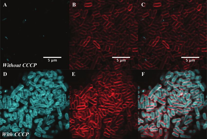
RSC
Chemical Biology
View Article Online
PAPER View Journal | View Issue
Fluorescent macrolide probes – synthesis
This article is licensed under a Creative Commons Attribution 3.0 Unported Licence.
and use in evaluation of bacterial resistance†
Open Access Article. Published on 17 November 2020. Downloaded on 12/1/2021 1:51:06 AM.
Cite this: RSC Chem. Biol., 2020,
1, 395
M. Rhia L. Stone, a Urszula Łapińska, b Stefano Pagliara, b Muriel Masi, c
Joanne T. Blanchfield, d Matthew A. Cooper a and Mark A. T. Blaskovich *a
The emerging crisis of antibiotic resistance requires a multi-pronged approach in order to avert the
onset of a post-antibiotic age. Studies of antibiotic uptake and localisation in live cells may inform the
design of improved drugs and help develop a better understanding of bacterial resistance and
persistence. To facilitate this research, we have synthesised fluorescent derivatives of the macrolide
antibiotic erythromycin. These analogues exhibit a similar spectrum of antibiotic activity to the parent
drug and are capable of labelling both Gram-positive and -negative bacteria for microscopy. The probes
localise intracellularly, with uptake in Gram-negative bacteria dependent on the level of efflux pump
activity. A plate-based assay established to quantify bacterial labelling and localisation demonstrated that
Received 9th July 2020, the probes were taken up by both susceptible and resistant bacteria. Significant intra-strain and -species
Accepted 28th October 2020 differences were observed in these preliminary studies. In order to examine uptake in real-time, the probe
DOI: 10.1039/d0cb00118j was used in single-cell microfluidic microscopy, revealing previously unseen heterogeneity of uptake in
populations of susceptible bacteria. These studies illustrate the potential of fluorescent macrolide probes to
rsc.li/rsc-chembio characterise and explore drug uptake and efflux in bacteria.
Introduction macrolides falls into one of four mechanistic categories: modifi-
cation of the macrolide target (the 50 s subunit of the bacterial
Macrolide antibiotics are a natural product-class of drugs ribosome); target protection (removal of macrolide from the active
discovered in the mid-twentieth century following isolation site); macrolide efflux (reduces intracellular concentration); or
from bacteria such as Saccharopolyspora erythrea. The arche-
typal macrolide, erythromycin (1, Fig. 1), entered clinical use in
1952. Since then, more than 10 semi-synthetic macrolides (e.g.
roxithromycin 2 and azithromycin 3, Fig. 1) have been devel-
oped and now see wide-spread clinical use.1
Macrolides are generally active against Gram-positive and
-negative cocci (e.g. Staphylococci, Streptococci, Neisseria
meningitidis), Gram-positive and some -negative bacilli (e.g.
Bacilli, Bordatella), atypical bacteria (e.g. Mycobacterium avium
complex, Helicobacter pylori), and intracellular pathogens (e.g.
Legionella, Chlamydia).2 One factor increasingly limiting their
use is acquired macrolide resistance, which was observed only a
few years after their introduction into the clinic.1 Resistance to the
a
Centre for Superbug Solutions, Institute for Molecular Bioscience, The University
of Queensland, 306 Carmody Road, St Lucia 4072, Brisbane, Australia.
E-mail: m.blaskovich@imb.uq.edu.au
b
Living Systems Institute, University of Exeter, Exeter EX4 4QD, UK
c
Université Paris-Saclay, CEA, CNRS, Institute for Integrative Biology of the Cell
(I2BC), 911198, Gif-sur-Yvette, France
d
School of Chemistry and Molecular Biosciences, The University of Queensland,
68 Cooper Road, St Lucia 4072, Brisbane, Australia
† Electronic supplementary information (ESI) available. See DOI: 10.1039/ Fig. 1 Common macrolide antibiotics erythromycin 1, roxithromycin 2,
d0cb00118j azithromycin 3, and generic probe structure 4.
This journal is © The Royal Society of Chemistry 2020 RSC Chem. Biol., 2020, 1, 395--404 | 395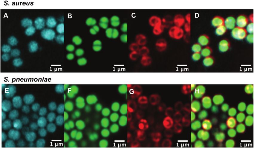
View Article Online
Paper RSC Chemical Biology
antibiotic modification.3 Chief amongst these mechanisms are compounds (with a 7-nitrobenz-2-oxa-1,3-diazol-4-yl (NBD)
ribosome modifications effected by the erm (erythromycin fluorophore), which were used to evaluate azithromycin dis-
ribosome methylation) enzymes, which mono- or di-methylate tribution in mammalian cells and whole animals.25 Alone of
the ribosome, interfering with macrolide binding; and the mef the previously reported fluorescent macrolides, those prepared
and mrs efflux pumps, which decrease intracellular concentra- by Matijasic et al. were confirmed to retain the antibacterial
tions of the antibiotic via active export.1 Many pathogenic activity of the parent drug, a critical step in ensuring that
bacteria in the clinic now possess one or more of these the probe accurately represents the parent in studies. However,
resistance mechanisms, with their dissemination between bac- neither uptake nor localisation in bacterial cells was assessed
teria facilitated by plasmid transmission.4 in their work. To date, no work has been reported utilising
This article is licensed under a Creative Commons Attribution 3.0 Unported Licence.
In order to combat macrolide resistance and develop new fluorescent macrolides for studying antibiotic resistance;
Open Access Article. Published on 17 November 2020. Downloaded on 12/1/2021 1:51:06 AM.
therapies for resistant bacteria, we need better tools to improve indeed there are relatively few reports on the use of any
our understanding of drug mode of action and resistance macrolide-derived fluorescent probes.26 In order to address
mechanisms, especially in terms of drug accumulation and this gap, in this paper we report on the synthesis and character-
efflux. One set of tools that have found increasing utility in isation of two novel fluorescent derivatives of erythromycin, where
antibiotic resistance research are fluorescent antibiotics.5 a common antibiotic core is linked to two different fluorophores.
Although innately fluorescent or fluorescent analogues of anti- We assessed their activity against both Gram-positive and Gram-
biotics were used in the second half of the 20th century to negative bacteria, showing that both probes retained good to
investigate the mode of action of antibiotics,5 they have been moderate levels of antimicrobial activity. In order to assess their
applied sparingly to help address the modern crisis of resis- utility for the study of antibiotic-bacterial interactions, particularly
tance. Examples of probes that have been successfully utilised investigations of resistance mechanisms, we tested their ability to
include fluorescent glycopeptides6 and b-lactams.7–9 However, label bacteria using a combination of spectrofluorometry and
instead of phenotypic studies, characterisation of resistance microscopy, at both single cell and bulk population levels.
has shifted towards – omics studies, where changes at the For bulk measurements we used spectrophotometry,
genetic, epigenetic and transcriptomic levels are analysed.5 because this technique is especially useful for quantitative
Where intracellular distribution studies have been conducted, analysis. It is fast, inexpensive, readily available, and compa-
they have largely relied on radioactive analogues, which are tible with plate-based, high-throughput assays, and allowed us
costly, difficult to access, and require specialist equipment.10–19 to investigate antimicrobial resistance at the ensemble level.
Neither of these techniques readily allow for analysis at We also employed high-resolution microscopy to gain a more
single-cell resolution, though some single cell sequencing detailed view of interactions between bacteria and the probes
reports are beginning to appear.20,21 Despite the success of at a single cell level. We then bridged both techniques with
these approaches, there remains a significant appeal in simple single-cell microfluidics, where we again gained insights into
tools that are readily accessible and do not require a large antimicrobial resistance at the single cell level but were able to
time or resource investment, or which can be utilised in simultaneously monitor thousands of individual cells.
combination with more modern technology, such as high-
resolution microscopy and single cell microfluidics.
There have been several fluorescent macrolides reported in
the literature, however none of these have seen widespread Results and discussion
use. In the 1970s, Pestka and colleagues used a fluorescein-
erythromycin conjugate to study interactions with the Synthesis
ribosome,22,23 and were able to determine which subunit the We utilised the same strategy that we have previously applied to
probe bound to, along with information about the binding functionalise other antibiotics,27–29 whereby we first installed
kinetics. These assays were carried out on isolated ribosomes, an azide ‘handle’ to which different fluorophores can be readily
so it is unclear how the fluorescent macrolide behaved in live attached using a copper activated azide–alkyne cycloaddition
bacteria. This is a common flaw with many antibiotic-derived (CuAAC) reaction. This reaction is highly selective, compatible
probes – their antibacterial activity is often not reported and the with unprotected functional groups, and yields a stable, bio-
degree of cell penetration unknown.5 With the fluorescein substi- compatible triazole linker. A key component of this tactic is to
tuent being a moderate-sized ligand on its own (M = 332 g mol 1), it select a site for modification that does not perturb the anti-
is likely that it would interfere with entry of the conjugate into bacterial activity. Although several macrolide sites have been
the cell. Three decades later, Li et al. developed boron- modified in the literature, the ketone at the 9-position of
dipyrromethene (BODIPY)-erythromycin probes that were also erythromycin was selected as the point of attachment due to
used to study interactions at the ribosome.24 Again, antibacter- the simple, high-yielding procedures available (4, Fig. 1). This
ial activity was not measured, so it is unclear how effectively ketone has been observed to not make significant interactions
these probes mimic the parent antibiotic. The authors used the with the ribosomal active site in crystal structure studies.30
fluorescent macrolide to develop a high-throughput screening To this end, erythromycin 1 was treated with hydroxylamine
protocol to identify new antibiotic candidates. More recently, to yield oxime 5 in quantitative yield (Scheme 1).31 A linker was
Matijasic et al. reported several azithromycin-based fluorescent then installed by alkylation with 1-bromo-4-chlorobutane.
396 | RSC Chem. Biol., 2020, 1, 395--404 This journal is © The Royal Society of Chemistry 2020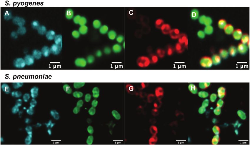
View Article Online

                                                                                                  RSC Chemical Biology                                                                                                                       Paper
            This article is licensed under a Creative Commons Attribution 3.0 Unported Licence.
Open Access Article. Published on 17 November 2020. Downloaded on 12/1/2021 1:51:06 AM.

                                                                                                  Scheme 1 Synthesis of roxithromycin-azide 6 from erythromycin 1, via formation of oxime intermediate 5, which is then O-alkylated with 1-bromo-4-
                                                                                                  chlorobutane, followed by displacement of the terminal halide with azide.

                                                                                                  Substitution of the terminal halide with sodium azide, yielding           MW of 316, 276 and 463 g mol 1 respectively). The smaller
                                                                                                  roxi-C4–N3 6 in 25% yield from the oxime.                                 substituent size reduces the likelihood that attaching it to the
                                                                                                      With azide 6 in hand, azide–alkyne click reactions were               antibiotic core will reduce bacterial uptake of the conjugate.
                                                                                                  carried out using alkyne-fluorophores 7 and 8 derived from                Furthermore, conjugation of the NBD fluorophore was found
                                                                                                  NBD and 7-dimethylaminocoumarin-4-acetic acid (DMACA)                     to not impair biological activity for azithromycin,25 hence
                                                                                                  respectively, to form fluorescent macrolides 9 (roxi-C4-Tz-NBD)           was considered a promising candidate for attachment by click
                                                                                                  and 10 (roxi-C4-Tz-DMACA) in moderate yields (Scheme 2). Ele-             chemistry.
                                                                                                  vated temperature was required to facilitate dissolution, and the
                                                                                                  modest isolated yields (36–37%) were due to product losses                Antibacterial activity
                                                                                                  during purification on a small scale, rather than incomplete              In order to confirm that the fluorescent probes 9 and 10 retained
                                                                                                  reaction. The NBD and DMACA fluorophores were chosen due                  the antibiotic activity of the parent drug, minimum inhibitory
                                                                                                  to their relatively small size (molecular weight of fluorophore only      concentrations (MICs) were measured against a panel of sus-
                                                                                                  165 and 231 g mol 1 respectively) compared to other fluorophores          ceptible bacteria (Table 1). Here, roxi-C4–N3 6 displayed good to
                                                                                                  such as fluorescein, BODIPY-FL, or rhodamine B (fluorophore               excellent antibacterial activity against all Gram-positive cocci

                                                                                                  Scheme 2   Synthesis of fluorescent macrolides 9 and 10 from azide 6 and fluorophore-alkynes 7 and 8.

                                                                                                  This journal is © The Royal Society of Chemistry 2020                                                RSC Chem. Biol., 2020, 1, 395--404 | 397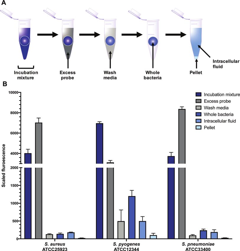
View Article Online
Paper RSC Chemical Biology
Table 1 Minimum inhibitory concentrations (MICs, in mg mL 1) of erythromycin (1), roxithromycin (2), roxi-C4–N3 (6), roxi-C4-Tz-NBD (9), and roxi-C4-
Tz-DMACA (10) against a panel of American Type Culture Collection (ATTC) susceptible bacteria, measured by broth microdilution in cation-adjusted
Mueller Hinton broth (CAMHB), n Z 4
Species Strain Ery 1 Roxi 2 Roxi-N3 6 Roxi-NBD 9 Roxi-DMACA 10
S. aureus ATCC25923 0.25–0.5 0.25–2 1–4 1–4 8–16
S. epidermidis ATCC12228 0.25–1 0.5–1 0.031–0.063 0.5–1 16–64
S. pneumoniae ATCC33400 0.25–0.5 0.5–2 1–2 1–2 8–32
S. pyogenes ATCC12344 r0.016 r0.016 r0.016 0.031–0.063 0.13
E. faecium ATCC35667 0.25–2 1–8 2–8 16 64
B. subtilis ATCC6051 0.13–0.25 0.25–2 0.5–1 1–2 8 -16
This article is licensed under a Creative Commons Attribution 3.0 Unported Licence.
E. coli ATCC25922 64 Z64 Z64 Z64 Z64
Open Access Article. Published on 17 November 2020. Downloaded on 12/1/2021 1:51:06 AM.
AG102a Z64 Z64 Z64 Z64 Z64
a
mar1, upregulated Acr-AB efflux pump.
and bacilli tested, with MICs generally at most 8-fold less Intracellular labelling and localisation
potent compared to the parent drugs erythromycin 1 and With antibiotic activity established, live-cell confocal micro-
roxithromycin 2. Similarly, NBD probe 9 exhibited good to scopy was performed in S. aureus and susceptible and resistant
moderate antibacterial activity, with very similar activity as Streptococci to examine localisation and labelling and
the azide 6 against Staphylococci and Streptococci (e.g. 4- to gain insight about the macrolide resistance mechanisms in
8-fold less active than the parent roxithromycin). The DMACA resistant Streptococci strains. Bacteria were incubated with
probe 10 was also active at inhibiting bacterial growth, though fluorescent probes 9 and 10, along with the red membrane
was less potent than the other derivatives tested, with approxi- dye FM4-64FX and a complementary nucleic acid dye (Syto-9,
mately 4- to 16-fold increased MIC compared to the NBD probe. green; or Hoechst-33342, blue). Despite its reduced antibacter-
However, both probes showed similar relative changes in ial potency, Roxi-C4-Tz-DMACA 10 was found to provide super-
potency across the different strains tested as the parent anti- ior labelling for microscopy and was able to be used at
biotic. For example, consistent with the lack of activity of concentrations as low as 1 nM in some species. The reason
erythromycin and roxithromycin (MIC Z 64 mg mL 1), none for this difference from NBD probe 9 is unclear but given the
of the macrolide probes showed substantial inhibitory activity similarity in MIC (Tables 1 and 2) and uptake results from the
against the one Gram-negative species tested, Escherichia coli quantitative assays (see Fig. 5, 7 and Table 3 below), the results
(E. coli). obtained from 10 should be representative of both probes, and
A panel of resistant Streptococci with either defined the parent antibiotic. Susceptible strains of Staphylococcus
(erm(B)+, mef(e)+) or undefined mechanisms of resistance were aureus (S. aureus) and S. pneumoniae were first examined
then tested (Table 2). The Streptococcus pneumoniae (S. pneu- (Fig. 2), with S. pyogenes (Fig. S1, ESI†) showing similar results,
moniae) strains were found to be highly resistant to all the where it was found that 10 localised intracellularly. This is in
macrolides tested, with MICs of Z64 mg mL 1 recorded. This contrast to the fluorophore-alkynes, which did not significantly
was also the case for S. pyogenes ATCC BAA-1412, however the accumulate in the bacteria (Fig. S2, ESI†).
MICs against ATCC BAA-1414 were only mildly elevated (still After confirming that the macrolide probe 10 successfully
above breakpoint for roxithromycin 2).32 Overall, the azide reached the bacterial cytoplasm in susceptible strains, we
intermediate and fluorescent probes had similar antibiotic analysed its localization in resistant S. pyogenes and S. pneumo-
activity profiles as compared to the parent drugs, with greatly niae. Surprisingly, we found that 10 was again found to localise in
increased MICs against the macrolide-resistant bacteria. The the cytoplasm (Fig. 3 and Fig. S5, ESI†), including in mef(e)+
MIC values were used to inform appropriate concentrations for S. pneumoniae. The latter strain, equipped with upregulated
subsequent assays. mef efflux pumps, appeared to show patterns of localised intensity
Table 2 Minimum inhibitory concentrations (MICs, in mg mL 1) of erythromycin (1), roxithromycin (2), roxi-C4–N3 (6), roxi-C4-Tz-NBD (9), and roxi-C4-
Tz-DMACA (10) against a panel of susceptible (for comparison) and resistant Streptococci, measured by broth microdilution in brain-heart infusion (BHI),
nZ3
Species Strain Ery 1 Roxi 2 Roxi-N3 6 Roxi-NBD 9 Roxi-DMACA 10
S. pneumoniae ATCC 33400 0.25–0.5 0.25–2 1–4 1–4 8–16
ATCC 700677a Z64 16–Z64 16–Z64 4–Z64 8–Z64
ATCC 700676bc Z64 Z64 Z64 Z64 Z64
S. pyogenes ATCC 12344 r0.016 r0.016 r0.016 0.031–0.063 0.13
ATCC BAA-1412 Z64 Z64 Z64 Z64 Z64
ATCC BAA-1414 0.031–0.13 1–2 0.063–1 r0.016–0.063 1
a b
erm(B)+. mef(e)+. c 5% CO2.
398 | RSC Chem. Biol., 2020, 1, 395--404 This journal is © The Royal Society of Chemistry 2020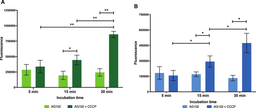
View Article Online

                                                                                                  RSC Chemical Biology                                                                                                                        Paper
            This article is licensed under a Creative Commons Attribution 3.0 Unported Licence.
Open Access Article. Published on 17 November 2020. Downloaded on 12/1/2021 1:51:06 AM.

                                                                                                  Fig. 2 Confocal microscopic images of (A–D) live susceptible S. aureus (ATCC 25923) and (E–H) susceptible S. pneumoniae (ATCC 33400) labelled
                                                                                                  with (A and E) roxi-C4-Tz-DMACA 10 (10 mM); (B and F) Syto-9 (green nucleic acid dye, 5 mM); (C and G) FM4-64FX (red membrane dye, 5 mg mL 1); and
                                                                                                  (D and H) overlaid.

                                                                                                  Fig. 3 Confocal microscopic images of (A–D) live resistant S. pyogenes (ATCC BAA-1412) and (E and F) resistant S. pneumoniae (ATCC 700676) labelled
                                                                                                  with (A and E) roxi-C4-Tz-DMACA 10 (10 mM); (B and F) Syto-9 (green nucleic acid dye, 5 mM); (C and G) FM4-64FX (red membrane dye, 5 mg mL 1); and
                                                                                                  (D and H) overlaid.

                                                                                                  when compared to the corresponding susceptible strain, but still           the measurements above with studies carried out using our
                                                                                                  had substantial internal labelling. In order to assess whether this        previously established single-cell microfluidics-microscopy
                                                                                                  difference in labelling was due to increased efflux in the resistant          assay.34–37 This technique enables single-cell studies to address
                                                                                                  strain, samples were incubated with the protonophore carbonyl              the impact of phenotypic heterogeneity38,39 on intracellular
                                                                                                  cyanide m-chlorophenylhydrazine (CCCP), which acts by disabling            drug accumulation and efficacy.40,41 As a proof of concept, we
                                                                                                  the proton motive force driving efflux pumps.33 In this case we              examined the real-time uptake of roxi-C4-Tz-NBD 9 into E. coli
                                                                                                  found no change in the localisation or intensity of labelling              (BW25113, a K12 strain) and susceptible S. aureus (MSSA476).
                                                                                                  compared to the susceptible strain ATCC 33400.                             Here it was found that the probe was rapidly taken up by
                                                                                                                                                                             S. aureus cells, with uptake initiated near-simultaneously across
                                                                                                                                                                             all measured cells, although there was significant variation in
                                                                                                  Dynamics of intracellular accumulation                                     the speed and extent of uptake (Fig. 4A). In contrast, there was a
                                                                                                  Although confocal microscopy allows for observation of the                 long lag period before any E. coli became fluorescent (Fig. 4B).
                                                                                                  precise intracellular localisation of the macrolide probes, it is          Even when the E. coli did take on the probe, both initiation and
                                                                                                  not suitable for quantifying the dynamics of antibiotic accu-              rate of uptake showed a substantial variability in response.
                                                                                                  mulation in individual bacteria. Therefore, we complemented                The spread of fluorescence behaviour indicates that in both

                                                                                                  This journal is © The Royal Society of Chemistry 2020                                                 RSC Chem. Biol., 2020, 1, 395--404 | 399
View Article Online
Paper RSC Chemical Biology
This article is licensed under a Creative Commons Attribution 3.0 Unported Licence.
Open Access Article. Published on 17 November 2020. Downloaded on 12/1/2021 1:51:06 AM.
Fig. 4 Accumulation of roxi-C4-Tz-NBD 9 in susceptible S. aureus (MSSA476) (A–C) and E. coli (BW25113) (D–F) cells as monitored by single-cell
microfluidics over time. Fluorescence of individual cells is tracked in (A and D), with the average shown in bold with symbols. Times of snapshots (B, C and
E, F) are indicated with dashed vertical lines. Brightfield (B and E) and the corresponding fluorescent (C and F) images are shown at early (2200 and
4000 s) and late (4400 and 12 000 s) timepoints for both species.
species examined, there is inherent population variability in mode of resistance, and help guide the development of strate-
response to the roxi-NBD probe 9. For E. coli, there were also gies to overcome efflux.
cells which did not accumulate any probe, despite being To start the examination of bacterial efflux of fluorescent
surrounded by bacteria with substantial uptake (see Fig. 4E macrolides 9 and 10, the model organism E. coli AG102 was
and F, e.g. right hand panels, third channel, 2nd bacterium used, in which efflux pump AcrAB is overproduced (mar1
from the bottom is visible in the brightfield microscopy image, mutant). In order to assess whether the roxi-probes were
but is not fluorescent despite cells on either side with high subject to efflux in this model, intracellular accumulation was
levels of fluorescence). The intracellular variation is striking. measured by spectrophotometry over time in the presence
This suggests heterogeneity in the macrolide response, poten- and absence of CCCP (Fig. 5). Mid-log phase bacteria were
tially due to differences in levels of native efflux pumps or in incubated with the macrolide probes (with or without CCCP),
the abundance of ribosomes that are the intracellular targets of with aliquots withdrawn at each time point. Labelled bacteria
macrolides, or a quiescent subpopulation. These new probes were then collected and lysed overnight using glycine-HCl. The
and time-dependent data open the way for deepening our lysed bacteria were centrifuged, and the fluorescence of the
understanding of subsets of bacteria, including persister and decantate measured using a plate reader. As expected, neither
viable but non culturable cells,40,42 that can transiently survive roxi-C4-Tz-NBD 9 or roxi-C4-Tz-DMACA 10 showed significant
antibiotic exposure and contribute to recalcitrant infections43 internalisation into AG102 in the absence of CCCP. In contrast,
and the development of antibiotic resistance.44 both probes showed a time-dependent accumulation when efflux
was abolished. This finding indicates both that the fluorescent
macrolides are indeed efflux pump substrates (potentially the
Dynamics of efflux AcrAB pump in this strain of E. coli) and their internal accumula-
In order to gain further insight on the molecular mechanisms tion can be used as a marker for the presence of efflux.
underlying reduced intracellular accumulation of our macro- To confirm this, live cell microscopy was carried out with
lide probes, we used ensemble measurements to quantify AG102 incubated with roxi-C4-Tz-DMACA 9 with or without
efflux, one of the major forms of resistance to macrolides. CCCP (Fig. 6). Here, the same pattern of labelling was observed,
We hoped that the macrolide probes could be used as tools with the efflux-active cells not showing significant uptake of the
to identify efflux upregulation in resistant bacteria, study this probe, but CCCP-treated efflux-negative cells having considerable
400 | RSC Chem. Biol., 2020, 1, 395--404 This journal is © The Royal Society of Chemistry 2020View Article Online
RSC Chemical Biology Paper
This article is licensed under a Creative Commons Attribution 3.0 Unported Licence.
Open Access Article. Published on 17 November 2020. Downloaded on 12/1/2021 1:51:06 AM.
Fig. 5 Intracellular accumulation of macrolide probes with NBD 9 (A) and DMACA 10 (B) fluorophores (50 mM) in AG102 E. coli in the absence and
presence of efflux inhibitor CCCP (10 mM), n = 3, * = p o 0.05, ** = p o 0.01.
Fig. 6 Airyscan confocal microscopy of AG102 E. coli without (A–C) or with (D–F) treatment with 10 mM CCCP: (A and D) DMACA probe 10 (5 mM),
(B and E) FM4-64FX (red membrane stain), and (C and F) overlaid.
internal probe fluorescence. Gratifyingly, this lack of significant was then centrifuged. Another aliquot was taken of the super-
labelling in efflux-active E. coli qualitatively corroborates the natant, which contained excess probe not taken up by the
single-cell data concerning the dynamics of macrolide accumula- bacteria (Fig. 7). The bacteria were then washed, and portions
tion presented in Fig. 4. Although eventual uptake was seen in of both the wash decantate (containing loosely bound probe
microfluidic analysis, this was not until after 80 min, whereas the that could be washed off, or probe that may have been rapidly
microscopy samples were only incubated with probes for 30 min. effluxed after uptake) and the washed bacteria were taken for
sampling (see ESI† for more detail). Finally, the remaining
Quantification of intracellular labelling washed bacteria were lysed using lysozyme, detergent (bacterial
In order to quantitatively evaluate intracellular labelling in a protein extraction reagent, B-PER) and freeze-thawing, then
high-throughput bulk-population measurement, a plate- based centrifuged to separate the intracellular fluid (IF) (e.g. with any
assay was developed using S. aureus, S. pneumoniae, and free probe inside the bacteria) from the remaining pellet (with
S. pyogenes, building on existing work in the literature.45 probe bound to cell membrane or intracellular components).
Bacteria were grown to mid-log phase, then treated with probes All fractions were then assessed for fluorescence. In order to
9 or 10, along with controls of alkynes 7 and 8, and roxithro- transform the raw data into meaningful values, several normal-
mycin 2. Aliquots were taken of this incubation mixture, which isations were conducted (see ESI†). At the 1 mM concentration
This journal is © The Royal Society of Chemistry 2020 RSC Chem. Biol., 2020, 1, 395--404 | 401View Article Online
Paper RSC Chemical Biology
This article is licensed under a Creative Commons Attribution 3.0 Unported Licence.
Open Access Article. Published on 17 November 2020. Downloaded on 12/1/2021 1:51:06 AM.
Fig. 7 Scaled fluorescence of roxi-C4-Tz-DMACA 10 in aliquots (incubation mixture, excess probe, wash media, whole bacteria, intracellular fluid,
pellet) of different bacteria: susceptible S. aureus and susceptible S. pyogenes and S. pneumoniae.
used in the assay (B1 mg mL 1), roxi-C4-Tz-DMACA 10 was In order to quantify the uptake of the probes, we calculated
found to mostly elute in the excess probe aliquot, indicating the percentage of the (reconstructed) full signal that ended up
this is beyond the saturation limit of the bacteria (Fig. 6). Given in the intracellular fluid (for details see ESI†). The percentage of
that the concentration to which the bacteria were exposed is probe taken up by the bacteria was found to be highly variable
around (susceptible) or below (resistant) the MIC, it indicates between different species, ranging from 2% of the total added
that uptake is a significant limiting factor in antibacterial in susceptible S. aureus to as much as 50% in S. pyogenes
activity. The next step, washing, did not lead to significant (Table S6, ESI†). In general, the values obtained for the two
loss of fluorescence, showing that once absorbed, the probe is fluorescent macrolide probes were similar, and in contrast the
not readily removed. As similarly low fluorescence levels were fluorophore-alkynes showed very little uptake in any of the
seen in the wash of efflux active strains, it can be inferred that bacteria tested.
substantial macrolide efflux does not occur over this time- As mentioned previously, the macrolide probes localised in
frame. From the whole washed bacteria (medium blue bars in the cytosol. Of the combined cytosol and pellet fluorescence
Fig. 6), lysis revealed that the probe fluorescence was primarily obtained after lysis, the IF portion contributed greater than
localised in the intracellular fluid, rather than bound to cellular 75% in most cases (Table 3). The percentages for roxi-C4-Tz-
debris in the pellet fraction. This is consistent with the probes DMACA 10 and roxi-C4-Tz-NBD 9 were generally very compar-
targeting the ribosomes, which are normally located in the able in S. aureus and S. pneumoniae, but in S. pyogenes the NBD
intracellular fluid. The labelling and localisation of roxi-C4-Tz- probe 9 consistently showed lower levels of accumulation,
NBD 9 proved to be very similar to its DMACA counterpart 10 indicating a potential species-selective bias between the NBD
(Fig. S4, ESI†). As before, most of the fluorescence was observed in vs. DMACA probes.
the excess aliquot, though this was less prominent in S. pyogenes, Comparing the macrolide sensitive and resistant Strepto-
possibly due to increased uptake of the NBD probe. Fluorophore cocci, contrasting trends were again observed between S. pyo-
alkynes 7 and 8 were also tested as controls, but they washed off genes and S. pneumoniae. ATCC BAA-1412 resistant S. pyogenes
the bacteria with practically no uptake from the full to whole showed a reduced fluorescence in the IF aliquot, potentially
aliquots, confirming that the roxithromycin fluorescent probes indicating less of an attraction for this subcellular localisation.
are acting in a target specific fashion (Fig. S4, ESI†). In contrast, the mef(e)+ S. pneumoniae ATCC 700676 showed a
402 | RSC Chem. Biol., 2020, 1, 395--404 This journal is © The Royal Society of Chemistry 2020View Article Online
RSC Chemical Biology Paper
Table 3 Percentage of fluorescence of probes 9 and 10 in the IF from This preliminary work illustrates the potential utility of the
(IF + pellet) macrolide fluorescent probes in illuminating the interactions
Average IF/(IF + pellet) (%) between antibiotics and bacteria, providing new insight into
a mechanisms of resistance. Fluorescent probes enable the
Species ATCC S/R DMACA probe 10 NBD probe 9
researcher to visualise target interactions in a practical manner,
S. aureus 25923 S 89 87 and can be applied to experiments that range from studies of
S. pyogenes 12344 S 83 64
BAA-1412 R 68 38 isolated targets through to investigations of mixed populations.
BAA-1414 R 83 78 Future implications include quantitatively distinguishing
S. pneumoniae 33400 S 87 90 modes of macrolide resistance and uncovering heterogeneity
This article is licensed under a Creative Commons Attribution 3.0 Unported Licence.
700677b R 94 99
in antibiotic response amongst bacterial populations.
Open Access Article. Published on 17 November 2020. Downloaded on 12/1/2021 1:51:06 AM.
700676c R 96 96
a b
S: susceptible, R: resistant. erm(B)+. c mef(e)+; italics: highly variable
due to poor absorption.
Conflicts of interest
There are no conflicts to declare.
somewhat increased preference for localisation of the probe
in the IF compared to the susceptible strain (Table 3) and a
similar overall uptake (IF and pellet, Fig. S2, ESI†), an unex- Acknowledgements
pected result given that greater efflux should lead to reduced
M. R. L. S. was supported by an Australian Postgraduate Award
intracellular levels, if the efflux is responsible for the macrolide
and an Institute for Molecular Biosciences Research Advance-
resistance. However, the result is consistent with the uptake
ment Award. U. L. and S. P. were supported through a MRC
seen in confocal microscopy (Fig. 3 vs. Fig. 2), suggesting
Proximity to Discovery EXCITEME2 grant (MCPC17189) and a
that the macrolide probe may not actually be a substrate for
Gordon and Betty Moore Foundation Marine Microbiology
the S. pneumoniae efflux pumps, with the higher MIC due to an
Initiative grant (GBMF5514). M. M. was supported by Campus
alternative resistance mechanism.
France the Programme Hubert Curien FASIC 2018 (No. 41621QL).
These results, albeit preliminary, illustrate the power of this
M. A. C. also holds a fractional professorial research fellow
assay in generating data on the localisation of antibiotics, especially
appointment at the University of Queensland, with his remain-
when applied to investigations of resistance. Combining qualitative
ing time as CEO of Inflazome Ltd, a company developing drugs
high-resolution fluorescence microscopy with quantitative
to address clinical unmet needs in inflammatory disease.
single-cell and bulk-population methods and mechanistic-
M. A. T. B. was supported in part by Wellcome Trust Strategic
specific fluorescent probes can provide a rich understanding
Grant WT1104797/Z/14/Z and NHMRC Development grant
of the chemical biology of the interactions between bacteria
APP1113719. Microscopy was performed at the Australian
and antibiotics.
Cancer Research Foundation (ACRF)/Institute for Molecular
Bioscience Cancer Biology Imaging Facility, which was estab-
lished with the support of the ACRF. Collaboration visits
Conclusion between the Queensland and Exeter team were funded via a
Two novel fluorescent derivatives of roxithromycin have been QUEX award. The authors would like to thank Dr Nicholas
synthesised from erythromycin, via site-selective modification Condon and Mark Walker from the IMB Microscopy team for
to add an azide substituent which was then utilised for azide– assistance and advice; and Felix Kaspar & Kari Sullivan for
alkyne ‘click’ chemistry to link two different colour fluoro- content and figure suggestions. The mar1 E. coli strain was
phores with an alkyne substituent. The resultant probes provided by M. M., the rest were purchased from ATCC.
retained the same pattern of antibiotic activity as the parent
drugs, and labelled both Gram-positive and -negative bacteria References
when visualised by confocal microscopy. Fluorescence was
observed to be localised inside the bacteria, with greatly increased 1 C. Fyfe, T. H. Grossman, K. Kerstein and J. Sutcliffe, Cold
uptake observed in efflux-inhibited Gram-negative bacteria. Spring Harbor Perspect. Med., 2016, 6(10), a025395.
However, reduced uptake was not seen in efflux upregulated 2 A. Bryskier, J. Antimicrob. Chemother., 1998, 41, 1–21.
Gram-positive bacteria. In order to examine the dynamics of the 3 T. Golkar, M. Zielinski, A. M. Berghuis and A. M. Berghuis,
accumulation of these probes, single-cell microfluidics was Front. Microbiol., 2018, 9, 1942.
used, revealing significant heterogeneity within populations 4 M. Matsuoka, M. Inoue, Y. Endo and Y. Nakajima, FEMS
that may relate to persistence and/or resistance. Lastly, uptake Microbiol. Lett., 2003, 220, 287–293.
was quantified on the bulk-scale by a plate-based assay. The 5 M. R. L. Stone, M. S. Butler, W. Phetsang, M. A. Cooper and
probes were taken up by the bacteria and mostly localised in M. A. T. Blaskovich, Trends Biotechnol., 2018, 36, 523–536.
the intracellular fluid. Significant differences were seen between 6 K. Tiyanont, T. Doan, M. B. Lazarus, X. Fang, D. Z. Rudner
different species and strains, with unexpected trends in some and S. Walker, Proc. Natl. Acad. Sci. U. S. A., 2006, 103,
strains with upregulated efflux, indicating scope for future work. 11033–11038.
This journal is © The Royal Society of Chemistry 2020 RSC Chem. Biol., 2020, 1, 395--404 | 403View Article Online
Paper RSC Chemical Biology
7 S. Sharifzadeh, F. Dempwolff, D. B. Kearns and E. E. Carlson, 27 W. Phetsang, M. A. T. Blaskovich, M. S. Butler, J. X. Huang,
ACS Chem. Biol., 2020, 15, 1242–1251. J. Zuegg, S. K. Mamidyala, S. Ramu, A. M. Kavanagh and
8 J. Vashist, V. Tiwari, R. Das, A. Kapil and M. R. Rajeswari, M. A. Cooper, Bioorg. Med. Chem., 2014, 22, 4490–4498.
Indian J. Med. Res., 2011, 133, 332–338. 28 W. Phetsang, R. Pelingon, M. S. Butler, S. Kc, M. E. Pitt,
9 T. Jarzembowski, K. Wisniewska, A. Jozwik, E. Bryl and G. Kaeslin, M. A. Cooper and M. A. T. Blaskovich, ACS Infect.
J. Witkowski, Curr. Microbiol., 2008, 57, 167–169. Dis., 2016, 2, 688–701.
10 S. Kojima and H. Nikaido, Proc. Natl. Acad. Sci. U. S. A., 2013, 29 M. R. L. Stone, M. Masi, W. Phetsang, J.-M. Pagès, M. A.
110, E2629. Cooper and M. A. T. Blaskovich, MedChemComm, 2019, 10,
11 L. J. V. Piddock, Y. F. Jin, V. Ricci and A. E. Asuquo, 901–906.
This article is licensed under a Creative Commons Attribution 3.0 Unported Licence.
J. Antimicrob. Chemother., 1999, 43, 61–70. 30 J. A. Dunkle, L. Xiong, A. S. Mankin and J. H. D. Cate, Proc.
Open Access Article. Published on 17 November 2020. Downloaded on 12/1/2021 1:51:06 AM.
12 Y. Zhou, C. Joubran, L. Miller-Vedam, V. Isabella, A. Nayar, Natl. Acad. Sci. U. S. A., 2010, 107, 17152.
S. Tentarelli and A. Miller, Anal. Chem., 2015, 87, 3579–3584. 31 J. C. Gasc, S. Gouin D’Ambrieres, A. Lutz and J. F. Chantot,
13 M. F. Richter, B. S. Drown, A. P. Riley, A. Garcia, T. Shirai, J. Antibiot., 1991, 44, 313–330.
R. L. Svec and P. J. Hergenrother, Nature, 2017, 545, 32 European Committee on Antimicrobial Susceptibility Test-
299–304. ing, Breakpoint Table for Interpretation of MICs and Zone
14 T. D. Davis, C. J. Gerry and D. S. Tan, ACS Chem. Biol., 2014, Diameters, Version 8.0, http://eucast.org, 2018.
9, 2535–2544. 33 H. Cho, Y. Oh, S. Park and Y. Lee, J. Microbiol., 2001, 39,
15 H. Prochnow, V. Fetz, S.-K. Hotop, M. A. Garcı́a-Rivera, 62–66.
A. Heumann and M. Brönstrup, Anal. Chem., 2019, 91, 34 U. Łapińska, G. Glover, P. Capilla-Lasheras, A. J. Young and
1863–1872. S. Pagliara, Philos. Trans. R. Soc., B, 2019, 374, 20180442.
16 H. Heidari-Torkabadi, T. Che, M. N. Lombardo, 35 A. Smith, J. Metz and S. Pagliara, Sci. Rep., 2019, 9, 10123.
D. L. Wright, A. C. Anderson and P. R. Carey, Biochemistry, 36 R. A. Bamford, A. Smith, J. Metz, G. Glover, R. W. Titball and
2015, 54, 2719–2726. S. Pagliara, BMC Biol., 2017, 15, 121.
17 H. Tian, D. A. Six, T. Krucker, J. A. Leeds and N. Winograd, 37 J. Cama, M. Voliotis, J. Metz, A. Smith, J. Iannucci, U. F.
Anal. Chem., 2017, 89, 5050–5057. Keyser, K. Tsaneva-Atanasova and S. Pagliara, Lab Chip,
18 J. Vergalli, E. Dumont, B. Cinquin, L. Maigre, J. Pajovic, 2020, 20, 2765–2775, DOI: 10.1039/D0LC00242A.
E. Bacqué, M. Mourez, M. Réfrégiers and J.-M. Pagès, Sci. 38 M. Ackermann, Nat. Rev. Microbiol., 2015, 13, 497–508.
Rep., 2017, 7, 9821. 39 M. E. Lidstrom and M. C. Konopka, Nat. Chem. Biol., 2010,
19 J. Vergalli, E. Dumont, J. Pajović, B. Cinquin, L. Maigre, 6, 705–712.
M. Masi, M. Réfrégiers and J.-M. Pagés, Nat. Protoc., 2018, 40 N. Q. Balaban, S. Helaine, K. Lewis, M. Ackermann,
13, 1348–1361. B. Aldridge, D. I. Andersson, M. P. Brynildsen, D. Bumann,
20 Z. Ma, P. M. Chu, Y. Su, Y. Yu, H. Wen, X. Fu and S. Huang, A. Camilli, J. J. Collins, C. Dehio, S. Fortune, J.-M. Ghigo,
Quant. Biol., 2019, 7, 171–181. W.-D. Hardt, A. Harms, M. Heinemann, D. T. Hung, U. Jenal,
21 T. Woyke, D. F. R. Doud and F. Schulz, Nat. Methods, 2017, B. R. Levin, J. Michiels, G. Storz, M.-W. Tan, T. Tenson, L. Van
14, 1045–1054. Melderen and A. Zinkernagel, Nat. Rev. Microbiol., 2019, 17,
22 R. Langlois, C. R. Cantor, R. Vince and S. Pestka, Biochem- 441–448.
istry, 1977, 16, 2349–2356. 41 D. Wilmaerts, E. M. Windels, N. Verstraeten and J. Michiels,
23 R. Vince, D. Weiss and S. Pestka, Antimicrob. Agents Che- Trends Genet., 2019, 35, 401–411.
mother., 1976, 9, 131–136. 42 M. Ayrapetyan, T. C. Williams and J. D. Oliver, Trends
24 J. Li, I. H. Kim, E. D. Roche, D. Beeman, A. S. Lynch, Microbiol., 2015, 23, 7–13.
C. Z. Ding and Z. Ma, Bioorg. Med. Chem. Lett., 2006, 16, 43 L. R. Mulcahy, J. L. Burns, S. Lory and K. Lewis, J. Bacteriol.,
794–797. 2010, 192, 6191.
25 M. Matijasic, V. M. Kos, K. Nujic, S. Cuzic, J. Padovan, 44 E. M. Windels, J. E. Michiels, M. Fauvart, T. Wenseleers, B. Van
G. Kragol, S. Alihodzic, B. Mildner, D. Verbanac and den Bergh and J. Michiels, ISME J., 2019, 13, 1239–1251.
V. E. Haber, Pharmacol. Res., 2012, 66, 332–342. 45 E. Dumont, J. Vergalli, J.-M. Pages, L. Conraux, C. Taillier,
26 M. R. L. Stone, M. S. Butler, W. Phetsang, M. A. Cooper and A. Vassort, M. Mourez, J. Pajovic and M. Refregiers,
M. A. T. Blaskovich, Trends Biotechnol., 2018, 36, 523–536. J. Antimicrob. Chemother., 2019, 74, 58–65.
404 | RSC Chem. Biol., 2020, 1, 395--404 This journal is © The Royal Society of Chemistry 2020You can also read